hemoglobin (hba1c) values but iron deficiency anemia,hemoglobin (hba1c) values but iron deficiency...
TRANSCRIPT
ABSTRACT
There are many types of anemia that affect Glycated
hemoglobin (HbA1c) values but iron deficiency anemia,
one of the most common, has been proved to show
higher than true values of HbA1c. The mechanism of
how iron deficiency anemia affects HbA1c has yet to be
understood. Several studies have been conducted in
order to unravel the mechanisms but there still remains
a dearth of information. Future research needs to focus
on the mechanistic reasons why HbA1c is higher in
patients with iron deficiency anemia in particular. This
can pave the way for possible large-scale studies to
address the HbA1c enhancing effect and the mechanism
of increased HbA glycation in iron deficiency properly.
KEY WORDS :
Diabetes; Hemoglobin; Iron deficiency Anemia; HbA1c
Name of the Authors:
Dr.Anil Batta
Professor & Head, Deptt. Of Medical Biochemistry GGS Medical College / Baba Farid University of Health Sciences, Faridkot.( INDIA )
Advance Research Journal of Multi-Disciplinary Discoveries ISSN NO : 2456-1045
ISSN CODE: 2456-1045 (Online)
(ICV-MDS/Impact Value): 2.31
(GIF) Impact Factor: 1.272
Copyright@IJF 2016
Journal Code: ARJMD/MDS/V-7.0/I-1/C-9/NOV-2016
Website: www.journalresearchijf.com
Received: 21.11.2016
Accepted: 28.11.2016
Date of Publication: 05.12.2016
Page: 55-60
A unit of International Journal Foundation Page I 55
Citation of the Article
Research Article
IRON DEFICIENCY ANEMIA AND GLYCATED HEMOGLOBIN
Dr. Jain K.K , Dr. Praveen J. & Dr.Kothari D.P. (2016, December 5). Iron deficiency Anemia and Glycated Hemoglobin. Advance Research Journal of Multidisciplinary Discoveries.,Vol. 7.0, C9, PP. 55-60. ISSN-2456-1045. from http://www.journalresearchijf.com
e
www.journalresearchijf.com
I 07

I . INTRODUCTION
Glycated hemoglobin is produced by a ketoamine reaction between glucose and the N-terminal valine of both β-chains of the hemoglobin molecule. The major form of Glycated hemoglobin is hemoglobin A1c (HbA1c) [i,ii]. The measurement of Glycated hemoglobin is the standard method for assessing the long-term glycemic control. When plasma glucose is consistently elevated, the nonenzymatic glycation of hemoglobin increases; this alteration reflects the glycemic history over the previous 2–3 months, since erythrocytes have an average lifespan of 120 days [iii,iv]. The HbA1c fraction is abnormally elevated in patients with chronic hyperglycemic diabetes mellitus and it correlates positively with the metabolic control [v]. According to the American Diabetes Association (ADA) guidelines, the value of HbA1c should be kept below 7% in all the diabetics [vi]. The values which are greater than 7% indicate an increased chance of progression to the diabetic complications, especially the microvascular ones. HbA1c is majorly affected by the blood glucose levels alone. However, certain studies have proven that the HbA1c levels are altered by various other coexisting factors, along with diabetes, especially that of iron deficiency anemia, which is a major public health problem in developing countries like India.The two known factors which can modulate the glycation of proteins are the prevailing concentration of glucose and the half life of the protein [x]. But evidences in the literature have documented increased Glycated protein levels in some non-diabetic pathological states, like iron deficiency anemia. Some authors have also found that on supplementation with iron therapy, there was a significant decrease in the levels of Glycated hemoglobin [xi]. Evidence has accumulated, which supports the hypothesis that the glycation reaction, apart from the traditional chronic hyperglycemia, can be modulated by the iron status of the patient. If the degree of glycation of other proteins in anemic patients was similar to that of the Glycated hemoglobin, it would have important clinical implications. Thus, the objective of the present study was to determine whether the HbA1c levels were increased among the anemic patients without diabetes. If so, the iron deficiency had to be corrected before any diagnostic or therapeutic decision was made based on the HbA1c level. Iron deficiency anemia (IDA) is one of the most common types of anaemia found worldwide.
Advance Research Journal of Multi-Disciplinary Discoveries ISSN NO : 2456-1045
A unit of International Journal Foundation Page I 56
REVIEW OF LITERATURE
Hemoglobin A1c (HbA1c) is a Glycated form of hemoglobin
(Hb) that is formed when the NH2-terminal valine residue
of the β chain of globin is Glycated [i]. It is most often used
to assess glycemic control over the previous three months.
HbA1c and other Glycated hemoglobin’s constitute the
HbA1 fraction of adult hemoglobin (HbA). HbA1c is the
predominant hemoglobin found in HbA1 fractions [i]. Since
2010, there has been a change in using HbA1c for more
than just glycemic control and it is now accepted for the
diagnosis of diabetes [ii]. The Diabetes Control and
Complications Trial (DCCT) and the United Kingdom
Prospective Diabetes Study (UKPDS) were two major
clinical trials that showed a clear link between metabolic
control (as measured by HbA1c) and the risk of chronic
diabetes complications [iii,iv].
Worldwide diabetic centers recommend and use specific
HbA1c targets in terms of DCCT/UKPDS HbA1c. Elevated
hemoglobin F, which is also associated with thalassemia
syndromes also affects some assay methods [xi]. Other
factors that affect HbA1c assays include hemolytic anemia,
haemoglobinopathies, chronic blood loss, pregnancy,
opiate consumption, vitamin C ingestion, vitamin E
ingestion, chronic ingestion of salicylates, alcoholism,
uremia, hyperbilirubinemia, aberrant lipid profiles,
hemolytic anemia and also in recovery from acute blood
loss [xi-xii]. There are also ethnic and racial differences in
HbA1c. Studies that compare HbA1c in groups with type 2
diabetes (T2DM) patients have shown higher HbA1c levels
in African Americans, Hispanics, and Asian/Pacific Islanders
when compared to Caucasians. Factors that might affect
glycemia such as age, gender, adiposity, fasting and post-
load glucose were all taken into account. In fully adjusted
models, up to 8% of the variance in HbA1c was observed
[20].
Advance Research Journal of Multi-Disciplinary Discoveries ISSN NO : 2456-1045
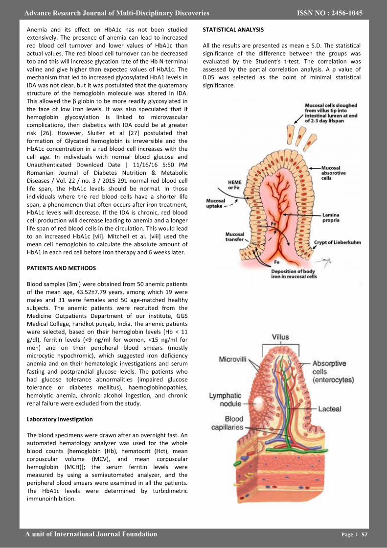
Anemia and its effect on HbA1c has not been studied extensively. The presence of anemia can lead to increased red blood cell turnover and lower values of HbA1c than actual values. The red blood cell turnover can be decreased too and this will increase glycation rate of the Hb N-terminal valine and give higher than expected values of HbA1c. The mechanism that led to increased glycosylated HbA1 levels in IDA was not clear, but it was postulated that the quaternary structure of the hemoglobin molecule was altered in IDA. This allowed the β globin to be more readily glycosylated in the face of low iron levels. It was also speculated that if hemoglobin glycosylation is linked to microvascular complications, then diabetics with IDA could be at greater risk [26]. However, Sluiter et al [27] postulated that formation of Glycated hemoglobin is irreversible and the HbA1c concentration in a red blood cell increases with the cell age. In individuals with normal blood glucose and Unauthenticated Download Date | 11/16/16 5:50 PM Romanian Journal of Diabetes Nutrition & Metabolic Diseases / Vol. 22 / no. 3 / 2015 291 normal red blood cell life span, the HbA1c levels should be normal. In those individuals where the red blood cells have a shorter life span, a phenomenon that often occurs after iron treatment, HbA1c levels will decrease. If the IDA is chronic, red blood cell production will decrease leading to anemia and a longer life span of red blood cells in the circulation. This would lead to an increased HbA1c [vii]. Mitchell et al. [viii] used the mean cell hemoglobin to calculate the absolute amount of HbA1 in each red cell before iron therapy and 6 weeks later. PATIENTS AND METHODS Blood samples (3ml) were obtained from 50 anemic patients of the mean age, 43.52±7.79 years, among which 19 were males and 31 were females and 50 age-matched healthy subjects. The anemic patients were recruited from the Medicine Outpatients Department of our institute, GGS Medical College, Faridkot punjab, India. The anemic patients were selected, based on their hemoglobin levels (Hb < 11 g/dl), ferritin levels (<9 ng/ml for women, <15 ng/ml for men) and on their peripheral blood smears (mostly microcytic hypochromic), which suggested iron deficiency anemia and on their hematologic investigations and serum fasting and postprandial glucose levels. The patients who had glucose tolerance abnormalities (impaired glucose tolerance or diabetes mellitus), haemoglobinopathies, hemolytic anemia, chronic alcohol ingestion, and chronic renal failure were excluded from the study. Laboratory investigation The blood specimens were drawn after an overnight fast. An automated hematology analyzer was used for the whole blood counts [hemoglobin (Hb), hematocrit (Hct), mean corpuscular volume (MCV), and mean corpuscular hemoglobin (MCH)]; the serum ferritin levels were measured by using a semiautomated analyzer, and the peripheral blood smears were examined in all the patients. The HbA1c levels were determined by turbidimetric immunoinhibition.
STATISTICAL ANALYSIS All the results are presented as mean ± S.D. The statistical significance of the difference between the groups was evaluated by the Student’s t-test. The correlation was assessed by the partial correlation analysis. A p value of 0.05 was selected as the point of minimal statistical significance.
A unit of International Journal Foundation Page I 57
A unit of International Journal Foundation Page I 58
Advance Research Journal of Multi-Disciplinary Discoveries ISSN NO : 2456-1045
OBSERVATION/RESULTS It was postulated that in normal individuals, there is a balance between HbA and serum glucose and if the glucose remained constant, a decrease in Hb concentration could cause an increase in the Glycated fraction [34]. A study that compared non diabetics with IDA to control groups found a higher mean HbA1c in IDA individuals. HbA1c significantly decreased after a 3 month course of iron therapy [3, 5]. Glycosylated hemoglobin has also been studied for its potential use as an index to distinguish between IDA and thalassemia minor. In a study on non-diabetic individuals, HbA1c levels were measured in β thalassemia, IDA and healthy controls. Median glycosylated hemoglobin was lower in β-thalassemia minor than IDA patients but there was no difference between IDA and control groups. In addition, in the IDA groups there was no significant correlation between HbA1c and other hematological parameters. The normal HbA1c concentrations in IDA could be due to normal red blood cell survival rate and normal levels of glycosylated hemoglobin in mature red blood cells [iii, vi]. Menopause has also been studied in respect with its link to IDA and HbA1c. Koga et al12 evaluated red blood cell indices and Glycated hemoglobin in pre-menopausal women and showed that RBC count is positively associated with HbA1c, but the case is the opposite for Hb, MCH and MCV. In the post-menopausal group, none of the indices could be linked to HbA1c. The MCH index had the highest correlation coefficient for association with HbA1c in premenopausal women because a decrease of 1 pg in MCH corresponded to an increase of approximately 0.03% in HbA1c [iii, vii]. In IDA, MCV and MCH modifications are observed before total Hb and RBC count are affected [6]. In premenopausal women, menstrual blood loss can cause IDA. It was concluded that the relatively iron deficient state of premenopausal women with lower MCH would be responsible for the higher observed HbA1c levels. In IDA, a decrease in Hb concentration could lead to an increase in the glycation fraction [iii, iv]. The HbA1c catabolism could be reduced in premenopausal women with lower MCH because if the red blood cell life span is increased then the HbA1c levels also increase. However, the study was not able to conclude this because information on red blood cell life span was contradictory [39-41]. Later Koga et al. [iv, v] conducted a study to relate iron metabolism indices with HbA1c in premenopausal women. The women had normal glucose tolerance and were grouped into those with IDA or iron deficient state (IDS) and the normal iron state (NIS). HbA1c levels were shown to be inversely associated with serum iron, serum transferrin saturation and serum ferritin. HbA1c levels were significantly higher in IDA and IDS than in the NIS group. It was concluded that iron deficiency increases HbA1c in premenopausal women regardless if the anemia is yet present or not [iv, ii]. Pregnant women have also been studied to find a correlation between HbA1c levels and glycemia in late pregnancy. In this stage, most women already have IDA. Using erythrocyte indices and iron metabolism indices, two studies by Hashimoto et al. [iv, iii] included non-diabetic pregnant women and later diabetic pregnant women not supplemented with iron. In both studies, the HbA1c levels were significantly increased in late pregnancy. It was concluded that the late pregnancy
increase in HbA1c levels was because of the IDA presence at this stage [iv]. A study involving non-diabetic pregnant women with and without IDA and an age matched control group analyzed the influence of iron metabolism indices on HbA1c and found a significant correlation between HbA1c and red blood cell and iron metabolic indices. Amongst the women who had confirmed IDA, HbA1c, OGTT (oral glucose tolerance test), red blood cell and iron metabolic indices were measured before and after iron treatment. The results indicated that HbA1c levels were higher in women with IDA, and after iron supplementation, the HbA1c levels decreased. A significant correlation between HbA1c and red blood cell and iron metabolic indices was observed [iv]. In the National Health and Nutrition Examination Survey (NHANES), observations of iron deficiency and HbA1c levels amongst non-diabetic adults showed that IDA increased the HbA1c level slightly and this occurred at the lower end of HbA1c levels i.e. between < 100 g/L and 5.72% in participants with Hb > 170 g/L. The adjusted mean concentrations of HbA1c were 5.56% and 5.46% among participants with and without iron deficiency, respectively (P = 0.095). They suggested, in contrast to previous studies, that IDA had little population effect on HbA1c. The difference in concentrations of HbA1c between extremes of concentrations of Hb was 0.2%. It was concluded that people with anemia who are close to the diagnostic threshold should be retested or undergo another diagnostic method [iv, vi]. Another more recent study showed that when comparing HbA1c levels in patients with IDA, it was found that the mean baseline level of HbA1c in anemic patients is lower and increases after treatment. The subsequent increase in HbA1c after iron supplementing was in contrast to the studies conducted previously. This was explained by the fact that their study group belonged to a lower socio-economic level where the cause of IDA was nutritional deficiency, rather than bleeding and malabsorption, and this could have affected the results [49]. Finally an Indian study that analyzed the effect of glycemic and non-glycemic parameters on HbA1c concentrations showed that if HbA1c is used to diagnose prediabetes and diabetes in iron deficient populations, this Unauthenticated Download Date | 11/16/16 5:50 PM 294 Romanian Journal of Diabetes Nutrition & Metabolic Diseases / Vol. 22 / no. 3 / 2015 leads to a false high prevalence [v]. Additionally other hematological parameters that predict higher HbA1c are anemia and red blood cell indices of IDA such as microcytosis, low MCH, low MCHC (mean corpuscular hemoglobin concentration) or high RDW (red blood cell distribution width) and low ferritin concentrations [vii].
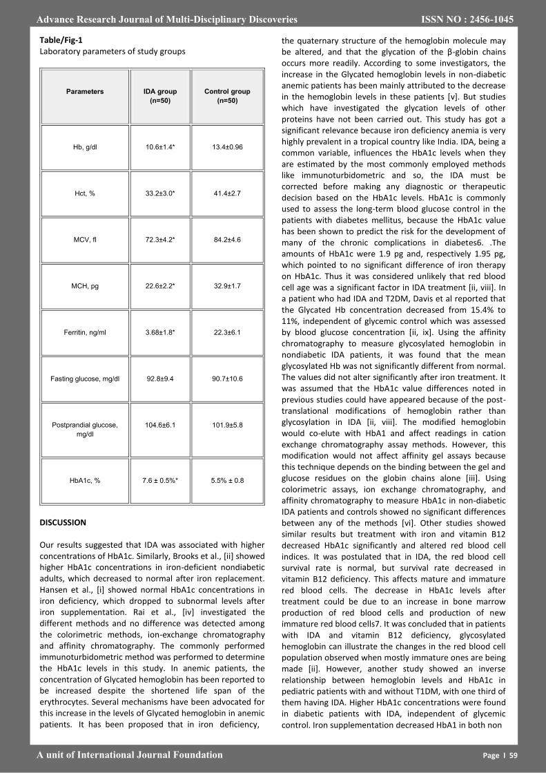
Table/Fig-1 Laboratory parameters of study groups
Parameters IDA group
(n=50)
Control group
(n=50)
Hb, g/dl 10.6±1.4* 13.4±0.96
Hct, % 33.2±3.0* 41.4±2.7
MCV, fl 72.3±4.2* 84.2±4.6
MCH, pg 22.6±2.2* 32.9±1.7
Ferritin, ng/ml 3.68±1.8* 22.3±6.1
Fasting glucose, mg/dl 92.8±9.4 90.7±10.6
Postprandial glucose,
mg/dl
104.6±6.1 101.9±5.8
HbA1c, % 7.6 ± 0.5%* 5.5% ± 0.8
DISCUSSION Our results suggested that IDA was associated with higher concentrations of HbA1c. Similarly, Brooks et al., [ii] showed higher HbA1c concentrations in iron-deficient nondiabetic adults, which decreased to normal after iron replacement. Hansen et al., [i] showed normal HbA1c concentrations in iron deficiency, which dropped to subnormal levels after iron supplementation. Rai et al., [iv] investigated the different methods and no difference was detected among the colorimetric methods, ion-exchange chromatography and affinity chromatography. The commonly performed immunoturbidometric method was performed to determine the HbA1c levels in this study. In anemic patients, the concentration of Glycated hemoglobin has been reported to be increased despite the shortened life span of the erythrocytes. Several mechanisms have been advocated for this increase in the levels of Glycated hemoglobin in anemic patients. It has been proposed that in iron deficiency,
Advance Research Journal of Multi-Disciplinary Discoveries ISSN NO : 2456-1045
A unit of International Journal Foundation Page I 59
the quaternary structure of the hemoglobin molecule may be altered, and that the glycation of the β-globin chains occurs more readily. According to some investigators, the increase in the Glycated hemoglobin levels in non-diabetic anemic patients has been mainly attributed to the decrease in the hemoglobin levels in these patients [v]. But studies which have investigated the glycation levels of other proteins have not been carried out. This study has got a significant relevance because iron deficiency anemia is very highly prevalent in a tropical country like India. IDA, being a common variable, influences the HbA1c levels when they are estimated by the most commonly employed methods like immunoturbidometric and so, the IDA must be corrected before making any diagnostic or therapeutic decision based on the HbA1c levels. HbA1c is commonly used to assess the long-term blood glucose control in the patients with diabetes mellitus, because the HbA1c value has been shown to predict the risk for the development of many of the chronic complications in diabetes6. .The amounts of HbA1c were 1.9 pg and, respectively 1.95 pg, which pointed to no significant difference of iron therapy on HbA1c. Thus it was considered unlikely that red blood cell age was a significant factor in IDA treatment [ii, viii]. In a patient who had IDA and T2DM, Davis et al reported that the Glycated Hb concentration decreased from 15.4% to 11%, independent of glycemic control which was assessed by blood glucose concentration [ii, ix]. Using the affinity chromatography to measure glycosylated hemoglobin in nondiabetic IDA patients, it was found that the mean glycosylated Hb was not significantly different from normal. The values did not alter significantly after iron treatment. It was assumed that the HbA1c value differences noted in previous studies could have appeared because of the post-translational modifications of hemoglobin rather than glycosylation in IDA [ii, viii]. The modified hemoglobin would co-elute with HbA1 and affect readings in cation exchange chromatography assay methods. However, this modification would not affect affinity gel assays because this technique depends on the binding between the gel and glucose residues on the globin chains alone [iii]. Using colorimetric assays, ion exchange chromatography, and affinity chromatography to measure HbA1c in non-diabetic IDA patients and controls showed no significant differences between any of the methods [vi]. Other studies showed similar results but treatment with iron and vitamin B12 decreased HbA1c significantly and altered red blood cell indices. It was postulated that in IDA, the red blood cell survival rate is normal, but survival rate decreased in vitamin B12 deficiency. This affects mature and immature red blood cells. The decrease in HbA1c levels after treatment could be due to an increase in bone marrow production of red blood cells and production of new immature red blood cells7. It was concluded that in patients with IDA and vitamin B12 deficiency, glycosylated hemoglobin can illustrate the changes in the red blood cell population observed when mostly immature ones are being made [ii]. However, another study showed an inverse relationship between hemoglobin levels and HbA1c in pediatric patients with and without T1DM, with one third of them having IDA. Higher HbA1c concentrations were found in diabetic patients with IDA, independent of glycemic control. Iron supplementation decreased HbA1 in both non
Advance Research Journal of Multi-Disciplinary Discoveries ISSN NO : 2456-1045
A unit of International Journal Foundation Page I 60
REFERENCES
[i] De Rosa MC, Sanna MT, Messana I, Castagno-la M, Galtieri A, Tellone E, et al. Glycated human haemoglobin (HbA1c): Functional characteristics and molecular modeling studies. Biophys Chem.1998; 72:323–35.
[ii] Peterson KP, Pavlovich JG, Goldstein D, Little R, England J, Peterson CM. What is haemoglobin A1c An analysis of glycated haemoglobins by electrospray ionization mass spectrometry. Clin Chem.1998;44:1951–58. .
[iii] Shapiro R, McManus M, Garrick L, McDonald MJ, Bunn HF. The nonenzymatic glycolisation of human haemoglobin at multiple sites. Metabolism. 1979; 28((suppl)):427–30.
[iv] Braunwald E, Fauci AS, Kasper DL, Hauser SL, Longo DL, Jameson JL. Diabetes mellitus. Harrison’s Principles of Internal Medicine. (15) 2001; 22:2105–09.
[v] Kim C, Bullard KM, Herman WH, Beckles GL. The association between iron deficiency and the HbA1c levels among adults without diabetes in the National Health and Nutrition Examination Survey, 1999–2006. Diabetes Care. 2010; 33:780–85.
[vi] Mayer TK, Freedman ZR. Protein glycosylation in diabetes mellitus: a review of the laboratory measurements and of their clinical utilities. Clin Chim Acta. 1983; 127:147e84.
[vii] Coban E, Ozdogan M, Timuragaoglu A. The effect of iron deficiency anemia on the levels of haemoglobin A1c in nondiabetic patients. Acta Haematol. 2004; 112:126–28.
[viii] Lapolla A, Traldi P, Fedele D. The importance of measuring the products of the non enzymatic glycation of proteins. Clin Biochem. 2005; 38:103e15.
[ix] Brownlee M. The negative consequences of glycation. Metabolism. 2000; 49:9e13.
[x] Mayer TK, Freedman ZR. Protein glycosylation in diabetes mellitus: a review of the laboratory measurements and their clinical utilities. Clin Chim Acta. 1983; 127:147e84.
[xi] Chowdhury TA, Lasker SS. The elevated Glycated hemoglobin in nondiabetic patients is associated with an increased mortality in myocardial infarction. Post grad Med J. 1998; 74:480e1.
[xii] Brooks AP, Metcalfe J, Day JL, Edwards MS. Iron deficiency and glycosylated hemoglobin A1. Lancet. 1980; 19(ii):141.
*****
A unit of International Journal Foundation Page I 07
diabetic and diabetic groups. This could be because of young red blood cells in the blood appear after iron therapy and lead to a “dilution effect” and lowering of HbA1c. The group stated there was no correlation between HbA1c and any other parameter than Hb. Structural or affinity changes in Hb, rather than iron concentration, reflect in changes of HbA1c [v]. Studies conducted in the 2000’s and beyond the effect of IDA on Hb subtype levels has also been studied using complete blood counts. This was done in a study using IDA individuals before and after iron therapy. Iron treatment improved RBC, Hb, Hct (hematocrit), MCV Unauthenticated Download Date | 11/16/16 5:50 PM 292 Romanian Journal of Diabetes Nutrition & Metabolic Diseases / Vol. 22 / no. 3 / 2015 (mean corpuscular volume) and MCH (mean corpuscular volume) values but HbA1c levels decreased as much as 17%8 Iron replacement used in IDA diabetics could increase the reliability of HbA1c measurements.
CONCLUSION
Our results showed that iron deficiency was associated with higher proportions of HbA1c, which could cause problems in the diagnosis of uncontrolled diabetes mellitus in iron-deficient patients. The iron status must be considered during the interpretation of the HbA1c concentrations in Diabetes mellitus. The iron replacement therapy is thus especially important in diabetic patients with iron deficiency, as it would also increase the reliability of the HbA1c determinations. Worldwide, iron deficiency is the most common nutritional deficiency and this is especially true for the low and middle income countries. In these regions, diabetes is also a rapidly increasing phenomenon. HbA1c is a popular tool for the screening and diagnosing of diabetes. Other tests such as the OGTT are time consuming and repeated laboratory tests can become expensive for poorer populations. Nonglycaemic factors that affect assays such as conditions that affect red blood cell turnover affect HbA1c readings. Most studies indicate that, although other forms of anemia lower HbA1c, iron deficiency anemia appears to have an enhancing effect on HbA1c. The mechanism of how IDA affects HbA1c remains elusive which indicates the need for further research to validate existing theories. Until large scale studies are conducted and the mechanism of increased HbA glycation in IDA states is not understood, we conclude that the reliability of HbA1c in IDA is questionable for diagnosing diabetes. People with anemia who are close to the diagnostic threshold may need to be retested or to be diagnosed with an alternative method. HbA1c values that do not correlate with the clinical scenario or in diabetics with IDA should be approached with caution.