hd7 - urogenital tract i 3/2/09 - columbia university · hd7 - urogenital tract i 3/2/09 10 ......
TRANSCRIPT

HD7 - Urogenital Tract I 3/2/09
1
Cathy [email protected] Human Development Spring 2009
Part I. Kidney development Part II. UGT development
nephrons in the kidney generate urine that is propelled to the ureters and then to the bladder for storage and excretion
The Urinary outflow tract:
• monitors and regulates extra-cellular fluids • excretes harmful substances in urine, including
nitrogenous wastes (urea) • returns useful substances to bloodstream • maintain balance of water, electrolytes (salts), acids,
and pH in the body fluids
glomeruli
tubules
The human kidney has about 500,000 nephrons that filter and modify the blood
At birth the collecting duct system has tens of thousands of branches
ub tip
CD CD
The papilla contains bundles of nephrons and CDs

HD7 - Urogenital Tract I 3/2/09
2
The urogenital system derives predominantly from intermediate mesoderm pronephros in an early embryo
The pronephros is a functional kidney in some species but regresses in mammals
Mesonephros is functional in frogs and birds
A metanephros is always drained exclusively by one duct, the ureter. In birds in reptiles the ureter separates from the nephric duct (Wolffian duct) and enters the cloaca. In mammals, the ureter separates from the nephric duct and enters the bladder
renal development begins when the ureteric bud invades kidney mesenchyme (the metanephric blastema)
kidney
ureter
common nephric duct
Wolffian duct
urogenital sinus
trigone
bladder
urethra
As the embryo grows, the ureters lengthen, and the kidneys rotate and ascend along the dorsal body wall

HD7 - Urogenital Tract I 3/2/09
3
• branching morphogenesis and nephron formation last until just after birth
• occur exclusively in the peripheral domain beneath the renal capsule
• new generations of nephrons and ureter branches displace older generations inward
• further differentiation occurs in inner domains at a distance from the renal capsule
The kidney is radially patterned Nephron formation
From “The Kidney�Mesenchymal nephron progenitors aggregate at ub tips and transdifferentiate
into epithelial cell types that comprise the nephron
stroma
nephron progenitors
ureteric bud tips
NEPHRONS
COLLECTING DUCT SYSTEM
CAPSULE/INTERSITIUM
RECIPROCAL SIGNALING BETWEEN STROMA, NEPHRON PROGENITORS AND URETERIC BUD TIPS GIVES RISE TO CELL TYPES IN THE MATURE KIDNEY
Nephron progenitors form nephrons
The ureteric bud forms the CD system
Melissa Little Lab
Reciprocal Signaling is required for branching morphogenesis and for nephron differentiation
co-culture experiments demonstrate reciprocal signaling between ureteric bud epithelial and nephron progenitors
nephron induction
branching morphogenesis
from: The kidney: Eds, Vize et al., 2003)
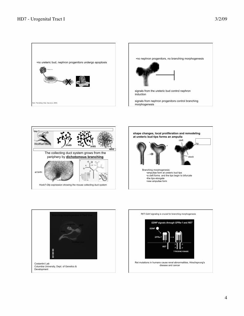
HD7 - Urogenital Tract I 3/2/09
4
• no ureteric bud, nephron progenitors undergo apoptosis
X
from: The kidney: Eds, Vize et al., 2003)
• no nephron progenitors, no branching morphogenesis
signals from the ureteric bud control nephron induction
signals from nephron progenitors control branching morphogenesis
The collecting duct system grows from the periphery by dichotomous branching
ub
2ndG 3rdG 4thG
Wolffian duct
at birth:
day 1
Hoxb7-Gfp expression showing the mouse collecting duct system
shape changes, local proliferation and remodeling at ureteric bud tips forms an ampulla
Branching morphogenesis: • ampullae form at ureteric bud tips • a cleft forms and the tips begin to bifurcate • the tips elongate • new ampullae form
cleft
stock
tip
ampulla
stalk
Wolffian duct
cleft
Costantini Lab Columbia University, Dept. of Genetics & Development
Branching morphogenesis in Real time
Ret mutations in humans cause renal abnormalities, Hirschsprung's disease and cancer
RET-Gdnf signaling is crucial for branching morphogenesis

HD7 - Urogenital Tract I 3/2/09
5
Mutations in Ret, Gdnf or Gfra1 result in renal agenesis or hypoplasia Gdnf secreted by nephron progenitors binds to Ret via the Ret
co-receptor (Gfra1) inducing branching
Gdnf NEPHRON PROGENITORS
Ret/Gfra1
Gdnf
Ret/Gfra1
URETERIC BUD TIP
STROMAL CELLS SECRETE RETINOIC ACID THAT IS REQUIRED FOR BRANCHING AND RET EXPRESSION IN UB TIPS
UB NP
STROMA
RA Control
AMPULLA FORMATION DEPENDS ON RA
TIME-LAPSE PHOTOGRAPHY OF HOXB7-GFP KIDNEYS GROWN WITH AND WITHOUT RA
+Retinoic acid 72h -Retinoic acid 72h
Hoxb7-Gfp kidney rudiments
Retinoic acid controls ureteric bud patterning
VITAMIN A CONTROLS BRANCHING VIA RET

HD7 - Urogenital Tract I 3/2/09
6
URETERIC BUD
Gdnf
STROMA
NEPHRON PROGENITORS
RA Ret/Gfra1
STROMAL CELL SECRETE RETINOIC ACID RETINOIC ACID INDUCES RET EXPRESSION IN NEARBY URTERIC BUD CELLS
RA secreted from stromal cells controls Ret expression In the ureteric bud
ureteric bud (RET)
stroma (vitamin A)
How does Ret signaling control branching?
ureteric bud cells must express Ret to contribute to a tip
Ret null cells are excluded from the tip domain- Presence in the tip depends on levels of Ret-Gdnf signaling?
How do nephrons form?
Nephron formation
From “The Kidney�Mesenchymal nephron progenitors aggregate at ub tips and transdifferentiate
into epithelial cell types that comprise the nephron

HD7 - Urogenital Tract I 3/2/09
7
NEPHRONS FORM EXCLUSIVELY AT URETERIC BUD TIPS IN RESPONSE TO LOCAL SIGNALS FROM URETERIC BUD CELLS
nephron progenitors
ureteric bud tip
Nephron progenitors condense at ub tips, aggregate
and trans-differentiate into epithelial cells that make up the renal vesicle, Comma and S-shaped bodies
from: The kidney: Eds, Vize et al., 2003)
Nephron polarity is established at early stages
WT1 in podocytes
Notch activation: Delta, jagged or serrate ligands on an adjacent cell bind Notch. The intracellular domain of Notch is cleaved, goes to the nucleus and induces transcriptional activation of Notch target genes.
Notch signaling controls nephron patterning along the P-D axis
Glomerular differentiation is arrested in Notch 2 mutant mice
Cheng et al, 2007
Notch2 is expressed in the developing nephron;
WT
WT
Notch2-
Notch2-
Part II. The lower urinary tract

HD7 - Urogenital Tract I 3/2/09
8
nephrons in the kidney generate urine that is propelled to the ureters and then to the bladder for storage and excretion DURING DEVELOPMENT THE URETER MOVES FROM THE
WOLFFIAN DUCT TO THE BLADDER
hydronephrosis in utero
Defective ureter insertion can cause obstruction and damage the kidney Severe reflux can cause end stage renal disease
The trigone is the site of the anti-reflux mechanism the ureteral valve is part of the trigone and is an anti-reflux mechanism that prevents urine back flow (reflux)
smooth muscle actin ureter epithelium
muscle
ureter
intra-mural ureter
Ureterl valve function depends on insertion of the ureter orifice at the proper position in the bladder neck (trigone)
intra-mural ureter
extra-mural ureter
detrusor
sheath

HD7 - Urogenital Tract I 3/2/09
9
THE TRIGONE IS MORPHOGLOGICALLY DISTINCT FROM THE BLADDER AND IS THOUGHT TO BE DERRIVED FROM THE COMMON NEPHRIC DUCT
The trigone contains an anti-reflux valve
Accepted model of ureter transposition
formation of the trigone from the common nephric duct repositions the ureters in the bladder
Larsen's Embryology
Mackie-Stephens hypothesis: the final position of the ureter with respect to the Trigone depends on the site of its formation on the Wolffian duct
Abnormal connections between the ureter orifice and trigone are associated with vesicoureteral reflux and obstruction
Test the Mackie Stephens hypothesis experimentally
kidney
ureter Wolffian duct
common nephric duct
expression of green fluorescent protein in the mouse common nephric duct enables us to follow its fate during
ureter insertion
using mouse models to re-assess the mechanism of ureter transposition:
The common nephric duct appears to regress rather than expand
what happens to the common nephric duct during ureter transposition?

HD7 - Urogenital Tract I 3/2/09
10
CND cells are absorbed into the urogenital sinus epithelium APOPTOSIS OF THE CND ENABLES THE URETER TO
SEPARATE AND REPOSITION IN THE BLADDER
THE CND UNDERGOES APOPTOSIS AND IS UNLIKELY TO FORM THE BLADDER TRIGONE
ONCE THE CND UNDERGOES APOPTOSIS THE URETER ORIFICE FUSES WITH THE BLADDER EPI AND IS MOVED TO ITS FINAL INSERTION SITE AS THE BLADDER EXPANDS
Ureter transposition depends on apoptosis of the common nephric duct
apoptotic common nephric duct cells
A revised model of ureter transposition
apoptosis of the common nephric duct enables the ureter orifice to detach from the Wolffian duct
the common nephric duct is absorbed into the expanding urogenital sinus. The ureter makes direct contact with and inserts into the urogenital sinus
continued growth and expansion of the urogenital sinus moves the ureter orifice further anterior to the bladder neck
sprouty, slit-2, retinoid excess
Impaired retinoid signaling, Ret
Calcineurin B (peristalsis)
sonic hedgehog (muscle)
uroplakin (epithelium)
Tbx18
abnormal position of the ureter orifice
abnormal peristalsis
Physical vs Functional obstruction

HD7 - Urogenital Tract I 3/2/09
11
Intrinsic ureteral abnormalities can cause obstruction
URETER PERISTALSIS IS MYOGENIC, MEDIATED BY SM IN THE URETERAL COAT
J. Clin. Invest. 113:1051-1058 (2004). Ching-Pin Chang, et al.
kidney ureter
bladder
Impaired peristalsis is a cause of obstruction (functional obstruction)
smooth muscle actin uroplakin
• a transitional epithelium expressing uroplakin lines the ureters
• The ureter smooth muscle coat mediates myogenic peristalsis • defective smooth muscle formation or mutations in uroplakins cause functional obstruction
Loss of Tbx18 results in megaureter
Airek et al.
Tbx18 is selectively expressed in peri-ureteral mesenchyme where it is required for ureter radial patterning
WT WT Tbx18- Tbx18-
Airek et al. wt
Tbx18-
The ureter is radially patterned by epithelial mesenchymal signals
Loss of Tbx18 expression in periureteral mesenchyme results in smooth muscle defects as well as epithelial abnormalities

HD7 - Urogenital Tract I 3/2/09
12
What signaling pathways are important for bladder formation?
The Bladder The bladder epithelium is lined with
plaques made from uroplakins that form a water-proof barrier
smooth muscle of the detrusor and rugae (folds) in the urothelium allow the bladder to expand and contract
Detrusor muscle
urothelium ureter
rugae
The urogenital sinus forms the bladder and urethra in both sexes
The urorectal septum partitions the cloaca into the urogenital sinus (ventral) and hindgut (dorsal)
Larsen's Embryology, 6th Edition
cloaca
urachus
hindgut
urorectal septum
from: The kidney: Eds, Vize et al., 2003)
Shh Ptch1 Gli1
wt
shh
Haraguchi et al., 2007
Sonic Hedgehog is localized in the bladder/urethral epi Patched and Gli1, downstream shh targets are localized in bladder/urethral mes
Shh is required for bladder formation