a numerical study of magnetic nanoparticles …jestec.taylors.edu.my/vol 12 issue 2 february...
TRANSCRIPT
Journal of Engineering Science and Technology Vol. 12, No. 2 (2017) 405 - 422 © School of Engineering, Taylor’s University
405
A NUMERICAL STUDY OF MAGNETIC NANOPARTICLES HYPERTHERMIA WITH ALTERNATING MAGNETIC FIELD UNDER INFLUENCE OF CONVECTION HEAT TRANSFER
MOSTAFA ZAKARIAPOUR1,*, MOHAMMAD HOSSEIN HAMEDI
1,
NASSER FATOURAEE2
1Department of Mechanical Engineering, K.N.T. University of Technology,
Pardis street, Tehran, Iran 2Department of Biomedical Engineering, Amir KabirUniversity of Technology,
Hafez street, Tehran, Iran
*Corresponding Author: [email protected]
Abstract
In this paper, a numerical study has been conducted to understand the heating
effects of the magnetic nanoparticles in the tumor hyperthermia in order to
reach a desirable temperature in the tumor. The developed numerical method
has been utilized to obtain the temperature distribution and magnetic induction
value using the bioheat and Maxwell equations inside a cylindrical geometry
including the tumor and healthy tissue while the perfusion and metabolism rates
have been considered. Results show that among all the parameters effected on
temperature rise, the diameter of the nanoparticles (ranging from 5,5.5,6 nm)
has the maximum effect, the strength of the applied alternating current (AC)
magnetic field (ranging from 50, 62,75 mT) has the minimum effect, and the
volume fraction (ranging from 0.0004,0.0006,0.0008) and the frequency of the
applied AC magnetic field (ranging from 300,400,500 kHz) result in increasing
the temperature relatively. The temperature rise for a temperature-dependent
metabolism is larger than a temperature-independent metabolism. Among the
materials investigated in this study, FePt has the most pronounced effect.
Keywords: Tumor, Hyperthermia, Bioheat, Magnetic nanoparticles, Induction.
1. Introduction
Hyperthermia is the procedure of temperature increase of cancerous tissue to 42-
46˚C for therapeutic reason. It has been shown in many studies; high temperature
can cause direct damage to cancerous cells or sensitize them to other cancer treat-
406 M. Zakariapour et al.
Journal of Engineering Science and Technology February 2017, Vol. 12(2)
Nomenclatures
c Specific heat of tissue, J/kg.K
cb Specific heat of blood, J/kg.K
D Diameter of MNP, m
f Frequency of magnetic field, Hz
H0 Amplitude of magnetic field, A/m
K Magnetocrystalline anisotropy, J/m3
k Boltzmann constant, J/K
kt Thermal conductivity, W/m.K
Md Domain magnetization, A/m
P Power density, W/m3
Qmetabolism Metabolic heat generation, W/m3
T Temperature of tissue, K
Ta Arterial temperature, K
To Initial temperature of tissue, K
t Time, s
VH Hydrodynamic volume of MNP, m3
VM Volume of MNP, m3
wb Perfusion rate of blood, m3/s/m
3
Greek Symbols
µ0 Permeability of free space, T.m/A
τ Effective relaxation time, s
τB Brownian relaxation time, s
τN Neel relaxation time, s
φ Volume fraction of MNPs
χ0 Equilibrium susceptibility
Subscripts
1,2 Number of layers
M Magnetic particle
ment modalities [1]. Hyperthermia is a thermal therapy of tumor, through
elevating the target tissue temperature in the human body, which therefore has
fewer side effects than that of the traditional chemotherapy or radiotherapy. It has
been well established that sustained temperature above 420C will cause necrosis
of living cells [2-5]. Hyperthermia is usually used with other forms of cancer
therapy, such as radiation therapy and chemotherapy. For instance, it has been
demonstrated that cytotoxicity of many chemotherapeutic agents is maximized at
temperatures between 40.5˚C and 43.0˚C [6]. Hyperthermia also enhances the
radio-sensitivity in hypoxic, low-pH areas of cancerous tissues [7]. Hyperthermia
is a viable cancer treatment for localized malignant tumors and its success has
been reported for head and neck cancer, breast cancer, urogenital tract cancer,
melanoma and sarcoma [8]. The effectiveness of hyperthermia treatment is
related to the temperature achieved during the treatment. An ideal hyperthermia
should destroy the tumor cells, without damaging the surrounding normal tissue.
Hypertherrmia can be performed by laser beam, microwave, ultrasound or
magnetic nanoparticle delivery to the tumor regions [9-13].
A Numerical Study of Magnetic Nanoparticles Hyperthermia with Alternating . . . . 407
Journal of Engineering Science and Technology February 2017, Vol. 12(2)
In the recent years, magnetic fluid hyperthermia (MFH) has been used due to
the advantages of cancer hyperthermia therapy. In MFH, a nanofluid containing
the magnetic nanoparticles (MNPs) is injected directly into the tumor or is
injected to the tumor vasculature. An alternating magnetic field, is then applied
to the target region, and then MNPs generate heat, according to Néel relaxation
and Brownian rotation. The heat generated increases the tumor temperature. The
tumor tissue is then destroyed by raising its temperature to about 42.50C whereas
healthy cells will be safe at lower temperatures [14-16].
The temperature rise due to magnetic nanoparticles therapy in the tissue,
strongly depends on the properties of the magnetic material used, the frequency
and the strength of the applied magnetic field, the blood perfusion in the tissue,
the duration of application of magnetic field and the volume fraction of magnetic
nanoparticles [17-20].
Hergt [21-23] studied about various magnetic nanoparticles with respect to
optimization of the specific loss power (SLP) for application in tumor hyperthermia.
Rosensweig [24] developed dissipation relationships based on the rotational
relaxation of single domain magnetic particles dispersed in a liquid matrix. Kim et
al. [25] made theoretical calculations of heat generation as a function of
Manganese Iron Oxid (MnFe2O4) nanoparticle diameter and compared these
specific absorption rate (SAR) values with experimental data. Their work shows
only the effect of nanoparticles in heat generation. Belc et al. [26] investigated the
effect of high AC magnetic fields on MNPs for magnetic hyperthermia and
radiation applications with just two nanoparticles and in a single computational
region. Dhar et al. [27] made an analytical study of temperature control in
hyperthermia by microwave to attain a desirable temperature at any point during a
fixed time, constant perfusion rate and by controlling optimally time dependent
heating power. Lin et al.[12] developed a hybrid numerical scheme for solving the
transient bioheat equation in spherical coordinates with constant perfusion rate
and in one dimensional state. The increase in temperature of biological tissues is
estimated for the heating effect of Iron-Platinum (FePt) MNPs. Yong et al. [28]
explored the three-dimensional (3-D) electromagnetic (EM) field and transient
temperature field induced by two external plate electrodes in the human body
containing a tumor during hyperthermia with micro/nano magnetic particles
which tissue and perfusion properties are variable with location. Narasimhan et al.
[29] studied transient simulations of heat transfer in human eye undergoing laser
surgery in single computational region and with constant perfusion rate in 2-D
coordinates. Kettering et al. [30] showed the possibility of temperature increase
due to MNPs accumulation in tumor. They found that a larger heating effect
occurs after exposing to an alternating magnetic field
Maenosono and Saita [11] investigated the theoretical assessment of
chemically disordered fcc-phase (face-centered cubic) FePt MNPs as heating
elements for magnetic hyperthermia by combining the heat generation model and
the bioheat transfer equation. To show the heating capability of these MNPs,
heating capability of these MNPs compared with other MNPs such as magnetite.
Consequently, fcc FePt MNPs were found to have a superior heating capability as
compared to other MNPs such as magnetite. Bagaria and Johnson [17] considered
the tissue model as a two-finite concentric spherical region together with the
blood perfusion effect.

408 M. Zakariapour et al.
Journal of Engineering Science and Technology February 2017, Vol. 12(2)
Salloum et al. [18, 19] performed an experimental study in a tissue-equivalent
agarose gel and evaluated magnetic nanofluid transport and heat distribution in
the gel. The SAR distribution showed that the nanoparticles distribution in the gel
is not uniform so that the concentration of the nanoparticles close to the injection
site is higher than others. Golneshan and Lahonian [31] studied the effect of
MNPs dispersion on temperature distribution in a tumor and surrounding healthy
tissue, during MFH. In this work, the Pennes bioheat equation (BHE) in a
spherical tissue with Neumann curved boundary condition has been solved. The
effects of blood perfusion, metabolism heat generation as well as MNPs heat
dissipation in an alternating magnetic field as the source term, have been
considered. To solve the Pennes BHE, Lattice Boltzmann Method (LBM) has
been used and results were compared with analytical ones. Attar et al. [32] studied
the dispersion of nanoparticles inside the tumor to investigate the tissues
temperature profiles. The problem is solved for polar coordinate in a cylindrical
tumor. Also the heating effect of magnetic fluid in a porcine liver tissue was
experimentally examined. Numerical transient solutions were found to be in good
agreement with experimental data. They found that the injected nanoparticles do
not usually distribute uniformly throughout the entire tumor.
Singh et al. [33] investigated hyperthermia with laser induced heating of a
tumor in a 2-D axisymmetric tissue embedded with gold-silica nanoshells in the
tumor. Effects of power density, laser exposure time, beam radius, blood vessel
diameter and volume fractions of nanoshells on temperature spread in the tissue
were analysed.
In view of all-above mentioned paragraphs, it is evident that heating the
colloidal magnetic fluid (ferrofluid) due to time-varying magnetic induction and
real boundary condition in tumor and normal tissue has not been considerably
studied. In this study we investigate the effect of physical characteristics of
nanoparticles, characteristics of applied magnetic field as well as characteristics
of tissue on heat transformation of human body in hyperthermia. It should be
mentioned that here evaporation rate from surface of tissue in contact with
surrounding space has also been considered.
2. Mathematical Formulation and Boundary Conditions
In order to find the temperature distribution during hyperthermia, it is essential to
solve the energy equation within the tumor domain with real boundary conditions.
Figure 1 shows the geometry and dimensions of the problem. It assumes that our
model simulates skin cancer starting from skin surface and ending to a defined
depth. The Pennes bioheat transfer equation [34] for a tumor and tissue can be
written as follows respectively
( ) P+Q+T-Twcρ+Tk=t
Tcρ metabolismabbbt∇∇∂
∂11 (1)
( )metabolismabbbt Q+T-Twcρ+Tk=
t
Tcρ ∇∇∂
∂22 (2)
where.T is the temperature, t is the time, wb, ρb, cb and Ta are the perfusion, the
density, the specific heat and the temperature of the blood, Qmetabolism and P are the

A Numerical Study of Magnetic Nanoparticles Hyperthermia with Alternating . . . . 409
Journal of Engineering Science and Technology February 2017, Vol. 12(2)
metabolic heat generation of the tissue and the distributed volumetric heat source
due to spatial heating of MNPs.
Considering the evaporation term in the skin, heat equation can be written as
follows [35]
( )Emetabolismabbbt QPQ+T-Twcρ+Tk=
t
Tcρ -+∇∇∂
∂11 (3)
in which QE is refered to the amount of evaporation‘which meanes is the amount
of water consumption in the skin per second’ is defined as follows. [35]
dt
dWαQE -= (4)
where α, is the latent heat of water, which is equal to 2260 kj/kg and W is the
density of water in tissue and is, only temperature dependent. Eq. (3) will be
obtained as follows
( ) PQ+T-Twcρ+Tk=t
Tcρ metabolismabbbt +∇∇∂
∂)( '
(5)
TWρ
αc
T
W
ρ
αcc ′-=
∂
∂-=′ (6)
))42.3
106-exp(-1(×778=)(
TTW (7)
In 2-D cylindrical coordinates we have
( ) PQ+T-Twcρ+z
Tk
r
Tr
rr
k=
t
Tcρ metabolismabbbt +
∂
∂+
∂
∂
∂
∂
∂
∂)′( 2
2
(8)
From Fig. 1, the boundary conditions are as follows. L, the height of the
healthy tissue, R, the radius of the healthy tissue, h, the height of the tumor tissue
and a, the radius of the tumor tissue
at z=0→dz
dTkTh -=)T-( ∞ (9)
at z=L→ 0=∂
∂
z
T (10)
at r=R→ CT 037= (11)
The initial boundary condition for temperature is also as follows
at t=0→ CT o37= (12)
in which, the specific heat (c1) and density (ρ1) (for the tumor in Eq. (1)) is
consisted of the tissue (with index 2) and the nanoparticles (with index M) and
with volume fraction of φ and given by [29]
)-1(+=
)-1(+=
21
21
φcc φc
φρφρρ
M
M (13)
410 M. Zakariapour et al.
Journal of Engineering Science and Technology February 2017, Vol. 12(2)
The power dissipation density for a mono-dispersion with constant
susceptibility is expressed as follows [11]
2
2000
)τfπ(2+1
τf π2f HXπμ=P (14)
in which µ0=4π×107 Tm/A is the permeability of free space, Χ0=(µ0φ
2Md
2VM)/kT
the susceptibility (here is assumed magnetic field independent and Md is the
domain magnetization of a suspended particle), H0 is the strength of applied AC
magnetic field and f is the cyclic frequency of applied AC magnetic field.
Because the Brownian and Néel processes take place, the effective relaxation
time, τ, is defined as follows [11]
BN
BN
ττ
τττ
+= (15)
where τN is the Néel relaxation time and τB is the Brownian relaxation time. The first
mechanism existing in this phenomenon is the Brownian mechanism of relaxation,
the magnetic moment is locked to the crystal axis and when the magnetic moment
aligns with the field, the particle rotates as well. A second mechanism exists (Néel
relaxation) in which the magnetic moment rotates within the crystal. To achieve
high heating rates, the Néel relaxation must not be dominated. The Brownian time
constant is given by the following relationship [11]
kT
Vητ
HB
3= (16)
where η is the viscosity of the carrier liquid, VH=π(D+2)3/6 is the hydrodynamic
volume of the particle, D is the nanoparticle maximum diameter, kb is the
Boltzmann constant, and T is the absolute temperature. The Néel relaxation time,
denoted τN, is given by the following expression due to Brown [11]
( )/kT KVττ MN exp= 0 (17)
where K is the anisotropy constant, VM =π(D)3/6 is the volume of the particle, T is
the absolute temperature, and τ0 is the time constant, (τ0=10−9
s).
By uncoupling Maxwell equations, here we consider the magnetic induction
formula as follows [36]
Bt
Bσμ ∇=∂
∂2
0 (18)
where B is the magnetic induction field, μ0 is the magnetic permeability and σ is
the electrical conductivity of the medium. Magnetic induction field is related to
the strength of applied AC magnetic field as follows[36]
)+(= dMHμB 0 (19)
and we have
at r=a→ mTB 73= (20)

A Numerical Study of Magnetic Nanoparticles Hyperthermia with Alternating . . . . 411
Journal of Engineering Science and Technology February 2017, Vol. 12(2)
Fig. 1. Geometry and dimensions of the problem in cylindrical coordinate.
3. Numerical Method
To solve Eqs. (3) and (18), a finite difference method (FDM) is used in which the
second order forward difference is used for coordinate-dependent terms and an
implicit approach is also used to discretize the time-dependent terms. To do this
task, Eqs. (3) and (18) are solved simultaneously, i.e., firstly Eq. (18) is solved
and the value of magnetic induction in all over the computational domain and all
times is obtained. Then, the Eq. (19) is solved using the obtained magnetic
inductions from Eq. (18) and considering the value of the thermal power
calculated by Eq. (14). Finally, temperature at different times and locations is
obtained by solving Eq. (3).
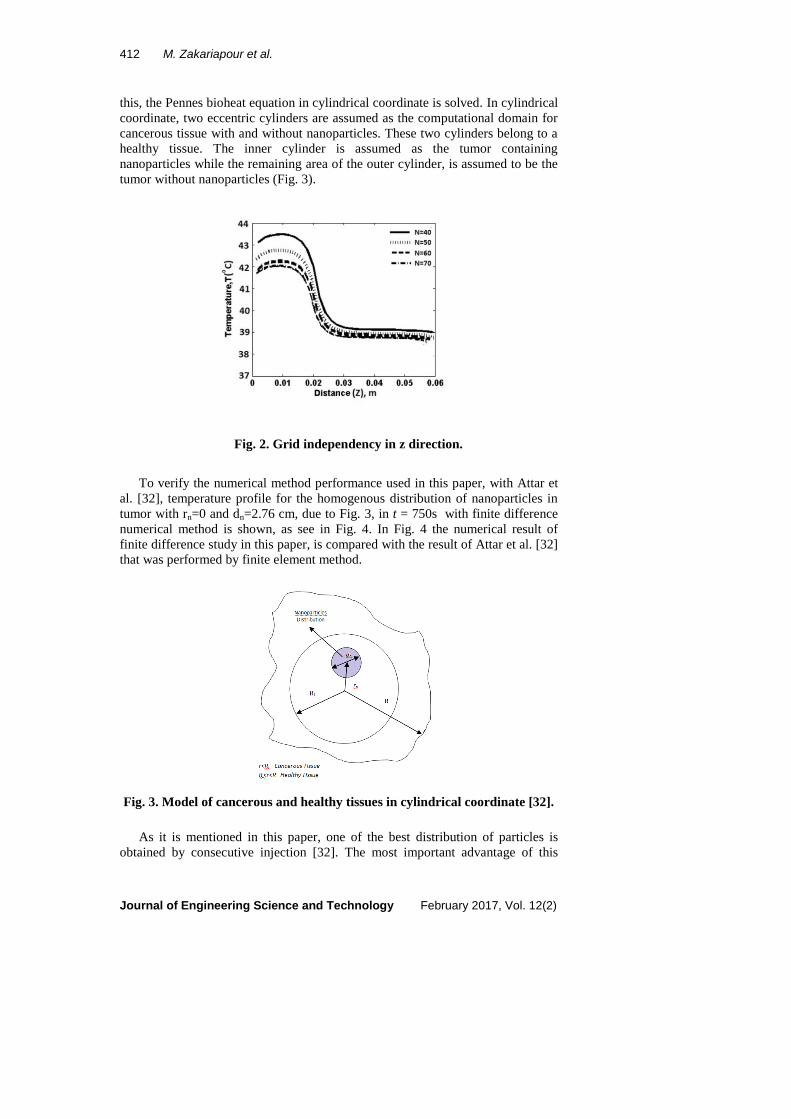
4. Grid Independency
As it was explained in section 2, in this study the described problem is two
dimensional and governing equations are discretized along the r and z directions
shown in Fig. 1. In order to make sure that the results are independent from the
grid resolution, Eq. (3) is solved on four computational grids i.e. 40×40, 50×50,
60×60 and 70×70. The results for temperature along the z direction and relevant
to the mentioned grids are shown in Fig. 2. As the figure indicates, because of
nearness of the results of 60×60 and 70×70 grids, choosing the 70×70 grid as the
independent grid is completely reasonable. The time step is Δt=0.5s.
5. Verification of the code
In this section, for verifying the results, this paper is compared with a numerical
research which performed in hyperthermia with MFH therapy [32]. To evaluate
r
z
L h
R
412 M. Zakariapour et al.
Journal of Engineering Science and Technology February 2017, Vol. 12(2)
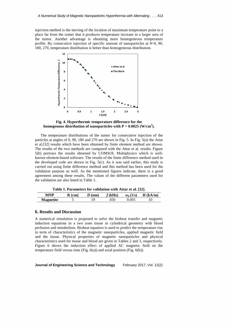
this, the Pennes bioheat equation in cylindrical coordinate is solved. In cylindrical
coordinate, two eccentric cylinders are assumed as the computational domain for
cancerous tissue with and without nanoparticles. These two cylinders belong to a
healthy tissue. The inner cylinder is assumed as the tumor containing
nanoparticles while the remaining area of the outer cylinder, is assumed to be the
tumor without nanoparticles (Fig. 3).
Fig. 2. Grid independency in z direction.
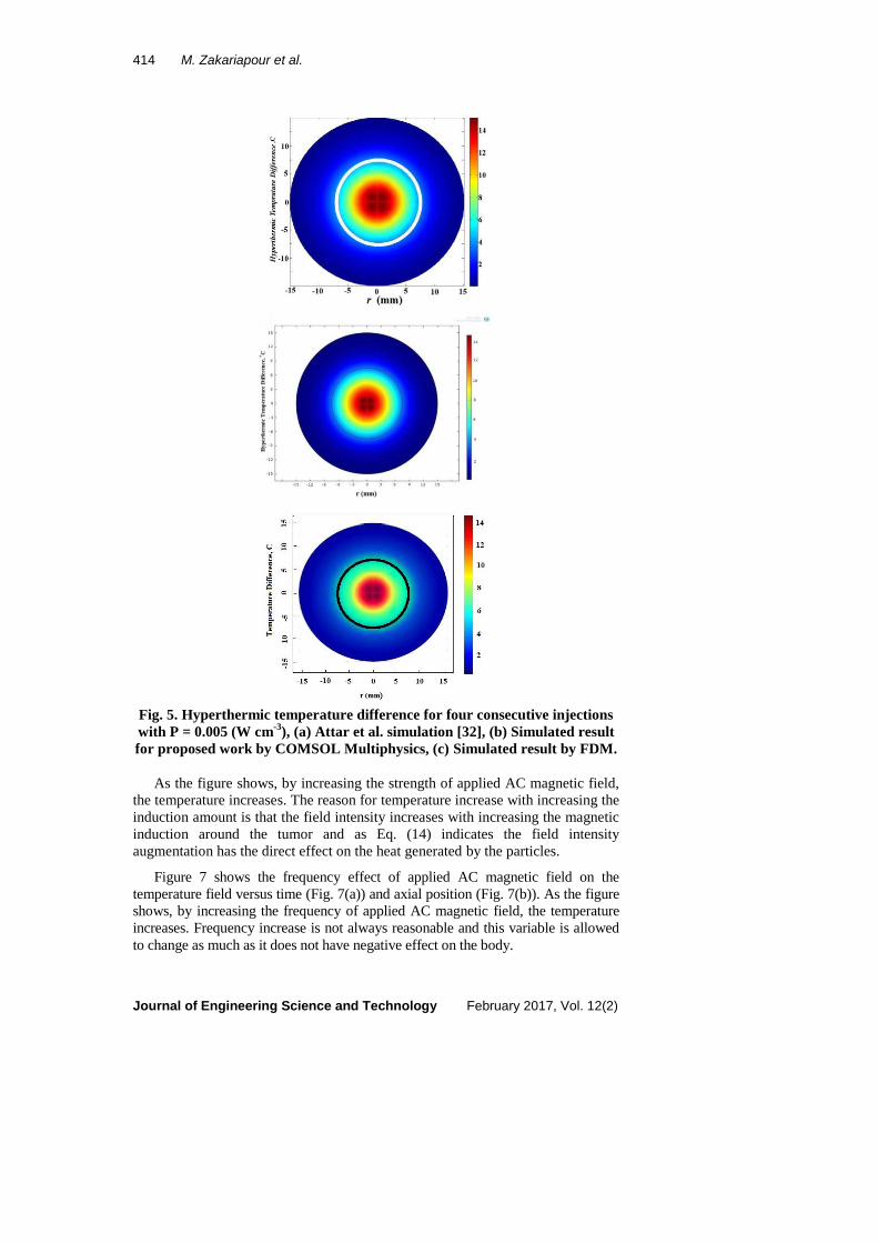
To verify the numerical method performance used in this paper, with Attar et
al. [32], temperature profile for the homogenous distribution of nanoparticles in
tumor with rn=0 and dn=2.76 cm, due to Fig. 3, in t = 750s with finite difference
numerical method is shown, as see in Fig. 4. In Fig. 4 the numerical result of
finite difference study in this paper, is compared with the result of Attar et al. [32]
that was performed by finite element method.
Fig. 3. Model of cancerous and healthy tissues in cylindrical coordinate [32].
As it is mentioned in this paper, one of the best distribution of particles is
obtained by consecutive injection [32]. The most important advantage of this
A Numerical Study of Magnetic Nanoparticles Hyperthermia with Alternating . . . . 413
Journal of Engineering Science and Technology February 2017, Vol. 12(2)
injection method is the moving of the location of maximum temperature point to a
place far from the center that it produces temperature increase in a larger area of
the tumor. Another advantage is obtaining more homogeneous temperature
profile. By consecutive injection of specific amount of nanoparticles at θ=0, 90,
180, 270, temperature distribution is better than homogeneous distribution.
Fig. 4. Hyperthermic temperature difference for the
homogenous distribution of nanoparticles with P = 0.0025 (W/cm3).
The temperature distributions of the tumor for consecutive injection of the
particles at angles of 0, 90, 180 and 270 are shown in Fig. 5. In Fig. 5(a) the Attar
et al.[32] results which have been obtained by finite element method are shown.
The results of the two methods are compared with the Attar et al. results. Figure
5(b) portrays the results obtained by COMSOL Multiphysics which is well-
known element-based software. The results of the finite difference method used in
the developed code are shown in Fig. 5(c). As it was said earlier, this study is
carried out using finite difference method and this method has been used for the
validation purpose as well. As the mentioned figures indicate, there is a good
agreement among these results. The values of the different parameters used for
the validation are also listed in Table 1.
Table 1. Parameters for validation with Attar et al. [32].
MNP R (cm) D (nm) f (kHz) wb (1/s) H (kA/m)
Magnetite 5 19 450 0.001 10
6. Results and Discussion
A numerical simulation is proposed to solve the bioheat transfer and magnetic
induction equations in a two zone tissue in cylindrical geometry with blood
perfusion and metabolism. Bioheat equation is used to predict the temperature rise
in term of characteristics of the magnetic nanoparticles, applied magnetic field
and the tissue. Physical properties of magnetic nanoparticles and physical
characteristics used for tissue and blood are given in Tables 2 and 3, respectively.
Figure 6 shows the induction effect of applied AC magnetic field on the
temperature field versus time (Fig. 6(a)) and axial position (Fig. 6(b)).
414 M. Zakariapour et al.
Journal of Engineering Science and Technology February 2017, Vol. 12(2)
Fig. 5. Hyperthermic temperature difference for four consecutive injections
with P = 0.005 (W cm-3
), (a) Attar et al. simulation [32], (b) Simulated result
for proposed work by COMSOL Multiphysics, (c) Simulated result by FDM.
As the figure shows, by increasing the strength of applied AC magnetic field,
the temperature increases. The reason for temperature increase with increasing the
induction amount is that the field intensity increases with increasing the magnetic
induction around the tumor and as Eq. (14) indicates the field intensity
augmentation has the direct effect on the heat generated by the particles.
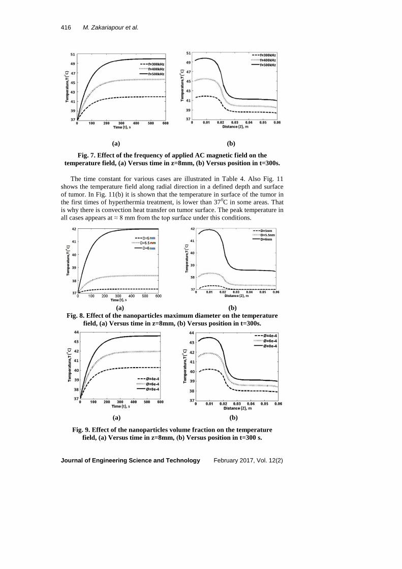
Figure 7 shows the frequency effect of applied AC magnetic field on the
temperature field versus time (Fig. 7(a)) and axial position (Fig. 7(b)). As the figure
shows, by increasing the frequency of applied AC magnetic field, the temperature
increases. Frequency increase is not always reasonable and this variable is allowed
to change as much as it does not have negative effect on the body.

A Numerical Study of Magnetic Nanoparticles Hyperthermia with Alternating . . . . 415
Journal of Engineering Science and Technology February 2017, Vol. 12(2)
Table 2. Physical properties of magnetic solids [11].
Magnetic
solid
Chemical
formula
Md
(kAm-1
)
K
(kJm-3
)
c
(J kg-1
K-1
)
ρ
(kgm-3
)
Dmax
(nm)
Maghemite Fe2O3 414 4.7 746 4600 23.5
Magnetite FeO Fe2O3 446 9 670 5180 19
Cobalt
ferrite
FeCo 1790 1.5 172 8140 340
Platinum
ferrite
LI0 FePt
1140 206 327 15200 9
Table 3. Physical characteristics used for tissue and blood [11].
ρt
(kg/m3)
cp,t
(J kg-1K-1)
ρb
(kgm-3)
cp,b
(J/kg.K)
kt
(W/m.K)
Ta
(0C)
wb
(1/s)
σ
(S/m)
1060 3600 1000 4180 0.5 37 0.0064 0.37
(a) (b)
Fig. 6. Effect of the strength of applied AC magnetic field on the temperature
field, (a) Versus time in z=8mm, (b) Versus position in t=300s.
Figure 8 shows the effect of nanoparticles maximum diameter on the
temperature field versus time (Fig. 8(a)) and axial position (Fig. 8(b)). As the
figure shows, by increasing the nanoparticles maximum diameter, the temperature
increases. Nanoparticle diameter is the most important parameter in hyperthermia.
Both Magnetic permeability and thermal power increases with increasing the
particle diameter.
Figure 9 shows the effect of nanoparticles volume fraction on the temperature
field versus time (Fig. 9(a)) and axial position (Fig. 9(b)). As the figure shows, by
increasing the nanoparticles volume fraction, the temperature increases. Figure 10
shows the effect of tissue metabolism and perfusion rate on the temperature field
versus time and axial position. As the figure shows, temperature dependent
modeling of metabolism and perfusion rates lead to lower temperature values and
the effect of temperature dependent perfusion rate is higher than temperature
dependent metabolism rate.
416 M. Zakariapour et al.
Journal of Engineering Science and Technology February 2017, Vol. 12(2)
(a) (b)
Fig. 7. Effect of the frequency of applied AC magnetic field on the
temperature field, (a) Versus time in z=8mm, (b) Versus position in t=300s.
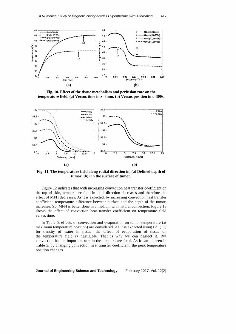
The time constant for various cases are illustrated in Table 4. Also Fig. 11
shows the temperature field along radial direction in a defined depth and surface
of tumor. In Fig. 11(b) it is shown that the temperature in surface of the tumor in
the first times of hyperthermia treatment, is lower than 370C in some areas. That
is why there is convection heat transfer on tumor surface. The peak temperature in
all cases appears at ≈ 8 mm from the top surface under this conditions.
(a) (b)
Fig. 8. Effect of the nanoparticles maximum diameter on the temperature
field, (a) Versus time in z=8mm, (b) Versus position in t=300s.
(a) (b)
Fig. 9. Effect of the nanoparticles volume fraction on the temperature
field, (a) Versus time in z=8mm, )b) Versus position in t=300 s.
A Numerical Study of Magnetic Nanoparticles Hyperthermia with Alternating . . . . 417
Journal of Engineering Science and Technology February 2017, Vol. 12(2)
(a) (b)
Fig. 10. Effect of the tissue metabolism and perfusion rate on the
temperature field, )a) Versus time in z=8mm, (b) Versus position in t=300s.
(a) (b)
Fig. 11. The temperature field along radial direction in, )a) Defined depth of
tumor, (b) On the surface of tumor.
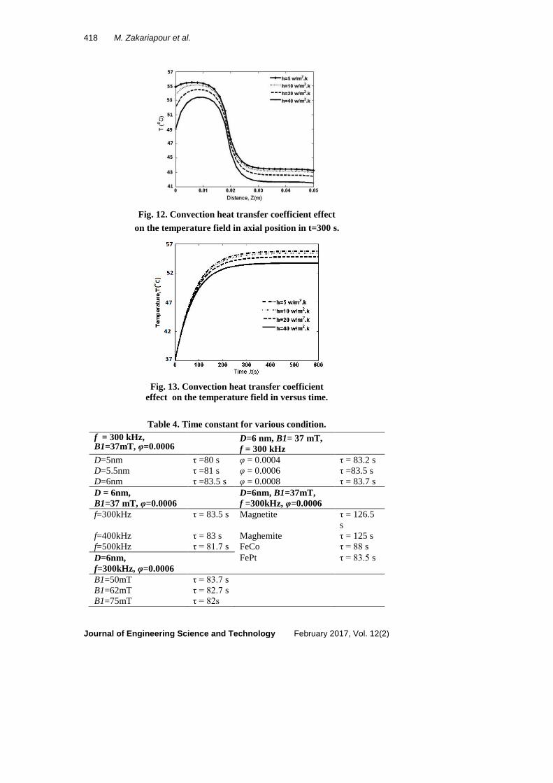
Figure 12 indicates that with increasing convection heat transfer coefficient on
the top of skin, temperature field in axial direction decreases and therefore the
effect of MFH decreases. As it is expected, by increasing convection heat transfer
coefficient, temperature difference between surface and the depth of the tumor,
increases. So, MFH is better done in a medium with natural convection. Figure 13
shows the effect of convection heat transfer coefficient on temperature field
versus time.
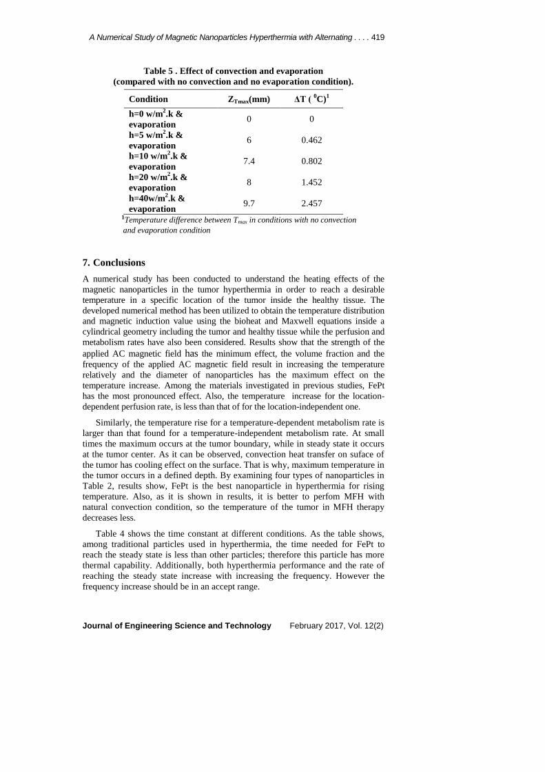
In Table 5. effects of convection and evaporation on tumor temperature (at
maximum temperature position) are considered. As it is expected using Eq. (11)
for density of water in tissue, the effect of evaporation of tissue on
the temperature field is negligible. That is why we can neglect it. But
convection has an important role in the temperature field. As it can be seen in
Table 5, by changing convection heat transfer coefficient, the peak temperature
position changes.
418 M. Zakariapour et al.
Journal of Engineering Science and Technology February 2017, Vol. 12(2)
Fig. 12. Convection heat transfer coefficient effect
on the temperature field in axial position in t=300 s.
Fig. 13. Convection heat transfer coefficient
effect on the temperature field in versus time.
Table 4. Time constant for various condition.
f = 300 kHz, B1=37mT, φ=0.0006
D=6 nm, B1= 37 mT,
f = 300 kHz
D=5nm τ =80 s φ = 0.0004 τ = 83.2 s
D=5.5nm τ =81 s φ = 0.0006 τ =83.5 s
D=6nm τ =83.5 s φ = 0.0008 τ = 83.7 s
D = 6nm,
B1=37 mT, φ=0.0006
D=6nm, B1=37mT,
f =300kHz, φ=0.0006
f=300kHz τ = 83.5 s Magnetite τ = 126.5
s
f=400kHz τ = 83 s Maghemite τ = 125 s
f=500kHz τ = 81.7 s FeCo τ = 88 s
D=6nm,
f=300kHz, φ=0.0006
FePt τ = 83.5 s
B1=50mT τ = 83.7 s
B1=62mT τ = 82.7 s
B1=75mT τ = 82s
A Numerical Study of Magnetic Nanoparticles Hyperthermia with Alternating . . . . 419
Journal of Engineering Science and Technology February 2017, Vol. 12(2)
Table 5 . Effect of convection and evaporation
(compared with no convection and no evaporation condition).
Condition ZTmax(mm) ΔT ( 0C)
1
h=0 w/m2.k &
evaporation 0 0
h=5 w/m2.k &
evaporation 6 0.462
h=10 w/m2.k &
evaporation 7.4 0.802
h=20 w/m2.k &
evaporation 8 1.452
h=40w/m2.k &
evaporation 9.7 2.457
1Temperature difference between Tmax in conditions with no convection
and evaporation condition
7. Conclusions
A numerical study has been conducted to understand the heating effects of the
magnetic nanoparticles in the tumor hyperthermia in order to reach a desirable
temperature in a specific location of the tumor inside the healthy tissue. The
developed numerical method has been utilized to obtain the temperature distribution
and magnetic induction value using the bioheat and Maxwell equations inside a
cylindrical geometry including the tumor and healthy tissue while the perfusion and
metabolism rates have also been considered. Results show that the strength of the
applied AC magnetic field has the minimum effect, the volume fraction and the
frequency of the applied AC magnetic field result in increasing the temperature
relatively and the diameter of nanoparticles has the maximum effect on the
temperature increase. Among the materials investigated in previous studies, FePt
has the most pronounced effect. Also, the temperature increase for the location-
dependent perfusion rate, is less than that of for the location-independent one. Similarly, the temperature rise for a temperature-dependent metabolism rate is
larger than that found for a temperature-independent metabolism rate. At small
times the maximum occurs at the tumor boundary, while in steady state it occurs
at the tumor center. As it can be observed, convection heat transfer on suface of
the tumor has cooling effect on the surface. That is why, maximum temperature in
the tumor occurs in a defined depth. By examining four types of nanoparticles in
Table 2, results show, FePt is the best nanoparticle in hyperthermia for rising
temperature. Also, as it is shown in results, it is better to perfom MFH with
natural convection condition, so the temperature of the tumor in MFH therapy
decreases less.
Table 4 shows the time constant at different conditions. As the table shows,
among traditional particles used in hyperthermia, the time needed for FePt to
reach the steady state is less than other particles; therefore this particle has more
thermal capability. Additionally, both hyperthermia performance and the rate of
reaching the steady state increase with increasing the frequency. However the
frequency increase should be in an accept range.

420 M. Zakariapour et al.
Journal of Engineering Science and Technology February 2017, Vol. 12(2)
References
1. Falk, M.H.; and Issels, R.D. (2001). Hyperthermia in oncology. International
Journal of Hyperthermia, 17,1-18.
2. Cavaliere, R.; Ciocatto, E.C.; Gionanella, B.C.; Heidelberger, C.; Johnson,
R.O.; Margottini, M.; Mondovi, B.; Moricca, G.; and Fanelli, A.R. (1967).
Selective heat sensitivity of cancer cells biochemical and clinical studies.
Cancer, 20, 1351-1381.
3. Robinson, J.E.; Wizenberg, M.J.; and Mccready, W.A. (1974). Combined
hyperthermia and radiation, an alternative to heavy particle therapy for
reduced oxygen enhancement ratios. Nature, 251, 521-522.
4. Steeves, R.A. (1992). Hyperthermia in cancer therapy: Where are we today
and where are we going? Bulletin of the New York Academy of Medicine, 68,
342-350.
5. Dewey, W. (1994). Arrhenius relationships from molecule and cell to clinic.
International Journal of Hyperthermia, 10, 457-483.
6. Issels, R.D. (2008). Hyperthermia adds to chemotherapy, European Journal
of Cancer, 44, 2546-2554.
7. Song, C.W.; Shakil, A.; Griffin, R.J.; and Okajima, K. (1997). Improvement
of tumor oxygenation status by mild temperature hyperthermia alone or in
combination with carbogen, Seminars in Oncology, 24, 626-632.
8. van der Zee, J. (2002). Heating the patient: a promising approach? Annals of
Oncology, 13, 1173-1184.
9. Moroz, P.; Jones, S.K.; and Gray, B.N. (2002). Magnetically mediated
hyperthermia: Current status and future directions. International Journal of
Hyperthermia, 18, 267-284.
10. Lagendijk, J.J.W. (2000). Hyperthermia treatment planning. Physics in
Medicine and Biology, 45, 61-76.
11. Maenosono, S.; and Saita, S. (2006). Theoretical assessment of FePt
nanoparticles as heating elements for magnetic hyperthermia. IEEE
Transactions on Magnetic , 42, 1638-1642.
12. Lin, Ch.T.; and Liu, K.Ch. (2009(. Estimation for the heating effect of
magnetic nanoparticles in perfused tissues. International Communication in
Heat and Mass Transfer, 36, 241-244.
13. Andrä, W.; D’Ambly, C.G.; Hergt, R.; Hilger, I..; and Kaiser, W.A. (1999).
Temperature distribution as function of time around a small spherical heat
source of local magnetic hyperthermia. Journal of Magnetism and Magnetic
Materials, 194, 197-203.
14. Jordan, A.; Scholz, R.; Wust, P.; Schirra, H.; Schiestel, T.; Schmidt, H.; and
Felix, R. (1999). Endocytosis of dextran and silancoated magnetite
nanoparticles and the effect of intracellular hyperthermia on human
mammary carcinoma cells in vitro. Journal of Magnetism and Magnetic
Materials, 194, 185-196.
15. Jordan, A.; Scholz, R.; Maier-Hauff, K.; Johannsen, M.; Wust, P.; Nadobny,
J.; Schirra, H.; Schmidt, H.; Deger, S.; Loening, S.; Lanksch, W.; and Felix,
R. (2001). Presentation of a new magnetic field therapy system for the
A Numerical Study of Magnetic Nanoparticles Hyperthermia with Alternating . . . . 421
Journal of Engineering Science and Technology February 2017, Vol. 12(2)
treatment of human solid tumors with magnetic fluid hyperthermia. Journal
of Magnetism and Magnetic Materials, 225, 118-126.
16. Thiesen, B.; and Jordan, A. (2008). Clinical applications of magnetic
nanoparticles for hyperthermia. International Journal of Hyperthermia, 24,
467-474.
17. Bagaria, H.G.; and Johnson, D.T. (2005). Transient solution to the bioheat
equation and optimization for magnetic fluid hyperthermia treatment.
International Journal of Hyperthermia, 21, 57-75.
18. Salloum, M.; Ma, R.H.; Weeks, D.; and Zhu, L. (2008). Controlling
nanoparticle delivery in magnetic nanoparticle hyperthermia for cancer
treatment: Experimental study in agarose gel. International Journal of
Hyperthermia, 24, 337-345.
19. Salloum, M.; Ma, R.H.; and Zhu, L. (2008). An in-vivo experimental study of
temperature elevations in animal tissue during magnetic nanoparticle
hyperthermia. International Journal of Hyperthermia, 24, 589-601.
20. Bellizzi, G.; and Bucci, O.M. (2010). On the optimal choice of the exposure
conditions and the nanoparticle features in magnetic nanoparticle
hyperthermia. International Journal of Hyperthermia, 26, 389-403.
21. Hergt, R. (1998). Physical limits of hyperthermia using magnetic fine
particle, IEEE Transactions on Magnetic, 34, 3745-3754.
22. Hergt, R. (2006). Magnetic particle hyperthermia: nanoparticle magnetism
and materials development for cancer therapy, Journal of Physical Sciences,
18, 2919-2934.
23. Hergt, R. (2007). Magnetic particle hyperthermia-biophysical limitations of a
visionary tumor therapy, Journal of Magnetism and Magnetic Materials, 311,
187-192.
24. Rosensweig, R.E. (2002). Heating the magnetic fluid with alternating
magnetic field. Journal of Magnetism and Magnetic Materials, 252, 370-374.
25. Kim, D.H.; Thai, Y.T.; Nikles, D.E.; and Brazel, C.S. (2009). Heating the
aqueous dispersions containing MnFe2O4 nanoparticles by radio-frequency
magnetic field induction. IEEE Transactions on Magnetic, 45, 64-70.
26. Belc, D.; Haik, Y.; Chen, C.J.; Roberts, R.; and Arora, R. (2004). The effect
of high AC magnetic field on magnetic nanoparticles for magnetic
hyperthermia and radiation/chemotherapy applications. Biomedical Circuits
and Systems, IEEE International Workshop on.
27. Dhar, P.; Dhar, R.; and Dhar, R. (2009). An analytical study of temperature
control in hyperthermia by microwave. Journal of Physical Sciences, 13,
39-56.
28. Lv, Y.G.; Deng, Z.Sh.; and Liu, J. (2005). 3-D numerical study on the
induced heating effects of embedded micro/nanoparticles on human body
subject to external medical electromagnetic field. IEEE Transactions on
Nanobioscience , 4, 284-292.
29. Narasimhan, A.; Jha, K.K.; and Gopal, L. (2010). Transient simulations of
heat transfer in human eye undergoing laser surgery. International Journal of
Heat and Mass Transfer, 53, 482-490.
422 M. Zakariapour et al.
Journal of Engineering Science and Technology February 2017, Vol. 12(2)
30. Kettering, M.; Winter, J.; Zeisberger, M.; Bremer, S.S.; Oehring, H.;
Bergemann, C.; Alexiou, C.; Hergt, R.; Halbhuber, K.J.; Kaiser, W.A.; and
Hilger, I. (2007). Magnetic nanoparticles as bimodal tools in magnetically
induced labeling and magnetic heating of tumor cells: an in vitro study.
Nanotechnology, 18.
31. Golneshan, A.A.; and Lahonian, M. (2011). The effect of magnetic
nanoparticle dispersion on temperature distribution in a spherical tissue in
magnetic fluid hyperthermia using the lattice Boltzmann method.
International Journal of Hyperthermia, 27, 266-274.
32. Attar, M.M.; Haghpanahi, M.; Amanpour, S.; and Mohaqeq, M. (2014).
Analysis of bioheat transfer equation for hyperthermia cancer treatment.
Journal of Mechanical Science and Technology, 28, 763-771.
33. Singh, R.; Das, K.; and Mishra, S.C. (2014). Laser-induced hyperthermia of
nanoshell mediated vascularized tissue- A numerical study. Journal of
Thermal Biology, 44, 55-62.
34. Pennes, H.H. (1948). Analyzing tissue and arterial blood temperatures in
resting the human forearm. Journal of Applied Physiology, 1, 93-122.
35. Yang, D.; Converse, M.C.; Mahvi, D.M.; and Webster, J.G. (2007). Expanding
the bioheat equation to include tissue internal water evaporation during heating.
IEEE Transactions on Biomedical Engineering, 54, 1382-1388.
36. Ghassemi, M.; and Pasandeh, R. (2003). Thermal and electromagnetic
analysis of an electromagnetic launcher. IEEE Transactions on Magnetics,
39, 1819-1822.