hantavirus infection: a review and global update - … · (hfrs) and hantavirus pulmonary syndrome...
TRANSCRIPT

Review Article Hantavirus Infection: a review and global update Zhenqiang Bi, Pierre B.H. Formenty, Cathy E. Roth Biorisk Reduction for Dangerous Pathogens, Department of Epidemic and Pandemic Alert and Response, World Health Organization, Geneva, Switzerland.
Abstract Hantaviruses have the potential to cause two different types of diseases in human: hemorrhagic fever with renal syndrome (HFRS) and hantavirus pulmonary syndrome (HPS). HFRS, initially described clinically at the turn of the 20
th century, occurs
endemically in the Asian and European continents, while HPS, recognized as a clinical entity since 1993, represents the prototype of emerging diseases occurring in the Western hemisphere. Approximately 150,000 to 200,000 cases of HFRS are hospitalized each year world wide, with most of the cases occurring in the developing countries. The case fatality rate of HFRS varies from <1% to 12% depending on the viruses. Although HPS is much smaller in number than HFRS, with approximately 200 HPS cases per year in the Americas, the average case fatality rate is 40%. The reported cases of hantaviral infection is increasing in many countries and new hantavirus strains have been increasingly identified worldwide, which constitutes a public health problem of increasing global concern. Hantaviral infection might be underestimated due to its asymptomatic and non-specific mild infection, and the lack of simple standardized laboratory diagnostics in hospitals, especially in the developing countries. This review summarizes the current knowledge on virology, epidemiology, clinical manifestation, laboratory diagnostics, treatment and prevention of hantaviruses and hantaviral infections. Key Words: hantavirus, hemorrhagic fever with renal syndrome, HFRS, hantavirus pulmonary syndrome, HPS, rodent control, vaccine. J Infect Developing Countries 2008; 2(1):3-23. Received 19 September 2007 - Accepted 7 December 2007. Copyright © 2007 Bi et al. This is an open access article distributed under the Creative Commons Attribution License, which permits unrestricted use, distribution, and reproduction in any medium, provided the original work is properly cited.
Introduction
Since the first hantavirus, Hantaan virus (HTNV), was isolated in 1976 [1], many other hantaviruses have been identified [2], with at least 22 being pathogenic to humans. Hantaviruses have the potential to cause two different types of diseases in humans: hemorrhagic fever with renal syndrome (HFRS) and hantavirus pulmonary syndrome (HPS). HFRS, which includes diseases formerly known as Korean hemorrhagic fever (KHF), epidemic hemorrhagic fever (EHF) and nephropathia epidemica (NE), denotes a group of clinically similar illnesses that occur throughout the Eurasian landmass and adjoining areas [3]. After about 30 years of its first modern description in the Russia Federation [4], HFRS came to the attention of the world when approximately 3,200 cases were reported from 1951 to 1954 among American soldiers deployed in the Republic of Korea [5]. Hantavirus once again attracted the attention of the world in 1993, when it was identified to be the etiologic agent of HPS outbreak in the Four Corners region of the United States [6]. Since most
HPS deaths are caused by myocardial dysfunction and hypoperfusion rather than hypoxia, some investigators have recently begun to use the term hantavirus cardiopulmonary syndrome (HCPS) [7]. We will use HPS instead of HCPS in the context of the review. Herein, we provide a global update of hantaviral infections focusing on virology, global prevalence, the hantaviruses causing human infections, clinical features and treatment, laboratory diagnostics and control and prevention. Virology
Hantaviruses, negative-sense single-strained RNA viruses, represent a separate genus in the Bunyaviridae family. However, in contrast to other genera of the Bunyaviridae, hantavirus is transmitted to human not by arthropod but from contact with persistently infected rodents and their excreta [2]. The genus consists of different species, which are also called "serotype", "genotype" or simply "hantavirus". Hantavirus generally grows slowly even inoculated in the most susceptible vero cell E6 clone, and usually results

Bi et al - Hantavirus infection review
4
J Infect Developing Countries 2008; 2(1): 3-23.
in little or no cytopathic effect (CPE). Electron microscopic studies of thin sections of postmortem tissue samples show the hantaviral particles to have a morphology fairly typical of members of the Bunyaviridae family, forming predominantly spherical or irregular particles 80 to 120 nm in diameter. Elongated particles averaging about 170 nm in length have also been observed, which is atypical of viruses of the Bunyaviridae family [2,3]. The Virions have surface structures in an unusual square grid-like pattern with each morphologic unit being about 8 nm in diameter when viewed by negative staining. Like other enveloped viruses, hantaviruses are readily inactivated by heat, detergents, UV irradiation, organic solvents and hypochlorite solutions [8].
As all members of the Bunyaviridae family, the consensus terminal nucleotide sequences of the L, M and S genome segments of Hantavirus are AUCAUCAUCUG… at the 3'-end [9, 10] and UAGUAGUA… at the 5'-end (http://www.ncbi.nlm.nih.gov/ICTVdb/Ictv/ index.htm). The Hantavirus coding strategy is the simplest of the five genera of the Bunyaviridae, with all the three segments encoding only one protein in the virus complementary sense. The genome consists of a large [6530-6550 nucleotide (nt)] segment (L) encoding for the protein or viral RNA-dependent RNA polymerase; a medium (3613-3707 nt) segment (M) encoding for a polyprotein cleaved by co-translational cleavage to form two viral glycoproteins, Gn and Gc; and a small (1696-2083 nt) segment (S) encoding for the nucleocapsid (N) protein.
Hantavirus produces chronic persistent infection in the host rodent [11,12]. Transmission of these viruses across a species barrier results in human infections. Virus type correlates closely with disease severity. The pathogenicity of Hantavirus remains incompletely understood. It has been demonstrated that some pathogenic Hantavirus enters cells via beta-3 integrins, which are present on the surfaces of endothelial cells, macrophages and platelets [13,14]; however, a recent study showed that beta-3 integrins did not play a critical role for respiratory epithelial cell monolayer integrity [15]. The hallmark feature of hantaviral infections is capillary leak. Although Hantavirus has been shown to replicate in cultured human endothelial cells and is present in endothelial cells in HFRS and HPS patients
[16,17], there is considerable evidence that immunopathology, especially T-cell responses rather than direct viral cytopathology, is responsible for its pathogenesis [18,19]. Cytokines such as tumor necrosis factor-α (TNF-α), interleukin (IL)-6, IL-10, and γ-interferon may also play some roles in pathogenesis of hantaviral infection [20,21]. Global Prevalence of Hantavirus Infections
Hantavirus causes a significant number of human illnesses, making it a global public health threat (Figure 1). Approximately 150,000 to 200,000 patients with HFRS are hospitalized each year throughout the world [22]. On average, approximately 200 cases of HPS per year are reported in the Americas, and although the number of cases is much smaller in number than that of HFRS, its average case fatality is about 40% [2]. Figure 1. Global geographic distribution of hantavirus pulmonary syndrome and hemorrhagic fever with renal syndrome.
Asia
In Asia, clinical cases of HFRS caused by HTNV and Seoul Virus (SEOV) have been identified mainly in China, the Republic of Korea, and the Far East Region of the Russia Federation (Table 1). In China, more than 1,400,000 clinical cases, with about 45,000 deaths, were reported over the period 1950 to 2001 [23], which makes China the most endemic country of HFRS with 40,000 to 60,000 cases annually reported in recent years, accounting for 70% to 90% of the total HFRS cases reported in the world [24]. In the Republic of Korea, a total of 14,309 HFRS patients were hospitalized from 1951 to 1986, with one third being soldiers [25]. In a recent study [26], it was noted that the yearly number of HFRS patients has decreased from 1,234 cases in 1991

Bi et al - Hantavirus infection review
5
J Infect Developing Countries 2008; 2(1): 3-23.
to 750 cases in 1998 since a vaccination campaign began in 1991. The number of hospitalized HFRS patients has declined to 100 to 300 per year in recent years in the Republic of Korea [27]. In the Asian region of the Russia Federation, approximately 100 to 200 HFRS cases are reported each year in recent years [28], which only account for 3% of the total number of HFRS cases in the country [29]. In Japan, although there have been no HFRS cases reported since 1985, HFRS outbreaks were reported from the 1960s to the 1970s, and seropositive R. norvegicus have been identified in ports and reclaimed areas in different locations through the country [30]. Table 1. Hantavirus infection (HFRS) in selected countries in Asia.
Countries/districts Reported Cases Seroprevalence
China 40000-60000/year 1%-12%
Hong Kong SAR 1 in 1987 4%
Province of Taiwan 14 as of 2005 6.2% (2.5%-
1.6%)
Republic of Korea 300-400/year 2.7%-12.5%**
Far East Russia Federation
100-300/year
Japan None since 1985 0.48%
India 2 fatal in 2000 5.50%
Indonesia 11 cases in 2002
Malaysia 3 as of 2001 2.52%
Singapore 3 as of 1996 2%-8%
Thailand 1 in 2005 5.3%-33.3%
Sri Lanka 14 in1988 5.60%
Cambodia 8.20%#
Israel 2%-12.3%*
Kuwait 7% (6%-11%)
Laos 10%
Philippines 6.1%(5.1%-7.6)
Viet Nam 5.40%
*The seroprevalence among hemodialysis patients, mild to moderate renal failure patients, and health population were 12.3%, 9% and 2%, respectively **Seroprevalence among civilians in endemic areas. The seroprevalence rate in Korea military personnel is 2.1 to 16.4 # seroprevalence in rodents.
Clinical cases of HFRS have also been
reported in other countries and districts in Asia [31-36] (Table 1). Serological investigation indicated that there were strong evidences of hantaviral infections in humans in Israeli, Kuwait, Laos, Malaysia, Philippines, Viet Nam and the China Hong Kong Special Administrative Region [37-42]. Hantaviral infection was also demonstrated in rodents in Cambodia, though there has been no report of human infections [43] (Table 1).
Europe NE, a mild form of HFRS described in Sweden
since 1934, remains the most prevalent hantaviral disease in Western and Central Europe (Table 2). The causative agent of NE, Puumala virus (PUUV), was first isolated in bank voles (Clethrionomys glareolus) in Puumala, Finland, in the early 1980s [44]. Nowadays, more than 9,000 cases per year are reported in Europe (Table 2), with an increasing tendency [45-48].
In Finland, a total of 8,184 laboratory confirmed cases were reported from March 1995 to February 2002 [45]. Recently, peak years have been at 3-year intervals, with 2,300 cases, 2,603 cases and 2526 cases recorded in the years1999, 2002 and 2005 respectively (Markus Brummer-Korvenkontio, Anti Vaheri and Olli Vapalahti, personal communication). With an average PUUV seroprevalence of 5% in humans, it was estimated that about 5,000 infections occurred in Finland on a yearly basis [49]. In Sweden, it has been estimated that at least 4,500 human infections occurred there in 1998, but only one eighth were diagnosed and reported [50]. In Norway, about 100 cases of HFRS are reported each year [3].
In Belgium, two major HFRS outbreaks were reported in 1995-1996 and 1999, with 217 and 124 cases respectively [51], and an increase was observed in recent years [48]. In France, 320 cases were diagnosed from the first recognition of HFRS in 1982 to the end of 1992. A sharp increase has been observed since 2003, with 128 cases reported in that year, and 115 cases were reported in 2005 [46,47]. In Germany, an annual number of about 200 clinical hantavirus infections were reported from 2001 to 2003 [52], and an increase in hantavirus infections was observed in 2005, with at least 258 cases reported in the first half of the year [47].
In Austria, approximately 15 HFRS cases have been diagnosed per year, with a maximum of 72 cases in 2004 [53]. In Hungary, 235 clinical cases of hantaviral infection were confirmed between 1992 and 2000, and the overall seroprevalence was 10% in the general population [54]. In Bulgaria, 399 HFRS cases were diagnosed between 1954 and 1988 with a fatality of 15.8%, and a significant rise in the incidence and mortality was observed in 1987-1988 when 36.7% of the patients died [55]. However, the etiology of HFRS in Bulgaria still needs to be further characterized.

Bi et al - Hantavirus infection review
6
J Infect Developing Countries 2008; 2(1): 3-23.
In Czech Republic, HFRS cases including a fatal case have been reported since 1992, and Dobrava virus (DOBV), PUUV and Tula virus (TULV) have been co-circulating, with seroprevalence of antibodies against HTNV and PUUV being 1% and 1.4% respectively [56]. In Estonia, only a few HFRS cases are officially reported each year; however, the overall hantaviral seroprevalence rate is as high as 9.1%, with those for PUUV, Saaremaa/Dobrava virus (SAAV/DOBV) and un-typed viruses being 5.1%, 3.4% and 0.6% respectively [57].
In the Russia Federation, around 123,000 HFRS cases were reported from 1990 to 2006, with an average yearly incidence of around 7,200 cases [58].
In 1997, a large-scale outbreak of HFRS caused by PUUV occurred in the Republic of Bashkortostan, resulting in 10,057 reported cases including 503 children, with an attack rate of 2.87% [59]. The highest prevalence of 58/100,000 was reported in Bashkortostan [3], where 2,723 cases were recorded in 2006, with a 40% increase over the previous year (http://depts.washington.edu/ einet/newsbrief230.html? article=2848#2848).
In the Balkans, and particularly the Federal Republic of Yugoslavia, HFRS outbreaks have been recorded since the early 1950s with a case fatality rate of 5% to 10%, with Dobrava virus (DOBV) being the predominant pathogenic virus [60]. An epidemic with more than 2,000 severe cases was reported in the Federal Republic of Yugoslavia from 1995 to 1996 [61]. In Croatia, 235 HFRS cases, of which 62.6% were soldiers, were reported from 1987 to 2001 [62], whereas 317 HFRS cases were diagnosed during an epidemic in 2002 [63]. In Serbia and Montenegro, a large epidemic with 128 laboratory-confirmed cases occurred in 2002, and since then, more than 30 cases have been serologically confirmed each year [64]. In Greece, most human infections are sporadic, but outbreaks were also reported. From 1982, when the first case of HFRS was described, to May 2001, there were 210 cases diagnosed in the country with a case fatality of 9% [65,66].
There have been reports of clinical hantaviral infections in Albania [67], Denmark [68], Luxembourg [48], Poland [69], Romania [70], Slovakia [71], Switzerland [72], the Netherlands [73] and the United Kingdom [74] (Table 2).
Table 2. Human hantavirus infections in Europe.
Region
PU
UV
1
DO
BV
2
SA
AV
3
Cases/ year
Seroprevalence (%)
Europe Russia
+ + 7000 6
Finland + 2000 5
Sweden + 300 8.0 in Northern
part
Germany + + 100 1-3
France + 100 2.5
Belgium + 100 1.5
Norway + 100 24**
Serbia/ Montenegro
+ + 30
Hungary + + 30 10
Austria + 15 1.2 (0.02-1.8)
Croatia + + 15 1.6
Slovenia + + + 15 1.7
Netherlands + 10 0.7 in blood donor
Bulgaria 10
Denmark + + 10 1 in some areas
Slovakia + + 10
Bosnia-Herzegovina
+ + 10 5
Greece + + 10 4
Luxembourg + 5
Switzerland 1▲
0-1.9▲▲
Poland 1▲▲▲
Estonia + + 9.1
Latvia + + 4
Lithuania + + 0.7 in blood
donors Czech Republic
+ + 1-2
Portugal 1
Italy 0-10.7 in high-
risks #
Albania +
Moldova + 7.8
Spain 0.06-4.54 1 PUUV=Puumala virus; 2 DOBV=Dobrava virus; 3 SAAV= Saaremaa virus *In foresters in Ile-de-France; **In patients without symptoms of NE in Hattfjelldal, an endemic area ▲Only one HFRS case possibly related to Tula virus was reported in Switzerland. ▲▲ 0% in forestry workers, 0.5% in blood donors, 0.8% in farmers, 1.1% in hunters and 1.9% in soldiers. ▲▲▲only one serologically confirmed case reported. #Hantaan-like virus infection, with a rate of 2.3% in "healthy" population.
Sero-epidemiological surveys have
demonstrated human hantaviral infections in Italy [75], Latvia [76], Lithuania [77], Portugal [3], the

Bi et al - Hantavirus infection review
7
J Infect Developing Countries 2008; 2(1): 3-23.
Republic of Moldova [78], and Spain [79], though no clinical cases have been reported in these countries (Table 2). Recently, Puumala virus infection was serologically demonstrated for the first time by using immunofluorescence assays in Turkish Microtus voles [80]. Americas
HPS constitutes a public health problem, especially in South America, where an increasing number of HPS cases are reported each year. Since HPS was first recognized as a hantaviral disease in the Four Corner areas of the United States in 1993 [6], clinical cases have also been confirmed in the Americas in Argentina, Bolivia, Brazil, Canada, Chile, Panama, Paraguay, United States and Uruguay [81-83], with more than 2,000 cases of HPS recorded in the Region of Americas from 1993 to 2004.
In Argentina, the first case of HPS was confirmed by virus detection in 1995 [84]. Three clusters involving 29 cases and a severe outbreak with 18 HPS cases were later reported in 1995 and 1996 respectively [85]. By the end of 2006, a total of 841 cases were reported in the country [86]. In Bolivia, through 2002, at least 10 HPS cases were reported with 6 deaths [87]. By the end of 2004, 36 cases had been reported in the country (http://www.paho.org). In Brazil, The first case of HPS was reported in a family cluster in 1993 [88], and 855 HPS cases were reported between 1993 and 2006 with a 39.3% case fatality [89]. In Chile, since the first identification of HPS in 1995 [90], 352 cases of HPS had been reported by August 4, 2006, with a case fatality rate of 33% (http://epi.minsal.cl/epi/html/bolets/reportes/Hantavirus/Hantavirus.pdf). In Uruguay, the first evidence of the circulation of these viruses came from a study of serum specimens collected from blood donors between 1985 and 1987 [91]. Since then, more than 60 cases of HPS have been confirmed [92]
The first cluster of HPS in Central America occurred from late December 1999 to February 2000 in Los Santos Province in Panama. Through 2006, there were 85 cases of HPS reported in Panama with a CFR of 17.6% [93] In Paraguay, the first outbreak of HPS occurred in 1995 [87], and through 2004, there had been 99 cases of HPS in that country (http://www.paho.org). The
overall seroprevalence of hantaviral infections in the Chaco area of Panama was 43% [95].
In the Caribbean region, a single case of HPS was serologically confirmed in eastern Venezuela recently, and a low prevalence (1.7%) but wide distribution of hantaviral infections was demonstrated in the country [96]. Human infections in Colombia [90] and rodent infection with Sin Nombre-like hantaviruses in Costa Rica, Mexico and Peru have been reported [98-100].
In Canada, cases of HPS are rare, fewer than 8 being reported per year, with the first Canadian case of HPS identified retrospectively back to 1989 [83]. A 26% case fatality of HPS was reported in Northern Alberta, Canada [101]. In the United States of America, HPS was retrospectively traced back to as early as 1975 [102], but HPS was first recognized in May 1993 [6]. As of 10 May 2006, CDC-USA has confirmed 438 cases of HPS reported from 30 states; 35% of these cases were fatal [103].
The HFRS-causing Old World hantaviruses are widely distributed in Asian and European countries. However, the Old World hantaviruses, especially the SEOV, have also been detected in the United States, Canada, and South American countries, although few clinical HFRS cases have been reported on the American continent [104,105]. Recently, Seoul- or Puumala-like hantavirus infections were reported in humans and rodents in the Caribbean country, Barbados [106]. Africa
There had been no data available on hantavirus infections in Africa until 1984 when Gonzalez et al. demonstrated serological evidence of hantaviral infections in human and rodents in Benin, Burkino Faso, the Central African Republic and Gabon [107]. Since then, human hantavirus infections have also been demonstrated in Senegal, Nigeria, Djibouti, and Egypt through serological survey [108-111]. Recently, the first African hantavirus, Sangassou virus (SANGV), was identified in Guinea [112]. To our best knowledge, only one case of HFRS has been reported in Africa [113]; however, the potential pathogenicity of SANGV or other African hantaviruses cannot be ignored. HFRS may be confused with other severe diseases (leptospirosis, rickettsiosis, other viral hemorrhagic fevers, plague, severe pneumonia, sepsis) or may

Bi et al - Hantavirus infection review
8
J Infect Developing Countries 2008; 2(1): 3-23.
be unrecognized because it is difficult to access laboratories for diagnosis in African countries. Hantaviruses causing Human Infections HFRS-associated Hantaviruses
Presently, there are seven hantaviruses associated with HFRS (Table 3). The most severe form of HFRS is caused by HTNV in Asia and DOBV in the Federal Republic of Yugoslavia region and other parts of Europe [2,3], with a mortality rate from 3% to12%. Saaremaa virus (SAAV), with Apodemus agrarius as the rodent reservoir in Europe, was recently separated from DOBV and recognized as a distinct causative agent of a mild form of HFRS [114]. PUUV, the most variable hantavirus, causes the mild form of HFR through central and northern Europe, the Russia Federation and the Balkans with a mortality of 0.1-0.4%.
SEOV infections are responsible for the moderate form of HFRS, with a CFR of 1-2% [25]. Though SEOV is distributed worldwide in Rattus norvegicus and R. rattus, and SEOV antibody has been detected in rats and people in Asia, Europe, the Americas and Africa, HFRS caused by SEOV is confined mainly to Asia. The few human HFRS cases in Africa [113] and America [115] were not demonstrated to be associated with SEOV, and no clear evidence of SEOV-related HFRS is available in Europe except from laboratory infections [116]. Amur and Far East viruses were recently identified in severe HFRS cases in the far eastern region of the Russia Federation [117,118], and need to be further characterized. HPS-associated Hantaviruses
Since Sin Nombre virus (SNV) was first identified as the causative agent of HPS in the US [6], at least 15 hantaviruses have been associated with HPS (Table 3).
The HPS-related New World hantaviruses are mainly confined to the Americas (Figure 1). SNV is the most common cause of HPS in Canada and United States, while New York virus (NYV), Monongahela (MONV), Bayou (BAYV) and Black Creek Canal (BCCV) cause HPS only in the United States [83,119]. This reflects the geographical distribution of Peromyscus in contrast to the reservoirs for these other viruses.
Table 3. Hantaviruses associated with human infections.
Rodent reservoir subfamily, virus
Associated diseases
Reservoir species
Geographical distribution
of virus
Murinae
Hantaan HFRS Apodemus agrarius
China, Korea, Russia
Dobrava HFRS Apodemus flavicollis
Balkans
Saaremaa HFRS Apodemus agrarius
Europe
Seoul HFRS Rattus norvegicus
World wide
Amur HFRS Apodemus peninsulae
Far East Russia
Arvicolinae
Puumala HFRS Clethrionomys glareolus
Europe
Sigmodontinae
Sin Nombre HPS Peromyscus maniculatus
USA, Canada
New York HPS Peromyscus leucopus
USA
Monongahela HPS Peromyscus maniculatus nubiterrae
Eastern USA
Bayou HPS Oryzomys palustris
Southeastern USA
Black Creek Canal HPS Sigmodon hispidus
USA (Florida)
Laguna Negra HPS Calomys laucha
Paraguay, Bolivia
Andes HPS Oligoryzomys longicaudatus
Argentina, Chile, Uruguay
Orán HPS Oligoryzomys longicaudatus
Northern Argentina
Choclo HPS Oligoryzomys fulvescens
Panama
Río Mamoré HPS Neacomys spinosus
Bolivia
Lechiguanas HPS Oligoryzomys flavescens
Argentina
Araraquara HPS Bolomys lasiurus
Brazil
Juquitiba HPS Oligoryzomys nigripes
Brazil
Unknown
Far East HFRS - Far East Russia
Castelo dos Sonhos HPS - Brazil
Hu39694 HPS - Argentina
Andes virus (ANDV), characterized in
Argentina in 1995 on the basis of specimens from a patient who died of HPS [81], has been responsible for most HPS cases recorded in Argentina, Chile, and Uruguay. Languna Negra virus (LNV) causes HPS in Bolivia and Paraguay. Oran, Lechiguanas and Hu39694 were found only in Argentina, while Araaquara, Castelo dos Sonhos and Juquitiba are all Brazilian isolates

Bi et al - Hantavirus infection review
9
J Infect Developing Countries 2008; 2(1): 3-23.
[81]. Recently, Andes and Juquitiba were detected in Northeastern Argentina and Eastern Paraguay [120]. Choclo virus, identified in Panama, was the first isolate of hantavirus causing HPS in Central America [121]. Hantaviruses with Disease Potentials Tula virus (TULV) is widely spread in rodents from central and eastern European, in Austria, Belgium, Croatia, Poland, the Russia Federation, Serbia, and Slovakia [122-128]. TULV has been linked to human infections in the Czech Republic, Switzerland and Germany [129-131]. However, association of TULV with human diseases has not been unequivocally demonstrated. The discovery of Topografov virus (TOPV) in Siberian lemming suggested that the historic disease "Lemming fever" may be hantavirus-mediated and may account for an outbreak of disease in German troops in 1942 [132]. Nevertheless, there is no current evidence linking TOPV with human diseases [133]. The Thottapalayam virus (TPMV) was isolated from an insectivore, Suncus murinus, in India in 1964 [134], and hantaan-like virus infections were reported in the country [31]. However, the fundamental biology of TMPV, including its true natural host and pathogenicity to humans, is unclear [135]. The Thailand virus (THAIV) was originally isolated from greater bandicoot rats (Bandicota indica) in 1985 [136]. Recently, the first clinical case of infection with THAIV (or at least a hantavirus closely related antigentically to THAIV), with symptoms consistent with HFRS, was reported in Thailand, suggesting that THAIV may represent an additional causative agent of HFRS [137]. The newly identified SOOV from Apodemus peninsulae was believed to be responsible for part of the remaining HFRS cases unrelated to HTN and SEOV viruses in Korea [27], but the confirmed HFRS cases caused by SOOV need to be further investigated. Recently, the presence of the hantavirus SANGV was convincingly demonstrated in African wood mouse [112], but its association with human infections remains unclear. Epidemiology and Transmission Reservoir hosts
Hantaviruses have been identified in rodents of the subfamilies Murinae, Arvicolinae, Sigmodontinae and at least one Insectivore
(Suncus murinus) [2]. Murinae comprise the largest number of Old World rodents and are the reservoirs for HNTV, DOBV, SAAV, SEOV and Amur virus, which cause HFRS and other hantaviruses that are not associated with human disease. Arvicolinae are voles and the reservoirs for PUUV, which has been associated with mild HFRS in Europe, and for Prospect Hill virus and related viruses in the United States, which have not been demonstrated to cause human infection. Sigmodontinae are the largest group of New World rats and mice that are the hosts of SNV and numerous other New World viruses. Each rodent subfamily carries phyelogenetically distinct viruses, some of which are human pathogens but some others are not human pathogens.
Each hantavirus appears to have a single predominant natural reservoir (Table 3). With rare exception, the phylogenetic interrelationships among the viruses and those of their predominant host show remarkable concordance [2,3]. All known hantaviruses, except TPMV [5] and the several newly identified viruses including Tangganya virus (TGNV) [138], Seewis virus (SWSV) [139], Ripley virus (RPLV) [140] and the Cao Bang virus (CBNV) [141], have their own murid rodents host. However, hantaviruses other than these viruses have been detected in insectivores such as Suncus murinus, in bats, cats and birds [142-145]. Serological evidence of hantavirus infection was also demonstrated in dogs and pigs [146-147]. It is not clear whether these species are persistently infected or just spillover hosts, i.e., a secondary host infected through contact with the primary host [5]. Transmissions
Transmission of Hantavirus is thought to occur mainly through contact with infected animal excreta, i.e. saliva, urine and feces. Though the aerosol route of infection is undoubtedly the most common means of transmission among rodents and to humans [2,3,5], virus transmission by bite occurs among rodents [148,149] and may also result in human infection. Mites (gamasid mites and chigger mites) have been found PCR positive for HTNV in China [150] and for Bayou virus in USA [151], indicating that they may also play some roles in transmission of both the Old World and New World hantaviruses. There are no cases suggesting transmission of SNV from infected

Bi et al - Hantavirus infection review
10
J Infect Developing Countries 2008; 2(1): 3-23.
mother to infants in uterus or via breast feeding [152-153], and no vertical SNV infection was shown in deer mouse [154]. However, it was demonstrated in cotton rats that BCCV, an HPS associated virus, could be vertically transmitted [155]. Recent studies showed that HFRS virus could cause intrauterine infection and lead to fetal death [156], but no post natal deformity in babies naturally born from infected mothers was observed [157]. Hantaviral infection associated with animal manipulation was well recognized through the world [2,3,5]. In a recent study, prolonged survival of PUUV was observed and indirect transmission of PUUV among rodents was demonstrated, providing evidence for hantavirus transmission via environment [158].
Person-to-person transmission of hantaviruses had not been documented until 1996, when possible inter-human transmission was demonstrated in an Andes-virus-associated HPS outbreak in Argentina [159]. The evidence supporting inter-human transmission of ANDV is increasing, although there are also contrary reports [160,161]. Pinna et al. reported inter-human transmission in a cluster of three HPS cases caused by AND South lineage [162]. Person-to-person transmission was demonstrated for HPS caused by AND Cent BsAs, another lineage of the virus [163]. Biological evidence for person-to-person transmission of Andes virus was demonstrated in an HPS outbreak [164]. Recently, the analyses of the clusters of hantavirus infection in Southern Argentina [165] and a prospective study in Chile [166] provided further evidence of interpersonal transmission. However, it seems that inter-human transmission is confined to ANDV, and further investigation is needed to explore whether such a transmission mechanism exists among other hantaviruses. Epidemiological features
Hantaviral infections are predominant in rural areas, though HFRS caused by SEOV occurs in urban areas [2]. Asymptomatic or non-specific mild infections result in underestimation of the number of hantavirus infections. The ratio of sub-clinical to clinical infection is 5:1 to 10:1 in Europe, and the ratio could be as high as 14:1 to 20:1 for some hantaviruses [5]. An eight-year study in Finland demonstrated that the estimated ratio of diagnosed HFRS cases was only 13% (4% to 30% for
different areas), leaving at least 70% of the PUUV infection undiagnosed because they were sub-clinical or showed only minimal or atypical symptoms [49]. A study in China showed a ratio of clinical to sub-clinical infection of 1:5.4 in some rural areas [167]. Previously published studies found no antibodies against SNV, the first HPS-causing hantavirus identified in the US, in a high-risk population [168,169]. However, recent studies found 0.5% to 40.4% seroprevalence rates of antibody SNV [170,171], and a serological survey in Brazil demonstrated 14.3% seroprevalence rate for antibody against ANDV [172], another HPS-causing hantavirus in the southern Americas. High prevalence of antibodies against other HPS-causing hantavirus were also reported by different investigators despite rare clinical cases of HSP in the populations [95,173]. It is therefore suggested that these viruses, like HFRS-causing viruses, may cause unrecognized infections, either asymptomatic or subclinical infection, in addition to HPS.
Occupation is a dominant factor, with animal trappers, forestry workers, farmers and military personnel at highest risk [174]. Epidemiological investigations have linked viral exposure to activities such as heavy farm work, threshing, sleeping on the ground, military exercises, and lower socioeconomic status [5]. For both HFRS and HPS, infection is more common in males than in females, with a M:F ratio of 2:1 to 3:1, with most of the cases occurring within the 20-40 age group [5], although infections can occasionally occur in children [152]. The time- and space-distribution of hantavirus infection mirrors the distribution and fluctuation of their rodent hosts [5,175]. In Finland, the bank vole population follows a 3 to 4 year cycle, often with two consecutive high density years, which is believed to have led to the patterns of PUU infection in human in the country [49]. In Sweden, the years of highest incidence of HFRS show a period pattern coincident with that of bank vole population dynamics with approximately a 3-year interval between peaks [50]. Such human infection-rodent relationships have been observed in China [19]. Clinical Features and Management Hemorrhagic Fever with Renal Syndrome
Hemorrhagic Fever with Renal Syndrome (HFRS) manifests as mild, moderate, or severe

Bi et al - Hantavirus infection review
11
J Infect Developing Countries 2008; 2(1): 3-23.
disease, depending in part on the causative virus [3,165]. In general, after infection with HTNV or DOBV , there is a 2- to 3-week incubation period followed by a typical five-period clinical course, namely, febrile period, hypotensive period, oliguric period, diuretic period and convalescent period (Figure 2). HFRS due to HTNV and SEOV presents clinical features involving multiple organs in the neurological, gastrointestinal, and cardiovascular systems, giving rise to a wide range of symptoms and signs [2]. It was recently observed that DOBV infection could cause unusually extended pulmonary involvement [176].Though HFRS caused by HTNV and DOBV has a relatively high case fatality from 3% to12%, it did not increase mortality rate among survivors in later life, as demonstrated in a case control study in Korean War veterans [177]. However, there is still some impact on selected morbidity outcomes, such as hypertension and renal dysfunctions.
NE is a mild form of HFRS caused by PUUV with a CFR of 0.1% to 0.4% [178]. NE patients, after 1 to 8 weeks of incubation period, typically have an acute onset of disease, with hallmarks of fever, abdominal pain and/or back pain and/or headache, and signs of renal involvement, but they do not necessarily have the five distinct clinical phases of HFRS caused by HTNV and DOBV. NE usually have a favorable prognosis. However, PUUV infection could lead to a severe disease, including disseminated encephalomyelitis, and hypopituitarism [179,180]. Respiratory manifestations and chest X-ray abnormalities were shown in 30% and 50% of PUUV patients respectively [181]. HPS-like severe pulmonary impairment has also been observed in HFRS patients caused by PUUV [182]. In addition, PUUV infection might cause long-term mild to moderate impairment of renal functions [183]. Hantavirus Pulmonary Syndrome HPS is a more severe disease than HFRS, with a mortality rate of about 40% [2]. The clinical course of HPS is divided into three periods: the febrile prodrome, cardiopulmonary stage and convalescence (Figure 3). There is a 14- to 17-day incubation period after exposure followed by the prodrome phase typically lasting 3-6 days with myalgia, malaise, and fever of abrupt onset in the absence of cough and coryza. Other early
symptoms include gastrointestinal disturbance, headache and chills. Figure 2. Clinical course and symptoms of hemorrhagic fever with renal syndrome.
The febrile phase of SNV-induced HPS is followed by a cardiopulmonary phase characterized by acute onset of pulmonary edema [184]. At this stage, cough is generally present, and gastrointestinal manifestations may dominate the clinical presentations.
Tachypnea, tachycardia, and postural hypotension are typical, whereas chest examination is not impressive. Like HFRS, the most common hematologic abnormalities at the late phase of HPS are thrombocytopenia and leukocytosis [185].

Bi et al - Hantavirus infection review
12
J Infect Developing Countries 2008; 2(1): 3-23.
Figure 3. Clinical course of hantavirus pulmonary syndrome.
In severe cases, significant myocardial
depression occurs, resulting in low cardiac output and hypotension [185]. Pulmonary evaluation will be distinctly abnormal, with low PAO2 level or low pulse oximetry findings [2]. Like HFRS, symptoms of HPS vary somewhat depending on the causative agents. Renal disease and myositis appear to be more common in cases with Bayou virus and Black Creek virus infection in the United States [186]. Neuropsychological impairments were also observed in some survivors of HPS [187]. There have been confirmed cases of SNV infections without cardiopulmonary involvement [188], indicating the diversity of HPS clinical presentations. Clinical Management
Currently, there is no specific therapy available for both HFRS and HPS; therefore, the cornerstone of treatment remains supportive measures. The management must include early admission to an intensive care unit where blood and tissue oxygenation, cardiac output, central blood pressure and cerebral pressure can be
monitored [165]. Maintaining fluids balance is very important; it must be carefully monitored according to the patient's fluid status, amount of diuresis, and kidney function. Usually one or two haemodialysis sessions are needed for HFRS treatment, while mechanical ventilation when indicated and appropriate use of pressures are crucial to HPS patients [3]. Extracorporeal membrane oxygenation has been found useful as a rescue therapy in patients with severe HPS [189]. Although corticosteroids are not standard of care in the treatment of hantaviral infection, steroid was used to treat severe HFRS and HPS cases. Recently, two HPS cases caused by PUU virus were successfully treated with corticosteroids combined with continuous veno-venous hemodiafiltration [182]. However, the utility of steroids in treatment of hantavirus infections needs further systematic evaluation. High levels of neutralizing antibodies was detected in people long after recovery from SNV infection [7], and the protective capacity of immune serum against HPS caused by ANDV has been demonstrated in animal experiments [190,191], suggesting the possibility of passive immunotherapy at the early stage of HPS, as it is with other hemorrhagic fever [192].
Ribavirin was demonstrated to have anti-hantaviral effect both in vitro and in vivo by causing error catastrophe during hantavirus replication [194]. Ribavirin is often used in treatment of HFRS in China and clinical trials there have shown that Ribavirin therapy can significantly reduce the mortality and the risk of entering the oliguric phase and experiencing hemorrhage [194]. Ribavirin is included in the WHO Model List of Essential Medicines for HFRS treatment in 2007 (http://mednet3.who.int/EML/expcom/expcom15/expertcomm15.htm). However, an open-label trial of Ribavirin failed to demonstrate a significant improvement in outcome in HPS patients [195], and the subsequent placebo-controlled, double-blind trial suggested that Ribavirin was probably not effective in the treatment of HPS in the cardiopulmonary stage [196]. It seems that Ribavirin should not be recommended for routine therapy of HPS until further evidence is documented. Recently, synergistic antiviral effectiveness of the combined use of amixine, an interferon inducer, and Ribavirin was demonstrated in experimental HFRS in suckling

Bi et al - Hantavirus infection review
13
J Infect Developing Countries 2008; 2(1): 3-23.
albino mice [197], and the evidence that hantaviruses are prone to error catastrophe opens the door to the development of new therapeutic strategies. Laboratory Diagnostics
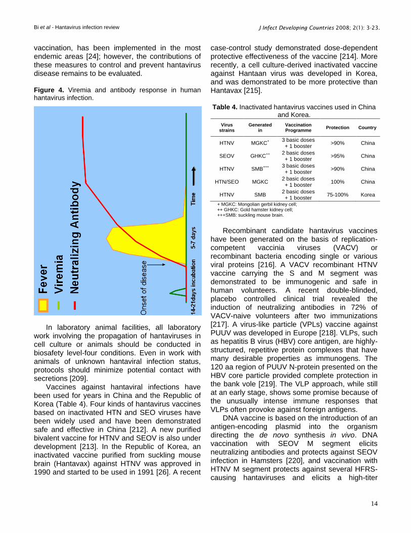
The diagnosis of hantaviral infections is based on clinical and epidemiological information and laboratory tests. However, it is almost impossible in the individual case with moderate to mild clinical symptoms to diagnose hantavirus infection on clinical grounds only. Laboratory diagnosis of acute hantavirus infection has to be primarily based on serology, since the viraemia of human hantavirus infections is short-termed and viral RNA cannot be regularly detected in the blood or urine of patients in hospitals [198], although the virus is readily detectable by rt-PCR during acute illness in research laboratories [199]. However, high levels of virus-specific antibody are usually detected at the onset of hantavirus diseases (Figure 4), and the serological assay is readily accessible to most hospitals.
A wide array of technologies have been used to detect antibodies to hantaviruses, using cultured and/or purified native-virus preparations or recombinant proteins expressed in bacteria, yeast or insect cells. Both indirect fluorescent assay (IFA) and enzyme immunoassay (EIA) are widely used for detection of specific IgM or low-avidity IgG antibodies [200]. IgM detecting methods are important tools for diagnosis of acute infections, especially in endemic areas with a high prevalence of virus-specific IgG due to previous infections. The μ-captured ELISA to detect IgM antibodies, using viral native or recombinant N antigens, should be preferred because it is superior to IFA and solid phase ELISA in terms of sensitivity [201]. Western blot assays can be used, which are generally in agreement with those of the IgM-capture format for acute infections. In addition, the immunochromatographic 5-min IgM-antibody test has been developed for rapid diagnosis [202].
The IFA test remains popular in Europe and Asia, perhaps in part because it is so easily performed, but such tests are intrinsically limited by problems with specificity, especially with inexperienced users. Although ELISA is optimal for a highly specific serological confirmation of hantaviral infections, the antibody responses usually cross-react strongly between different
hantaviruses [203], indicating that ELISA or other serological tests such as IFA or immunoblotting cannot be used for serotyping. The plaque reduction neutralization test (PRNT) is considered to be the gold standard serological test and, it can be used to discriminate between different species of Hantavirus [204]. However, the PRNT test with infectious hantavirus should be done in a biosafety level-three laboratory, which is a serious limitation for many investigators. For post-mortem confirmations, detection of hantavirus antigens can be done by immunohistochemistry testing of formalin-fixed tissues with appropriately specific monoclonal or polyclonal antibodies [205]. Nucleic acid tests and reverse transcriptional-polymerase chain reaction (RT-PCR) can detect hantaviral RNA in fresh-frozen lung tissues, blood clots or nucleated blood cells, and can be used for hantavirus confirmation and genotyping [206]. Recently, one-step assays for detecting hantaviruses based on real-time RT-PCR were developed and proved to be of high levels of sensitivity, specificity and reproducibility [207]. Virus isolation is extremely rare from human sources and thus is not considered an option for diagnostic purpose of hantavirus infections. Control and Prevention The most effective way of controlling hantaviral diseases is to reduce human exposure to infected rodents and their excrement. Monitoring of hantavirus prevalence in rodent populations may give some warning of expected increase in the numbers of human cases [208]. CDC recommendations call for rodent-proofing of homes, reduction of rodent cover around houses, minimization of food available for rodents, trapping in and around dwellings, and the careful disposal of dead rodents [209]. Removal of rodents from a non-rodent-proofed ranch building did not reduce rodent infestation, while the application of simple rodent-proofing measures to dwellings can decrease the frequency and intensity of rodent intrusion, thereby reducing the risk of HPS among rural residents [210,211]. Similarly, workplaces and conditions in agriculture, forestry and military activities should be modified when possible to reduce human-rodent exposure. In China, a comprehensive preventive strategy against hantavirus infection, including health education and promotion, rodent control, surveillance, and
Bi et al - Hantavirus infection review
14
J Infect Developing Countries 2008; 2(1): 3-23.
vaccination, has been implemented in the most endemic areas [24]; however, the contributions of these measures to control and prevent hantavirus disease remains to be evaluated.
Figure 4. Viremia and antibody response in human hantavirus infection.
In laboratory animal facilities, all laboratory work involving the propagation of hantaviruses in cell culture or animals should be conducted in biosafety level-four conditions. Even in work with animals of unknown hantaviral infection status, protocols should minimize potential contact with secretions [209].
Vaccines against hantaviral infections have been used for years in China and the Republic of Korea (Table 4). Four kinds of hantavirus vaccines based on inactivated HTN and SEO viruses have been widely used and have been demonstrated safe and effective in China [212]. A new purified bivalent vaccine for HTNV and SEOV is also under development [213]. In the Republic of Korea, an inactivated vaccine purified from suckling mouse brain (Hantavax) against HTNV was approved in 1990 and started to be used in 1991 [26]. A recent
case-control study demonstrated dose-dependent protective effectiveness of the vaccine [214]. More recently, a cell culture-derived inactivated vaccine against Hantaan virus was developed in Korea, and was demonstrated to be more protective than Hantavax [215]. Table 4. Inactivated hantavirus vaccines used in China
and Korea.
Virus strains
Generated in
Vaccination Programme
Protection Country
HTNV MGKC+
3 basic doses + 1 booster
>90% China
SEOV GHKC++
2 basic doses + 1 booster
>95% China
HTNV SMB+++
3 basic doses + 1 booster
>90% China
HTN/SEO MGKC 2 basic doses + 1 booster
100% China
HTNV SMB 2 basic doses + 1 booster
75-100% Korea
+ MGKC: Mongolian gerbil kidney cell; ++ GHKC: Gold hamster kidney cell; +++SMB: suckling mouse brain.
Recombinant candidate hantavirus vaccines
have been generated on the basis of replication-competent vaccinia viruses (VACV) or recombinant bacteria encoding single or various viral proteins [216]. A VACV recombinant HTNV vaccine carrying the S and M segment was demonstrated to be immunogenic and safe in human volunteers. A recent double-blinded, placebo controlled clinical trial revealed the induction of neutralizing antibodies in 72% of VACV-naive volunteers after two immunizations [217]. A virus-like particle (VPLs) vaccine against PUUV was developed in Europe [218]. VLPs, such as hepatitis B virus (HBV) core antigen, are highly-structured, repetitive protein complexes that have many desirable properties as immunogens. The 120 aa region of PUUV N-protein presented on the HBV core particle provided complete protection in the bank vole [219]. The VLP approach, while still at an early stage, shows some promise because of the unusually intense immune responses that VLPs often provoke against foreign antigens.
DNA vaccine is based on the introduction of an antigen-encoding plasmid into the organism directing the de novo synthesis in vivo. DNA vaccination with SEOV M segment elicits neutralizing antibodies and protects against SEOV infection in Hamsters [220], and vaccination with HTNV M segment protects against several HFRS-causing hantaviruses and elicits a high-titer

Bi et al - Hantavirus infection review
15
J Infect Developing Countries 2008; 2(1): 3-23.
neutralizing antibody response in Rhesus monkey [221]. DNA vaccines against SNV and ANDV have also been shown to be effective in mice models [222,223]. Recently, a bivalent HTN/AND viral DNA vaccine was constructed and has been shown to elicit a potent memory response, and to elicit antibody responses that neutralize viruses causing both HFRS and HPS [224].
The high yield of stable and highly pure nucleocapsid proteins of HTNV, PUUV and DOBV in yeast Saccharomyces cerevisiae represents a useful tool for vaccine development against hantavirus infection [225]. Hantaviral recombinant proteins, which could also be obtained from transgenic plants, were able to elicit specific immune responses [226]. In addition, through the development of reverse genetics for negative-stranded RNA viruses [227], it should be possible in the future to generate attenuated hantavirus vaccine with defined alterations/deletions in pathogenicity-related regions. Concluding Remarks
Over the past few decades, the understanding and recognition of hantaviral infections through the world has greatly improved. The number of recognized viruses continues to increase, as does the spectrum of hantaviral infections. Though newly detected, Hantavirus is an old disease. Environmental changes may affect the geographic distribution, abundance, and dynamics of the rodent carrier, and hence the epidemiology of hantavirus infections. It is generally accepted that hantaviruses are distributed worldwide, but the distribution of specific virus remains to be further investigated. With the development of more rapid and sensitive tests, and increased clinician awareness, human hantaviral infections will presumably be detected in new areas, and new rodent species might be found to carry yet unknown viruses. There is still a long way to go to find an effective treatment for hantavirus infections, and the long-term prognosis of hantaviral infections and the pathogenicity of certain virus species remain to be established. Prevention can be partially achieved by rodent avoidance, but real protection will require a safe and effective multivalent vaccine or a vaccine adapted to local conditions.
Acknowledgements The author is a staff member of the World Health Organization.
The author alone is responsible for the views expresses in this publication and they do not necessarily represent the decisions or the stated policy of the World Health Organization.
References 1. Lee HW, Lee PW, Johnson KM (1978) Isolation of the
etiologic agent of Korean hemorrhagic fever. J Infect Dis 137: 298-308.
2. Lednicky JA (2003) Hantavirus: a short review. Arch Pathol Lab Med 127: 30-35.
3. Vapalahti O, Mustonen J Lundkvist A, Henttonen H, Plyusnin A, Vaheri A (2003) Hantavirus infections in Europe. The Lancet Infect Dis 3: 653-752.
4. Sirotin BZ, Keiser NP (2001) On the history of the study of hemorrhagic fever with renal syndrome in eastern Russia. Nephrol Dial Transplant 16: 1288-90.
5. Hart CA, Bennett M (1999) Hantavirus infections: epidemiology and pathogenesis. Microbes and Infections 1:1229-1237.
6. Nichol ST, Spiropoulou CF, Morzunov S, Rollin PE, Ksiazek TG, Feldmann H, Sanchez A, Childs J, Zaki S, Peters CJ (1993) Genetic identification of a hantavirus associated with an outbreak of acute respiratory illness. Science 262: 914-917.
7. Ye C, Prescott J, Nofchissey R, Goade D, Hjelle B (2004) Neutralizing antibodies and Sin Nombre virus RNA after recovery from hantavirus cardiopulmonary syndrome. Emerg Infect Dis 10: 478-482.
8. Kraus AA, Priemer C, Heider H, Kruger DH, Ulrich R (2005) Inactivation of Hantaan virus-containing samples for subsequent investigations outside biosafety level 3 facilities. Intervirology 48: 255-261.
9. McCaughey C, Hart CA (2000) Hantavirus. J Med Microbiol 49: 587-599.
10. Schmaljohn CS, Dalrymple JM. (1983) Analysis of Hantaan virus RNA: evidence for a new genus of bunyaviridae. Virology. 131:482-91.
11. Easterbrook JD, Zink MC, Klein SL (2007) Regulatory T cells enhance persistence of the zoonotic pathogen Seoul virus in its reservoir host. Proc Natl Acad Sci U S A. 104: 15502-7.
12. Schountz T, Prescott J, Cogswell AC, Oko L, Mirowsky-Garcia K, Galvez AP, Hjelle B (2007) Regulatory T cell-like responses in deer mice persistently infected with Sin Nombre virus. Proc Natl Acad Sci U S A. 104:15496-501.
13. Larson RS, Brown DC, Ye C, Hjelle B (2005) Peptide antagonists that inhibit Sin Nombre virus and hantaan virus entry through the beta3-integrin receptor. J Virol 79: 7319-7326.
14. Song JW, Song KJ, Baek LJ, Frost B, Poncz M, Park K (2005) In vivo characterization of the integrin beta3 as a receptor for Hantaan virus cellular entry. Exp Mol Med. 37:121-7.
15. Rowe RK, Pekosz A (2006) Bidirectional virus secretion and nonciliated cell tropism following Andes virus infection of primary airway epithelial cell cultures. J Virol. 80: 1087-97.
16. Cosgriff TM (1991) Mechanisms of disease in Hantavirus infection: pathophysiology of hemorrhagic fever with renal syndrome. Rev Infect Dis 13: 97-107.

Bi et al - Hantavirus infection review
16
J Infect Developing Countries 2008; 2(1): 3-23.
17. Zaki SR, Greer PW, Coffield LM, Zaki SR, Greer PW, Coffield LM, Goldsmith CS, Nolte KB, Foucar K, Feddersen RM, Zumwalt RE, Miller GL, Khan AS (1995) Hantavirus pulmonary syndrome: pathogenesis of an emerging infectious disease. Am J Pathol 146: 552-579.
18. Terajima M, Vapalahti O, Van Epps HL, Vaheri A, Ennis FA (2004) Immune responses to Puumala virus infection and the pathogenesis of nephropathia epidemica. Microbes Infect 6: 238-245.
19. Borges AA, Campos GM, Moreli ML, Souza RL, Aquino VH, Saggioro FP, Figueiredo LT (2006) Hantavirus cardiopulmonary syndrome: immune response and pathogenesis. Microbes Infect 8: 2324-2330.
20. Mori M, Rothman AL, Kurane I, Montoya JM, Nolte KB, Norman JE, Waite DC, Koster FT, Ennis FA (1999) High levels of cytokine-producing cells in the lung tissues of patients with fatal hantavirus pulmonary syndrome. J Infect Dis 179: 295-302.
21. Makela S, Mustonen J, Ala-Houhala I, Hurme M, Partanen J, Vapalahti O, Vaheri A, Pasternack A. (2002) Human leukocyte antigen-B8-DR3 is a more important risk factor for severe Puumala hantavirus infection than the tumor necrosis factor-alpha(-308) G/A polymorphism. J Infect Dis 86: 843-846.
22. Lee HW (1996). Epidemiology and pathogenesis of haemorrhagic fever with renal syndrome. In: Elliot RM, ,editor, The Bunyaviridae. New York: Plenum Press. 253-267.
23. Wu GH (2003) Progress of epidemiological studies on hemorrhagic fever with renal syndrome in China. Chin J Epidemiol 24: 413-415.
24. Zhang YZ, Xiao DL, Wang Y, Wang HX, Sun L, Tao XX, Qu YG. (2004) The epidemic characteristics and preventive measures of hemorrhagic fever with renal syndrome in China. Chin J Epidemiol 25: 466-469.
25. Lee HW (1989). Hemorrhagic fever with renal syndrome in Korea. Rev Infect Dis 11 Suppl 4: 864-876.
26. Cho HW, Howard CR, Lee HW (2002) Review of an inactivated vaccine against hantaviruses. Intervirology 45: 328-333.
27. Baek LJ, Kariwa H, Lokugamage K Yoshimatsu K, Arikawa J, Takashima I, Kang JI, Moon SS, Chung SY, Kim EJ, Kang HJ, Song KJ, Klein TA, Yanagihara R, Song JW (2006) Soochong virus: an antigenically and genetically distinct Hantavirus isolated from Apodemus peninusulae in Korea. J Med Virol 78: 290-297.
28. Yashina LN, Patrushev NA, Ivanov LI Slonova RA, Mishin VP, Kompanez GG, Zdanovskaya NI, Kuzina II, Safronov PF, Chizhikov VE, Schmaljohn C, Netesov SV (2000) Genetic diversity of Hantavirus associated with hemorrhagic fever with renal syndrome in the Far East of Russia. Virus Res 70: 31-44.
29. Tkachenko E, Dzagurova T, Bashkirtsev V, Bernshtein A, Apekina N, Sedova N, Okulova N, Korotina N, Nabatnikov P, Malkin A, Smirnov A, Morozov V, Yunicheva Y, Klempa B, Kruger D ( 2007 ) Epidemiology of hemorrhagic fever with renal syndrome in european Russia. The 7th International Conference on HFRS, HPS and Hantaviruses, Buenos Aires, Argentina. June 13-15, Abstract book, Page 17.
30. Lokugamage N, Kariwa H, Lokugamage K, Iwasa MA, Hagiya T, Yoshii K, Tachi A, Ando S, Fukushima H, Tsuchiya K, Iwasaki T, Araki K, Yoshimatsu K, Arikawa J,
Mizutani T, Osawa K, Sato H, Takashima I (2004) Epizootiological and epidemiological study of hantavirus infection in Japan. Microbiol Immunol 48 :843-851.
31. Clement J, Maes P, Muthusethupathi M, Nainan G, van Ranst M. (2006) First evidence of fatal hantavirus nephropathy in India, mimicking leptospirosis. Nephrol Dial Transplant 21: 826-827.
32. Groen J, Suharti C, Koraka P van Gorp EC, Sutaryo J, Lundkvist A, Osterhaus AD (2002) Serological evidence of human hantavirus infections in Indonesia. Infection 30: 326-327.
33. Chan KP, Chan YC, Doraisingham S (1996) A severe case of hemorrhagic fever with renal syndrome in Singapore. Southeast Asian J Trop Med Public Health 27: 408-10.
34. Vitarana T, Colombage G, Bandaranayake V, Lee HW.( 1988) Hantavirus diseases in Sri Lanka. The Lancet 2: 1263.
35. Tai PW, Chen LC, Huang CH. Hanta hemorrhagic fever with renal syndrome: a case report and review. J Microbiol Immunol Infect. 2005;38(3):221-223.
36. Suputthamongkol Y, Nitatpattana N, Chayakullkeeree M, Palabodeewat S, Yoksan S, Gonzalez JP (2005) Hantavirus infection in Thailand: first clinical case report. Southeast Asian J Trop Med Public Health 36: 700-703
37. George J, Patnaik M, Bakshi E, Levy Y, Ben-David A, Ahmed A, Peter JB, Shoenfeld Y (1998) Hantavirus seropositive in Israeli patients with renal failure. Viral Immunol 11:103-108.
38. Pacsa AS, Elbishbishi EA, Chaturvedi, Chu KY, Mustafa AS (2002) Hantavirus-specific antibodies in rodents and human living in Kuwait. FEMS Immunol Med Microbiol 33: 139-42.
39. Rollin PE, Nawrocka E, Rodhain F (1986) Serological data on hemorrhagic fever with renal syndrome in Southeast Asia. Bull Soc Pathol Exot Filiales 79: 473-475.
40. Lam SK, Chua KB, Myshrall T, Devi S, Zainal D, Afifi SA, Nerome K, Chu YK, Lee HW (2001) Serological evidence of hantavirus infections in Malaysia. Southeast Asian J Trop Med Public Health 32: 809-813.
41. Quelapio ID, Villa L, Clarin M, Bacosa M, Tupasi TE (2000) Seroepidemiology of Hantavirus in the Philippines. Int J Infect Dis 4: 104-107.
42. Shortridge KF, Lee HW, LeDuc JW, Wong TW, Chau GW, Rosen L (1987) Serological evidence of Hantaan-related viruses in Hong Kong. Trans R Soc Trop Med Hyg 81: 400-2.
43. Reynes JM, Soares JL, Hue T, Bouloy M, Sun S, Kruy SL, Flye Sainte Marie F, Zeller H (2003) Evidence of the presence of Seoul virus in Cambodia. Microbes Infect 5: 769-773.
44. Brummer-Korevakontio M, Vaheri A, Hove T, von Bonsdorff CH, Vuorimies J, Manni T, Penttinen K, N. Oker-Blom, Lähdevirta (1980) Nephropathia epidemica: detection of antigen in bank voles and serologic diagnosis of human infections. J Infect Dis 141: 131-134.
45. Rose AM, Vapalahti O, Lyytikäinen, Nuorti P (2003) Patterns of puumala virus infections in Finland. Euro Surveillance 8: 9-13.
46. Mailles A, Vaillant V, Haeghbaert S, Fradet MR, Capek I, Zeller H (2005). Increase of Hantavirus infections in France, 2003. Med Mal Infect 35: 68-72.

Bi et al - Hantavirus infection review
17
J Infect Developing Countries 2008; 2(1): 3-23.
47. Mailles A, Sin MA, Ducoffre G, Heyman P, Koch J, Zeller H (2005) Larger than usual increase in cases of hantavirus infections in Belgium, France and Germany, June 2005. Euro Surveillance 10: E050721.4.
48. Schneider F and Mossong J (2005) Increased hantavirus infections in Luxembourg, August 2005. Euro Surveillance 10: E050825.1.
49. Brummer-Korvenkontio M, Vapalahti O, Henttonen H, Koskela P, Kuusisto P, Vaheri A (1999) Epidemiological study of nephropathia epidemica in Finland 1989-96. Scand J Infect Dis 31: 427-435.
50. Olsson GE, Dalerum F, Hörnfeldt B, Elgh F, Palo TR, Juto P, Ahlm (2003) Human hantavirus infections, Sweden. Emerg Infect Dis 9: 1395-1401.
51. Heyman P, Vervoort T, Colson P, Chu YK, Avsic-Zupanc T, Lundkvist A (1999) A major outbreak of hantavirus infection in Belgium in 1995 and 1996. Epidemiol Infect 122: 447-453.
52. Ulrich R, Meisel H, Schutt M, Schmidt J, Kunz A, Klempa B, Niedrig M, Pauli G, Kruger DH, Koch J. Prevalence of hantavirus infections in Germany (2004) Bundesgesundheitsblatt Gesundheitsforschung Gesundheitsschutz 47: 661-670.
53. Plyusnina A, Aberle SW, Aberle JH, Plyusnin A (2006) Genetic analysis of Puumala hantavirus strains from Austria. Scand J Infect Dis 38: 512-519.
54. Ferenczi E, Racz G, Szekeres J, Balog K, Toth E, Takacs M, Csire M, Mezey I, Berencsi G, Faludi G (2003) New data for the public health importance of hantaviruses in Hungary. Orv Hetil 144: 467-474.
55. Vasilenko S, Brodvarova I, Topov Y, Shindaro L , Kebedzhiev G (1990) Hemorrhagic fever with renal syndrome in Bulgaria: isolation of hantaviruses and epidemiologic considerations. Arv Virol. 33 Suppl 1: 63-67.
56. Pejcoch M, Kriz B (2003). Hantavirus in the Czech Republic. Emerg Infect Dis 9: 756-7.
57. Golovljova I, Sjolander KB, Lindegren G, Vene S, Vasilenko V, Plyusnin A, Lundkvist A (2002) Hantaviruses in Estonia. J Med Virol 68: 589-598.
58. Maksyoma I (2007) Hemorrhagic fever with renal syndrome in Russia. The 7th International Conference on HFRS, HPS and Hantaviruses, Buenos Aires, Argentina. June 13-15
59. Nurgaleeva RG, Tkachenko EA, Stepanenko AG, Mustafin IM, Kireev SG, Dzagurova TK, Dekonenko AE, Klimchuk LA (1999) An epidemiological analysis of hemorrhagic fever with renal syndrome morbidity in the Republic of Bashkortostan in 1997. Zh Mikrobiol Epidemiol Immunobiol 6: 45-49.
60. Lukac V, Obradovic M, Gligic A, Stojanovic R, Rakovic-Savcic L, Diglisic G, Drndarevic D, Dordevic D (1990) Hemorrhagic fever with renal syndrome in Yugoslavia 1951-1988. Vojnosanit Pregl 47: 242-248.
61. Skataric V, Dimitrijevic J, Jovanovoc D, Marie M, Kovacevic Z, Glogic A. (1998) Clinical features of hemorrhagic fever with renal syndrome in Yugoslavia 1996. The Fourth International Conference of HFRS and hantaviruses, Atlanta, USA, March 5-7
62. Mulić R, Ropac D (2002) Epidemiologic characteristics and military implications of hemorrhagic fever with renal syndrome in Croatia. Croat Med J. 43:581-6.
63. Mulic R, Ropac D, Gizdic Z, Sikic N (2003) What is new in the epidemiologic characteristics of hemorrhagic fever with renal syndrome in Croatia? Acta Med Croatica 57:399-405.
64. Papa A, Bojovic B, Antoniadis A (2006) Hantaviruses in Serbia and Montenegro. Emerg Infect Dis 12: 1015-1018
65. Papa A, Antoniadis A (2001) Hantavirus infections in Greece--an update. Eur J Epidemiol 17:189-94.
66. Papa A, Nemirov K, Henttonen H, Niemimaa J, Antoniadis A, Vaheri A, Plyusnin A, Vapalahti O (2001) Isolation of Dobrava virus from Apodemus flavicollis in Greece. J Clin Microbiol 39: 2291-2293.
67. Eltari E, Nuti M, Hasko I, Gina A (1978) Hemorrhagic fever with renal syndrome in a case in northern Albania. Lancet 2:1211.
68. Nemirov K, Andersen HK, Leirs H, Henttonen H, Vaheri A, Lundkvist A, Plyusnin A (2004) Saaremaa hantavirus in Denmark. J Clin Virol 30: 254-257.
69. Knap JP, Brzostek T, Raczka A, Burzynski W, Litarska U (2006) A case of haemorrhagic fever with renal syndrome. Pol Merkur Lekarski 21: 474-476.
70. Manasia M, Olinic O, Zagreanu I, Serban A (1977) Hemorrhagic fever with renal syndrome. Report of 11 observations. Int Urol Nephrol 9: 177-84.
71. Sibold C, Ulrich R, Labuda M, Lundkvist A, Martens H, Schutt M, Gerke P, Leitmeyer K, Meisel H, Kruger DH (2001) Dobrava hantavirus causes hemorrhagic fever with renal syndrome in central Europe and is carried by two different Apodemus mice species. J Med Virol 63: 158-167.
72. Schultze D, Fierz W, Matter H, Bankoul S, Niedrig M, Schmiedl A (2007) Cross-sectional survey on hantavirus seroprevalence in Canton St. Gallen, Switzerland. Swiss Med Wkly 137: 21-26.
73. Dillingh SJ, Jira P, Morroy G, Wolters B, Beutler J, Schneeberger PM (2006) Two patients with Hantavirus infection in The Netherlands; substantial increase in incidence in neighbouring countries Ned Tijdschr Geneeskd 10; 150: 1303-1306.
74. Pether JV, Lloyd G (1993) The clinical spectrum of human hantavirus infection in Somerset, UK. Epidemiol Infect 111: 171-5.
75. Kallio-Kokko H, Laakkonen J, Rizzoli A, Tagliapietra V, Cattadori I, Perkins SE, Hudson PJ, Cristofolini A, Versini W, Vapalahti O, Vaheri A, Henttonen H (2006) Hantavirus and arenavirus antibody prevalence in rodents and humans in Trentino, Northern Italy. Epidemiol Infect 134: 830-836.
76. Lundkvist A, Lindegren G, Brus Sjolander K, Mavtchoutko V, Vene S, Plyusnin A, Kalnina V (2002) Hantavirus infections in Latvia. Eur J Clin Microbiol Infect Dis 21:626-629.
77. Sandmann S, Meisel H, Razanskiene A, Wolbert A, Pohl B, Krüger DH, Sasnauskas K, Ulrich R (2005) Detection of human hantavirus infections in Lithuania. Infection 33: 66-72.
78. Mikhailenko AG, Tkachenko EA, Smirnov IIa, Malkin AE, Chebotar' IF (1994) Results of serologic screening for hemorrhagic fever with kidney syndrome in Moldova. Vopr Virusol 39: 260-262.
79. Lledo L, Klingstrom J, Gegundez MI, Plyusnina A, Vapalahti O, Saz JV, Beltran M, Sjolander KB, Vaheri A, Plyusnin A, Lundkvist A (2003) Hantavirus infections in

Bi et al - Hantavirus infection review
18
J Infect Developing Countries 2008; 2(1): 3-23.
Spain: analysis of sera from the general population and from patients with pneumonia, renal disease and hepatitis. J Clin Virol 27: 296-307.
80. Laakkonen J, Kallio-Kokko H, Oktem MA, Blasdell K, Plyusnina A, Niemimaa J, Karataş A, Plyusnin A, Vaheri A, Henttonen H (2006) Serological survey for viral pathogens in Turkish rodents. J Wildl Dis 42: 672-676.
81. Padula PJ, Colavecchia SB, Martinez VP, Gonzalez Della Valle MO, Edelstein A, Miguel SD, Russi J, Riquelme JM, Colucci N, Almiron M, Rabinovich RD (2000) Genetic diversity, distribution, and serological features of hantavirus infection in five countries in South America. J Clin Microbiol 38: 3029-3035.
82. Johnson AM, de Souza LT, Ferreira IB, Pereira LE, Ksiazek TG, Rollin PE, Peters CJ, Nichol ST (1999) Genetic investigation of novel hantaviruses causing fatal HPS in Brazil. J Med Virol 59: 527-535.
83. Weir E. Hantavirus: 'tis the season. CMAJ 2005; 17:147. 84. Lopez N, Padula P, Rossi C, Lazaro ME, Franze-
Fernandez MT (1996) Genetic identification of a new hantavirus causing severe pulmonary syndrome in Argentina. Virology 220: 223-226.
85. Levis S, Morzunov SP, Rowe JE, Enria D, Pini N, Calderon G, Sabattini M, St Jeor SC (1998) Genetic diversity and epidemiology of hantaviruses in Argentina. I Infect Dis 177: 529-538.
86. Capria SG, Elder M, Cacace ML, Cortes J, Bruno MR, Farace MI, Padula PJ (2007) Hantavirus pulmonary syndrome in Argentina, 1995-2006. The 7th International Conference on HFRS, HPS and Hantaviruses, Buenos Aires, Argentina. June 13-15, Abstract book, Page 22
87. Carroll DS, Mills JN, Montgomery JM, Bausch DG, Blair PJ, Burans JP, Felices V, Gianella A, Iihoshi N, Nichol ST, Olson JG, Rogers DS, Salazar M, Ksiazek TG (2005) Hantavirus pulmonary syndrome in Central Bolivia: relationships between reservoir hosts, habitats, and viral genotypes. Am J Trop Med Hyg 72: 42-46.
88. Moreli ML, Sousa RL, Figueiredo LT (2004). Detection of Brazilian hantavirus by reverse transcription polymerase chain reaction amplification of N gene in patients with hantavirus cardiopulmonary syndrome. Mem Inst Oswaldo Cruz 99: 633-638.
89. Da Silva MV (2007) Brazilian hantavirus: clinical manifestions and case-fatality rate. The 7th International Conference on HFRS, HPS and Hantaviruses, Buenos Aires, Argentina. June 13-15, Abstract book, Page 43).
90. Espinoza R, Vial P, Noriega LM, Johnson A, Nichol ST, Rollin PE, Wells R, Zaki S, Reynolds E, Ksiazek TG (1998) Hantavirus pulmonary syndrome in a Chilean patient with recent travel in Bolivia. Emerg Infect Dis 4: 93-95.
91. Weissenbacher MC, Cura E, Segura EL, Hortal M, Baek LJ, Chu YK, Lee HW (1996). Serological evidence of human Hantavirus infection in Argentina, Bolivia and Uruguay. Medicina (B Aires) 56: 17-22.
92. Delfraro A, Tome L, Elia GD, Glara M, Rego N, Achaval F, Arbiza J (2007) Hantavirus reservoir hosts and genotypies circulating in uruguay. The 7th International Conference on HFRS, HPS and Hantaviruses, Buenos Aires, Argentina. June 13-15, Abstract book, Page 118.
93. Armien B, Armien A, Pascale JM, Avila M, Gonzalez P, Munoz C, Mendoza O, Guevara Y, Koster F, Salazer J, Hjelle B, Yates T, Mertz G, Gracia F, Glass G (2007)
Human and rodent epidemiology of hantavirus in Panama 2000-2006. The 7th International Conference on HFRS, HPS and Hantaviruses, Buenos Aires, Argentina. June 13-15, Abstract book, Page 97).
94. Williams RJ, Bryan RT, Mills JN, Palma RE, Vera I, De Velasquez F, Baez E, Schmidt WE, Figueroa RE, Peters CJ, Zaki SR, Khan AS, Ksiazek TG (1997) An outbreak of hantavirus pulmonary syndrome in western Paraguay. Am J Trop Med Hyg 57: 274-282.
95. Ferrer JF, Galligan D, Esteban E, Rey V, Murua A, Gutierrez S, Gonzalez L, Thakuri M, Feldman L, Poiesz B, Jonsson C (2003) Hantavirus infection in people inhabiting a highly endemic region of the Gran Chaco territory, Paraguay: association with Trypanosoma cruzi infection, epidemiological features and haematological characteristics. Ann Trop Med Parasitol 97:269-280.
96. Rivas YJ, Moros Z, Moron D, Uzcátegui MG, Durán Z, Pujol FH, Liprandi F, Ludert JE (2003) The seroprevalences of anti-hantavirus IgG antibodies among selected Venezuelan populations. Ann Trop Med Parasitol 97: 61-67.
97. Mattar S, Parra M (2004) Serologic evidence of hantavirus infection in humans, Colombia. Emerg Infect Dis 10: 2263-2264.
98. Hjelle B, Anderson B, Torrez-Martinez N, Song W, Gannon WL, Yates TL (1995) Prevalence and geographic genetic variation of hantaviruses of New World harvest mice (Reithrodontomys): identification of a divergent genotype from a Costa Rican Reithrodontomys mexicanus. Virology 207: 452-459.
99. Suzan G, Ceballos G, Mills J, Ksiazek TG, Yates T (2001) Serologic evidence of hantavirus infection in sigmodontine rodents in Mexico. J Wildl Dis 37:391-393.
100. Powers AM, Mercer DR, Watts DM, Guzman H, Fulhorst CF, Popov VL, Tesh RB (1999) Isolation and genetic characterization of a hantavirus (Bunyaviridae: Hantavirus) from a rodent, Oligoryzomys microtis (Muridae), collected in northeastern Peru. Am J Trop Med Hyg 61: 92-98.
101. Verity R, Prasad E, Grimsrud K, Artsob H, Drebot M, Miedzinski L, Preiksaitis J (2000) Hantavirus pulmonary syndrome in northern Alberta, Canada: clinical and laboratory findings for 19 cases. Clin Infect Dis 31: 942-946.
102. Wilson C, Hjelle B, Jenison S (1994) Probable hantavirus pulmonary syndrome that occurred in New Mexico in 1975. Ann Intern Med 120: 813.
103. Engelthaler D, Levy C, Ettestad P, Kruger K, Schuermann J, Leslie M (2006) Hantavirus Pulmonary Syndrome --- Five States, 2006. MMWR 55:627-629.
104. Diglisic G, Rossi CA, Doti A, Walshe DK. (1999) Seroprevalence study of Hantavirus infection in the community based population. Md Med J 48: 303-306.
105. LeDuc JW, Smith GA, Pinheiro FP, Vasconcelos PF, Rosa ES, Maiztegui JI. (1985) Isolation of a Hantaan-related virus from Brazilian rats and serologic evidence of its widespread distribution in South America. Am J Trop Med Hyg 34: 810-815.
106. Groen J, Koraka P, Edwards CN, Branch SL, Douglas KO, Osterhaus AD, Levett PN (2002) Serological evidence of hantavirus in humans and rodents in Barbados. J Infect 45: 109-110.

Bi et al - Hantavirus infection review
19
J Infect Developing Countries 2008; 2(1): 3-23.
107. Gonzalez JP, McCormick JB, Baudon D, Gautun JP, Meunier DY, Dournon E, Georges AJ. (1984) Serological evidence for Hantaan-related virus in Africa. Lancet 2: 1036-1037.
108. Saluzzo JF, Digoutte JP, Adam F, Bauer SP, McCormick JB (1985) Serological evidence for Hantaan-related virus infection in rodents and man in Senegal. Trans R Soc Trop Med Hyg 79: 874-875
109. Tomori O, Morikawa S, Matsuura Y, Kitamura T (1986) Antibody to Japanese strains of haemorrhagic fever with renal syndrome (HFRS) virus in Nigerian sera. Trans R Soc Trop Med Hyg; 80: 1008-1009.
110. Rodier G, Soliman AK, Bouloumie J, Kremer D. (1993)Presence of antibodies to hantavirus in rat plus human populations of Djibouti. Trans R Soc Trop Med Hyg 87: 160-161.
111. Botros BA, Sobh M, Wierzba T, Arthur RR, Mohareb EW, Frenck R, El Refaie A, Mahmoud I, Chapman GD, Graham RR (2004) Prevalence of hantavirus antibody in patients with chronic renal disease in Egypt. Trans R Soc Trop Med Hyg 98: 331-336.
112. Klempa B, Fichet-Calvet E, Lecompte E, Auste B, Aniskin V, Meisel H, Denys C, Koivogui L, ter Meulen J, Kruger DH. (2006) Hantavirus in African Wood Mouse, Guinea. Emerg Infect Dis 12: 838-840.
113. Coulaud X, Chouaib E, Georges AJ, Rollin P, Gonzalez JP. (1987) First human case of haemorrhagic fever with renal syndrome in the Central African Republic. Trans R Soc Trop Med Hyg 81: 686.
114. Sironen T, Vaheri A, Plyusnin A (2005) Phylogenetic evidence for the distinction of Saaremaa and Dobrava hantaviruses. Virol J 2: 90-95.
115. Glass GE, Watson AJ, LeDuc JW, Childs JE. (1994) Domestic cases of hemorrhagic fever with renal syndrome in the United States. Nephron 68: 48-51.
116. Heyman P, Plyusnina A, Berny P, Cochez C, Artois M, Zizi M, Pirnay JP, Plyusnin A. (2004) Seoul hantavirus in Europe: first demonstration of the virus genome in wild Rattus norvegicus captured in France. Eur J Clin Microbiol Infect Dis 23: 711-717.
117. Yashina L, Mishin V, Zdanovskaya N, Schmaljohn C, Ivanov L. (2001) A newly discovered variant of a hantavirus in Apodemus peninsulae, far Eastern Russia. Emerg Infect Dis 7: 912-913.
118. Lokugamage K, Kariwa H, Lokugamage N, Miyamoto H, Iwasa M, Hagiya T, Araki K, Tachi A, Mizutani T, Yoshimatsu K, Arikawa J, Takashima I. (2004) Genetic and antigenic characterization of the Amur virus associated with hemorrhagic fever with renal syndrome. Virus Res 101: 127-134.
119. McIntyre NE, Chu YK, Owen RD, Abuzeineh A, De la Sancha N, Dick CW, Holsomback T, Nisbett RA, Jonsson C (2005) A Longitudinal study of Bayou virus, hosts, and habitat. Am J Trop Med Hyg 73: 1043-1049.
120. Padula P, Martinez VP, Bellomo C, Maidana S, San Juan J, Tagliaferri P, Bargardi S, Vazquez C, Colucci N, Estévez J, Almiron M (2007) Pathogenic hantaviruses, northeastern Argentina and eastern Paraguay. Emerg Infect Dis. 13:1211-4.
121. Vincent MJ, Quiroz E, Gracia F, Sanchez AJ, Ksiazek TG, Kitsutani PT, Ruedas LA, Tinnin DS, Caceres L, Garcia A, Rollin PE, Mills JN, Peters CJ, Nichol ST (2000) Hantavirus pulmonary syndrome in Panama:
identification of novel hantaviruses and their likely reservoirs. Virology 277: 14-19.
122. Bowen MD, Gelbmann W, Ksiazek TG, Nichol ST, Nowotny N (1997) Puumala virus and two genetic variants of Tula virus are present in Austrian rodents. J Med Virol 53: 174-181.
123. Heyman P, Klingstrom J, de Jaegere F, Klingstrom J, Vandenvelde C, Lundkvist A, Rozenfeld F, Zizi M. (2002)Tula hantavirus in Belgium. Epidemiol Infect 128: 251-256.
124. Scharninghausen JJ, Pfeffer M, Meyer H, (2002) Genetic evidence for tula virus in Microtus arvalis and Microtus agrestis populations in Croatia. Vector Borne Zoonotic Dis 2: 19-27.
125. Song JW, Baek LJ, Song KJ, Skrok A, Markowski J, Bratosiewicz-Wasik J, Kordek R, Liberski PP, Yanagihara R. (2004) Characterization of Tula virus from common voles (microtus arvalis) in Poland: evidence for geographic-specific phylogenetic clustering. Virus Genes 29: 239-247.
126. Plyusnin A, Vapalahti O, Lankinen H, Lehväslaiho H, Apekina N, Myasnikov Y, Kallio-Kokko H, Henttonen H, Lundkvist A, Brummer-Korvenkontio M (1994) Tula virus: a newly detected hantavirus carried by European common voles. J Virol 68: 7833-7839.
127. Song JW, Gligic A, Yanagihara R (2002) Identification of Tula hantavirus in Pitymys subterraneus captured in the Cacak region of Serbia-Yugoslavia. Int J Infect Dis 6: 31-36.
128. Sibold C, Meisel H, Kruger DH, Schulz A, Cifire F, Ulrich R, Kozuch O, Labuda M, Kruger DH (1999) Recombination in Tula hantavirus evolution: analysis of genetic lineages from Slovakia. J Virol 73: 667-675.
129. Vapalahti O, Lundkvist A, Kukkonen SK, Cheng Y, Gilljam M, Kanerva M, Manni T, Pejcoch M, Niemimaa J, Kaikusalo A, Henttonen H, Vaheri A, Plyusnin A. (1996) Isolation and characterization of Tula virus, a distinct serotype in the genus Hantavirus, family Bunyaviridae. J Gen Virol 77: 3063-3067.
130. Schultze D, Lundkvist A, Blauenstein U, Heyman P. (2002) Tula virus infection associated with fever and exanthema after a wild rodent bite. Eur J Clin Microbiol Infect Dis 21: 304-306.
131. Klempa B, Meisel H, Rath S, Bartel J, Ulrich R, Kruger DH. (2003) Occurrence of renal and pulmonary syndrome in a region of northeast Germany where Tula hantavirus circulates. J Clin Microbiol 41: 4894-4897.
132. Clement J, Heyman P, McKenna P, Colson P, Avsic-Zupanc T. (1997) The hantavirue of Europe: from the bedside to the beach. Emerg Infect Dis 3: 205-211.
133. McMaughey C and Hart CA. Hantavirus (2000) J Med Microbiol 49: 587-599.
134. Carey, D. E., R. Reuben, K. N. Panicker, R. E. Shope, and R. M. Myers (1971) Thottapalayam virus: a presumptive arbovirus isolated from a shrew in India. Indian J. Med. Res. 59:1758-1760.
135. Okumura M, Yoshimatsu K, Kumperasart S, Nakamura I, Ogino M, Taruishi M, Sungdee A, Pattamadilok S, Ibrahim IN, Erlina S, Agui T, Yanagihara R, Arikawa J (2007) Development of serological assays for Thottapalayam virus, an insectivore-borne Hantavirus. Clin Vaccine Immunol. 14:173-81.

Bi et al - Hantavirus infection review
20
J Infect Developing Countries 2008; 2(1): 3-23.
136. Elwell MR, Ward GS, Tingpalapong M, LeDuc JW, 1985. Serologic evidence of Hantaan-like virus in rodents and man in Thailand. Southeast Asian J Trop Med Public Health 16: 349– 354.
137. Pattamadilok S, Lee BH, Kumperasart S, Yoshimatsu K, Okumura M, Nakamura I, Araki K, Khoprasert Y, Dangsupa P, Panlar P, Jandrig B, Krüger DH, Klempa B, Jäkel T, Schmidt J, Ulrich R, Kariwa H, Arikawa J (2006) Geographical distribution of hantaviruses in Thailand and potential human health significance of Thailand virus. Am J Trop Med Hyg. 75:994-1002.
138. Klempa B, Fichet-Calvet E, Lecompte E, Auste B, Aniskin V, Meisel H, Barrière P, Koivogui L, ter Meulen J, Krüger DH (2007) Novel Hantavirus Sequences in Shrew, Guinea. Emerg Infect Dis. 13:520-2.
139. Song JW, Gu SH, Bennett SN, Arai S, Puorger M, Hilbe M, Yanagihara R (2007) Seewis virus, a genetically distinct hantavirus in the Eurasian common shrew (Sorex araneus). Virol J. 4(1):114.
140. Arai S, Song JW, Sumibcay L, Bennett SN, Nerurkar VR, Parmenter C, Cook JA, Yates TL, and Yanagihara R (2007) Hantavirus in Northern Short-tailed Shrew, United States. Emerg Infect Dis. 13:1420-23.
141. Song JW, Kang HJ, Song KJ, Truong TT, Bennett SN, Arai S, Truong NU, and Yanagihara R (2007) Newfound Hantavirus in Chinese Mole Shrew, Vietnam. Emerg Infect Dis. 13:1784-87.
142. Tang YW, Xu ZY, Zhu ZY Tsai TF (1985) Isolation of haemorrhagic fever with renal syndrome virus from Suncus murinus, an insectivore. The Lancet 1: 513-514
143. Jung YT, Kim GR (1995) Genomic characterization of M and S RNA segments of hantaviruses isolated from bats. Acta Virol 39: 231-233.
144. Luo ZZ, Li SQ, Peng DS, Wang YY, He H, Li YZ, Zhou N, Ni DS, Li YX, Xue ZY (1985) Isolation and characterization of epidemic haemorrhagic fever virus from domestic cats. Chin J Microbiol Immunol 5: 79-81.
145. Slonova RA, Tkachenko EA, Kushnarev EL, Dzagurova TK, Astakova TI (1992) Hantavirus isolation from birds. Acta Virol 36: 493.
146. Malecki TM, Jillson GP, Thilsted JP, Elrod J, Torrez-Martinez N, Hjelle B (1998) Serologic survey for hantavirus infection in domestic animals and coyotes from New Mexico and northeastern Arizona. J Am Vet Med Assoc 212: 970-973.
147. Yang Z, Liu Y, Peng Z (1998) Epidemiologic and experimental studies on epidemic haemorrhagic fever virus in pigs. Chin J Epidemol 19: 218-220.
148. Glass G, Childs J, Korch G, LeDuc JW (1988) Association of intraspecific wounding with hantaviral infection in wild rats (Rattus norvegicus). Epidemiol Infect 101: 459-472.
149. Hinson ER, Shone SM, Zink MC, Glass GE, Klein SL (2004) Wounding: the primary mode of Seoul virus transmission among male Norway rats. Am J Trop Med Hyg 70: 310-317.
150. Zhang Y, Zhu J, Deng XZ, Wu GH, Wang JJ, Zhang JJ, Xing AH, Wu JW (2003) Detection of hantaan virus from gamasid mite and chigger mite by molecular biological methods. Zhonghua Shi Yan He Lin Chuang Bing Du Xue Za Zh 17: 107-111.
151. Houck MA, Qin H, Roberts HR (2001) Hantavirus transmission: potential role of ectoparasites. Vector Borne Zoonotic Dis 1: 75-79.
152. Overturf GD (2005) Clinical sin nombre hantaviral infections in children. Pediatr Infect Dis J 24: 373-374
153. Howard MJ, Doyle TJ, Koster FT, Zaki SR, Khan AS, Petersen EA, Peters CJ, Bryan RT (1999) Hantavirus pulmonary syndrome in pregnancy. Clin Infect Dis 29: 1538-1544.
154. Botten J, Mirowsky K, Ye C, Gottlieb K, Saavedra M, Ponce L, Hjelle B (2002) Shedding and intracage transmission of Sin Nombre hantavirus in the deer mouse (Peromyscus maniculatus) model. J Virol 76: 7587-7594
155. Hutchinson KL, Rollin PE, Shieh WJ, Zaki S, Greer PW, Peters CJ (2000) Transmission of Black Creek Canal virus between cotton rats. J Med Virol 60: 70-76.
156. Ma RM, Xiao H, Jing XT, Lao TT (2003) Hemorrhagic fever with renal syndrome presenting with intrauterine fetal death. A case report. J Reprod Med 48: 661-664.
157. Peng H, Tang S and Qi X (2002) Clinical study on intrauterine hemorrhagic fever with renal syndrome virus infection. Chin J Exp Clin Virol. 16:281-2.
158. Kallio ER, Klingstrom J, Gustafsson E, Manni T, Vaheri A, Henttonen H, Vapalahti O, Lundkvist A (2006) Prolonged survival of Puumala hantavirus outside the host: evidence for indirect transmission via the environment. J Gen Virol 87: 2127-2134.
159. Enria D, Padula P, Segura EL, Pini N, Edelstein A, Posse CR, Weissenbacher MC (1996) Hantavirus pulmonary syndrome in Argentina. Possibility of person to person transmission. Medicina (B Aires) 56: 709-711.
160. Chaparro J, Vega J, Terry W, Vera JL, Barra B, Meyer R, Peters CJ, Khan AS, Ksiazek TG (1998) Assessment of person-to-person transmission of hantavirus pulmonary syndrome in a Chilean hospital setting. J Hosp Infect 40: 281-285.
161. Vitek CR, Breiman RF, Ksiazek TG, Rollin PE, McLaughlin JC, Umland ET, Nolte KB, Loera A, Sewell CM, Peters CJ (1996) Evidence against person-to-person transmission of hantavirus to health care workers. Clin Infect Dis 22: 824-826.
162. Pinna DM, Martinez VP, Bellomo CM, López C, Padula P. (2004) New epidemiologic and molecular evidence of person to person transmission of hantavirus Andes Sout. Medicina (B Aires) 64: 43-46.
163. Martinez VP, Bellomo C, San Juan J, Pinna D, Forlenza R, Elder M, Padula PJ (2005) Person-to-person transmission of Andes virus. Emerg Infect Dis 11: 1848-1853.
164. Padula PJ, Edelstein A, Miguel SD, Lopez NM, Rossi CM, Rabinovich RD (1998) Hantavirus pulmonary syndrome outbreak in Argentina: molecular evidence of person to person transmission of Andes virus. Viology 241: 323-330.
165. Lázaro ME, Cantoni GE, Calanni LM, Resa AJ, Herrero ER, Iacono MA, Enria DA, González Cappa SM (2007) Clusters of hantavirus infection, southern Argentina. Emerg Infect Dis 13: 104-110.
166. Ferres M, Vial P, Marco C, Yanez L, Godoy P, Castillo C, Hjelle B, Delgado I, Lee SJ, Mertz GJ; Andes Virus Household Contacts Study Group (2007) Prospective evaluation of household contacts of persons with

Bi et al - Hantavirus infection review
21
J Infect Developing Countries 2008; 2(1): 3-23.
hantavirus cardiopulmonary syndrome in Chile. J Infect Dis. 195:1563-71.
167. Ruo SL, Li YL, Tong Z, Ma QR, Liu ZL, Tang YW, Ye KL, McCormick JB, Fisher-Hoch SP, Xu ZY (1994) Retrospective and prospective studies of hemorrhagic fever with renal syndrome in rural China. J Infect Dis 170: 527-534.
168. Vitek CR, Ksiazek TG, Peters CJ, Breiman RF (1996) Evidence against infection with hantaviruses among forest and park workers in the southwestern United States. Clin Infect Dis. 23:283-5.
169. Zeitz PS, Graber JM, Voorhees RA, Kioski C, Shands LA, Ksiazek TG, Jenison S, Khabbaz RF (1997) Assessment of occupational risk for hantavirus infection in Arizona and New Mexico. J Occup Environ Med. 39:463-7.
170. Fulhorst CF, Milazzo ML, Armstrong LR, Childs JE, Rollin PE, Khabbaz R, Peters CJ, Ksiazek TG. (2007) Hantavirus and arenavirus antibodies in persons with occupational rodent exposure. Emerg Infect Dis. 13:532-538.
171. Ferrer JF, Jonsson CB, Esteban E, Galligan D, Basombrio MA, Peralta-Ramos M, Bharadwaj M, Torrez-Martinez N, Callahan J, Segovia A, Hjelle B. (1998) High prevalence of hantavirus infection in Indian communities of the Paraguayan and Argentinean Gran Chaco. Am J Trop Med Hyg. 59:438-44.
172. Campos GM, Moro de Sousa RL, Badra SJ, Pane C, Gomes UA, Figueiredo LT (2003) Serological survey of hantavirus in Jardinopolis County, Brazil. J Med Virol. 71: 417-22.
173. Armien B, Pascale JM, Bayard V, Munoz C, Mosca I, Guerrero G, Armien A, Quiroz E, Castillo Z, Zaldivar Y, Gracia F, Hjelle B, Koster F. (2004) High seroprevalence of hantavirus infection on the Azuero peninsula of Panama. Am J Trop Med Hyg. 70:682-7.
174. Zeitz PS, Graber JM, Voorhees RA, Kioski C, Shands LA, Ksiazek TG, Jenison S, Khabbaz RF (1997) Assessment of occupational risk for hantavirus infection in Arizona and New Mexico. J Occup Environ Med 39: 463-467.
175. Suzán G, Giermakowski JT, Marcé E, Suzán-Azpiri H, Armién B, Yates TL. (2006) Modeling hantavirus reservoir species dominance in high seroprevalence areas on the Azuero Peninsula of Panama. Am J Trop Med Hyg. 74:1103-10.
176. Schutt M, Meisel H, Kruger DH, Ulrich R, Dalhoff K, Dodt C (2004) Life-threatening Dobrava hantavirus infection with unusually extended pulmonary involvement. Clin Nephrol 62: 54-57.
177. Mathes RW, Page WF, Crawford HM, McBean AM, Miller RN (2005) Long-term sequelae of hemorrhagic fever with renal syndrome attributable to Hantaan virus in Korean War veterans. Mil Med 170: 315-319.
178. Settergren B (2000) Clinical aspects of nephropathia epidemica (Puumala virus infection) in Europe: a review. Scand J Infect Dis 32: 125-132.
179. Krause R, Aberle S, Haberl R, Daxbock F, Wenisch C (2003) Puumala virus infection with acute disseminated encephalomyelitis and multiorgan failure. Emerg Infect Dis 9: 603-605.
180. Hautala T, Sironen T, Vapalahti O, Pääkkö E, Särkioja T, Salmela PI, Vaheri A, Plyusnin A, Kauma H (2002) Hypophyseal hemorrhage and panhypopituitarism during Puumala Virus Infection: Magnetic Resonance Imaging
and detection of viral antigen in the hypophysis. Clin Infect Dis 35: 96-101.
181. Nguyen AT, Penalba C, Bernadac P, Jaafar S, Kessler M, Canton P, Hoen B (2001) Respiratory manifestations of hemorrhagic fever with renal syndrome. Retrospective study of 129 cases in Champagne-Ardenne and Lorraine. Presse Med 30: 55-58.
182. Seitsonen E, Hynninen M, Kolho E, Kallio-Kokko H, Pettilä V (2006) Corticosteroids combined with continuous veno-venous hemodiafiltration for treatment of hantavirus pulmonary syndrome caused by Puumala virus infection. Eur J Clin Microbiol Infect Dis 25: 261-266.
183. Novo R, Gagnadoux MF, Le Guenno Y, Gubler MC, Niaudet P, Guyot C, Broyer M (1999) Chronic renal failure after Puumala virus infection. Pediatr Nephrol 13: 934-935.
184. Hallin GW, Simpson SQ, Crowell RE, James DS, Koster FT, Mertz GJ, Levy H. (1996) Cardiopulmonary manifestations of hantavirus pulmonary syndrome. Crit Care Med 24: 252-258.
185. Raboni SM, Rubio G, DE Borba L, Zeferino A, Skraba I, Goldenberg S, Dos Santos CN (2005) Clinical survey of hantavirus in southern Brazil and the development of specific molecular diagnosis tools. Am J Trop Med Hyg 72: 800-804.
186. Hjelle B, Goade D, Torrez-Martinez N, Lang-Williams M, Kim J, Harris RL, Rawlings JA (1996) Hantavirus pulmonary syndrome, renal insufficiency, and myositis associated with infection by Bayou hantavirus. Clin Infect Dis 23: 495-500.
187. Hopkins RO, Larson-Lohr V, Weaver LK, Bigler ED (1998) Neuropsychological impairments following hantavirus pulmonary syndrome. J Int Neuropsychol Soc 4: 190-196.
188. Kitsutani PT, Denton RW, Fritz CL, Murray RA, Todd RL, Pape WJ, Wyatt Frampton J, Young JC, Khan AS, Peters CJ, Ksiazek TG (1999) Acute Sin Nombre hantavirus infection without pulmonary syndrome, United States. Emerg Infect Dis 5: 701-705.
189. Crowley MR, Katz RW, Kessler R, Simpson SQ, Levy H, Hallin GW, Cappon J, Krahling JB, Wernly J (1998) Successful treatment of adults with severe Hantavirus pulmonary syndrome with extracorporeal membrane oxygenation. Crit Care Med 26: 409-414.
190. Custer DM, Thompson E, Schmaljohn CS, Ksiazek TG, Hooper JW (2003) Active and passive vaccination against hantavirus pulmonary syndrome with Andes virus M genome segment-based DNA vaccine. J Virol. 77:9894-905.
191. Hooper JW, Ferro AM, Wahl-Jensen V. (2007) Immune Serum Produced by DNA Vaccination Protects Hamsters Against Lethal Respiratory Challenge with Andes Virus. J Virol. Nov 21 [Epub ahead of print].
192. Enria D, Maiztegui JI (1994) Antiviral treatment of Argentine hemorrhagic fever. Antiviral Res 23: 23-31.
193. Severson WE, Schmaljohn CS, Javadian A, Jonsson CB (2003) Ribavirin causes error catastrophe during Hantaan virus replication. J Virol 77: 481-488.
194. Huggins JW, Hsiang CM, Cosgriff TM, Guang MY, Smith JI, Wu ZO, LeDuc JW, Zheng ZM, Meegan JM, Wang QN (1991) Prospective, double-blind, concurrent, placebo-controlled clinical trial of intravenous Ribavirin therapy of

Bi et al - Hantavirus infection review
22
J Infect Developing Countries 2008; 2(1): 3-23.
hemorrhagic fever with renal syndrome. J Infect Dis 164: 1119-1127.
195. Chapman LE, Mertz GJ, Peters CJ, Jolson HM, Khan AS, Ksiazek TG, Koster FT, Baum KF, Rollin PE, Pavia AT, Holman RC, Christenson JC, Rubin PJ, Behrman RE, Bell LJ, Simpson GL, Sadek RF. (1999) Intravenous ribavirin for hantavirus pulmonary syndrome: safety and tolerance during 1 year of open-label experience. Antivir Ther 4: 211-219.
196. Mertz GJ, Miedzinski L, Goade D, Pavia AT, Hjelle B, Hansbarger CO, Levy H, Koster FT, Baum K, Lindemulder A, Wang W, Riser L, Fernandez H, Whitley RJ, Collaborative Antiviral Study Group (2004) Placebo-controlled, double-blind trial of intravenous ribavirin for the treatment of hantavirus cardiopulmonary syndrome in North America. Clin Infect Dis 39: 1307-1313.
197. Loginova SIa, Koval'chuk AV, Borisevich SV, Kopylova NK, Pashchenko IuI, Khamitov RA, Maksimov VA, Shuster AM (2005) Antiviral effectiveness of the combined use of amixine and virasole in experimental hemorrhagic fever with renal syndrome in sucking albino mice. Vopr Virusol 50: 30-32.
198. Plyusnin A, Mustonen J, Asikainen K, Plyusnina A, Niemimaa J, Henttonen H, Vaheri A (1999) Analysis of puumala hantavirus genome in patients with nephropathia epidemica and rodent carriers from the sites of infection. J Med Virol 59: 397-405.
199. Kramski M, Meisel H, Klempa B, Krüger DH, Pauli G, Nitsche A (2007) Detection and typing of human pathogenic hantaviruses by real-time reverse transcription-PCR and pyrosequencing. Clin Chem. 53:1899-905.
200. Biel SS, Donoso Mantke O, Lemmer K, Vaheri A, Lundkvist A, Emmerich P, Hukic M, Niedrig M (2003) Quality control measures for the serological diagnosis of hantavirus infections. J Clin Virol 28: 248-256.
201. Kallio-Kokko H, Vapalahti O, Lundkvist A, Vaheri A (1998) Evaluation of Puumala virus IgG and IgM enzyme immunoassays based on recombinant baculovirus-expressed nucleocapsid protein for early nephropathia epidemica diagnosis. Clin Diagn Virol 10: 83-90.
202. Hujakka H, Koistinen V, Kuronen I, Eerikainen P, Parviainen M, Lundkvist A, Vaheri A, Vapalahti O, Narvanen A (2003) Diagnostic rapid tests for acute hantavirus infections: specific tests for Hantaan, Dobrava and Puumala viruses versus a hantavirus combination test. J Virol Methods 108: 117-122.
203. Lundkvist A, Hukic M, Horling J, Gilljam M, Nichol S, Niklasson B (1997) Puumala and Dobrava viruses cause hemorrhagic fever with renal syndrome in Bosnia-Herzegovina: evidence of highly cross-neutralizing antibody responses in early patient sera. J Med Virol 53: 51-59.
204. Lee PW, Gibbs CJ Jr, Gajdusek DC, Yanagihara R (1985) Serotypic classification of hantaviruses by indirect immunofluorescent antibody and plaque reduction neutralization tests. J Clin Microbiol 22: 940-944.
205. Tennstedt C, Wunschmann S, Schmitz H, Schreiber M (1994) Pathological-anatomical findings after serologic and molecular biologic evidence of Hantaan virus infection. Zentralbl Pathol 140: 173-180.
206. Ahn C, Cho JT, Lee JG, Lim CS, Kim YY, Han JS, Kim S, Lee JS (2000) Detection of Hantaan and Seoul viruses by
reverse transcriptase-polymerase chain reaction (RT-PCR) and restriction fragment length polymorphism (RFLP) in renal syndrome patients with hemorrhagic fever. Clin Nephrol 53: 79-89.
207. Aitichou M, Saleh SS, McElroy AK, Schmaljohn C, Ibrahim MS (2005) Identification of Dobrava, Hantaan, Seoul, and Puumala viruses by one-step real-time RT-PCR. J Virol Methods 124: 21-26.
208. Mills JN (2005) Regulation of rodent-borne viruses in the natural host: implications for human disease. Arch Virol Suppl 19:45-57.
209. Centers for Disease Control and Prevention (1993) Hantavirus infection--southwestern United States: interim recommendations for risk reduction. MMWR Recomm Rep 42: 1-13.
210. Douglass RJ, Kuenzi AJ, Williams CY, Douglass SJ, Mills JN (2003) Removing deer mice from buildings and the risk for human exposure to Sin Nombre virus. Emerg Infect Dis 9: 390-392.
211. Glass GE, Johnson JS, Hodenbach GA, Disalvo CL, Peters CJ, Childs JE, Mills JN (1997) Experimental evaluation of rodent exclusion methods to reduce hantavirus transmission to humans in rural housing. Am J Trop Med Hyg 56: 359-364.
212. Chen HX, Luo ZZ, Zhang JJ, Hantavirus Vaccine Efficacy Evaluation Working Group (2002) Large scale field evaluation on vaccines of hemorrhagic fever with renal syndrome in China. Chin J Epidemiol 23: 145-147.
213. Dong GM, Han L, An Q, Liu WX, Kong Y, Yang LH (2005) Immunization effect of purified bivalent vaccine to haemorrhagic fever with renal syndrome manufactured from primary cultured hamster kidney cells. Chin Med J (Engl) 118: 766-768.
214. Park K, Kim CS, Moon KT (2004) Protective effectiveness of hantavirus vaccine. Emerg Infect Dis 10:2218-2220.
215. Choi Y, Ahn CJ, Seong KM, Jung MY, Ahn BY (2003) Inactivated Hantaan virus vaccine derived from suspension culture of Vero cells. Vaccine 21: 1867-1873
216. Schmaljohn CS, Hasty SE, Dalrymple JM (1992) Preparation of candidate vaccinia-vectored vaccines for haemorrhagic fever with renal syndrome. Vaccine 10: 10-13.
217. McClain DJ, Summers PL, Harrison SA, Schmaljohn AL, Schmaljohn CS (2000) Clinical evaluation of a vaccinia-vectored Hantaan virus vaccine. J Med Virol 60: 77-85.
218. Ulrich R, Koletzki D, Lachmann S, Lundkvist A, Zankl A, Kazaks A, Kurth A, Gelderblom HR, Borisova G, Meisel H, Kruger DH (1999) New chimaeric hepatitis B virus core particles carrying hantavirus (serotype Puumala) epitopes: immunogenicity and protection against virus challenge. J Biotechnol 73: 141-153.
219. Koletzki D, Lundkvist A, Sjolander KB, Gelderblom HR, Niedrig M, Meisel H, Kruger DH, Ulrich R (2000) Puumala (PUU) hantavirus strain differences and insertion positions in the hepatitis B virus core antigen influence B-cell immunogenicity and protective potential of core-derived particles. Virology 276: 364-375.
220. Hooper JW, Kamrud KI, Elgh F, Custer D, Schmaljohn CS (1999) DNA vaccination with hantavirus M segment elicits neutralizing antibodies and protects against Seoul virus infection. Virology 255: 269-278.
221. Hooper JW, Custer DM, Thompson E, Schmaljohn CS (2001) DNA vaccination with the Hantaan virus M gene

Bi et al - Hantavirus infection review
23
J Infect Developing Countries 2008; 2(1): 3-23.
protects Hamsters against three of four HFRS hantaviruses and elicits a high-titer neutralizing antibody response in Rhesus monkeys. J Virol 75: 8469-8477.
222. Custer DM, Thompson E, Schmaljohn CS, Ksiazek TG, Hooper JW (2003) Active and passive vaccination against hantavirus pulmonary syndrome with Andes virus M genome segment-based DNA vaccine. J Virol 77: 9894-9905.
223. Hooper JW, Custer DM, Smith J, Wahl-Jensen V (2006) Hantaan/Andes virus DNA vaccine elicits a broadly cross-reactive neutralizing antibody response in nonhuman primates. Virology 347: 208-216.
224. Bharadwaj M, Mirowsky K, Ye C, Masten B, Yee J, Lyons CR, Hjelle B (2002) Genetic vaccines protect against Sin Nombre hantavirus challenge in the deer mouse (Peromyscus maniculatus). J Gen Virol 83: 1745-1751.
225. Razanskiene A, Schmidt J, Geldmacher A, Ritzi A, Niedrig M, Lundkvist A, Krüger DH, Meisel H, Sasnauskas K, Ulrich R (2004) High yields of stable and highly pure nucleocapsid proteins of different hantaviruses can be generated in the yeast Saccharomyces cerevisiae. J Biotechnol 111:319-333.
226. Khattak S, Darai G, Sule S, Rosen-Wolff A (2002) Characterization of expression of Puumala virus nucleocapsid protein in transgenic plants. Intervirology 45: 334-339.
227. Palese P, Zheng H, Engelhardt OG, Garcia-Sastre A (1996) Negative-strand RNA viruses: genetic engineering and applications. Proc Natl Acad Sci USA 93: 11354-11358.
Corresponding Author: Zhenqiang Bi, Biorisk Reduction for Dangerous Pathogens, Department of Epidemic and Pandemic Alert and Response, World Health Organization, 20 Avenue Appia, CH-1211 Geneva 27, Switzerland, Tel: +41 22 791 1085, Fax: +41 22 791 4198, e-mail: [email protected] or [email protected]. Conflict of interest: No conflict of interest is declared.