hannibal 2012
TRANSCRIPT

Developmental Biology 361 (2012) 427–438
Contents lists available at SciVerse ScienceDirect
Developmental Biology
j ourna l homepage: www.e lsev ie r .com/deve lopmenta lb io logy
Evolution of Developmental Control Mechanisms
The functional relationship between ectodermal and mesodermal segmentation inthe crustacean, Parhyale hawaiensis
Roberta L. Hannibal a,1, Alivia L. Price a,b, Nipam H. Patel a,⁎a Departments of Molecular and Cell Biology and Integrative Biology, University of California - Berkeley, CA 94720, USAb Department of Molecular Genetics and Cell Biology, Committee on Developmental Biology, University of Chicago, IL 60637, USA
⁎ Corresponding author at: Department of Molecula#3200, Berkeley, CA 94720-3200, USA. Fax: +1 510 64
E-mail address: [email protected] (N.H. Pa1 Present address: Department of Genetics, Stanford
CA 94305, USA.
0012-1606/$ – see front matter © 2011 Elsevier Inc. Alldoi:10.1016/j.ydbio.2011.09.033
a b s t r a c t
a r t i c l e i n f oArticle history:Received for publication 30 May 2011Revised 27 September 2011Accepted 27 September 2011Available online 12 October 2011
Keywords:Parhyale hawaiensisSegmentationEvolutionArthropodMesodermEctoderm
In arthropods, annelids and chordates, segmentation of the body axis encompasses both ectodermal and me-sodermal derivatives. In vertebrates, trunk mesoderm segments autonomously and induces segmental ar-rangement of the ectoderm-derived nervous system. In contrast, in the arthropod Drosophila melanogaster,the ectoderm segments autonomously and mesoderm segmentation is at least partially dependent on the ec-toderm. While segmentation has been proposed to be a feature of the common ancestor of vertebrates andarthropods, considering vertebrates and Drosophila alone, it is impossible to conclude whether the ancestralprimary segmented tissue was the ectoderm or the mesoderm. Furthermore, much of Drosophila segmenta-tion occurs before gastrulation and thus may not accurately represent the mechanisms of segmentation in allarthropods. To better understand the relationship between segmented germ layers in arthropods, we askedwhether segmentation is an intrinsic property of the ectoderm and/or the mesoderm in the crustacean Parhyalehawaiensis by ablating either the ectoderm or the mesoderm and then assaying for segmentation in the remain-ing tissue layer. We found that the ectoderm segments autonomously. However, mesoderm segmentation re-quires at least a permissive signal from the ectoderm. Although mesodermal stem cells undergo normalrounds of division in the absence of ectoderm, they do not migrate properly in respect to migration directionand distance. In addition, their progeny neither divide nor express the mesoderm segmentation markersPh-twist and Ph-Even-skipped. As segmentation is ectoderm-dependent in both Parhyale and holometabola in-sects, we hypothesize that segmentation is primarily a property of the ectoderm in pancrustacea.
© 2011 Elsevier Inc. All rights reserved.
Introduction
Arthropods, annelids and chordates all display segmented trunk re-gions where segments are formed from derivatives of both the ecto-derm and mesoderm. Trunk segmentation has best been studied inthe vertebrates and in the arthropod Drosophila melanogaster. In verte-brates, trunk mesodermal segments, or somites, are added sequentiallyfrom a posterior growth zone of paraxial mesoderm (Dequéant andPourquié 2008). As patterning genes are still cyclically expressed inparaxial mesoderm explants, mesoderm segmentation is autonomous(Correia and Conlon, 2000; Palmeirim et al., 1998). Ectoderm-derivedaxons also develop in a segmental pattern in the vertebrate trunk, aseach axon runs through the anterior half of each somite (Keynes andStern, 1984). Unlike somites however, trunk axons do not display in-trinsic segmental pattern. (Detwiler, 1934; Keynes and Stern, 1984;Lewis et al., 1981). Thus vertebrate trunk mesoderm is autonomously
r and Cell Biology, 519A LSA3 5022.tel).University School of Medicine,
rights reserved.
patterned and also induces segmental arrangement of the adjacentectoderm-derived nervous system.
In contrast to the vertebrate trunk, segments form almost simulta-neously along the anterior–posterior axis of Drosophila (Akam, 1987).During the blastoderm stage, the entire embryo is subdivided intoprogressively smaller units by a molecular cascade, ultimately givingrise to the characteristic pattern of larval segments. As ectodermalsegmental pattern is still apparent in mesoderm-deficient embryos,Drosophila ectoderm segments autonomously (Rao et al., 1991).
The question of whether Drosophila mesoderm segments autono-mously has been difficult to address. Mesoderm forms from themost ventral region of the blastoderm that invaginates during gastru-lation. Before gastrulation, stripes of segmental gene expression ex-tend around the entire circumference of the embryo and are presentin both the ectoderm and the mesoderm before the mesoderm is incontact with the ectoderm (Akam, 1987). In support of a model inwhich mesoderm is segmented independent of ectoderm, weak stain-ing of the mesoderm-specific marker bagpipe (bap) can be seen beforegastrulation in a segmental pattern (Azpiazu et al., 1996). However,functional experiments have shown that mesoderm segmentation isat least partially dependent on the ectoderm. The Drosophila heart is asegmented dorsal vessel that forms after gastrulation from the dorsal

428 R.L. Hannibal et al. / Developmental Biology 361 (2012) 427–438
most mesoderm (Frasch, 1999). Several secreted molecules are in-volved in patterning the heart, and these molecules are expressed inboth the ectoderm and the mesoderm. However, mesodermal expres-sion of thesemolecules gradually fades away and experiments using ge-netic mosaics suggest that the ectodermal contribution is critical fornormal patterning and pattern maintenance (Azpiazu et al., 1996,Frasch, 1999).
In summary, in Drosophila, ectoderm segments autonomously andprovides at least some patterning information to the mesoderm. Thisalso appears to be true for other holometabola insects. Ablation stud-ies in the lacewing Chrysopa perla and the potato beetle Leptinotarsadecelineata found that the ectoderm can segment autonomously(Bock, 1942; Haget, 1953). These studies also suggest that mesodermsegmentation requires the ectoderm. As only morphological segmen-tation was examined, however, mesoderm in ectoderm-ablated em-bryos could still have segmental pattern on a molecular level.
The trait of segmentation in vertebrates and arthropods has beenproposed to be inherited from a common, segmented ancestor (Peeland Akam, 2003; Peel, 2004; Pourquié, 2003; Tautz, 1994). Evidencefor a segmented common ancestor lies in comparisons of shared mo-lecular mechanisms of segmentation between groups, such as theNotch pathway (Gibb et al., 2010; Peel, 2004; Tautz, 1994), as wellas the apparent difficulty of separately evolving such a complex traitas segmentation in different lineages (Telford and Budd, 2003).While these are valid arguments to consider, the distinction betweenwhich tissue layer contains the primary segmental information invertebrates versus Drosophila makes it difficult to resolve whetherthe primary segmented tissue was the ectoderm or the mesodermin a common segmented ancestor. One way of resolving this issue isto examine other arthropods, since the initial step of Drosophila seg-mentation utilizes a derived mechanism and may not accurately rep-resent segmentation mechanisms in all arthropods (Sander, 1976). InDrosophila, much of segmentation takes place before gastrulation. Incontrast, in most insects and arthropods, segments are added pro-gressively from anterior to posterior only after gastrulation (Sander,1976). Therefore, Drosophilamay not reflect the ancestral mode of ar-thropod segmentation. It has even been hypothesized that ectodermsegmentation in crustaceans relies on the mesoderm as it does in ver-tebrates (Budd, 2001; Scholtz, 1990).
There are two lines of evidence supporting the hypothesis that, inarthropods, mesoderm segments autonomously and induces segmen-tation in the overlying ectoderm. First, comparative studies betweenfossil and extant arthropods suggest that mesoderm–ectoderm inter-actions in extant arthropods recapitulate the evolution and develop-ment of segmentation in arthropod ancestors. By examining fossilarthropods, Budd (2001) notes that the arthropod ancestor had anunsegmented ectoderm, but a segmented mesoderm. These animalshad short, stumpy legs with unsynchronized movement. As arthro-pod ancestors evolved longer legs, Budd (2001) suggests that it be-came critical to synchronize leg movement; in order to gain bettercontrol of limb movement, ectodermal epidermal plates for muscleattachment evolved. Therefore, Budd (2001) concludes that ectodermsegmentation evolved secondarily and was based upon segmentalpattern already established in the mesoderm. Finally, Budd (2001)suggests that the development of ancestral arthropods would reflectthis evolutionary scenario, in that the evolutionary older segmentalmesodermwould induce the evolutionarily younger ectoderm to seg-ment. Mesoderm may still segment autonomously and induce seg-mentation in the ectoderm in extant arthropods.
Second, Scholtz (1990) suggests that conservation of developmentof the segmental mesoderm amongst Malacostracan crustaceans im-plies a critical role for the mesoderm during segmentation. Malacos-tracans utilize a teloblastic mechanism of growth to produce segmentswhere large stem cells, teloblasts, undergo repeated divisions to cre-ate smaller segmental founder cells, blast cells. Comparative studiesof Malacostracan crustaceans found that eight mesodermal stem
cells, mesoteloblasts, produce segmental trunk mesoderm in all spe-cies examined. In contrast, formation of trunk ectoderm segmentsvaries between species. Some species utilize ectodermal stem cells,ectoteloblasts, while others do not. Moreover, the number of ectote-loblasts varies between species. Conservation of mesoderm segmen-tation is consistent with a critical developmental function, such asproviding an instructive signal to the segmental ectoderm.
To test the hypothesis that the mesoderm segments autonomous-ly and induces the ectoderm to segment, we investigated the func-tional relationship between the segmental ectoderm and mesodermin the Malacostracan crustacean Parhyale hawaiensis. While Malacos-tracans utilize a teloblastic mechanism of growth to produce seg-ments that has not yet been found outside this group, subsequentsegmentation steps are similar to other, non-Malacostracan crusta-ceans (Davis et al., 2005). In addition, Parhyale is a particularly excel-lent model system for arthropod segmentation because they areeasily raised in the laboratory, have a short generation time, and areamenable to various experimental manipulations (Browne et al.,2005; Liubicich et al., 2009; Pavlopoulos et al., 2009; Price and Patel,2008; Vargas-Vila et al., 2010). Molecular markers of the segmentalectoderm and mesoderm have been identified in Parhyale. The tran-scription factor Engrailed (En) provides a visual marker for ectodermsegmentation, as it is expressed in the anterior row of cells of eachparasegment (Browne et al., 2005; Martinez-Arias and Lawrence,1985; Patel et al., 1989). Ph-twist (Ph-twi; Price and Patel, 2008) andPh-Even-skipped (Ph-Eve; Vargas-Vila et al., 2010) are the earliestknown molecular markers of segmentation in the mesoderm, andcontinue to be expressed in a subset of the segmental mesodermthroughout later development. In addition, Parhyale embryos haveearly stereotypical cleavage patterns that allow targeting of reagentsto each segmental germ layer (Fig. 1; Gerberding et al., 2002).
To investigate interactions between the segmental ectoderm andmesoderm in Parhyale, we first observed how the formation of onesegment's worth of ectoderm and mesoderm is coordinated duringnormal development. Then, we asked whether segmentation is an in-trinsic property of one or both germ layers by ablating one layer andassaying for segmentation in the other. Contrary to the hypothesisthat the mesoderm drives segmentation in arthropods, we find that,in Parhyale, the ectoderm both segments autonomously and suppliesat least a permissive signal for mesoderm segmentation.
Materials and methods
Injection, photoablation and timelapse microscopy
P. hawaiensis rearing and staging were performed as described inBrowne et al., 2005. To follow lineages, specific blastomeres wereinjected following Gerberding et al., 2002 and Rehm et al., 2009with approximately 50 picoliters of 5 mg/mL fluorescein isothiocyanate(FITC) covalently liked to dextran (250,000 MW; Sigma), or cappedmRNA (SP6 and T7AmbionmMessageMachine kits) encoding a nuclearlocalized version of either DsRed red fluorescent protein (called DsRed-NLS; Price and Patel, 2008) or a tandem version of Tomato red fluores-cent protein (called tdTomato-NLS). Since both DsRed-NLS andtdTomato-NLS produce similar red, nuclear-localized, fluorescence,both are referred to as “DsRed-NLS”. While higher amounts of FITC-dextran are toxic to cells when exposed to UV light (see below), theamount of FITC-dextran used as a lineage tracer (approximately 50 plof 5 mg/mL) is non-toxic to cells, even when combined with UV light(Gerberding et al., 2002).
Photoablation of targeted cells were performed as described inPrice et al., 2010. First, approximately 100 pl of 25–50 mg/ml FITC-dextran was injected into the appropriate cell(s) at the eight-cellstage (Stage 4; 8 h). Second, to ablate these cells or their lineages atlater stages, the entire embryo was exposed to the fluorescein excita-tion wavelength (λ=488 nm) for 10–20 min. Cell death following
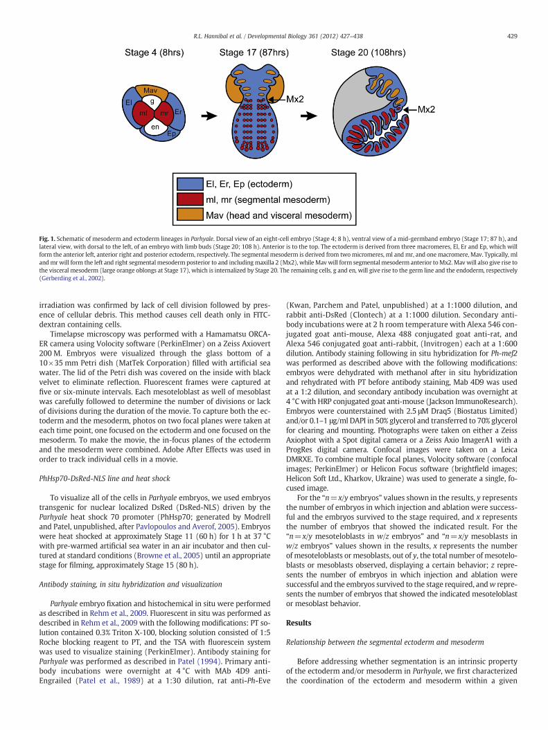
Fig. 1. Schematic of mesoderm and ectoderm lineages in Parhyale. Dorsal view of an eight-cell embryo (Stage 4; 8 h), ventral view of a mid-germband embryo (Stage 17; 87 h), andlateral view, with dorsal to the left, of an embryo with limb buds (Stage 20; 108 h). Anterior is to the top. The ectoderm is derived from three macromeres, El, Er and Ep, which willform the anterior left, anterior right and posterior ectoderm, respectively. The segmental mesoderm is derived from twomicromeres, ml and mr, and onemacromere, Mav. Typically, mlandmrwill form the left and right segmentalmesoderm posterior to and includingmaxilla 2 (Mx2), while Mavwill form segmentalmesoderm anterior toMx2.Mav will also give rise tothe visceral mesoderm (large orange oblongs at Stage 17), which is internalized by Stage 20. The remaining cells, g and en, will give rise to the germ line and the endoderm, respectively(Gerberding et al., 2002).
429R.L. Hannibal et al. / Developmental Biology 361 (2012) 427–438
irradiation was confirmed by lack of cell division followed by pres-ence of cellular debris. This method causes cell death only in FITC-dextran containing cells.
Timelapse microscopy was performed with a Hamamatsu ORCA-ER camera using Volocity software (PerkinElmer) on a Zeiss Axiovert200 M. Embryos were visualized through the glass bottom of a10×35 mm Petri dish (MatTek Corporation) filled with artificial seawater. The lid of the Petri dish was covered on the inside with blackvelvet to eliminate reflection. Fluorescent frames were captured atfive or six-minute intervals. Each mesoteloblast as well of mesoblastwas carefully followed to determine the number of divisions or lackof divisions during the duration of the movie. To capture both the ec-toderm and the mesoderm, photos on two focal planes were taken ateach time point, one focused on the ectoderm and one focused on themesoderm. To make the movie, the in-focus planes of the ectodermand the mesoderm were combined. Adobe After Effects was used inorder to track individual cells in a movie.
PhHsp70-DsRed-NLS line and heat shock
To visualize all of the cells in Parhyale embryos, we used embryostransgenic for nuclear localized DsRed (DsRed-NLS) driven by theParhyale heat shock 70 promoter (PhHsp70; generated by Modrelland Patel, unpublished, after Pavlopoulos and Averof, 2005). Embryoswere heat shocked at approximately Stage 11 (60 h) for 1 h at 37 °Cwith pre-warmed artificial sea water in an air incubator and then cul-tured at standard conditions (Browne et al., 2005) until an appropriatestage for filming, approximately Stage 15 (80 h).
Antibody staining, in situ hybridization and visualization
Parhyale embryo fixation and histochemical in situ were performedas described in Rehm et al., 2009. Fluorescent in situ was performed asdescribed in Rehm et al., 2009 with the following modifications: PT so-lution contained 0.3% Triton X-100, blocking solution consisted of 1:5Roche blocking reagent to PT, and the TSA with fluorescein systemwas used to visualize staining (PerkinElmer). Antibody staining forParhyale was performed as described in Patel (1994). Primary anti-body incubations were overnight at 4 °C with MAb 4D9 anti-Engrailed (Patel et al., 1989) at a 1:30 dilution, rat anti-Ph-Eve
(Kwan, Parchem and Patel, unpublished) at a 1:1000 dilution, andrabbit anti-DsRed (Clontech) at a 1:1000 dilution. Secondary anti-body incubations were at 2 h room temperature with Alexa 546 con-jugated goat anti-mouse, Alexa 488 conjugated goat anti-rat, andAlexa 546 conjugated goat anti-rabbit, (Invitrogen) each at a 1:600dilution. Antibody staining following in situ hybridization for Ph-mef2was performed as described above with the following modifications:embryos were dehydrated with methanol after in situ hybridizationand rehydrated with PT before antibody staining, Mab 4D9 was usedat a 1:2 dilution, and secondary antibody incubation was overnight at4 °Cwith HRP conjugated goat anti-mouse (Jackson ImmunoResearch).Embryos were counterstained with 2.5 μM Draq5 (Biostatus Limited)and/or 0.1–1 μg/ml DAPI in 50% glycerol and transferred to 70% glycerolfor clearing and mounting. Photographs were taken on either a ZeissAxiophot with a Spot digital camera or a Zeiss Axio ImagerA1 with aProgRes digital camera. Confocal images were taken on a LeicaDMRXE. To combine multiple focal planes, Volocity software (confocalimages; PerkinElmer) or Helicon Focus software (brightfield images;Helicon Soft Ltd., Kharkov, Ukraine) was used to generate a single, fo-cused image.
For the “n=x/y embryos” values shown in the results, y representsthe number of embryos in which injection and ablation were success-ful and the embryos survived to the stage required, and x representsthe number of embryos that showed the indicated result. For the“n=x/y mesoteloblasts in w/z embryos” and “n=x/y mesoblasts inw/z embryos” values shown in the results, x represents the numberof mesoteloblasts or mesoblasts, out of y, the total number of mesotelo-blasts or mesoblasts observed, displaying a certain behavior; z repre-sents the number of embryos in which injection and ablation weresuccessful and the embryos survived to the stage required, andw repre-sents the number of embryos that showed the indicated mesoteloblastor mesoblast behavior.
Results
Relationship between the segmental ectoderm and mesoderm
Before addressing whether segmentation is an intrinsic propertyof the ectoderm and/or mesoderm in Parhyale, we first characterizedthe coordination of the ectoderm and mesoderm within a given

430 R.L. Hannibal et al. / Developmental Biology 361 (2012) 427–438
segment. In order to address this relationship, we examined fixed em-bryos stained with DAPI via confocal microscopy. In addition, we fluo-rescently labeled the mesoderm and ectoderm in live embryos, andmonitored segmentation via timelapse microscopy. We filmed em-bryos when segmental tissue is both generated and patterned, Stage15 (80 h) to Stage 19 (96 h).
Our analysis corroborated previous research that separately char-acterized the formation of the segmental ectoderm and mesoderm inParhyale. At the eight-cell stage (Stage 4; 8 h), three cells will formthe anterior left, anterior right, and posterior ectoderm (El, Er andEp, respectively; Fig. 1), and two cells will form the left and right seg-mental trunk mesoderm (ml and mr, respectively; Fig. 1; Gerberdinget al., 2002). Segmental trunk ectoderm, from the posterior compart-ment of the mandible on, is formed by the organization of ectodermalcells into Parasegment Precursor Rows (PSPRs; SupplementaryMovies 1, 2; Browne et al., 2005). With the exception of rows 0 and1, which give rise to parts of the mandible and first maxillae, therest of the PSPRs divide in a stereotypicalmanner. First, the PSPRs dividealong the anterior–posterior axis to give rise to rows a/b and c/d. Then,rows a/b and c/d divide along the anterior–posterior axis to give rise torows a and b, and c and d, respectively. PSPRs form and divide in a pro-gressivemanner in the embryo, both anterior to posterior, andmedial tolateral. Segmental trunk mesoderm, from the second maxilla on, formsin a progressive manner from anterior to posterior through the migra-tion and division of eight mesodermal stem cells (derived from ml andmr) called mesoteloblasts (Supplementary Movies 1, 3, 4; Browne etal., 2005). The mesoteloblasts are arranged in four columns on eitherside of the midline and are designated one through four (M1–M4),with column one being closest to the midline. The mesoteloblasts giverise to the mesoblasts, which are the segmental founder cells, as eachrow of mesoblasts will give rise to one segment's worth of mesoderm.
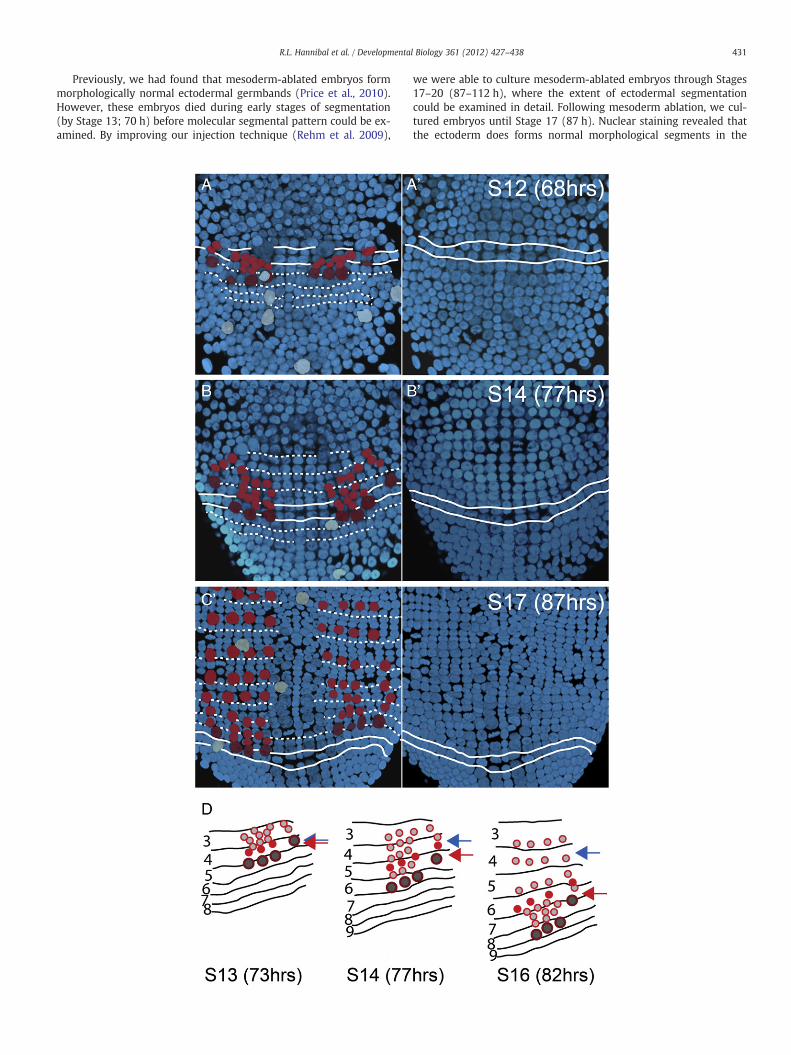
By examining segmentation simultaneously in both the ectodermand the mesoderm, we found that, prior to approximately Stage 13(72 h), the mesoteloblasts begin to migrate and divide beforethe first four PSPRs have begun to divide (n=8/8 embryos; Figs. 2A,A′). As development progresses, and the PSPRs begin to divide, the re-lationship between the mesoteloblasts and the PSPRs graduallychanges. After approximately Stage 14 (77 h), the mesoteloblasts(mesoderm) are either beneath or slightly anterior to the parasegmentprecursor row (PSPR; ectoderm) that is actively dividing (n=9/9 em-bryos; Fig. 2; Supplementary Movies 1–6). In summary, although themesoteloblasts begin to migrate and divide before the PSPRs begin todivide, PSPR division catches up to the position of the mesoteloblasts.The dynamic relationship between the mesoteloblasts and PSPRs sug-gests at least some autonomy for segmentingmesoderm and ectoderm.
As the mesoteloblasts migrate, they undergo a series of divisionsto produce the mesoblasts, the segmental founder cells (n=7/7 em-bryos; Fig. 2; Supplementary Movies 1–6). Since the mesoteloblastsproduce mesoblasts concurrent with PSPR divisions, we hypothesizedthat one parasegment's row of mesoblasts would be born under andremain associated with that parasegment's ectoderm. Surprisingly,we found that the mesoblasts do not stay associated with the ectoder-mal PSPR that they are born under (n=5/5 embryos; Fig. 2D; Supple-mentary Movies 1–6). Rather, a mesoteloblast initially generates twoto three mesoblasts, mesodermal parasegments, under only one PSPR,
Fig. 2.Mesoderm segmentation is not initially in registerwith ectoderm segmentation. (A–C′) CDorsal view. Ectoderm is blue; mesoteloblasts, mesoblasts and vitellophages are false colored dment boundaries and solid white lines delineate the most posterior PSPR that is actively dividithe dorsal view. Solid white lines delineate the most posterior PSPR that is actively dividing. (Atively dividing; the PSPR that is actively dividing is anterior to themesoblasts. (B, B′) Stage 14 (consist of at least four rows of cells associate with one parasegment′s worth of mesoderm. (DMesoteloblasts are large, dark gray circles outlined in dark red, while mesoblasts are either smis represented by solid red circle to highlight the relationship between the mesoblasts and thbut then migrates to become associated with ectodermal parasegment six. Red arrow denotebirth position (ectodermal parasegment four) of this row. Note that the fourth mesoblast in thments; ectodermal parasegments are numbered following Browne et al., 2005.
ectodermal parasegment. After birth, these mesoblasts migrate poste-riorly to associate with the progeny of one PSPR, and position them-selves under ectodermal rows c and d. While mesoblasts appear tomigrate to a specific PSPR all along the anterior–posterior axis, thegreatest distance between where the mesoblast is born and where itultimately ends up is in the anterior of the embryo. After mesoblastmigration, one discrete parasegment of mesoderm remains associatedwith one ectodermal parasegment. The mesoblasts always remain inbirth order, so that earlier bornmesoblasts are associated withmore an-terior ectodermal parasegments and later bornmesoblasts are associatedwithmore posterior ectodermal parasegments. Ourfinding that ectoder-mal andmesodermal progenitors undergo independent stereotypic divi-sions is consistent with a model in whichmesoderm and ectoderm haveautonomous segmental mechanisms.
Ectoderm and mesoderm have regional identity
Since the mesoderm and ectoderm present in a given parasegmentmay not be born in alignment, we hypothesized that ultimate positionmay be based on a shared regional cure. To test this hypothesis, we ex-amined Hox gene expression in the mesoblasts and ectodermal para-segments. Hox genes specify regional identity in many animals,including Parhyale (Liubicich et al., 2009; McGinnis and Krumlauf,1992).We focused on the Hox gene Ph-Ubx, as it is expressed in the eas-ily visualized anterior thoracic region (Liubicich et al., 2009). By per-forming fluorescent in situ hybridization for Ph-Ubx, we found thatPh-Ubx is expressed in PSPRs 4–8 and their progeny, similar to previous-ly published results (Liubicich et al., 2009; Figs. 3A, B). Supporting ourhypothesis, Ph-Ubx is expressed in mesoblasts that are initially out ofregister with ectodermal parasegments expressing Ph-Ubx, and con-tinues to be expressed in mesoblast progeny after mesoblasts havemoved into register with the ectoderm and have divided (Figs. 3C–D′).
Ectoderm segmentation does not require mesoderm
To test whether the ectoderm requires a signal from the meso-derm to segment properly, we photo-ablated all three mesodermalprecursor cells (Mav, ml and mr) at the eight-cell stage (Stage 4;8 h). Then we cultured these embryos until Stage 17 (87 h) and visu-alized segmentation in the ectoderm both morphologically, by stain-ing nuclei with DAPI, and molecularly, by staining for En, a markerof ectoderm segmentation (Browne et al., 2005; Patel et al., 1989).At this stage in control embryos, multiple PSPRs have formed andmany have undergone multiple rounds of division (Figs. 4A, A′).PSPR and progeny division are from both anterior to posterior andmedial to lateral, producing a chevron pattern (Fig. 4A; Browne etal., 2005). Rows a and b can be distinguished from rows c and d bytheir smaller cell size (Fig. 4.A′). Additionally, En is expressed in theanterior row, row a, of each parasegment following two rounds of di-vision (Figs. 4A, A′). Segmental En expression is also apparent in ecto-derm anterior to the PSPRs that does not form via PSPR division(Fig. 4A; Browne et al., 2005). This produces a reiterated, segmental,pattern of En-positive and En-negative rows of cells along the anteri-or–posterior axis of the embryo.
onfocal stacks of the posterior region of DAPI stained embryos, anterior to the top. (A, B, C)ark red, red, and light blue, respectively. Dashedwhite lines delineate ectodermal paraseg-ng. (A′, B′, C′) Ventral view of ectoderm, flipped horizontally to facilitate comparison with, A′) Stage 12 (68 h) embryo. Mesoteloblasts are not yet aligned with the PSPR that is ac-77 h) embryo. (C, C′) Stage 17 (87 h) embryo. The anterior ectodermal parasegments that) Diagram of three timepoints from timelapse movie (Supplementary Movies 2–3, 5–6).all, light gray circles outlined in red or are small, solid red circles. One row of mesoblastse ectoderm. The row of solid red mesoblasts is born under ectodermal parasegment four,s present position of the row of solid red mesoblasts in each panel, blue arrow denotesis row is not yet born in the first panel. Lines indicate boundaries of ectodermal paraseg-
431R.L. Hannibal et al. / Developmental Biology 361 (2012) 427–438
Previously, we had found that mesoderm-ablated embryos formmorphologically normal ectodermal germbands (Price et al., 2010).However, these embryos died during early stages of segmentation(by Stage 13; 70 h) before molecular segmental pattern could be ex-amined. By improving our injection technique (Rehm et al. 2009),
we were able to culture mesoderm-ablated embryos through Stages17–20 (87–112 h), where the extent of ectodermal segmentationcould be examined in detail. Following mesoderm ablation, we cul-tured embryos until Stage 17 (87 h). Nuclear staining revealed thatthe ectoderm does forms normal morphological segments in the

Fig. 3. Ph-Ultrabithorax (Ph-Ubx) is expressed in the ectoderm and the underlying mesoderm. (A–D′) Ventral views; anterior to the top. Overlay of Ph-Ubx (red) and DAPI nuclearstain (blue). Nuclear dots (red dots) indicate active transcription. (A) Stage 13 (73 h). Ph-Ubx is expressed in PSPRs 4–8. (B) Stage 18 (90 h). Ph-Ubx is expressed in thoracic seg-ments 2–8 (T2-T8). (C, C′) Confocal image of bracketed area in (A). (D, D′) Confocal image of bracketed area in (B); bracketed area includes the posterior of T3 and all of T4. (C, D)Ph-Ubx is expressed in the ectoderm. (C′, D′) Ph-Ubx is also expressed in the underlying mesoderm. Arrows point to mesoblasts (C′) and mesoblast progeny (D′) expressing Ph-Ubx.Mesoblasts are not yet aligned with a specific PSPR in (C′), while mesoblast progeny are aligned with the progeny of a specific PSPR, PSPR 7 (posterior of T4), in (D′).
432 R.L. Hannibal et al. / Developmental Biology 361 (2012) 427–438
absence of the mesoderm (n=13/13 embryos; Figs. 4C–D; Supple-mentary Movies 7 and 8). Mesoderm-ablated embryos displayed thesame pattern of PSPRs and subsequent PSPR divisions as control em-bryos. PSPRs formed and divided in a progressive manner, from bothanterior to posterior and medial to lateral (Figs. 4A, C). Additionally,similar to controls, rows a and b could be distinguished from rows cand d based on their smaller cell size (Figs. 4A′, C′). To molecularlyconfirm normal ectoderm segmentation we examined En expressionin mesoderm-ablated embryos. Antibody staining revealed proper ex-pression of En in the anterior row, row a, of each parasegment(n=13/13 embryos; Figs. 4C,C′). Enwas expressed in the same reiterat-ed, segmental, pattern of En positive and negative rows of cells as con-trols. These data show that ectoderm segmentation does not require themesoderm.
While the ectoderm does not require the mesoderm for initial seg-mental pattern, it could be required for later pattern maintenance. Asour mesoderm-ablated embryos died by Stage 20 (112 h), likely due
to the lack of visceral mesoderm crucial for the formation and func-tion of the digestive system, we used an alternative ablation strategyto assay for the dependence of ectoderm segmentation on the meso-derm later in development. In order to selectively ablate the trunksegmental mesoderm, while keeping the visceral mesoderm intact,we photo-ablated the progeny of the left and/or right mesodermalprecursor cells (ml and/or mr) right after the mesoteloblasts formed(Stage 10; 56 h). At the time of ablation, the progeny of ml/mr con-sists of all four mesoteloblasts as well as 0–2 mesoblasts per mesote-loblast, since some mesoteloblasts have already begun to divide bythe time all four mesoteloblasts have formed. In addition, the progenyof ml/mr also consists of approximately 5–30 cells that will form headand visceral mesoderm (Price and Patel, 2008). This is the earlieststage possible to ablate the progeny of ml and/or mr without progenyfrom the remaining mesodermal cell (Mav) compensating for theirloss (Price et al., 2010). We then cultured these embryos until Stage23 (142 h). At this stage, control embryos have developed all of

Fig. 4. The ectoderm segments autonomously. (A–D) Stage 15 (80 h) embryos, anterior to the top. Nuclear DAPI staining in blue. (A–B) Control embryo. (A, A′) Ventral view. (A′)Thoracic segments of embryo(A). Engrailed (En) protein (yellow) is present in the most anterior row (row a) of each parasegment once each PSPR has undergone two rounds ofdivision. (B) Dorsal view; confocal stack of (A′), showing mesoderm (false colored red cells) and vitellophages (false colored light blue cells) under the segmental ectoderm. (C–D)Mesoderm-ablated embryo. (C, C′) Ventral view. (C′) Thoracic segments of embryo (C). Just as in the control embryo, En protein is present in the most anterior row (row a) of eachparasegment once each PSPR has undergone two rounds of division. (D) Dorsal view; confocal stack of (C′), showing the lack of mesoderm in this embryo, although vitellophagesare still present. Dorsal views (B) and (D) were flipped horizontally to facilitate comparison with the overlying ectoderm. (E–H) Ventral view of Stage 23 (142 h) embryos, anteriorto the top. Nuclear DAPI staining in blue (E, G). (E–F) Control embryo. (G–H) Segmental trunk mesoderm-ablated embryo. (F, H) Posterior region of embryos (E′) and (G′), respec-tively. To generate segmental trunk mesoderm-ablated embryo, the mesoteloblasts, which produce the segmental trunk mesoderm, were ablated at their birth (Stage 10; 56 h).Head segmental mesoderm and visceral mesoderm, derived from Mav, are still present in this embryo. Ablation was confirmed by absence of Ph-mef2 (purple) in the trunkbody walls and limbs (compare E′ to G′). Residual Ph-mef2 staining in (G′, H) is visceral mesoderm derived from Mav. En protein (brown) is present in the posterior of each ecto-dermal segment (arrowheads in (F, H)) in both control and mesoderm-ablated embryos.
433R.L. Hannibal et al. / Developmental Biology 361 (2012) 427–438
their limbs, which are clearly organized in a segmental manner, andthese embryos also display distinct grooves between each trunk seg-ment (Fig. 4E). In addition, En is expressed both in the posterior ofeach ectodermal segment and in a subset of neurons in the trunk(Browne et al., 2005). To confirm ablation of the segmental mesoderm,we performed in situ hybridization for a marker of differentiated meso-derm, Ph-myocyte enhancing factor 2 (Ph-mef2; Price and Patel, 2008).Ph-mef2was largely absent in the ablated regions, except for trace stain-ing in visceral mesoderm derived from Mav (Fig. 4G′). We found thatmesoderm-ablated embryos displayed normal segmentation (n=6/6embryos; Fig. 4). Similar to controls, mesoderm-ablated embryos devel-oped segmented limbs and distinct grooves between each trunk seg-ment (Fig. 4G). In addition, mesoderm-ablated embryos displayedproper expression of En in the posterior of each limb (Fig. 4H) and inthe nervous system (Fig. 4G′). These data show that the ectodermdoes not require the mesoderm to form morphological segments, tomolecularly pattern these segments, or to maintain segmental patternlate in development.
Mesoteloblasts can divide autonomously, but mesoblast division requiresthe ectoderm
To test whether the mesoderm requires a signal from the ectodermto segment properly, we ablated the ectoderm and assayed for segmen-tation in the mesoderm. Because the ectodermal precursor cells (Er, Eland Ep) compose most of the volume of the embryo, ablation of allthree, or even two, of these cells results in death of the entire embryowithin 1 day of injection, 2 days before segmentation would normallybegin (Price et al., 2010). Ablation of only one ectodermal precursorcell at the eight-cell stage is also impractical for this study because prog-eny of the remaining ectodermal precursor cells compensate for the ab-lated cell by the time segmentation begins (Price et al., 2010). Toovercome these limitations, we photo-ablated the progeny of either
the left or right anterior ectodermal precursor cell (El or Er, respective-ly) after the mesoteloblasts formed, when the embryo can no longercompensate (Stage 10; 56 h; Price et al., 2010). Additionally, in orderto track the mesoderm on the ectoderm-ablated side of the embryo,we labeled the ipsilateral mesodermal precursor cell (either ml or mr)with the nuclear localized fluorescent lineage tracer DsRed-NLS.
To assay for mesoderm segmentation, we made movies of labeledmesoteloblasts in ectoderm-ablated embryos (Figs. 5A–F′; Supple-mentaryMovies 9 and 10). In control embryos,mesoteloblastsmigrat-ed posteriorly, progressing 1/3 the length of the embryo in a 10 hperiod (n=16/16 mesoteloblasts in 4/4 embryos). During this migra-tion, mesoteloblasts went through a series of asymmetric divisionsalong their anterior–posterior axis, producing one mesoblast every2 h (n=16/16 mesoteloblasts in 4/4 embryos). In ectoderm-ablatedembryos, however, most mesoteloblasts migrated in both posteriorand lateral directions (n=10/11 mesoteloblasts in 3/3 embryos) in-stead of only posteriorly (n=1/11 mesoteloblasts in 1/3 embryos).In addition, mesoteloblasts traveled less than 1/4 the length of the em-bryo in a 10 hour period (n=8/11 mesoteloblasts in 3/3 embryos). Ifthe mesoteloblasts came into contact with ectoderm from one of theremaining ectodermal lineages, El or Er, and/or Ep, they resumed posteri-ormigration and traveled 1/3 the length of the embryo in a 10 hour peri-od (n=3/11mesoteloblasts in 1/3 embryos; Fig. 5G). Similar to controls,mesoteloblasts in ectoderm-ablated embryos produced one mesoblastevery 2 h (n=11/11mesoteloblasts in 3/3 embryos; Figs. 5A–F′; Supple-mentary Movies 9 and 10). Additionally, mesoteloblasts in ectoderm-ablated embryos divided in the same anterior–posterior orientation asthey did in controls (n=11/11 mesoteloblasts in 3/3 embryos; Supple-mentary Movies 9 and 10). These data show that themesoteloblasts donot require the ectoderm for proper division, but do require the ecto-derm for proper migration. Proper migration may require the physicalsubstrate provided by the ectoderm, and/or could require a posteriormigration cue secreted from the ectoderm.

Fig. 5. Mesoteloblast division is autonomous, but mesoblast division requires the ectoderm. (A–F′) Stills from timelapse movie of mesoderm development in control and leftectoderm-ablated embryos Stage 14 (77 h) through Stage 18 (92 h). Ventral–lateral view; anterior to the top. Nuclei of left mesoderm are labeled with DsRed-NLS (white). (A′–C′ and D′–F′) In focus nuclei are false-colored to highlight mesoteloblasts and their progeny. Large circles denote mesoteloblasts, small circles denote mesoblasts. Mesoblasts arethe same color as the mesoteloblast from which they originated. (A–C′) Control embryo. (A–B′) Mesoteloblasts divide to give rise to mesoblasts while migrating posteriorly. Meso-blasts then undergo subsequent divisions (C, C′). Each bracket in C and C′ indicates one segment's worth of mesoderm where the mesoblasts have undergone multiple divisions.Arrows in B and C indicate the formation of a tailfold in the control embryo, which obscures more posterior mesoteloblasts and mesoblasts. (D–F′) Left ectoderm-ablated embryo.Mesoteloblasts continue to undergo divisions to give rise to mesoblasts, but do not migrate properly. In addition, mesoblasts born both before and after ectoderm ablation do notdivide (F, F′). While more mesoblasts are in focus in the left ectoderm-ablated embryo compared to the control embryo, these cells have not divided. Arrowhead in (F′) indicatesapproximate location of an orange lineage mesoblast that has gone out of focus. (G) Left side of fixed left ectoderm-ablated embryo; left mesoderm is labeled with and stained forDsRed-NLS (purple). Ventral view, anterior to the top. Mesoderm in the ablated region is in unorganized clumps (arrowheads). Once mesoderm from the ablated side comes intocontact with the posterior ectoderm, however, mesoderm is again spatially organized into rows (arrows; compare to control mesoderm in B, B′).
434 R.L. Hannibal et al. / Developmental Biology 361 (2012) 427–438
In contrast to the mesoteloblasts, the mesoblasts do not divide inthe absence of the ectoderm. Mesoblasts born both before and afterectoderm ablation never divide (n=9/9 and 15/15 mesoblasts, re-spectively, in 3/3 embryos; Figs. 5F, F′; Supplementary Movies 9 and10), indicating that a signal from the ectoderm is required later inmesoblast development, or for a longer period of time. We concludemesoteloblast division is autonomous, but division of their progeny,the mesoblasts, requires the ectoderm.
In the absence of ectoderm,mesoblasts do not express segmentationmarkers
Although mesoblast division requires the ectoderm, we hypothe-sized that mesoblasts may still have intrinsic segmental gene expres-sion without the ectoderm. For example, mesoblasts might still be
able to express segmental genes with the correct developmental timingeven if they are unable to divide. To test this hypothesis, we examinedexpression of molecular markers of mesoderm segmentation, Ph-twiand Ph-Eve (Fig. 6; Price and Patel, 2008; Vargas-Vila et al., 2010), inectoderm-ablated embryos. Not only are Ph-twi and Ph-Eve the earliestknownmolecularmarkers ofmesoderm segmentation, but they contin-ue to be expressed in a subset of the segmental mesoderm throughoutlater development. In control embryos, Ph-twi and Ph-Eve are expressedin the anterior daughters of mesoblasts in the second and fourthcolumns of mesoderm, respectively, in T2 andmore posterior segments.The second column of mesoderm is underneath the developing limbfield and the fourth column is the most dorsal mesoderm. Ph-twi andPh-Eve are also expressed in a similar segmental pattern in the meso-derm of Mx2 and T1. Later in development, Ph-twi and Ph-Eve are

435R.L. Hannibal et al. / Developmental Biology 361 (2012) 427–438
expressed in clusters of mesodermal cells in roughly the same positionsas in early development (Figs. 6A, B). Ph-twi is expressed at the base oflimb and in cells moving into the limbs (Price and Patel, 2008). Ph-Eveis expressed in a cluster of the dorsal-most mesodermal cells (Vargas-Vila et al., 2010). Based on comparison with similar Eve positive cells inDrosophila (Frasch et al., 1987), Ph-Eve positive cells likely contributeto the heart and dorsal somatic mesoderm.
In ectoderm-ablated embryos, however,we found that neither Ph-twinor Ph-Eve was expressed in the mesoderm on the ectoderm-ablatedside (n=6/6 embryos for Ph-twi and 5/5 embryos for Ph-Eve; Fig. 6).These data are consistent with two potential scenarios (Figs. 7 and 8).First, the ectoderm is required for both division and subsequent segmen-tal patterning of the mesoderm. Alternatively, the mesoderm may con-tain intrinsic segmental pattern, but the pattern is not visible until afterthe mesoblasts have divided, and this division requires a signal fromthe ectoderm.
Discussion
Ectoderm segmentation does not require the mesoderm
We have characterized the functional relationship between thesegmenting ectoderm and mesoderm in the crustacean P. hawaiensis.We asked whether segmentation is an intrinsic property of the ecto-derm and/or the mesoderm by ablating one layer and assaying both
Fig. 6. In the absence of ectoderm, mesoblasts do not express markers of mesoderm segmenmentation markers in a subset of the mesoderm throughout development. Overlay of Ph-EStage 18 (90 h). Solid white lines delineate midline; arrowheads point to bilateral expresslabeled El, Er and Ep, respectively. (C–D′) El or Er ablated embryos. Solid white lines delineand the posterior ectoderm. Left, right and posterior ectoderm are labeled El, Er and Ep, respembryos Stage 17 (87 h). The mesoderm on the ablated side of the embryo is labeled with DDAPI (blue). (C′) On the unablated (Er) side, Ph-Eve is expressed in a subset of the progeny oPh-Eve is not expressed in mesoblasts. Ph-Eve is expressed in one mesoteloblast near the pohas not migrated properly (arrows). Medial neural expression of Ph-Eve in the ectoderm is aalthough is present on the control side of older ectoderm-ablated embryos (data not shown)embryo Stage 18 (90 h). While this embryo is orientated with anterior to the top, it is curvedectoderm. (D) False color overlay of Ph-twi (yellow) over DAPI (blue). (D′) On the unablateMx2 and thoracic segments (arrowheads). On the ablated side (Er), Ph-twi is not expressemesoderm from the ablated side that now lies under the posterior ectoderm (arrows).
for morphological and molecular segmentation in the remaininglayer. We found that the ectoderm does not require the mesodermto form morphological segments, to molecularly pattern these seg-ments, or to maintain segmental pattern. Our data refute a hypothesisfrom arthropod literature that mesoderm segmentation evolved first,followed by ectoderm segmentation, and that this evolutionary sce-nario could be reflected in the developmental order and importanceof this tissue in extant animals (Budd, 2001). This evolutionary sce-nario is not reflected in the development of Parhyale.
Data from Parhyale also argue against another potential signalingsource of segmental pattern to the epidermal–ectoderm. Besides sug-gesting that ectoderm segmentation could be dependent on the me-soderm, Budd (2001) suggests that the neural-ectoderm couldinduce segmental pattern in the rest of the ectoderm. Similar to themesoderm, a segmented nervous system evolved prior to a segment-ed epidermis and this evolutionary scenario could be reflected in de-velopmental mechanisms of extant animals. In Parhyale, however,segmental pattern in the epidermal–ectoderm is apparent before ner-vous system formation, arguing against induction of pattern from theneural-ectoderm to the epidermal–ectoderm (Browne et al., 2005).
Although our data argue against Budd's evolutionary models, wehave only examined the functional relationship between the ecto-derm and mesoderm in Parhyale. Parhyale is an attractive system forthis study as its mesodermal and ectodermal lineages are separatedvery early in development, and are thus amendable to ablation and
tation. Ventral view; anterior to the top. (A, B) Control embryos bilaterally express seg-ve protein (green; A) and false color overlay of Ph-twi (yellow; B) over DAPI (blue) ation in maxilla 2 (Mx2) and thoracic mesoderm. Left, right and posterior ectoderm areate midline; dashed white lines delineate the boundary between the ablated ectodermectively. Ablated ectoderm is crossed out with a red “X”. (C, C′) Left ectoderm-ablated
sRed-NLS (red). (C) Overlay of Ph-Eve expression (green) and left mesoderm (red) overf the mesoblasts in Mx2 and thoracic segments (arrowheads). On the ablated side (El),sterior of the embryo, as well as in one mesoteloblast in the anterior of the embryo thatbsent on both the ablated and the control side of this embryo (compare (A) to (C, C′)),, suggesting a slight delay in nervous system formation. (D, D′) Right ectoderm-ablatedso that posterior is to the left due to differential growth caused by ablation of the right
d side (El), Ph-twi (purple) is expressed in a subset of the progeny of the mesoblasts ind in the ablated region of the embryo. However, Ph-twi is expressed in a subset of the

Fig. 7. The development of the segmental mesoderm involves an autonomous and anectoderm-dependent phase. Ventral view of left mesoderm (right side is a mirrorimage); anterior to the top, midline to the left. (A) Mesoteloblast division is not depen-dent on the ectoderm. The mesoteloblasts divide asymmetrically to give rise to themesoblasts. Each row of mesoblasts will give rise to one segment's worth of mesoderm(the production of two segment's worth of mesoderm is depicted here). (B) Mesoblastdivision is dependent on the ectoderm. The mesoblasts divide symmetrically, generatinganterior and posterior daughters. (C) Generation of segmental pattern in the mesodermmay be dependent on the ectoderm. After themesoblasts divide, the first knownmolecu-larmarkers of mesoderm segmentation are expressed. The second anterior daughter fromthemidline expresses Ph-twi and the fourth anterior daughter from themidline expressesPh-Eve, while their posterior sisters do not.
436 R.L. Hannibal et al. / Developmental Biology 361 (2012) 427–438
lineage-tracing experiments. Also, the necessary experimental toolswere available for this study, as Parhyale is an emerging model crusta-cean. However, the relationship between the ectoderm and mesodermmay have changed over evolutionary time andmay no longer reflect theevolution of the segmental ectoderm and mesoderm (Budd, 2001). Ad-ditionally, the ectoderm–mesoderm relationship in Parhyale, aMalacos-tracan, might not be the same as in non-Malacostracan crustaceans thatdo not utilize teloblasts to produce segments. While this is a concern,the absence of teloblasts in crustaceans outside of the Malacostracanscould be due to a scarcity of developmental data versus an actual lackof teloblasts in these species. Moreover, subsequent steps of segmenta-tion are relatively similar in species with and without teloblasts (Daviset al., 2005), suggesting that the relationship between the segmentingectoderm and mesoderm could be the same as well. Even if the ecto-derm–mesoderm relationship is similar amongst all pancrustaceans, itmay differ from the other arthropod groups, themyriapods and chelice-rates. In order to form amore comprehensive picture of the relationshipbetween the ectoderm and mesoderm in arthropods, more speciesshould be examined.
Scholtz also uses comparative data to suggest that the mesodermis the primary segmental germ layer in Malacostracan crustaceans(Scholtz, 1990). By comparing Malacostracans, Scholtz notes that seg-mental trunk mesoderm development is conserved, while segmentaltrunk ectoderm development varies, between species. This conserva-tion could suggest a greater importance of the segmental mesodermduring development, perhaps by inducing ectoderm segmentation.Instead, our data from the Malacostracan Parhyale suggest that thedevelopmental conservation of the segmental mesoderm and the de-velopmental plasticity of the segmental ectoderm in Malacostracansdo not correlate with their relative contributions to segmentation.
Development of segmental mesoderm in Parhyale involves both anautonomous and an ectoderm-dependent phase
We found that the ectoderm and mesoderm composing each para-segment are not initially associated with one another. After birth, oneparasegment of mesoderm migrates to associate with its correspond-ing parasegment of ectoderm. This raises the possibility that the me-soderm has some autonomy from the ectoderm, a hypothesis we haveconfirmedwith ablation experiments.Whenwe ablated either the left orright anterior ectoderm in Parhyale, we found that the mesoteloblastscontinued to divide at a normal rate and in the correct anterior–posteriororientation. Although themesoteloblasts often failed tomigrate proper-ly, this may be due to the lack of a physical substrate, rather than inter-cellular signals.
After this first, autonomous production of the segmental meso-derm (Fig. 7A) a second, ectoderm–dependent, phase of mesodermsegmentation occurs (Fig. 7B). We found that the progeny of themesoteloblasts, the mesoblasts, only divide after they are associatedwith one parasegment's worth of ectoderm. Our ablation experimentsshow that the mesoblasts fail to divide and do not express knownmarkers of later mesoderm segmentation in the absence of the ecto-derm. While we found that the mesoderm requires a signal fromthe ectoderm to formmature segments, future experiments are neededto address whether this ectodermal signal is instructive or permissive(Fig. 7C). A permissive signal would allow the mesoblasts to divide,but the mesoblasts themselves would contain segmental patterning in-formation, and would express markers of segmentation autonomouslyafter division occurred (Fig. 8A). Alternatively, an additional instructivesignal from the ectoderm could impart segmental pattern to the meso-derm (Fig. 8B). One way to distinguish between these possibilitieswould be to examine a protein expressed in either the anterior or pos-terior membrane of the mesoblasts, if such a protein were to be discov-ered. If mesoblasts have intrinsic segmental pattern, this marker wouldstill be present in its proper anterior–posterior location in ectoderm-ablated embryos.
Evolution of segmentation
There is considerable debate on whether trunk segmentation inthe arthropods, annelids and chordates is homologous, since eachphylum is more closely related to unsegmented phyla than to eachother (Davis and Patel, 1999; Peel and Akam, 2003; Seaver, 2003).One way to approach this question of homology is to compare devel-opment of trunk segmentation amongst these groups. If the commonancestor of these groups was segmented, perhaps the relationship be-tween the two segmental layers, the ectoderm and mesoderm, wouldbe the same. For example, the mesoderm could segment autono-mously and induce segmental pattern in the ectoderm in all threegroups.
In vertebrates, trunkmesoderm segments autonomously and inducessegmental arrangement of the overlying ectoderm-derived nervous sys-tem (Correia and Conlon, 2000; Detwiler, 1934; Keynes and Stern, 1984;Lewis et al., 1981; Palmeirim et al., 1998). This method of segmentationis conserved amongst all vertebrates examined. In contrast, in the

Fig. 8. The ectoderm provides the mesoderm with a permissive, and possibly instruc-tive, signal. Transverse view of the ectoderm and underlying mesoderm. Anterior tothe top, dorsal to the right, ventral to the left. Small arrows represent signals fromthe ectoderm to the mesoderm. Large arrows represent developmental progression.Mesoblasts require a permissive signal from the ectoderm in order to divide. (A) Meso-blast progeny may have intrinsic segmental pattern. (B) Mesoblast progeny may re-quire an instructive signal from the ectoderm for segmental pattern.
437R.L. Hannibal et al. / Developmental Biology 361 (2012) 427–438
holometabola insects Drosophila, Chrysopa and Leptinotarsa, the ecto-derm segments autonomously and is at least partially required for prop-er mesoderm segmentation (Azpiazu et al., 1996; Bock, 1942; Frasch,1999; Haget, 1953; Rao et al., 1991). Our ablation experiments showthat this is not restricted to the holometabola insects. In Parhyale, ecto-derm can also segment autonomously and, at the least, supplies a per-missive signal for mesoderm segmentation. Together, these datasuggest that segmentation is primarily a property of the ectoderm inthe pancrustacea (crustaceans plus insects; Regier et al., 2010). It willbe interesting to investigate segmentation in the other arthropod groups,specifically themyriapods and chelicerates, to definewhether segmenta-tion is primarily a property of the ectoderm in all arthropods.
Unlike vertebrates and pancrustaceans, in annelids, there does notappear to be one germ layer that has intrinsic segmental pattern andinduces the other to segment. A caveat to this statement is that theseinteractions have only been studied in two annelids, so a commonmethodmay emerge ifmore specieswere studied. In the leechHelobdellaand the sludge worm, Tubifex, segments are produced by the asym-metrical divisions of ectodermal and mesodermal stem cells (Gotoet al., 1999; Nakamoto et al., 2000; Weisblat and Shankland, 1985).In Helobdella, mesoderm and ectoderm are dependent on each otherfor segmental organization and pattern, but not for the production
and division of segmental precursor cells (Blair, 1982). Similar toHelob-della, in Tubifex, ectoderm segmental precursor cells are producedwith-out the mesoderm, but these cells are not arranged in a segmentalmanner (Nakamoto et al., 2000). However, in contrast to Helobdella,Tubifex mesoderm segmentation is autonomous (Goto et al., 1999). Asonly morphological segmentation was examined in these studies, mo-lecular segmental pattern could still be intrinsic.
Together, these data show that the germ layer containing segmen-tal information differs between arthropods and vertebrates, and evenamongst different species of annelids. This suggests that the tissuelayer that imparts segmental information might be easily acquired,modified and lost, and thereforemay not be a suitable developmental fea-ture for inferring instances of the independent evolution of segmentation.
Supplementary materials related to this article can be found on-line at doi:10.1016/j.ydbio.2011.09.033
Acknowledgments
We thank other members of the Patel Lab for providing the fol-lowing reagents: Elaine Kwan for the Ph-Eve antibody, Paul Liu forthe tdTomato-NLS construct, and Melinda Modrell for the Hsp70-DsRed-NLS Parhyale line. We thank Roger Tsien for the Tomato con-struct. We thank Crystal Chaw, Stephanie Gline, Paul Liu, and AndreaWills for helpful comments.
References
Akam, M., 1987. The molecular basis for metameric pattern in the Drosophila embryo.Development 101, 1–22.
Azpiazu, N., Lawrence, P.A., Vincent, J.P., Frasch, M., 1996. Segmentation and specifica-tion of the Drosophila mesoderm. Genes Dev. 10, 3183–3194.
Blair, S.S., 1982. Interactions between mesoderm and ectoderm in segment formationin the embryo of a glossiphoniid leech. Dev. Biol. 89, 389–396.
Bock, E., 1942. Wechselbeziehung zwischen den Keimblattern bei der Organbildungvon Chrysopa perla (L.). Wilhelm Roux Arch Entwickl. Mech. Org. 141, 159–247.
Browne, W.E., Price, A.L., Gerberding, M., Patel, N.H., 2005. Stages of embryonic devel-opment in the amphipod crustacean, Parhyale hawaiensis. Genesis 42, 124–149.
Budd, G.E., 2001. Why are arthropods segmented? Evol. Dev. 3, 332–342.Correia, K.M., Conlon, R.A., 2000. Surface ectoderm is necessary for the morphogenesis
of somites. Mech. Dev. 91, 19–30.Davis, G.K., Patel, N.H., 1999. The origin and evolution of segmentation. Trends Cell Biol.
9, M68–M72.Davis, G.K., D'Alessio, J.A., Patel, N.H., 2005. Pax3/7 genes reveal conservation and di-
vergence in the arthropod segmentation hierarchy. Dev. Biol. 285, 169–184.Dequéant, M.-L., Pourquié, O., 2008. Segmental patterning of the vertebrate embryonic
axis. Nat. Rev. Genet. 9, 370–382.Detwiler, S.R., 1934. An experimental study of spinal nerve segmentation in Amblys-
toma with reference to the plurisegmental contribution to the brachial plexus. J.Exp. Zool. 67, 395–441.
Frasch, M., 1999. Intersecting signaling and transcriptional pathways in Drosophilaheart specification. Semin. Cell Dev. Biol. 10, 61–71.
Frasch, M., Hoey, T., Rushlow, C., Doyle, H., Levine, M., 1987. Characterization and local-ization of the even-skipped protein of Drosophila. EMBO J. 6, 749–759.
Gerberding, M., Browne, W.E., Patel, N.H., 2002. Cell lineage analysis of the amphipodcrustacean Parhyale hawaiensis reveals an early restriction of cell fates. Develop-ment 129, 5789–5801.
Gibb, S., Maroto, M., Dale, J.K., 2010. The segmentation clock mechanism moves up anotch. Trends Cell Biol. 20, 593–600.
Goto, A., Kitamura, K., Shimizu, T., 1999. Cell lineage analysis of pattern formation inthe Tubifex embryo. I. Segmentation in the mesoderm. Int. J. Dev. Biol. 43, 317–327.
Haget, A., 1953. Analyse expérimentale des facteurs de la morphogenèse embryonnairechez le coléoptère Leptinotarsa. Bull. Biol. Fr. Belg. 87, 123–217.
Keynes, R.J., Stern, C.D., 1984. Segmentation in the vertebrate nervous system. Nature310, 786–789.
Lewis, J., Chevallier, A., Kieny, M., Wolpert, L., 1981. Muscle nerve branches do not de-velop in chick wings devoid of muscle. J. Embryol. Exp. Morphol. 64, 211–232.
Liubicich, D.M., Serano, J.M., Pavlopoulos, A., Kontarakis, Z., Protas,M.E., Kwan, E., Chatterjee,S., Tran, K.D., Averof,M., Patel, N.H., 2009. Knockdownof ParhyaleUltrabithorax recapit-ulates evolutionary changes in crustacean appendagemorphology. Proc. Natl. Acad. Sci.U. S. A. 106, 13892–13896.
Martinez-Arias, A., Lawrence, P.A., 1985. Parasegments and compartments in theDrosophilaembryo. Nature 313, 639–642.
McGinnis,W., Krumlauf, R., 1992. Homeobox genes and axial patterning. Cell 68, 283–302.Nakamoto, A., Arai, A., Shimizu, T., 2000. Cell lineage analysis of pattern formation in
the Tubifex embryo. II. Segmentation in the ectoderm. Int. J. Dev. Biol. 44, 797–805.Palmeirim, I., Dubrulle, J., Henrique, D., Ish-Horowicz, D., Pourquie, O., 1998. Uncou-
pling segmentation and somitogenesis in the chick presomitic mesoderm. Dev.Genet. 23, 77–85.
438 R.L. Hannibal et al. / Developmental Biology 361 (2012) 427–438
Patel, N.H., 1994. Imaging neuronal subsets and other cell types in whole-mount Dro-sophila embryos and larvae using antibody probes. Methods Cell Biol. 44, 445–487.
Patel, N., Martin-Blanco, E., Coleman, K., Poole, S., Ellis, M., Kornberg, T., Goodman, C.,1989. Expression of engrailed proteins in arthropods, annelids, and chordates.Cell 58, 955–968.
Pavlopoulos, A., Averof, M., 2005. Establishing genetic transformation for comparativedevelopmental studies in the crustacean Parhyale hawaiensis. Proc. Natl. Acad. Sci.U. S. A. 102, 7888–7893.
Pavlopoulos, A., Kontarakis, Z., Liubicich, D.M., Serano, J.M., Akam, M., Patel, N.H.,Averof, M., 2009. Probing the evolution of appendage specialization by Hox genemisexpression in an emerging model crustacean. Proc. Natl. Acad. Sci. U. S. A.106, 13897–13902.
Peel, A., 2004. The evolution of arthropod segmentation mechanisms. Bioessays 26,1108–1116.
Peel, A., Akam, M., 2003. Evolution of segmentation: rolling back the clock. Curr. Biol.13, R708–R710.
Pourquié, O., 2003. Vertebrate somitogenesis: a novel paradigm for animal segmenta-tion? Int. J. Dev. Biol. 47, 597–603.
Price, A.L., Patel, N.H., 2008. Investigating divergent mechanisms of mesoderm devel-opment in arthropods: the expression of Ph-twist and Ph-mef2 in Parhyalehawaiensis. J. Exp. Zool. B Mol. Dev. Evol. 310B, 24–40.
Price, A.L., Modrell, M.S., Hannibal, R.L., Patel, N.H., 2010. Mesoderm and ectoderm lineagesin the crustacean Parhyale hawaiensis display intra-germ layer compensation. Dev. Biol.341, 256–266.
Rao, Y., Vaessin, H., Jan, L.Y., Jan, Y.N., 1991. Neuroectoderm in Drosophila embryos isdependent on the mesoderm for positioning but not for formation. Genes Dev. 5,1577–1588.
Regier, J.C., Shultz, J.W., Zwick, A., Hussey, A., Ball, B., Wetzer, R., Martin, J.W., Cunning-ham, C.W., 2010. Arthropod relationships revealed by phylogenomic analysis ofnuclear protein-coding sequences. Nature 463, 1079–1083.
Rehm, E.J., Hannibal, R.L., Chaw, R.C., Vargas-Vila, M.A., Patel, N.H., 2009. The crustaceanParhyale hawaiensis: a new model for arthropod development. In: Crotty, D.A.,Gann, A. (Eds.), Emerging Model Organisms: a Laboratory Manual, Volume 1.Cold Spring Harbor Laboratory Press, New York, pp. 373–404.
Sander, K., 1976. Specification of the basic body pattern in insect embryogenesis. Adv.Insect Physiol. 12, 125–238.
Scholtz, G., 1990. The Formation, differentiation and segmentation of the post-naupliargerm band of the amphipod Gammarus pulex L. (Crustacea, Malacostraca, Peracar-ida). Proc. R. Soc. Lond. B 163–211.
Seaver, E.C., 2003. Segmentation: mono- or polyphyletic? Int. J. Dev. Biol. 47, 583–595.Tautz, D., 1994. Segmentation. Dev Cell. 7, 301–312.Telford, M.J., Budd, G.E., 2003. The place of phylogeny and cladistics in Evo-Devo re-
search. Int. J. Dev. Biol. 47, 479–490.Vargas-Vila, M.A., Hannibal, R.L., Parchem, R.J., Liu, P.Z., Patel, N.H., 2010. A prominent
requirement for single-minded and the ventral midline in the dorsoventral axis ofthe crustacean Parhyale hawaiensis. Development 137, 3469–3476.
Weisblat, D.A., Shankland, M., 1985. Cell lineage and segmentation in the leech. Philos.Trans. R. Soc. Lond. Ser. B 312, 39–56.