hanna c persson - göteborgs universitet · vii stroke är ett samlingsnamn för hjärninfarkt och...
TRANSCRIPT

i
Hanna C Persson
Department of Clinical Neuroscience and Rehabilitation
Institute of Neuroscience and Physiology
Sahlgrenska Academy at University of Gothenburg
Gothenburg 2015
ii
Cover illustration: Individual patterns of change over time in upper extremity
function, assessed with the Fugl-Meyer Assessment for Upper Extremity
(FMA-UE) from 3 days to one year post stroke onset.
Upper extremity functioning during the first year after stroke
© Hanna C Persson 2015
ISBN 978-91-628-9662-1
http://hdl.handle.net/2077/40884
Printed in Gothenburg, Sweden 2015
Aidla Trading AB/Kompendiet

iii
A comfort zone is a beautiful place, but nothing ever grows there.
Unknown
To Andreas, Axel and Elsa

iv
v
Hanna C Persson
Department of Clinical Neuroscience and Rehabilitation, Institute of Neuroscience and Physiology
Sahlgrenska Academy at University of Gothenburg Göteborg, Sweden
The overall aim of this thesis was to investigate upper extremity functioning
during the first year after stroke from different perspectives.
Methods. All patients with first ever stroke, admitted to a stroke unit within
72 hours after stroke incidence were included during a period of 18 months.
The prevalence of impaired upper extremity function was investigated within
72 hours. Differences in change over time in functioning (function and
activity) between patients with ischemic and hemorrhagic stroke were
explored. The possibility of a simple early assessment to predict the level of
upper extremity motor function required for a drinking task was investigated,
as well as the relationship between patient-perceived and assessed strength
capacity. The studies are a part of the SALGOT-study (The Stroke Arm
Longitudinal Study at the University of Gothenburg).
Main results. Of patients admitted to a stroke unit, 48% had impaired upper
extremity function within 72 hours after stroke onset. In patients with
impaired upper extremity function initially, those with hemorrhagic stroke
had a larger improvement from 1 to 3 months in their function and activity
compared to patients with ischemic stroke. Patients with hemorrhagic and
ischemic stroke improved function and activity to a similar level 3 months
and thereafter. Two items from the Action Research Arm Test (ARAT) used
at 3 days post stroke could accurately predict the level of motor function
required for a drinking task at three later time points during the first year post
stroke. Assessed grip strength capacity and perceived strength at 10 days post
stroke correlated highly, but some patients rated their strength differently
compared to the assessment of strength capacity.
Conclusions and clinical implications. Fewer patients than previously
described had impaired upper extremity function early after stroke which is

vi
of importance in planning of care and rehabilitation. In patients with impaired
upper extremity function, larger improvements of function and activity were
seen after 1 month in those patients with hemorrhagic stroke compared to
ischemic, but both stroke types reached a similar level at 3 months post
stroke. These results together with the finding that early prediction of
function is possible, and that a combination of patient-reported and objective
strength assessment early after stroke may be valuable in planning of care,
rehabilitation and goal setting, and therefore improve the overall
rehabilitation process.
Keywords: Stroke recovery, Upper extremity, Paresis, Outcome, Process
assessment, Stroke, Cerebral haemorrhage, Prognosis, Motor skills,
Movement, Rehabilitation, Treatment outcome, Muscle strength, Self-report
ISBN: 978-91-628-9662-1
http://hdl.handle.net/2077/40884

vii
Stroke är ett samlingsnamn för hjärninfarkt och hjärnblödning. Antalet
personer som insjuknar i stroke varierar i världen, och i Sverige drabbas
årligen 25-30 000 personer. Konsekvenser av en stroke kan se olika ut, men
det är vanligt med nedsatt funktion och känsel i arm eller ben, eller påverkan
på språk eller tankeförmåga och minne. Nedsatt arm- och handfunktion
påverkar möjligheten att utföra aktiviteter. I akut skede har enligt tidigare
studier 70-80% av patienterna nedsatt arm- och handfunktion, och ca 40% får
kvarstående problem. Störst återhämtning av funktioner sker vanligen de
första veckorna efter insjuknade, men individuella variationer förekommer.
Det övergripande syfte för avhandlingen var att studera funktion och
aktivitetsförmåga i arm och hand under det första året efter stroke utifrån
olika perspektiv. Avhandlingen omfattar fyra delarbeten, där patienter som
vårdas för stroke på en strokeenhet inom 72 timmar efter insjuknade ingår.
Samtliga delarbeten är en del av SALGOT-studien (The Stroke Arm
Longitudinal Study at the University of Gothenburg).
I Studie I undersöktes förekomst av nedsatt arm- och handfunktion initialt
efter insjuknade. Resultatet visade att 48% hade nedsatt arm- och
handfunktion inom 72 timmar efter insjuknade. Dessa patienter var äldre,
vårdades längre tid på strokeenheten och hade också en högre dödlighet än de
patienter med god arm- och handfunktion.
I Studie II undersöktes eventuella skillnader mellan patienter som fått en
infarkt eller blödning avseende återhämtning av motorisk funktion och
aktivitetsförmåga i arm och hand under första året efter insjuknade. Patienter
med blödning förbättrades mer de första 3 månaderna, jämfört med de som
fått en infarkt. Båda grupperna hade ungefär samma nivå vid 3 månader och
där efter. Högre ålder och mer uttalad stroke påverkade återhämtningen
negativt i båda grupperna.
I Studie III undersöktes om en kort klinisk bedömning efter 3 dagar
respektive 1 månad efter insjuknande, kunde förutsäga arm- och
handfunktion som motsvarar att kunna dricka ur ett glas. Två delmoment från
ett mer omfattande bedömningsinstrument användas. Den korta bedömningen
visades ha god förmåga att förutsäga motorisk funktion motsvarande att
kunna dricka ur ett glas efter 10 dagar, 1 månad och 1 år, och hade bäst

viii
precision om patienten vid första bedömningen hade någon arm-
handfunktion.
I Studie IV undersöktes överensstämmelse mellan patientens skattade styrka i
arm och hand med en klinisk mätning av greppstyrka 10 dagar efter stroke.
Majoriteten av patientens självskattade arm- och handstyrka överenstämde
med den mätningen av handstyrka, men mindre del av patienterna över- eller
underskattade sin styrka.
Sammanfattningsvis visar avhandlingen att färre än 50% av patienterna har
nedsatt arm- och handfunktion i akut skede efter stroke, vilket skiljer sig från
vad tidigare studier har visat. De patienter som hade nedsatt arm- och
handfunktion visade sig ha sämre återhämtning under första året om de var
äldre eller hade mer uttalad stroke. Patienter med blödning hade snabbare
återhämtning de första månaderna, jämfört med de med infarkt, men vid 3
månader hade båda nått likvärdig funktion- och aktivitetnivå. Vidare visades
att en kort, enkel, bedömning 3 dagar efter strokeinsjuknade kan förutsäga
motorisk funktion som motsvarar att kunna dricka ur ett glas. Att kombinera
patientskattning och klinisk funktionsbedömning tidigt efter stroke, ger olika
perspektiv på funktion och tydliggör patientens kännedom om sin egen
förmåga. Sammantaget kan resultaten bidra till ökad kunskap kring arm- och
handfunktion och aktivitet efter stroke, som kan användas för bättre planering
av vård och rehabilitering redan i tidigt skede efter strokeinsjuknande.
i
This thesis is based on the following studies, referred to in the text by their
Roman numerals.
I. Persson HC, Parziali M, Danielsson A, Sunnerhagen
KS. Outcome and upper extremity function within 72
hours after first occasion of stroke in an unselected
population at a stroke unit. A part of the SALGOT
study. BMC Neurol. 2012;12:162.
II. Persson HC, Opheim A, Lundgren-Nilsson Å, Alt
Murphy M, Danielsson A, Sunnerhagen KS. Differences
in recovery of upper extremity functioning after
ischemic and hemorrhagic stroke – a part of the
SALGOT study. Submitted manuscript.
III. Persson HC, Alt Murphy M, Danielsson A, Lundgren-
Nilsson Å, Sunnerhagen KS. A cohort study
investigating a simple, early assessment to predict upper
extremity function after stroke - a part of the SALGOT
study. BMC Neurol. 2015;15:92.
IV. Persson HC, Danielsson A, Sunnerhagen KS. A cross
sectional study of upper extremity strength ten days after
a stroke; relationship between patient-reported and
objective measures. BMC Neurol. 2015;15(1):178.
ii
ABBREVIATIONS .............................................................................................. V
DEFINITIONS IN SHORT .................................................................................. VII
INTRODUCTION ................................................................................................ 1
Stroke ............................................................................................................ 1
Stroke in change ...................................................................................... 2
Classification of diseases and health status .................................................. 3
Upper extremity ............................................................................................ 4
Upper extremity after stroke .................................................................... 4
Stroke recovery ............................................................................................. 5
Recovery of upper extremity ................................................................... 5
Stroke rehabilitation ...................................................................................... 6
Rehabilitation of upper extremity ............................................................ 7
Measuring upper extremity after stroke ........................................................ 8
Psychometrics of measurements ................................................................... 9
Lack of knowledge ..................................................................................... 10
AIM ................................................................................................................ 11
PATIENTS AND METHODS .............................................................................. 12
The study population ............................................................................. 12
Definitions of impaired upper extremity function ................................. 14
Study design .......................................................................................... 14
Outcome measures ...................................................................................... 15
Descriptive variables ............................................................................. 19
Assessment procedure and data acquisition ................................................ 20
Care and rehabilitation........................................................................... 23
Data analyses .............................................................................................. 23
Statistical analyses ................................................................................. 24
Statistical software ................................................................................. 27
Ethics .......................................................................................................... 27
iii
RESULTS ........................................................................................................ 28
Drop-outs and non-included patients .......................................................... 28
Prevalence of impaired upper extremity function ....................................... 29
Differences in recovery of functioning ....................................................... 31
Prediction of function ................................................................................. 33
Patients’ perception in relation to capacity measurement ........................... 34
Summary of results ..................................................................................... 35
DISCUSSION ................................................................................................... 37
Prevalence of impaired upper extremity function .................................. 37
Aspects of functioning ........................................................................... 38
Methodological considerations ................................................................... 40
Generalisability ...................................................................................... 43
Strengths and limitations ............................................................................ 44
Clinical implications ................................................................................... 45
CONCLUSION ................................................................................................. 46
FUTURE PERSPECTIVES .................................................................................. 47
ACKNOWLEDGEMENT .................................................................................... 48
REFERENCES .................................................................................................. 51

iv
v
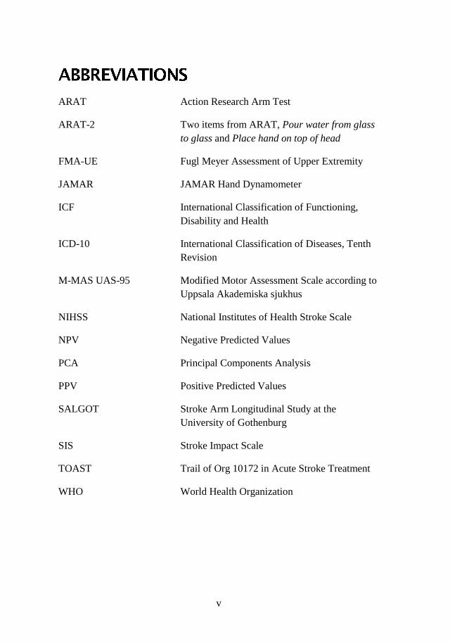
ARAT Action Research Arm Test
ARAT-2 Two items from ARAT, Pour water from glass
to glass and Place hand on top of head
FMA-UE Fugl Meyer Assessment of Upper Extremity
JAMAR JAMAR Hand Dynamometer
ICF International Classification of Functioning,
Disability and Health
ICD-10 International Classification of Diseases, Tenth
Revision
M-MAS UAS-95 Modified Motor Assessment Scale according to
Uppsala Akademiska sjukhus
NIHSS National Institutes of Health Stroke Scale
NPV Negative Predicted Values
PCA Principal Components Analysis
PPV Positive Predicted Values
SALGOT Stroke Arm Longitudinal Study at the
University of Gothenburg
SIS Stroke Impact Scale
TOAST Trail of Org 10172 in Acute Stroke Treatment
WHO World Health Organization

vi

vii
Activity
The execution of a task or action by an individual
(WHO, 2001).
Body Functions The physiological functions of body systems (WHO,
2001).
Body structures Anatomical parts of the body such as organs, limbs and
their components (WHO, 2001).
Capacity
Ability to execute a task or an action in a standardised
environmental (WHO, 2001).
Impairment
Problems in body function or structure as a significant
deviation or loss (WHO, 2001).
Functioning Umbrella term of Body Functions and Structures and
Activities and Participation, positive aspects (WHO,
2001).
Participation Involvement in a life situation (WHO, 2001).
Performance What a person does in his or her current environmental
(WHO, 2001).


Hanna C Persson
1
One in six people worldwide will have a stroke in their lifetime. Stroke has a
great impact not only on the quality of life of the person, but also on their
relatives and caregivers and could leave the person with multiple
impairments and complex needs1. As the demands for efficiency in stroke
care increases, knowledge of the prognosis of function and activity for
patients is of importance in order to optimize stroke management, to reduce
the suffering of individuals and their level of disability, as well as to use
patients and financial resources optimally.
Paresis in an upper extremity is a common impairment after stroke2,3
. Few
up-to-date studies, have investigated upper extremity functioning (function
and activity) in an unselected population during the first year after stroke
onset2-8
. The prevalence of impaired upper extremity, as well as different
aspects of upper extremity function and activity needs to be further explored.
Stroke includes three sub types; cerebral infarction (ischemic stroke),
intracerebral hemorrhage, and subarachnoid hemorrhage9,10
. According to the
World Health Organization (WHO), stroke is defined as rapidly developing
clinical signs of focal (at times global) disturbance of cerebral function,
lasting more than 24 hours or leading to death with no apparent cause other
than that of vascular origin11
. In 2013, a more detailed definition of the
stroke was published, including specific diagnosis according to imaging
findings9. However, in this thesis the WHO definition of stroke is used;
ischemic and intra cerebral hemorrhage (hemorrhagic) stroke are included in
the thesis and subarachnoid hemorrhage is excluded.
Every year about 15 million people worldwide suffer a stroke where of 75-
80% are living in low and middle-income countries12,13
. In contrast to a
decreased stroke incidence in high income countries, the prevalence of stroke
has increased in low and middle income countries12
. The prevalence of stroke
types also varies globally. In high income countries, hemorrhagic stroke
amounts to less than one third of the total stroke cases, compared to in low
and middle income countries where nearly half of all stroke diagnoses are
hemorrhagic14
. People suffering a hemorrhagic stroke are younger compared
to those with ischemic stroke12
. Six million people die from stroke yearly
around the world, which makes stroke the second leading cause of death15
.

Upper extremity functioning during the first year after stroke
2
The mortality of stroke is strongly influenced by each country’s economic
situation14
. The total burden of stroke has increased around the world12,13
, and
the burden of hemorrhagic stroke has been shown to be greater than of
ischemic stroke, even if the total number of hemorrhagic strokes were
lower12
. Many people lives with long term impairments after stroke which
has consequences that may restrict their possibility to participate in life as
they wish. Approximately 50% of patients who survive their stroke require at
least some assistance in their activities of daily living16
.
In Sweden, around 25-30 000 people suffer a stroke yearly17
, 18 000 of which
are due to a first ever stroke. In 2014 the mean age of suffering a stroke was
75.6 years, women on mean being five years older than men18
.The stroke
mortality in Sweden is 100 per 100 000 yearly17
, and stroke is the somatic
disease that accounts for the largest number of days spent in Swedish
hospitals.
The characteristics of people who suffer from a stroke seemed to have
changed over time, especially in high income countries. During recent
decades, a higher proportion of patients have received primary prevention,
such as lowering their blood pressure19
, which may have had an impact of
stroke severity. The number of patients that have been treated with
thrombolysis or thrombectomy18
, and the number of patients that have
received care at stroke units has increased20,21
. A higher proportion of patients
survives their stroke1,18
and is more likely to be discharged to their homes
after their hospital stay than patients were previously22
. Patients have also
been shown to be more independent prior to their stroke than previously12
.
There has also been a change in the hospital admittance rate after a stroke. In
accordance with the Swedish national guidelines23,24
and the Swedish national
stroke campaign25
, all patients with stroke symptoms should be immediately
admitted to hospital as well as treated in hospital. In many countries, patients
with no self-care problems or only mild motor impairments after their stroke
have previously been treated as outpatients8 and not admitted to hospital as a
part of the ordinary routine. Also the duration of the in-hospital stay in
Sweden and in many other high-income countries has decreased the recent
decades18,26
influencing demands on the care and the rehabilitation in the
hospitals as well as the outpatient treatment facilities.

Hanna C Persson
3
The WHOs International Classification of Diseases, Tenth Revision (ICD-
10), aims to standardize diagnostic classification of most diseases27
. In this
thesis, patients with stroke, diagnosis codes I61 intra cerebral hemorrhages
(hemorrhagic stroke) and I63 ischemic stroke according to the ICD-10, were
included.
As stroke is complex, multi-faceted and affects the total person, the bio-
psycho-social model of International Classification of Functioning, Disability
and Health (ICF)28
provided by WHO, could be a useful tool to capture the
many different facets of stroke. The ICF can be used when collecting and
summarising data in the clinic and in a research context. The model provides
a standardised language and theoretical framework for health and health-
related states28
. In the ICF model information is organised in two parts each
with two components; Part 1, functioning and disabilities including a) body
functions and structures, b) activities and participation. Part 2, contextual
factors including c) environmental factors d) personal factors (Figure 1).
Using the ICF model, the person’s life circumstances can be analysed in a
multi-perspective approach. A person’s functioning in a specific domain is
influenced by interaction between other factors or conditions (Figure 1).
The model from the International Classification of Functioning, Disability and Health Figure 1.
(ICF) illustrating the interactions between different components including contextual factors
The components body functions and structures, activities and participation
could be seen as functioning (positive) or disability (negative). Disability
includes impairments, activity limitation and participation restrictions.
Upper extremity functioning during the first year after stroke
4
A combination of the ICF and ICD-10 provides a broad picture on health and
health related conditions28
, since a diagnosis alone cannot explain a person’s
functional level, such as the ability to perform tasks in the environment29
. The
focus of this thesis has been stroke corresponding to ICD-10 codes I61 and
I63 and upper extremity functioning (body functions/structures and
activities).
Arm and hand movements are used in many common daily tasks, involved in
reaching, grasping and manipulation30
, and can be used in both unilateral or
bilateral tasks. The upper extremity has a large degrees of freedom and the
ability to take different positions. To perform a well-balanced and specific
task, all different parts of the upper extremity (such as sensory motor
function, coordination) need to contribute for optimal performance31,32
. The
hand function is complex and advanced, used to grasp objects of different
shapes and sizes and coordinate isolated or more complex movements. The
human hand has a unique function, where both position and length of the
thumb are of importance33
.
The most common impairment after stroke has been shown to be paresis2 in
the upper or lower limb. Clinically a paresis can be defined as a weakness
(impairment) in the extremity, resulting in slower, less accurate and less
efficient movements, compared to similar movements in persons with an
intact neurological system34,35
. Early after stroke the prevalence of upper
extremity impairment has shown to be present in 70-80% of the patients2,3,36
.
Later after stroke, approximately 40-50% has remaining upper extremity
impairments3,37,38
. Upper extremity impairments after stroke have shown to
have a significant impact on the person’s ability to perform an activity using
the upper limb and consequently negatively affected their quality of life39,40
.
As the impairment in upper extremity after stroke is common, focused
research into the area has been rated as a top-ten topic both by patients and
staff working with patients with stroke41
.
The severity of paresis strongly affects upper extremity function. Three
weeks post stroke, the severity of the paresis explained 88% of the variance
in upper extremity function, similarly at 3 months post stroke this was 80%31
.
The severity of paresis correlated with the ability to perform a movement or
an action31
. However, other factors than the severity of paresis may influence
the upper extremity function and activity, such as impaired sensory

Hanna C Persson
5
function30,42
, shoulder pain43
, spasticity31,44
, cognitive function45
or environ-
mental factors.
Recovery after stroke is a complex process including combination of
spontaneous and learning-dependent processes46,47
, and the recovery profiles
are characterized by high inter individual variability8. The major part of
recovery usually takes place within the first few months and the speed slows
down with time since stroke onset47-50
. Most of the motor recovery follows a
non-linear pattern, occurs in a limited time window within the early phase
post stroke, where the exact length of the time window is not yet known51
.
After the early phase post stroke, recovery is mainly focused on brain re-
organization, and neural plasticity allowing for damaged areas’ functions to
be taken over by other brain regions52
. Around 20-40% of the patients shows
increased neurological symptoms within the first days after stroke onset53
, but
most patients improve their function thereafter if no other complications
occur (such as an early recurrent stroke)47
. The learning-dependent process
includes restitution (restoring the functionality of damaged neural tissue),
substitution (reorganisation of partly spared neural pathways to relearn lost
functions) and compensation46,47
. The degree of stroke recovery can be
influenced by several factors, such as pre-stroke status, extent of the stroke,
type/s, treatment therapies and comorbidities52
. Within the first year after
stroke onset, patients suffering a hemorrhagic stroke are often more severely
impaired54,55
, have poorer long-term outcome56-58
and have higher mortality
than patients suffering an ischemic stroke. In addition, patients suffering a
hemorrhagic stroke have shown to have increased mortality in the long-term,
compared to a normal population59
. Other factors such as cognitive functions
(executive function, neglect, apraxia), language (aphasia), coping strategies
and the family situation53,60
may also influences the stroke recovery.
Many studies indicate that after 6-12 months upper extremity function
seldom continues to improve3,5,8,48,49,51
. Furthermore, even if the patient’s
capacity were high at discharge from hospital, the independence in activities
in daily living have shown to decrease between 6-12 months, and within the
same time frame, the need for support was expanded61
.
Consistent with general stroke recovery the major part of recovery of
function in the upper extremity appears within the first 3 months after
stroke3,62
, and thereafter little improvement has been shown3,50,62
. Maximum

Upper extremity functioning during the first year after stroke
6
arm function is achieved by 80% of the patients within 3 weeks after stroke
onset and by 95% of the patients within 9 weeks3. Patients with mild stroke
seem to have faster and to a higher extent recovery of motor function
compared to patients with greater impairments51,63
. It has been shown that a
40% improvement on an initial score of function (within first week after
stroke onset) could correspond to the level of function at 6 months post
stroke 50
.
The aim of rehabilitation is to regain the capacity of normal function and
activity or as close to normally as possible64
. The definition of rehabilitation
according to the WHO is: Rehabilitation of people with disabilities is a
process aimed at enabling them to reach and maintain their optimal physical,
sensory, intellectual, psychological and social functional levels.
Rehabilitation provides disabled people with the tools they need to attain
independence and self-determination65
. Rehabilitation is an active and
dynamic process intended to maximise functional ability and minimise
disability66
. Furthermore, the care and rehabilitation has striven to become
more person centred67,68
. The process of stroke rehabilitation should include:
1) assessment to identify the patient’s needs, 2) goal setting to define realistic
and attainable goals; 3) interventions to assist in the achievement of said
goals; and 4) reassessment to assess progress46
. A motivated and engaged
patient has greater ability to achieve good outcome from rehabilitation. The
rehabilitation process should start as soon as possible after stroke onset46,60
.
The stroke rehabilitation is included in the discipline of Rehabilitation
Medicine that focuses on patients with medical conditions that had led to
long lasting, often complex disabilities. The discipline of Rehabilitation
Medicine is a part of the patient’s total rehabilitation within the health care
system, and rehabilitation aims to build a bridge to a meaningful life for the
patient. Different professions work together in the discipline of Rehabilitation
Medicine, together with the patient, in a multidisciplinary team64,66
. The
complexity of the human in the rehabilitation process can be explored using
the framework provided by the ICF69
.
Working multidisciplinary with specialized nursing staff is an important
component in stroke rehabilitation, which initially takes place at stroke
units26,56,64
. A stroke unit is a ward exclusively for patients with stroke and is
the basis of high quality stroke care26
. In Sweden, a stroke unit could be
defined as a designated unit at the hospital for acute stroke care, with a team
approach that includes rehabilitation staff, team meetings and discharge
Hanna C Persson
7
planning. The routine care at stroke units includes a structured analysis of
function, activities and impairments as well as early mobilization and
rehabilitation26,60
. All the different aspects of physical, psychological,
cognitive and social consequences of stroke both for the patient and their
relatives, needs to be considered and taken into account by the
multidisciplinary team60
. Patients that have received organized inpatient
stroke unit care have shown to be more likely to survive, regain independence
and return home compared to patients with less-organized service26,56
.
One of the key disciplines in rehabilitation is physiotherapy70
. According to
the World Confederation for Physical Therapy (WCPT), Physiotherapy
provides services to individuals and populations to develop maintain and
restore maximum movement and functional ability throughout the lifespan.
This includes providing services in circumstances where movement and
function are threatened by ageing, injury, pain, diseases, disorders,
conditions or environmental factors. Functional movement is central to what
it means to be healthy71
. The body of evidence of physiotherapy treatments
following stroke is large and is growing70,72
. Motor control theory is a
cornerstone in physiotherapy rehabilitation after stroke. Several theories
exist, though the task-oriented approach presented by Wollacott and
Shumway-Cook73
has had a large impact on the field of rehabilitation and
physiotherapy. Training of motor control requires repeated actions (tasks)
and ongoing practice47,73,74
. The task-oriented approach of movement, focuses
on the interaction of three factors; the individual, the task and the
environment, all three of which are of important and interdependent73
. There
is good supporting evidence for task-specific or task-oriented training in all
phases after stroke70,75
, but it has not been shown to be superior to other
training concepts72,76
. Task-oriented approach includes the following steps; 1)
resolve, reduce or prevent impairment; 2) develop effective and efficient
task- specific strategies; 3) adapt functional goal-oriented strategies in order
to maximize participation and minimize disablement73
. Task-oriented training
assists the natural functional recovery and the intervention has been shown to
have the largest effect at the level to which it is targeted (according to
ICF)46,47
.
The optimal rehabilitation goal of the upper extremity function and activity
after a stroke may be to restore functional use and ability to participate in the
usual environment. Patients with increased stroke severity and more
dependency in activities in daily living has shown to also have reduced use of
upper extremity early after stroke77
and after 1 year78
. Different interventions
Upper extremity functioning during the first year after stroke
8
such as constraint-induced movement therapy (CIMT), repetitive task
practice, mirror therapy, mental practice, interventions for sensory
impairment and virtual reality can be useful at different time points during
stroke rehabilitation46,76
. Evidence based physiotherapy70,72
and occupational
therapy46,79
promote the rehabilitation of upper extremity functioning after a
stroke and aim to reduce impairments.
Measuring outcomes following stroke, has several purposes, such as being
useful in clinical decision making for individual patients, improving the care
and outcome for patients or providing data for research purpose80
. Systematic
routine measurements of impairments are critical for clinical decision
making. There is no single measure that is specific and sensitive to all aspects
of upper extremity function and activity, recovery or outcome post stroke.
Different outcome measures according ICF levels need to be included if
different aspects of function and activity are going to be captured1,31,80,81
.
Upper extremity measures can broadly be divided into two categories: 1)
performance measures (clinician rates or times a series of upper extremity
actions performed by the patients), and 2) self-reported measures (patient
respond to questionnaires)31
. Several performance measures capturing
function and activity are available to measure the outcomes of stroke
rehabilitation31,81,82
. Recently, six measures of upper extremity functioning
were identified, with high measurement quality and clinical utility, in an
overview of systematic reviews 81
. The authors’81
recommended the Fugl-
Meyer Assessment of Upper extremity83,84
at the level of Body function and
structures, and the Action Research Arm Test (ARAT)85,86
, Box and Block
Test (BBT), Chedoke Arm and Hand Activity Inventory (CAHAI), Wolf
Motor Function Test (WMFT) and ABILHAND at the level of activity.
Different measures seems to capture a similar perspective of function and
activity31
, indicating it should be possible to choose the instrument that is best
suited for the purpose for which context it is going to be used31
.
Self-reported tasks often reflect a person’s perception of their performance in
their own environment, and could therefore be seen as more complex
compared to if the task is assessed at a clinic87
. Performance based (including
capacity measures) and self-reported measures88
often cover different aspects
of function and activity, but increases if the same aspects were assessed88
.
Even if self-reported problems were covered by items or domains in an
outcome measure, discrepancy between patient-reported problems, and
problems assessed with performance measures were seen89
. Exploring
Hanna C Persson
9
different aspects of functional limitations improves clinical practice88,90
, it is
important that stroke care considers not only the obvious impairments
discovered in functional assessments88
, but also includes the patients’
perspective.
Outcome measures could also be used for the prediction of a functional
outcome or recovery pattern. Accurate prognostic models with 100%
certainty of the functional outcome after stroke are not yet available. A well-
validated model of upper extremity recovery that generates accurate
prediction of long-term use could be highly valuable in order to make
informed decisions about treatment91
. Function after stroke has shown to be
predictable within the first days, despite individual differences in recovery or
outcome7,47,92
. Age, the initial severity of motor impairment or stroke severity
are variables shown to be most important predictors of the upper extremity
functional outcome93-97
. A systematic review of prediction of upper extremity
recovery97
also showed that the number of motor-evoked and somatosensory-
evoked potentials were strongly associated with a better upper extremity
outcome. Measure of initial finger extension and shoulder abduction early
post stroke has shown to be able to predict upper extremity activity
corresponding to >10 points on the ARAT 6 months post stroke7,98,99
.
However, it is not clear if >10 points on the ARAT corresponds to an ability
to use the upper extremity in an activity100,101
. A combination of clinical
assessment and transcranial magnetic stimulation or imaging100,102,103
has also
shown to increase the accuracy of prediction.
When outcome measures are chosen to assess function and activity after
stroke, the psychometrics needs to be considered. The level of the scale needs
to be considered, there are four classic scale levels, nominal, ordinal, interval
and ratio. The levels are based on to what extent a measure corresponds to a
real number or a categorical system104
. Nominal scales provide classification
without order, such as gender or the Trial of Org 10172 in Acute Stroke
Treatment (TOAST)105
. Ordinal scales are measures in hierarchical order (as
not difficult at all, a little difficult, somewhat difficult), such as
questionnaires (Stroke Impact Scale) and assessment scales (such as FMA-
UE or ARAT), but where the distance and rank between categories are not
known106
. The interval and ratio scale levels, are ordered categories with
equal distance between items. Ratio scales have an absolute point of zero,
which interval measures do not have107
.
Upper extremity functioning during the first year after stroke
10
A standardized outcome measure should have good clinical utility81
, be
reliable, valid, and assess what it is intended to measures104
. Firstly, the
reliability includes that a measures should be repeatable, when administrated
more than one time or by another rater104,108
. Secondly, the validity includes
to what extent a measure investigates what it is intended to measure. The
validity comprises the appropriateness, meaningfulness and usefulness of the
measures and of the interference that can be made from the score. The
validity is achieved by accumulating evidence104,108
. One aspect of the
validity is the responsiveness, the measures ability to assess change over time
also including clinically important change (minimal clinical important
difference, MICD)108
. Furthermore, important factors when selecting
measures are the sensitivity to changes107
and the stability of items over
time109
. The predictive properties of an outcome measure can be calculated
using probabilities for a patient to be classified with or without a condition
(at a later time point), using sensitivity, specificity and predictive
values104,110,111
.
Early after stroke, upper extremity impairment has been reported as one of
the most common symptoms. Just as primary prevention has changed,
medical treatment and rehabilitation after a stroke are changing1,8,12,18,20,21,22
.
Knowledge regarding the prevalence of upper extremity impairment needs to
be updated. Upper extremity functioning after stroke needs to be explored in
different perspectives according to the ICF model28
. Both the patients’
perspective/s and performance measures should be used89,90,112,113
, as the
significance of person centred care is being emphasized within the field.
Longitudinal upper extremity functional change in ischemic or hemorrhagic
stroke has been sparsely investigated and findings have been
inconclusive54,55,114-117
. As hospitalisation time after stroke has become
shorter, a reliable, valid and easily performed assessment, that can be made
early post stroke and predict activity at a later time are needed in the clinic.
Also the coherence between patient-reported and performance based
measures in the very early phase after stroke has not yet been investigated.
Hanna C Persson
11
The overall aim of this thesis was to investigate upper extremity functioning
during the first year after stroke from different perspectives.
The specific aims were
I. To describe baseline characteristics, care pathway and
discharge status as well as frequency of impaired arm
and hand function in an unselected group of patients
with first occasion of stroke, admitted to a stroke unit
within 72 hours post stroke. A second aim was to
explore factors associated with impaired upper extremity
function and the impact on the patient’s outcome.
II. To assess if there were differences in extent of change in
upper extremity function and activity in patients with
ischemic versus hemorrhagic stroke during the first year.
III. To investigate whether a sub-set of items from Action
Research Arm Test (ARAT), administered at 3 days and
1 month post-stroke, could predict the level of upper
extremity motor function required for a drinking task, at
10 days and at 1 and 12 months post stroke.
IV. To investigate the relationship between perceived upper
extremity strength and clinically measured hand strength
at 10 days post-stroke.

Upper extremity functioning during the first year after stroke
12
This thesis comprises four quantitative studies; all are a part of the Stroke
Arm Longitudinal Study at the University of Gothenburg study (SALGOT-
study). The overall purpose of the SALGOT-study was to describe the
longitudinal recovery of upper extremity function and activity in a non-
selected sample with first ever clinical stroke, admitted to a stroke unit118
.
The inclusion of patients to the SALGOT-study required more time than
expected and in order to investigate possible reasons for the low inclusion
rate, Study I was conducted. The inclusion and exclusion criteria in Study I-
IV are shown in Table 1.
Table 1. Inclusion and exclusion criteria in Study I-IV.
Inclusion/Exclusion Criteria
Inclusion
Study I-IV
1. first ever clinical stroke, defined according to WHO criteria by
either imaging or clinical assessment10
2. received treatment in the stroke unit within 3 days (±1),
3. ≥18 years of age
4. resident in the Gothenburg urban area (within 35 km from the
hospital)
Inclusion Study II-IV 5. impaired upper extremity function 3 days after stroke onset
Exclusion Study I 1. upper extremity impairment prior to the stroke
Exclusion
Study II-IV
1. an upper-extremity injury/condition prior to the stroke, that limited
the functional use of the affected arm and/or hand
2. severe multi-impairment or diminished physical condition before
the stroke that will affect the arm function
3. short life expectancy
4. non-Swedish speaking prior to the stroke
Exclusion Study III 5. ≥66 points at FMA-UE83,84 at 3 days post stroke
Exclusion
Study IV
5. incomplete answers in the strength domain (domain one) of the
SIS119,120or incomplete objective measure of hand strength
(JAMAR hand dynamometer121)
Abbreviations: FMA-UE Fugl-Meyer Assessment Scale for Upper Extremity; SIS, Stroke
Impact Scale; WHO, World Health Organization
Patients were recruited to the Study I and to the SALGOT-study during a
period of 17.5 months, between February 4, 2009 and December 2, 2010,
with two breaks (in total 145 days) for administrative reasons, Figure 2.
Patients were consecutively enrolled to the studies from the largest of three
stroke units at the Sahlgrenska University Hospital in Gothenburg, Sweden.
Hanna C Persson
13
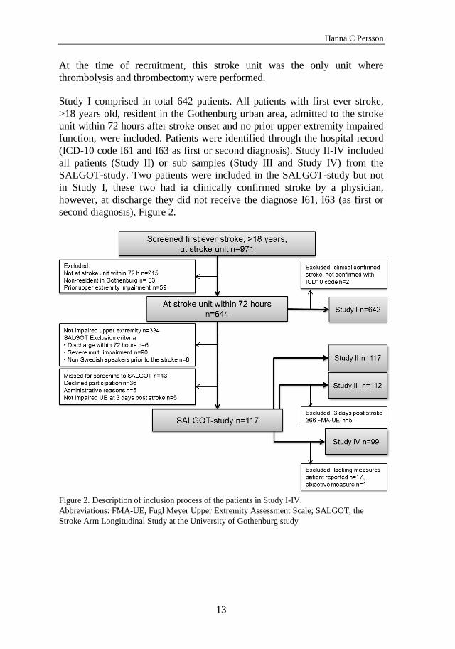
At the time of recruitment, this stroke unit was the only unit where
thrombolysis and thrombectomy were performed.
Study I comprised in total 642 patients. All patients with first ever stroke,
>18 years old, resident in the Gothenburg urban area, admitted to the stroke
unit within 72 hours after stroke onset and no prior upper extremity impaired
function, were included. Patients were identified through the hospital record
(ICD-10 code I61 and I63 as first or second diagnosis). Study II-IV included
all patients (Study II) or sub samples (Study III and Study IV) from the
SALGOT-study. Two patients were included in the SALGOT-study but not
in Study I, these two had ia clinically confirmed stroke by a physician,
however, at discharge they did not receive the diagnose I61, I63 (as first or
second diagnosis), Figure 2.
Description of inclusion process of the patients in Study I-IV. Figure 2.
Abbreviations: FMA-UE, Fugl Meyer Upper Extremity Assessment Scale; SALGOT, the
Stroke Arm Longitudinal Study at the University of Gothenburg study

Upper extremity functioning during the first year after stroke
14
Study I: Upper extremity function was assessed within 72 hours post stroke
onset using item F, G and H from the Modified Motor Assessment Scale
according to Uppsala Akademiska sjukhus(M-MAS UAS-95)122,123
. The three
items including upper arm function, hand movements and advanced hand
activity were summed to a score of 0-15 points122
. Impaired upper extremity
function was defined as a ≤14 points.
Study II-IV: Impaired upper extremity function, 3 days post stroke onset was
defined as <57 points on the ARAT (0-57 points)85,86
.
The SALGOT-study was planned with purpose to comprise the different
domains of the ICF28,118
. The study designs, analysis and data source of each
study as well as the specific aims in SALGOT-study118
are described in Table
2. The demographics of included patients in each study are provided in
Table 3.
Table 2. An overview of the study design
Study I II III IV
Design Cross sectional
study
Longitudinal
study
Cohort study Cross sectional
study
Analyses
Descriptive Multivariate Association
Prediction
Association
Agreement
Data source Patients charts,
The Riks-Stroke
Collaboration
SALGOT-data SALGOT-data
SALGOT-data
Specific aims
in SALGOT-
study
- Follow recovery
of upper
extremity by
using clinical
measures of
body function,
activity and
participation
after stroke,
(aim A).
To predict
function at 12
months by
analysis of data
gathered at first
week after onset
of stroke,
(aim D).
To gather the
assessments of
participants’ self-
perceived upper
extremity
function over the
first year after
stroke, (aim C).
Abbreviations: SALGOT, Stroke Arm Longitudinal Study at the University of Gothenburg
Hanna C Persson
15
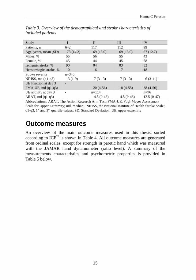
Table 3. Overview of the demographical and stroke characteristics of included patients
Study I II III IV
Patients, n 642 117 112 99
Age, years, mean (SD) 73 (14.2) 69 (13.0) 69 (13.0) 67 (12.7)
Males, %
Female, %
55
45
56
44
55
45
42
58
Ischemic stroke, %
Hemorrhagic stroke, %
90
10
84
16
83
17
82
18
Stroke severity
NIHSS, md (q1-q3)
n=345
3 (1-9)
7 (3-13)
7 (3-13)
6 (3-11)
UE function at day 3
FMA-UE, md (q1-q3)
-
20 (4-56)
18 (4-55)
38 (4-56)
UE activity at day 3
ARAT, md (q1-q3)
- n=114
4.5 (0-43)
4.5 (0-43)
n=96
12.5 (0-47)
Abbreviations: ARAT, The Action Research Arm Test; FMA-UE, Fugl-Meyer Assessment
Scale for Upper Extremity; md, median; NIHSS, the National Institute of Health Stroke Scale;
q1-q3, 1st and 3rd quartile values; SD, Standard Deviation; UE, upper extremity
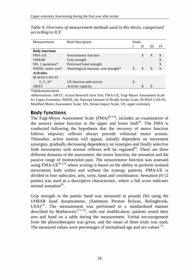
An overview of the main outcome measures used in this thesis, sorted
according to ICF28
is shown in Table 4. All outcome measures are generated
from ordinal scales, except for strength in paretic hand which was measured
with the JAMAR hand dynamometer (ratio level). A summary of the
measurements characteristics and psychometric properties is provided in
Table 5 below.
Upper extremity functioning during the first year after stroke
16
Table 4. Overview of measurement methods used in this thesis, categorized according to ICF
Measurements Brief description Study
I II III IV
Body functions
FMA-UE Sensorimotor function X X X
JAMAR Grip strength X
SIS, 2 questions* Perceived hand strength X
NIHSS, motor arm* Neurological outcome, arm strength* X X X X
Activities
M-MAS UAS-95
F, G, H*
UE function and activity
X
ARAT Activity capacity X X
*Subdomains/items
Abbreviations: ARAT, Action Research Arm Test; FMA-UE, Fugl-Meyer Assessment Scale
for Upper Extremity; NIHSS, the National Institute of Health Stroke Scale; M-MAS UAS-95,
Modified Motor Assessment Scale; SIS, Stroke Impact Scale; UE, upper extremity
The Fugl-Meyer Assessment Scale (FMA)83,124
, includes an examination of
the sensory motor function in the upper and lower limb83
. The FMA is
conducted following the hypothesis that the recovery of motor function
follows stepwise; reflexes always precede volitional motor actions.
Thereafter, active motion will appear, initially dependent on movement
synergies, gradually decreasing dependency on synergies and finally selective
limb movements with normal reflexes will be regained83
. There are three
different domains of the assessment; the motor function, the sensation and the
passive range of motion/joint pain. The sensorimotor function was assessed
using FMA-UE83,124
where scoring is based on the ability to perform isolated
movements both within and without the synergy patterns. FMA-UE is
divided in four subscales; arm, wrist, hand and coordination. Sensation (0-12
points) was used as a descriptive characteristic, where a full score indicates
normal sensation83
.
Grip strength in the paretic hand was measured in pounds (lb) using the
JAMAR hand dynamometer, (Sammons Preston Rolyan, Bolingbrook,
USA)121
. The measurement was performed in a standardized manner
described by Mathiowetz121,125
, with one modification; patients rested their
arm and hand on a table during the measurement. Verbal encouragement
from the physiotherapist was given, and the mean of three trials was used.
The measured values were percentages of normalised age and sex values125
.

Hanna C Persson
17
The Stroke Impact Scale (SIS) 3.0119,120
is a stroke specific patient-reported
health status measure. The SIS was developed on the basis of the perspectives
of patients, caregivers and health professionals119
. The SIS assess
multidimensional stroke outcomes and includes eight domains (strength,
hand function, activities of daily living, mobility, communication, emotion,
memory and thinking, participation)119,120
.
To assess outcome and degree of recovery for patients with stroke the
National Institute of Health Stroke Scale (NIHSS)126
was used. The NIHSS
comprises the following items; level of consciousness, eye movements,
visual tests, face, extremity strength, ataxia, sensory function, language and
speech, extinction and inattentions.
Upper extremity activity capacity was assessed using the Action Research
Arm Test (ARAT)85,86,127
which is intended to assess UE dexterity in basic
movements. The ARAT consists of 19 items scores that are summed to 4
hierarchical sub scores; gross motor, grasp, grip and pinch. The ARAT is
based on movement performance and on a time limit, and was performed
following a standard approach127,128
.
The Modified Motor Assessment Scale (M-MAS UAS-95)123
is developed
from the original version of Motor Assessment Scale122
designed to assess
everyday motor function in patients with stroke. The assessment scale was
developed on the basis of Carr and Shepherds theories of motor relearning
after stroke129
and assesse upper arm motor function and hand movements (F,
G, H), sitting balance, transfers such as lying to sitting standing and
walking123
.

Upper extremity functioning during the first year after stroke
18
Table 5. Overview of the characteristics and psychometric properties of the main measurements included in the thesis.
Body function Activities
FMA-UE
Motor function83
JAMAR121,125 SIS119,120 NIHSS126 ARAT85,86,127,128 M-MAS UAS-
95122,123
Type Observational
rating scale
Dynamometer,
grip strength
Self-reported
outcome
Observational
rating scale
Observational
rating scale
Observational
rating scale
Scale level Ordinal Ratio (Pound) Ordinal Ordinal Ordinal Ordinal
Range sum
score
0-66 Reference values Each domain 0-
100
0-46* 0-57 0-55
Number of
items
33 - 59 13 19 11
Item range 0-2 - 1-5 Varies, 0-2, 3 or 4 0-3 1-5
Reliability Excellent31,124,130 Excellent31 Excellent31,119,120 Adequate to
Excelent126,131,132
Excellent86,127,130 Excellent123
Validity
Excellent133-135 Adequate to
Excellent136
Excellent Strength
domain119
Varies, Adequate-
Excellent132
Excellent137 Adequate to
Excellent123
Responsiveness Large84,138 - Large sub-acute119 - Large137,138 -
MCID 7 points (10%) 139
11-13 pounds140 Varies, within
domains (4.5-17.8
points)141
2 points82,126 6 points (10%) 139 -
Abbreviations: ARAT, Action Research Arm Test; FMA-UE, Fugl-Meyer Assessment Scale for Upper Extremity; JAMAR, Jamar Hand
Dynamometer; M-MAS UAS-95, The Modified Motor Assessment Scale; MICD, minimal clinical important difference; NIHSS, the National Institute
of Health Stroke Scale; SIS, Stroke Impact Scale; UE, upper extremity.
* NIHSS, lower total score indicate less severe stroke.
Reliability: Excellent ≥0.75, Adequate 0.-074 ICC, kappa statistics142
Validity: Excellent ≥0.60, Adequate 0.31-0.59 Construct/convergent, concurrent, Excellent ≥0.90, Adequate 0.70-0.89 ROC, ACU142
Responsiveness < 0.5 small, 0.5-0.8 moderate, ≥0.8 large Standardised effect sizes, sensitive to change142

Hanna C Persson
19
Outcome measures/variables used for description of the included population
are described below and sorted according to ICF28
.
Consciousness at arrival to hospital (Study I) was assessed using a modified
version of the Reaction Level Scale (RLS-85)143,144
according to criteria from
the Riks-Stroke, The Swedish Stroke Register145
. The patient’s consciousness
was stratified into three categories; Alert and oriented (RLS 1), drowsy or
confused (RLS 2-3) and unconscious only responding to stimuli (RLS 4-8)145
.
The screening test Barrow Neurological Institute Screen for higher cerebral
function (BNIS)146
was developed to assess a variety of higher cerebral
function. The BNIS has been shown to have good validity in a Swedish
population with different neurological deceases (high sensitivity)147,148
, and to
be useful as screening test in patients with ischemic stroke149
. The BNIS pre-
screening including three initial items assessing arousal level alertness, basic
communication skills and level of cooperation was assessed (ordinal score 0-
9 points).
The modified Rankin Scale (mRS)150
0-6 points, evaluates the patient over-all
status after a stroke, at a seven grade ordinal scale. No symptoms at all
corresponds to 0, and 5 corresponds to severe disability and 6 corresponds to
death.
In order to detect cognitive impairment, a subset of items from the NIHSS
score was used, entitled the COG-4151
. The COG-4, is a comprehensive score
of four items from NIHSS; orientation (item 1b), executive function,
language and inattention. The COG-4 is used as screening of cognitive
impairment and are scored 0-9 points, where 0 indicates no cognitive
reduction151
.The COG-4 has been shown to have similar possibilities to
detect severe cognitive impairment as the Mini-Mental State Examination
(MMSE)151,152
.
The stroke location was recorded from patients’ charts, in Study I as
right/left/bilateral/unclear and in Study II-IV right/left/bilateral/brain
stem/unknown. Acute medical treatment (conservative, thrombolysis,
thrombectomy), length of stay at stroke unit and care pathway were collected
from patients’ charts. The amount of in- or outpatient rehabilitation received
Upper extremity functioning during the first year after stroke
20
was noted at each assessment. Daily support in activities of daily living pre
and post stroke, as well as mobility pre stroke was recorded from the Riks-
Stroke register145
.
The causes of the ischemic stroke were defined and sub-categorized
according to the classification system the SSS-TOAST (Stop Stroke Study
Trial of Org 10172)153
, where each category consisting of subgroups
according to evident, probable or possible. In a second step the SSS-TOAST
were convert to the original classification of TOAST; the Trial of Org 10172
in Acute Stroke Treatment (TOAST)105
.
Using clinical neurological findings, the Oxfordshire Community Stroke
Project classification (Bamford classification) divides ischemic stroke into
four sub-groups according to stroke location154
. The four groups are total
anterior circulation infarct, TACI; partial anterior circulation infarct PACI;
Lacunar anterior circulation infarct, LACI; and posterior circulation infarct,
POCI154
.
In Study I, data from patients who received care at the stroke unit were
gathered retrospectively from the medical charts. All patients with ICD-10
code I61 or I63 were screened for inclusion. More than 1800 charts were
screened and patients with first ever stroke were included and those with
recurrent stroke were omitted. The TOAST105
, the Bamford classification154
,
the NIHSS126
at onset and the mRS150
at discharge from hospital were
assessed from the patient’s medical chart. A systematic error resulted in 52
patients not being correctly classified according to the TOAST criteria;
therefore all charts were re-evaluated (by KSS) post publication of Study I.
Also, patients with missing NIHSS scores were evaluated (by KSS) post
publication of Study 1. Results from these evaluations are present within this
thesis.
According to clinical practice, the M-MAS UAS-95122,123
was performed as a
screening of general function as well as upper extremity function, by the
physiotherapists at the stroke unit within the first 3 days post stroke. The
results were noted on a screening sheet by the physiotherapists at the stroke
unit. In Study I the upper extremity function was determined from the
patient’s chart in by two of the authors (Study I, HCP and MP) and defined as
impaired or not impaired in the following steps:
Hanna C Persson
21
1) A documented assessment of the M-MAS UAS-95 (within
72 hours post stroke). The three items of upper arm function
(item F), hand movements (item G) and advanced hand
activities (item H) were summed, and impaired upper
extremity function correspond to M-MAS UAS-95 <14
points.
2) Evaluation of other documented standardized assessment of
upper extremity function (within 72 hours post stroke) by
physiotherapist, occupational therapist or physicians at the
stroke unit.
The upper extremity function was assessed with the M-MAS UAS-95 in
80.4% of patients, and in 19.6% of the patients, the two authors (HCP and
MP) assessed the patients’ charts according to the procedure. In a second
step, a comparison of the evaluated upper extremity function and NIHSS at
admission to hospital (sub score arm strength) was performed
Prior to start of inclusion to the SALGOT-study, a pilot study including five
patients from a convenience sample at the stroke unit was conducted. The
SALGOT-study protocol was revised (small changes) according to results of
the pilot study.
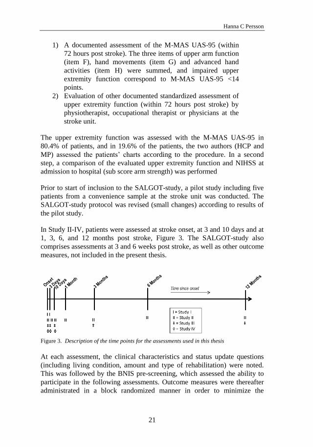
In Study II-IV, patients were assessed at stroke onset, at 3 and 10 days and at
1, 3, 6, and 12 months post stroke, Figure 3. The SALGOT-study also
comprises assessments at 3 and 6 weeks post stroke, as well as other outcome
measures, not included in the present thesis.
Description of the time points for the assessments used in this thesis Figure 3.
At each assessment, the clinical characteristics and status update questions
(including living condition, amount and type of rehabilitation) were noted.
This was followed by the BNIS pre-screening, which assessed the ability to
participate in the following assessments. Outcome measures were thereafter
administrated in a block randomized manner in order to minimize the

Upper extremity functioning during the first year after stroke
22
systematic bias. The two assessments of ARAT or JAMAR hand
dynamometer were conducted in a random order. Next, the FMA-UE was
assessed, and finally the SIS was recorded (Figure 4). The test order and the
reasons for missed or unsuccessful test results were recorded in a test
protocol.
Illustration of main outcome measures at each time point after stroke onset. Figure 4.
Abbreviations: ARAT, The Action Research Arm Test; FMA-UE, Fugl-Meyer Assessment
Scale for Upper Extremity; M-MAS UAS-95, The Modified Motor Assessment Scale;
NIHSS, the National Institute of Health Stroke Scale; SIS, the Stroke Impact Scale
Clinical characteristics were gathered at the first assessment, from the
patients’ charts, as well as from the Riks-Stroke145
in Study II-IV. Also in
Study II-IV values from TOAST, Bamford criteria and NIHSS at onset were
assessed from the patients’ charts.
In Study IV, 2 questions from the SIS 3.0119,120
strength domain (domain 1),
were used: In the past week, how would you rate the strength of your; 1a)
Arm that was most affected by your stroke? 1b) Grip of your hand that was
most affected by your stroke?. The patients rated their strength on a verbal,
five point ordinal scale from no strength at all (1) to a lot of strength (5).
The majority of assessments in the SALGOT-study were performed at the
hospital in a special test room, which was also used at the follow-up
assessments. If the patient was unable to travel to the test room, assessments
could be performed in the patient’s home or in their current care setting.
Three physiotherapists, not otherwise involved in the care, performed the
assessments (after training) following a standardized protocol118
. At the
follow-up assessments, the physiotherapists were not familiar with the results
from the previous assessment, except for the time point at 12 months, where
Hanna C Persson
23
the patient received information on their results from all the assessments
during the past year. If a patient not could contribute and participate in the
assessment at 1 month, the patient only was followed at the 1 year follow up.
The studies in this thesis did not interfere with the routine care rehabilitation
or care pathways of the included patents. Patients followed the ordinary
planning of rehabilitation and discharge procedure. Patients received
individually adjusted, functional, task-specific rehabilitation from the first
day in the stroke unit. Rehabilitation in this thesis includes physiotherapy
and/or occupational therapy and the amount of received rehabilitation at each
assessment was recorded. Inpatient rehabilitation was defined as ongoing
rehabilitation at the hospital (at stroke unit or rehabilitation unit), and
outpatient rehabilitation as rehabilitation received when patients’ lived at
their own homes or at community care units.
The guidelines for reporting observational studies, Strengthening of
Reporting of Observational Studies in Epidemiology (STROBE)155
were
followed.
Due to administrative reasons, 20 patients out of 117 in Study II and 18
patients out of 99 in Study III were not assessed using FMA-UE at 10 days
post stroke, but were assessed with all other outcome measures at 10 days. In
order to enable use of these 20 patients FMA-UE data, a score for each non-
assessed patient was estimated in the following manner: The mean change
from 3 to 10 days after onset for all patients assessed at both time points
(Study II, n=97, Study III n=94) was calculated. By adding the mean change
(this was 5 points) to each of the missing 20 (Study II) and 18 (Study III)
patients’ FMA-UE scores at 3 days, an estimated value at 10 days post stroke
was obtained. The estimated value at assessment at 10 days could not exceed
the score of the subsequent assessment.
In study III, the predictive validity of the FMA-UE≥32 to correctly classify a
patient’s motor ability to drink from a glass (the drinking task) was
investigated. In a previous publication from the SALGOT-data156
, patients
were included if they could perform a drinking task. The drinking task
included reaching, grasping, lifting the glass to drink a sip of water. All
patients included in that study156
had a score of ≥32 points on the FMA-UE,
and therefore this cut-off was used to classify the ability to drink from a
Upper extremity functioning during the first year after stroke
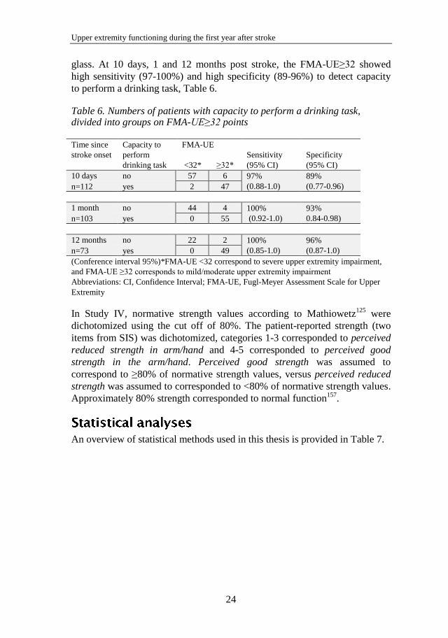
24
glass. At 10 days, 1 and 12 months post stroke, the FMA-UE≥32 showed
high sensitivity (97-100%) and high specificity (89-96%) to detect capacity
to perform a drinking task, Table 6.
Table 6. Numbers of patients with capacity to perform a drinking task, divided into groups on FMA-UE≥32 points
Time since
stroke onset
Capacity to
perform
drinking task
FMA-UE
Sensitivity
(95% CI)
Specificity
(95% CI)
<32*
≥32*
10 days no 57 6 97%
(0.88-1.0)
89%
(0.77-0.96) n=112 yes 2 47
1 month no 44 4 100%
(0.92-1.0)
93%
0.84-0.98) n=103 yes 0 55
12 months no 22 2 100%
(0.85-1.0)
96%
(0.87-1.0) n=73 yes 0 49
(Conference interval 95%)*FMA-UE <32 correspond to severe upper extremity impairment,
and FMA-UE ≥32 corresponds to mild/moderate upper extremity impairment
Abbreviations: CI, Confidence Interval; FMA-UE, Fugl-Meyer Assessment Scale for Upper
Extremity
In Study IV, normative strength values according to Mathiowetz125
were
dichotomized using the cut off of 80%. The patient-reported strength (two
items from SIS) was dichotomized, categories 1-3 corresponded to perceived
reduced strength in arm/hand and 4-5 corresponded to perceived good
strength in the arm/hand. Perceived good strength was assumed to
correspond to ≥80% of normative strength values, versus perceived reduced
strength was assumed to corresponded to <80% of normative strength values.
Approximately 80% strength corresponded to normal function157
.
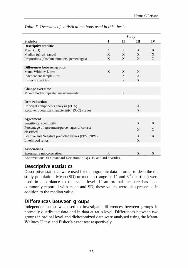
An overview of statistical methods used in this thesis is provided in Table 7.
Hanna C Persson
25
Table 7. Overview of statistical methods used in this thesis
Study
Statistics I II III IV
Descriptive statistic
Mean (SD) X X X X
Median (q1-q3, range) X X X X
Proportions (absolute numbers, percentages) X X X X
Differences between groups
Mann-Whitney U-test X X X
Independent sample t-test X X
Fisher’s exact test X X
Change over time
Mixed models repeated measurements X
Item reduction
Principal components analysis (PCA) X
Receiver operation characteristic (ROC) curves X
Agreement
Sensitivity, specificity X X
Percentage of agreement/percentages of correct
classified X X
Positive and Negative predicted values (PPV, NPV) X X
Likelihood ratios X
Associations
Spearman rank correlation X X X
Abbreviations: SD, Standard Deviation; q1-q3, 1st and 3rd quartiles,
Descriptive statistics were used for demographic data in order to describe the
study population. Mean (SD) or median (range or 1st and 3
rd quartiles) were
used in accordance to the scale level. If an ordinal measure has been
commonly reported with mean and SD, these values were also presented in
addition to the median value.
Independent t-test was used to investigate differences between groups in
normally distributed data and in data at ratio level. Differences between two
groups in ordinal level and dichotomized data were analysed using the Mann-
Whitney U test and Fisher’s exact test respectively.
Upper extremity functioning during the first year after stroke
26
There is no non-parametric statistical method that handles differences
between groups in longitudinal data. Therefore in Study II, in order to
investigate extent of recovery regarding functioning as well as to explore
differences between two types of stroke (hemorrhagic and ischemic stroke),
the Mixed models repeated measurement158-160
was used. The Mixed model
repeated measurements enables maximum use of data as it can handle
missing values, calculate individual changes over time as well as investigate
differences in change over time between groups. Two separate models were
used; one for motor function (FMA-UE) and one for activity capacity
(ARAT) both including the independent variables; type of stroke, age and
stroke severity. Differences between the two types of stroke were further
investigated using values generated from Mixed models repeated
measurements, where the impact of significant factors on change over time in
function and activity were explored. Differences in the extent of change in
motor function and activity capacity between the two types of stroke were
calculated at four time periods; from 3 days to 1, 3, and 12 months post
stroke respectively, as well as from 1 to 3 months. When calculating
differences in changes in FMA-UE or ARAT scores, significant factors (from
the Mixed models repeated measurements) were controlled for.
In order to achieve a clinical assessment with potential to be feasible in
routine care at the stroke unit early after stroke, an assessment that was short
and easy to perform was required. The hypothesis was; that ARAT has
potential to detect limitations in upper extremity, the minimal number of
items needed to capture most of the variation could be identified using
principal components analysis (PCA) based on the SALGOT-population
(n=117). Only components with eigenvalues ≥1 were selected and ARAT
items with loading values greater than 0.6 were considered, according to
Kaiser’s criterion159
. In the next step, the ARAT items with the most wide
variation in difficulty (out of possible items), were identified according to
Koh et al161
. The selection of the sub-set of ARAT items was carried out by
identification of an optimal cut-off level of the score and was valid to all time
points of assessments during the year. This was identified using receiver
operation characteristic (ROC) curves162
.
Agreement between two types of outcome measures was investigated with 2-
ways contingency tables. Sensitivity is the probability that a patient that has
problems is classified correctly; specificity is the probability that a patient
without problems is classified correctly. The predictive values could be

Hanna C Persson
27
positive (PPV), indicating the probability that a patient without problems is
classified as having problems, and negative (NPV) the probability that a
patients having problems is classified as having no problems104,110
. The
Likelihood ratios, indicates the overall value of information of the predictive
test and can be positive and negative111
. In Study III and IV sensitivity,
specificity, PPV, NPV, percentages of agreement/percentages of correctly
classified patients, and likelihood ratios (positive, negative) were investigated
at each time point including 95% confidence intervals (CIs)110
.
Spearmans rank correlation (rho) was used to investigate the associations of
impaired upper extremity function and patient’s care pathway (mortality,
length of stay, discharge) (Study I). In Study III, rho was used to investigate
relationship between ARAT items and in Study IV, correlations between
perceived arm and hand strength and the measured strength (capacity).
Statistical analyses were mainly performed in the IBM Statistical Package for
Social Sciences (SPSS version 21.0, for Windows). In Study II, the analysis
using the Mixed models repeated measurements was performed using the
Statistical Analysis Program, (SAS proc mix, version 9.3 SAS Institute Inc.,
Cary, N.C USA). The Confidence Intervals of likelihood ratios were analysed
using the Prop CIs Package in R version 3.1.1 (2014-07-10).
The SALGOT study received ethical approval by The Regional Ethical
Review Board in Gothenburg, with the reference number 225-08, approved
5th of May 2008, Study II-IV. In Study I, a complementary approval (to 225-
08) was needed, approval was received on the 10th of January 2011, reference
number T801-10. In Study I, complementary data were collected from the
patients’ charts, and according to Swedish law on Data Protection Authority
(Personuppgiftslagen, Swedish law No. SFS 1998:204) patients’ informed
consent is not mandatory when data is gathered for clinical use. Approval to
read the patients charts was given from the responsible authority at the
hospital and on the wards. All patients or next of kin gave informed oral and
written consent for participation in the SALGOT-study (Study II-IV). The
Helsinki Declaration163
was followed. The SALGOT-study (included Study
II-IV) was registered at the clinical trials.gov: NCT01115348, May 3, 2010.
Upper extremity functioning during the first year after stroke
28
The differences between included, non-included and drop-outs as well as the
main findings from Study I-IV are presented in this section.
Differences between included patients, drop-outs and non-included patients
from Study I to the SALGOT-study were investigated. There were no
differences in gender (p=0.477) or in stroke severity (p=0.157) in patients
that were missed for screening to SALGOT, declined participation and not
included due to administrative reasons (total n=84), compared to the
included patients in the SALGOT-study (n=117), Figure 2. However,
included patients were significantly younger (p=<0.001) with a mean age of
69.3 years, compared to the other group who had a mean age of 76.1 years.
The percentage of patients with ischemic stroke was lower (74%) in the
included patients compared with the other group (94%, p=0.029).
In Study I there were no differences in gender (p=0.134), type of stroke
(p=0.134) or stroke severity (p=0.491) of patents with prior upper extremity
impairment (n=59) compared to patients who were included in Study I
(n=642).
In Study II, there were no drop-out from assessments at 3 and 10 days post
stroke. The main reasons for drop-out at 12 months were in both ischemic
and hemorrhagic stroke death, withdrawal from study or new stroke. Eight
patients could not contribute in the assessment at 1 month and these patients
were only followed at the 12 month assessment; two out of these eight
patients participated in the follow-up at 12 months whereof both lived at
nursing homes and had full support in their activities of daily living and had
low or no function or activity in upper extremity; an additional two patients
survived the first year after stroke, of which one declined to participate, and
one had moved from the area at the 12 months follow-up; four patients died
within the first 12 months.
In Study III, there were no drop-out from assessments at 3 and 10 days post
stroke. At assessment at 1 and 12 months, 9 respective 39 patients did not
participate, and the reasons for non-assessed in each group are specified in
Figure 5. The reasons for drop-out at 12 months did not differ in patients with
initially severe or moderate/mild impairment regarding; type of stroke, stroke
severity, initial upper extremity function (severe or moderate/mild) or gender.

Hanna C Persson
29
However, the patients that participated at the 12 months assessment were
significantly (p=0.017) younger compared to the drop-outs.
Patients assessed and non-assessed including the reasons for drop-out (Study Figure 5.
III). The population was divided due to upper extremity function at stroke onset; severe
FMA-UE ≥32, mod/mild FMA-UE <32. Note that the vertical axis starts at 60%, all
patients below 60% participated. Abbreviations: Mod, moderate
In Study IV, no differences between included (n=99) and non-included
patients (n=18) from the SAGLOT-study were seen in gender and type of
stroke (ischemic or hemorrhagic stroke). However, the non-included patients
were older (p=0.02) and had a significant more severe stroke at onset (higher
NIHSS score, p= <0.001).
In total, 642 patients with first ever hemorrhagic or ischemic stroke, admitted
to the stroke unit at Sahlgrenska University hospital were included in Study I.
Re-evaluated values of the TOAST and NIHSS score (post publication of
Study I) are shown in Table 8, cardio-aortic embolism was the most common
cause of stroke (41%) followed by small artery occlusion (30%). The
frequency of cardio-aortic embolism and small artery occlusion were the two
categories of TOAST that showed different values (Table 8) compared to the
published results in Study 1. However, these differences did not affect the
main results in Study I. The NIHSS-score was re-evaluated in total 565
patients with a mean of 6.0, median of 3.0 points (min-max 0-33), Table 8,

Upper extremity functioning during the first year after stroke
30
similar results as present in Study I, n=345man 5.5, median 3.0, min-max
0.33).
Table 8. Characteristics of included patients in Study I, re-evaluated.
n = 642
TOAST-criteria,
causes of ischemic stroke,
n (%)
Large artery atherosclerosis
Cardio-aortic embolism
Small artery occlusion
Other causes
Undetermined cause
Unclear
102 (18)
236 (41)
171 (30)
25 (4)
14 (2)
27 (5)
NIHSS 0-42 points, n=565
Mean 5.5
Md 3.0
min-max 0-33
Abbreviations: NIHSS, the National Institute of Health Stroke Scale; Md, Median; TOAST,
the Trial of Org 10172 in Acute Stroke Treatment
The most frequent subtype according to Bamford classification was the
Lacunar stroke, present in 44% of the study population. Out of the patients
with first ever stroke, admitted to the stroke unit within 72 hours, 48% had
impaired upper extremity function, Study I, Figure 1.
Impaired upper extremity function correlated to higher age (p=<0.004). There
were no differences between patients (ischemic) who had received treatment
with thrombolysis (8.9%), or patients not treated with thrombolysis
(p=<0.482) regarding upper extremity
function assessed within 72 hours. Patients
with the Bamford classification TACI had
impaired upper extremity function more
often (p=<0.001) than patients classified as
LACI or PACI (p<0.031). Of the 642
patients admitted to stroke unit, 71 patients
(11.1%) died during the stay at the stroke
unit, and 571 (88.9%) survived, Figure 6.
Care pathway of included 642 Figure 6.
patients in Study I.
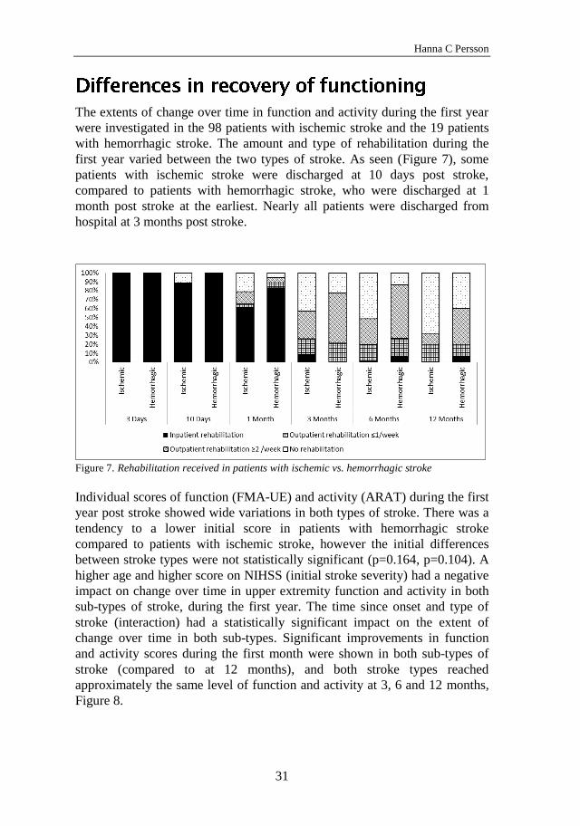
Hanna C Persson
31
The extents of change over time in function and activity during the first year
were investigated in the 98 patients with ischemic stroke and the 19 patients
with hemorrhagic stroke. The amount and type of rehabilitation during the
first year varied between the two types of stroke. As seen (Figure 7), some
patients with ischemic stroke were discharged at 10 days post stroke,
compared to patients with hemorrhagic stroke, who were discharged at 1
month post stroke at the earliest. Nearly all patients were discharged from
hospital at 3 months post stroke.
Rehabilitation received in patients with ischemic vs. hemorrhagic stroke Figure 7.
Individual scores of function (FMA-UE) and activity (ARAT) during the first
year post stroke showed wide variations in both types of stroke. There was a
tendency to a lower initial score in patients with hemorrhagic stroke
compared to patients with ischemic stroke, however the initial differences
between stroke types were not statistically significant (p=0.164, p=0.104). A
higher age and higher score on NIHSS (initial stroke severity) had a negative
impact on change over time in upper extremity function and activity in both
sub-types of stroke, during the first year. The time since onset and type of
stroke (interaction) had a statistically significant impact on the extent of
change over time in both sub-types. Significant improvements in function
and activity scores during the first month were shown in both sub-types of
stroke (compared to at 12 months), and both stroke types reached
approximately the same level of function and activity at 3, 6 and 12 months,
Figure 8.

Upper extremity functioning during the first year after stroke
32
The extent of change over time in function (FMA-UE) and activity (ARAT) Figure 8.
during the first year (mean scores), in hemorrhagic and ischemic stroke.
To further explore differences in the extent of change in function and activity
between patients with ischemic or hemorrhagic stroke, four time periods were
investigated using mean values of FMA-UE and ARAT, adjusted for
significant variables (age and initial stroke severity). Patients with
hemorrhagic stroke gained more functioning compared to patients with
ischemic stroke from onset to one year; in function 13.2 points on FMA-UE
(p=0.0015) and in activity 13.8 points on ARAT (p=0.002). The largest
differences (in the shortest time period) in the extent of change over time
between stroke types were seen between 1 to 3 months post stroke, where
patients with hemorrhagic stroke improved their motor function (FMA-UE
8.6 points) and their activity (ARAT 10.9 points) more than patients with
ischemic stroke. These findings indicate that patients with hemorrhagic
stroke may have a different pattern in change over time in the first 3 months
post stroke, compared to patients with ischemic stroke, but both groups reach
similar results at 3 months and thereafter.
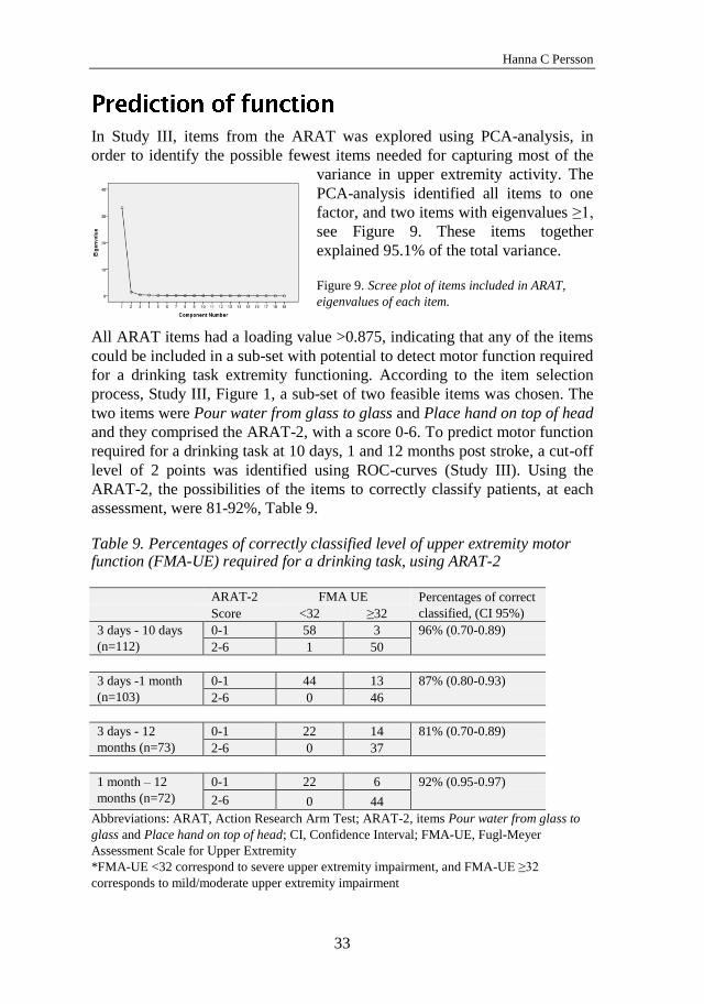
Hanna C Persson
33
In Study III, items from the ARAT was explored using PCA-analysis, in
order to identify the possible fewest items needed for capturing most of the
variance in upper extremity activity. The
PCA-analysis identified all items to one
factor, and two items with eigenvalues ≥1,
see Figure 9. These items together
explained 95.1% of the total variance.
Scree plot of items included in ARAT, Figure 9.
eigenvalues of each item.
All ARAT items had a loading value >0.875, indicating that any of the items
could be included in a sub-set with potential to detect motor function required
for a drinking task extremity functioning. According to the item selection
process, Study III, Figure 1, a sub-set of two feasible items was chosen. The
two items were Pour water from glass to glass and Place hand on top of head
and they comprised the ARAT-2, with a score 0-6. To predict motor function
required for a drinking task at 10 days, 1 and 12 months post stroke, a cut-off
level of 2 points was identified using ROC-curves (Study III). Using the
ARAT-2, the possibilities of the items to correctly classify patients, at each
assessment, were 81-92%, Table 9.
Table 9. Percentages of correctly classified level of upper extremity motor function (FMA-UE) required for a drinking task, using ARAT-2
ARAT-2 FMA UE Percentages of correct
classified, (CI 95%) Score <32 ≥32
3 days - 10 days
(n=112)
0-1 58 3 96% (0.70-0.89)
2-6 1 50
3 days -1 month
(n=103)
0-1 44 13 87% (0.80-0.93)
2-6 0 46
3 days - 12
months (n=73)
0-1 22 14 81% (0.70-0.89)
2-6 0 37
1 month – 12
months (n=72)
0-1 22 6 92% (0.95-0.97)
2-6 0 44
Abbreviations: ARAT, Action Research Arm Test; ARAT-2, items Pour water from glass to
glass and Place hand on top of head; CI, Confidence Interval; FMA-UE, Fugl-Meyer
Assessment Scale for Upper Extremity
*FMA-UE <32 correspond to severe upper extremity impairment, and FMA-UE ≥32
corresponds to mild/moderate upper extremity impairment

Upper extremity functioning during the first year after stroke
34
In summary, using ARAT-2 had a high probability of accurately predict the
motor function required for the drinking task in patients with >2 points (high
specificity, high NPV, high negative likelihood ratios), during the first year.
The lower percentages of correctly classified patients from 3 days to 12
months (81%), as well as lower specificity, lower PPV and lower positive
likelihood ratios indicated less acuracy in ARAT-2 to predict patients with
very little arm and hand activity (ARAT-2 ≤1) at 3 days post stroke. A more
detailed description of the ARAT-2 sensitivity, specificity, predictive values
and likelihood ratios at the different time points are present in Study III.
In Study IV, higher levels of perceived arm and grip strength were associated
with higher capacity measured grip strength (Study IV, Figure 1b) except for
in category 5 (a lot of strength), only rated by only four patients. The
correlations at 10 days post stroke in patient’s perception of strength in their
arm/hand and capacity measured grip strength were 0.82 (p=<0.001) and 0.87
(p=<0.001) respectively.
When dichotomizing patient-reported strength (dichotomized SIS) and
capacity measured strength (dichotomised at 80%), high sensitivity (0.86-
0.87) and high PPV (0.86-0.91) values were found (Study IV, Table 2). The
high PPV indicated that patients with normal (>80%) upper extremity
strength assessed their strength more correctly compared to those with
impaired strength. Lower specificity (0.62-0.77) and NPV (0.64-0.67) were
shown between the two measurements.

Hanna C Persson
35
The SALGOT-study aimed to describe recovery in function and activity
longitudinally from stroke onset to one year post stroke. The individual
scores in FMA-UE for each patient are illustrated in Figure 10, were a wide
dispersion in recovery of function could be seen over the first year after
stroke onset. A summary of main results from Studies I-IV, is shown in
Figure 11.
Individual scores at each assessment on Fugl-Meyer Assessment of Upper Figure 10.
Extremity, illustrating sensory motor function in upper extremity from day 3 to 12 months
post stroke. Each line represents one of the 117 patients in SALGOT-study.
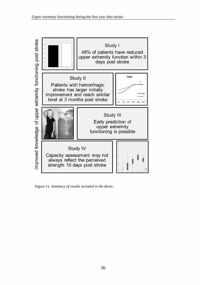
Upper extremity functioning during the first year after stroke
36
Summary of results included in the thesis. Figure 11.
Hanna C Persson
37
The focus of this thesis was to investigate upper extremity functioning after
stroke from different perspectives. All patients with their first ever stroke and
admitted to a stroke unit were included. Patients with impaired upper
extremity function were followed during the first year post stroke in the
longitudinal SALGOT-study. The data collected enabled analysis of upper
extremity function and activity in different aspects, in a cohort of patients
with stroke. This thesis has aimed to capture upper extremity functioning
according to the ICF domains, body function and activity using capacity and
patient-reported outcome measures.
The finding that 48% of all patients at the stroke unit had impaired upper
extremity function within 72 hours post stroke was lower than expected and
gave an explanation to why inclusion to SALGOT-study required more time
than expected (Study I). Patients with impaired upper extremity function
were older, had a more severe stroke and they also required a longer stay in
the stroke unit, compared to patients without upper extremity impairment.
The result that less than half of patients at a stroke unit had impaired upper
extremity function is a new finding, where previous studies have reported
frequencies of 70-80%2,3,36
. The difference in prevalence may have several
potential explanations. First; regarding the study population, only patients
admitted to the stroke unit within 72 hours with a first ever stroke were
included. This is in contrast to the Copenhagen study3 which also included
patients with recurrent stroke. Furthermore, patients’ who were admitted to
other wards at the hospital or needed neuro surgical care164
were not included
in Study I. Second; stroke care has changed since data collection for the
previous studies2,3,36
. It is reasonable to believe that decreased stroke
severity1,18
may have an impact on prevalence and on severity of upper
extremity function. Improved primary prevention19
, acute medical
treatment1,18
, an increasing numbers of patients receiving care at stroke units
may also affect the frequency of upper extremity impairment20,21,26
. Third;
even if the number of patients with stroke that are treated in hospitals with
stroke has not increased, people today might be more aware of the symptoms
of stroke23-25
, and immediately seek hospital care. These factors combined
may result in a larger number of patients with mild symptoms that is received
stroke unit care24
. Taken together, these findings are novel and need to be
confirmed in other studies with different samples of patients.
Upper extremity functioning during the first year after stroke
38
Study I also showed that the impaired upper extremity function was more
common in patients with a total anterior circulation stroke(p=<0.001), than in
other Bamford classifications. Total anterior circulation corresponds to higher
mortality and a larger infarct53
, and the correlation to impaired upper
extremity function, may not be surprising. A higher age was associated with
impaired upper extremity function which also correlated to a longer stay in
the stroke unit.
Stroke severity and age were shown to have significant negative impact on
the extent of change in upper extremity function and activity during the first
year, in both patients with ischemic and hemorrhagic stroke (Study II).
However the stroke severity and age did not explain the differences seen
between the two types of stroke in gained motor function and activity.
Patients with hemorrhagic stroke improved more in function and activity,
with the largest improvement seen between 1 month and 3 months. Similar to
others3,6,48,62
little improvement of function and activity were found beyond 3
months, where both stroke types reached approximately similar levels. A
potential reason for patients with hemorrhagic stroke showing the larger
improvement 1-3 months after stroke could be the stroke unit setting of the
SALGOT-study witch excluded those patients with more severe hemorrhagic
stroke. As wide inter-individual variations were shown in the change over
time within first year, this, together with the type of stroke needs to be
considered when planning for care and rehabilitation (to individual based), as
well as in goal setting. Furthermore, the different patterns due to type of
stroke from 3 days to 3 months could be of importance in prediction models
of upper extremity function or activity, but these findings should also be
confirmed in larger studies.
The level of motor function required for a drinking task at 10 days, 1 month
and 12 months was possible to predict 3 days post stroke in patients with
initial impaired upper extremity function (Study III). Two items from the
ARAT; Pour water from glass to glass and Place hand on top of head, called
the ARAT-2, demonstrated high predictive values, were easily performed and
have the potential to be clinically feasible to use within the first days post
stroke. An early time point of prediction is important if the test is going to be
used as guide for clinicians’, in goal setting, therapy selection or discharge
planning94,95
. The prediction test should also be short and easily performed94
as the time required to administer a measure strongly influences
implementation into clinical practice31
.

Hanna C Persson
39
The prediction model ARAT-2 has similarities to other prediction models.
Presence of finger extension and shoulder abduction early post stroke have
been shown to be important predictors7,98-100,103
, and are functions partly also
required to receive initiate points on ARAT-2. Just like the ARAT-2, these
models have a lower correct prediction in patients with an initially low motor
function (lower specificity)7,98-100,103,165
. Careful frequent follow-ups of
patients with an initially poor prognosis for upper extremity capacity have
been suggested165
to determine progress of recovery. A correct prediction
increases if the early clinical assessments are followed by neuroimaging of
patients with poor function102,166,167
. However, that model may be
complicated (and expensive), and complex models have shown to be a barrier
to implementation in routine care94
and were shown to have approximately
similar prediction166
as in ARAT-2.
The predictive properties of ARAT-2 were tested in an unselected sample of
patients, including patients with hemorrhagic stroke. The practical use of
prediction of upper extremity functioning is a research field that is
ongoing165
, but is currently focused on patients with ischemic stroke7,98-
100,103,165. Findings in Study II indicate that the pathway of recovery (function
and activity) may differ from onset to 3 months post stroke, but not
thereafter, due to stroke type. These findings may be of importance in the
continued research, as patients with hemorrhagic stroke seldom are included
in predictive models.
Different prediction studies have often used different outcome measures and
this complicates comparison between studies. The motor function required
for a drinking task (FMA-UE≥32) was used as the outcome in Study III, and
correlated highly to a real drinking task. An ARAT score greater than 10
points has also been used as outcome measure7, however, whether this cut-off
automatically reflects recovery of function or the ability to use the arm in a
meaningful activity, is descussed100,101,168
. To compare different prediction
models, in order to detect the best possible prediction model, could be a
subject for further studies, also comprising patients with hemorrhagic stroke.
Furthermore, how the lever of motor function required for a drinking task can
be transferred to other activities needs to be further explored.
A major part of a patient’s perceived upper extremity strength was confirmed
with grip strength capacity measurements (Study IV). However, those
patients’ who rated their strength differently with the results of grip-strength
capacity measures are important to follow-up more closely since their
perception may deviate in both directions, which requires flexibility in goal
setting and planning. Self-reported and capacity assessed function capture

Upper extremity functioning during the first year after stroke
40
different perspectives88
and the use of both types of measurements could be
appropriate early after stroke. The use of self-reported function early after
stroke onset is not yet as common as the use of capacity scores88
, and these
novel findings indicate that self-rating could be a complement to capacity
measures. The agreement of dichotomized perceived strength and capacity
assessed strength, showed lower NPV, indicated that patients might be
unaware of their strength reduction. There were 15-20% of the patients that
over- or under-estimated their strength 10 days post stroke. Awareness of the
own function and activity is required for a patient to be motivated in the
rehabilitation process169
. There might be differences between those patients
that over- and under estimate their function, which is needed to take into
account in planning the rehabilitation170
. Patients who over-estimate their
function need intervention aimed at increasing awareness (such as
feedback)171
, and those patients that under-estimated need to be encouraged
in performing activities that they are actually capable of doing. This may
emphasize why it could be important in clinical practice, to distinguish
between patients that overestimate their level of function and these who
underestimate170
.
There are several other potential factors that may influence a patient’s upper
extremity function and activity within the first year. The family support, the
use of the paretic arm, the patient’s capacity to cope in their new situation, or
the rehabilitation received are some factors that could be important53
.
Data in this were thesis was predominantly gathered using ordinal scales, and
sum-scores were analysed with non-parametric statistics. However, when
summing ordinal scales, some problems can arise80,106
. Scores have a
minimum and a maximum level, which gives a floor and a ceiling effect on
the scale and only patients who fall within the range of the scale can achieve
relevant measures106
. If a maximum score is reached early after stroke onset,
only the initially changes could be detected and none beyond that. The ARAT
and FMA-UE floor effects have shown to be larger early after stroke onset,
and the ceiling effect increases with time since onset130
. The ceiling effect in
both FMA-UE and ARAT was shown to be rather large in Study II and Study
III, as has been seen previously when using these scales172
. In order to
minimise the ceiling effect, measures at different levels according to the ICF
were used.

Hanna C Persson
41
It cannot be assumed that the scores from ordinal scales are proportional and
that the distance between and within item scale steps are equal80,106
. Despite
being aware of this, sum-scores of ordinal scales were used in this thesis. The
FMA-UE has demonstrated unidimensional hierarchy in the early and late
phases after stroke173,174
, also shown in the ARAT161
which is one
requirement for be used as a sum score in the clinical setting. Importantly,
unidimensional hierarchy is only one aspect of linearity of a scale.
Transformations of the scales to interval level measures using the mathematic
based Rasch model would improve the psychometric properties. Studies109,175-
177 have used the Rasch model in FMA-UE and ARAT, however, two of the
studies aimed to reduce the number of items176,178
and it is unclear if the
original form of the measure was used175,176,179
. Continued research is needed
on the psychometric properties of both regarding ARAT and FMA-UE.
Dichotomisation of ordinal sum scores was used in Study III and VI with two
different purposes. In Study III, the purpose was to explore a measure
(ARAT-2) with potential to be feasible in clinical routine, and to detect the
minimal level of function required for an activity. In Study IV the purpose
was to investigate differences between different types of outcome measures.
Dichotomisation of a summed ordinal scale score, could increase the
feasibility of it to be clinically useful, on the other hand it reduces
information, which could lead to a reduced ability to detect changes or shifts
that may be of clinical importances82
. The difficulties with ordinal scales and
potential un-equal distances between score levels and items, may be less of a
problem if the sum score is dichotomized. The importance to explore
correlations of cut-offs between commonly used measures168
or different
types of instruments according to ICF levels has been emphasised.
Furthermore, also to explore cut-offs to identify the ability to use the
impaired upper extremity in activities of daily living180
. The cut off of FMA-
UE≥32 (Study III) was used as corresponding to detect the motor function
required for a drinking task (activity) and was based on a previous study from
the SALGOT-population156
. Interestingly, this cut off was recently168
supported as indicating limited to full arm and hand capacity (corresponding
to >22 points on the ARAT). In Study IV, it was assumed that 80% of normal
strength157
corresponds to good perceived strength. Another study have
shown that 75% of normal strength181
could be plausible to correspond to
normal strength. If a different cut-off would have been chosen, this could
have influenced the result. Furthermore, also another cut-off of the two SIS
questions could have had impact on final results.
Another methodological issue was that the SIS has not been validated prior to
1 month post stroke120
. It is possible that patients early after stroke have not

Upper extremity functioning during the first year after stroke
42
been active enough to fully be aware of the consequences of their stroke. As
only two questions from the SIS were used, this might be a minor issue in
this thesis. Self-perceived hand function could also have been explored using
the hand function domain in SIS, commonly reported in studies later after
stroke onset90,101,112,182
. Ten days after stroke onset, the two questions from
SIS were assumed to have ecological validity and the two questions were
preferable compared to the full hand function domain.
The retrospective design of gathering data to Study I, reading patients’ charts
lead to incomplete data sets. Only 53% of the patients in Study I had a
NIHSS-score noted in their charts. In cases where the NIHSS-score was not
formally assessed, the score was evaluated from the patients chart, a
procedure which has shown to have a high degree of reliability and
validity183
. However, this re-assessment of 220 patients revealed only a small
change in mean value (5.5 compared to 6) compared to the published data in
Study I, and did not affect the main result. Also the TOAST categories were
re-evaluated post publication of Study I. The 52 patients not correctly
classified according to TOAST were a systematic error, and importantly this
did not affect the main results in Study I. Furthermore, regarding
classification of TOAST, the SSS-TOAST criterion153
was used. The use of
SSS-TOAST compared to ordinary TOAST may decreases the proportion of
patients with undetermined cause53
.
Grip strength was assessed using the JAMAR hand dynamometer. With a
hand held dynamometer it may be more difficult to detect low grip strength,
compared to the Grippit184
which is securely attached to a table and assesses
grip strength over a 10-second period185
. To increase the patients’ ability to
participate even when having low muscle strength, the weight of the arm was
off-loaded by resting on a table. This was different from the JAMAR
manual121
. Grip strength values were calculated from percentages of
normative values, but could also have been reported as percentage of the
unaffected side. However, as the “non-affected” arm may be affected as
well186
, normative values were preferred in the current study. Different ways
of reporting strength, such as percentage values of normative or the non-
affected arm, may affect the ability to compare results between studies.
In order to enable use of longitudinal data in 20 patients (Study II) in 18
patients (Study III) respectively, missing values of FMA-UE at 10 days were
estimated and imputed based on each patient’s individual score 3 days post
stroke, according to the described procedure. Importantly, these patients were
assessed with all other measures at 10 days post stroke, but not with FMA-
Hanna C Persson
43
UE. There are other possible procedures to impute values, but the chosen
procedure was considered as the most appropriate and gentle handling of
data, taking the patients’ change over time in function into account.
Some statistical issues also need to be discussed. To fully make use of
information gathered in longitudinal analysis (Study II), a multivariable
analysis with the Mixed models repeated measurements was used. This
model is based on parametric statistics, enabled longitudinal analysis of
extent of recovery in motor function (FMA-UE) and in activity (ARAT)
between type of stroke, and the impact of explanatory factors. A rule of
thumb states that for each included variable about 10 patients53,187
in each
category should be included. The relatively small number of patients with
hemorrhagic stroke enabled that only the most important variables according
to literature qualified. This may have led to incomplete models and
uncertainly of the accuracy of the results. Therefore further research is
needed in other samples of patients, and studies could also include other
explanatory factors such as the level of cognitive function, amount of
rehabilitation, comorbidity or medical treatment. Furthermore, the use of
parametric statistics with ordinal data limits the ability to draw conclusions of
the magnitude of the estimates.
Multivariate regression analysis of prediction models is common7,98-100,103
,
and was also considered in Study III and IV. A regression model would have
enabled correction for confounding variables158,162
, such as age or stroke
severity. Data needs to fulfil several criteria to be useful in a regression
model, such as, 10 patients per explanatory variable and about 5 patients in
each of the response-categories. When these considerations were taken into
account when planning Study III and IV, it was decided that regression
models would have been affected (by dichotomisation or excluded important
variables), and therefor 2-way contingency tables were used.
Research need to connect to other findings in order to be useful in a larger
context. In quantitative research, the generalizability of results may be
influenced by several factors, such as the study setting, selection of patients
and study drop-out.
The setting of the present studies is in a high income country, at a stroke unit
at a University Hospital, with tax-funded care and rehabilitation. This
influences the results22,188
, and should be considered when generalising any
results from the present studies. The selection process for Study I and the

Upper extremity functioning during the first year after stroke
44
SALGOT-study aimed to cover an unselected sample of patients with first
ever stroke. The detailed information of the included, excluded, non-included
and drop-outs, provides information about the generalisability. The included
patients in the SALGOT-study were younger compared to non-included
which need to be taken into account. Also, 90 patients were excluded from
the SALGOT-study due to severe multiple impairments. Furthermore, the
SALGOT-study inclusion criteria excluded patients with a more severe
stroke, those patients with stroke not admitted to the stroke unit164
and
patients with subarachnoid hemorrhage189
. A considerable effort was made in
order to minimise the study drop-out, but still from SALGOT-study at 12
months this was 35% (Study II, Study IV), compared to the estimated 30%118
.
The majority of the drop-out were deaths, new stroke or withdrawal from the
study (Study II, Study III).
The STROBE guidelines155
, were followed. The STROBE guidelines provide
an item checklist, aiming to improve the quality of reporting findings from
studies155
. This may also emphasizes the ability to generalise the findings. In
total, the generalisability of findings from these studies may be limited to
patients with stroke in high income countries that receive care at a stroke unit
and those who have a similar care and rehabilitation system as in the present
studies. Furthermore, patients in the SALGOT-study were younger and less
multi impaired compared to a general stroke unit population.
Strengths with this thesis are the investigation of the different aspects of
upper extremity function and activity during the first year after stroke. The
thesis includes clinical outcome measures that cover functioning at the level
of body functions and at activities according to ICF, however, there is a lack
of the participation perspective and more extensively self-reported measures.
Some of this information has been gathered within the SALGOT-study and
will be analysed in other studies.
The thesis comprises a well-defined group, with detailed information of the
included and the non-included patients as well as the drop-outs. The early
inclusion of patients with first ever stroke, both ischemic and hemorrhagic
stroke, as well as the duration of follow-ups during the first year are
strengths. The retrospective data collection in Study I, is a limitation, and led
to incomplete data sets. However, a strength in Study I is the inclusion of all
patients over that time period. The rate of drop-outs from the SALGOT-study
(Study II-IV) must be considered as a limitation however.

Hanna C Persson
45
Non-motor aspects after stroke have not been in the focus of this thesis and
unexplored factors may have had an impact on upper extremity function and
activity. Particularly common after stroke could be aphasia, neglect, and
cognitive deficits only briefly explored within this thesis. Other factors, that
impact on results, such as medical treatment with thrombolysis or
thrombectomy, motivation, sensory function or lower limb function, has not
been explored.
Findings from this thesis increase the knowledge regarding patients with
impaired upper extremity function after a first ever stroke and aim to improve
care and rehabilitation. Less than 50% of patients at a stroke unit had
impaired upper extremity function within 72 hours after stroke onset.
Impairment was correlated with a more severe stroke and a longer stay at the
stroke unit. The findings indicate that an early assessment of upper extremity
function is of importance in planning for the patient’s further need for
rehabilitation and care.
Furthermore, the findings in this thesis could be useful in the rehabilitation of
upper extremity function and activity after stroke. Age and stroke severity
significantly influenced the change over time, but did not explain the
differences seen between stroke types. Patients with hemorrhagic stroke had a
lower initial functional level and improved significantly more compared to
patients with ischemic stroke. This emphasizes the importance of
rehabilitation planning to be individual basis and include regular follow-ups
of both function and activity.
In order to predict the upper extremity motor function required for a drinking
task, the ARAT-2 has potential to be useful at 3 days post stroke at a stroke
unit. ARAT-2 requires no special equipment and the early prediction may
improve the planning of care as well as rehabilitation towards training or
compensations.
The discrepancy between patient-perceived strength and capacity assessed
strength could indicate a patient’s lack of awareness of their reduced grip
strength early after stroke onset. In the clinical setting, the use of a
combination of self-perceived function and capacity measures could improve
patients’ awareness of functioning or lack thereof which may further could
facilitate the rehabilitation process.
Upper extremity functioning during the first year after stroke
46
This thesis shows that 48% of patients in a stroke unit with
first ever stroke had impaired upper extremity function
within 72 hours after onset. Impaired upper extremity
function was associated with higher age, higher mortality
and longer stay at the stroke unit.
A steeper recovery pattern was seen for the patients with
hemorrhagic stroke compared to ischemic stroke, however
starting from a lower level of functioning. At 3 months and
thereafter, the differences between the groups were no
longer present. Poor initial motor function or activity
capacity could mislead expertise and exclude patients with
hemorrhagic stroke from intensive rehabilitation.
The ARAT-2, when assessed at 3 days post stroke had high
possibility to predict the level of motor function required for
a drinking task at 10 days, 1 and 12 months. The ARAT-2
demonstrated high predictive values, is easy to perform and
has potential to be clinically feasible.
Ten days after stroke, a patient’s perception of arm and hand
strength was not always reflected in objective capacity
strength. A combination of the measurements is needed to
enhance the setting of realistic goals early after stroke, and
to increase the knowledge of patient’s awareness of
function.
Hanna C Persson
47
Several issues and ideas for future research have arisen during the work on
this thesis
The prevalence of impaired upper extremity function needs
to be explored in other cohorts.
Larger samples of patients with hemorrhagic stroke should
be followed regarding change over time in upper extremity
function and activity.
The psychometric properties of the ARAT-2 and the
feasibility in a clinical setting need to be explored as well as
the predicative capability in a prospective study.
The patient perspective of upper extremity function and
activity should be continuously studied and qualitative
analysis could also be used. The patient’s perspective of
function and activity also needs to be captured very early
after stroke.
The SALGOT-study comprises more longitudinal data,
which enables the possibility of continued analysis of the
patient perspective of functioning. Furthermore, movement
analysis of longitudinal upper extremity function and
activity captured with kinematic analysis would provide
detailed information of the change over time.
Further research on the long-term consequences of impaired
upper extremity function and activity its impact on daily life
and ability to participate in society would be interesting.
In order to enable the use of data correctly, psychometric
analysis of the FMA-UE with transformations into interval
level measures is needed.

Upper extremity functioning during the first year after stroke
48
Mitt varmaste tack till alla patienter som deltagit i studierna. Bedömningar
med er blev det som kallas data, som analyserats, och formar ett resultat.
Utan er hade denna avhandling inte kunnat göras.
Jag vill tacka min huvudhandledare Katharina Stibrant Sunnerhagen. Tack
för att du tog dig an mig som doktorand, trodde på mig, för att du har lyssnat
på mina idéer, kommit med egna förslag och för att du har guidat mig genom
forskningens labyrinter. Tack för att du gett mig stort utrymme att utvecklas,
för stöttning på vägen och för att du delar med av din gedigna kunskap. Din
kapacitet och förmåga att se nya lösningar är helt otrolig. Tack för att du
möjliggör en kreativ forskarmiljö med stor möjlighet att utvecklas.
Tack även till min bihandledare Anna Danielsson, för stöd, vägledning och
för att du ger nya perspektiv. Att du har kommit med goda råd och förslag för
att komma vidare i forskningen har varit till stor hjälp. Din förmåga att ringa
in vad som är det viktiga, att du har trott på mig genom hela processen har
betytt mycket. Ett speciellt tack också för hjälp med förbättring av min
skriftliga engelska.
Tack Margit Alt Murphy, för alla diskussioner, funderingar, resor och för ett
gott samarbete när data till SALGOT-studien samlades in. Vi har under stora
delar av min doktorandtid delat rum, glädjeämnen, samt små och stora
problem. Som medförfattare har du kommit med många kloka förslag kring
forskning och skrivandet som har gett mig nya perspektiv.
Tack till Åsa Lundgren Nilsson och Arve Opheim, mina medförfattare för er
hjälp genom forskningen som har betytt mycket för att öka min förståelse
kring statistik och övrig forskning. Tack för hjälp och uppmuntran vid
bearbetning och skrivandet av delarbetena och ramen. Tack också till
medförfattare Marina Parziali, för samarbete i Studie I.
Gunnar Grimby, din långa erfarenhet är verkligen värdefull att få tillgång av.
Tack för möjligheten att fråga om råd, om för mig svåra frågor, och att från
dig få ett snabbt, ofta enkelt svar.
Ett stort tack till alla kollegor vid Forskargruppen vid Rehabiliterings-
medicin. Det är er kunskap, era skratt, er öppenhet, trygghet, och
tvärprofessionellitet som har gett mig glädje och energi att utvecklas vidare.
Tack för möjligheten att fundera tillsammans, att det alltid är någon av er
Hanna C Persson
49
som kan något om det som jag upplever som ett problem. Texter som jag haft
fått era kloka synpunkter på i per-review gruppen, stort tack! Ett stort tack till
mina resekompisar på kongresserna, som har fått stå ut med mina löprundor,
bestämda idéer om snabbaste vägen och annat. Ett speciellt tack till;
Elisabeth Brodin, Guna Berzina, Emma Kjörk, Åsa Nordin, Annie Palstam,
Carina Persson, Graciela Rovner, Lena Rafsten, och Karin Törnbom. Tack
till Kenneth Wikholmen, Sahar Wesali, Lovisa Carlsson och Jenny Snickars
för möjligheten att medverka i era projektarbeten. Tack också till Jurgen
Broeren för stöd i de första trevande forskarstegen. Det är många fler som
ingår i forskargruppen, som har pågående eller avslutade projekt, tack även
alla ni. Det är blandningen som gjort att jag har fått möjligheten!
Tack till Margit Alt Murphy, Eva-Lena Bustrén och Anna Danielsson för
hjälp vid insamlingen till SALGOT-materialet. Att vi tillsammans pusslade
för att få ihop tider till alla bedömningar gjorde det möjligt.
Tack Sara Lilliehöök och Harriet Egerlund, enhetschefer på Fysioterapi
Sahlgrenska, Sahlgrenska Universitetssjukhuset för er flexibla inställning till
min kombination (med inte alltid lång framförhållning) av klinik, forskning
och föräldraledighet. Tack för att ni gör detta möjligt! Tack också till Monica
Hedberg, tidigare enhetchef för att du uppmuntrade och trodde på mig från
allra första början.
Tack Åsa Sand verksamhetschef Fysioterapi och Arbetsterapi Sahlgrenska
Universitetssjukhuset samt tidigare verksamhetschef Eva Beckung, för att ni
inom verksamheten uppmuntrar nyfikenhet, kreativitet och forskning.
Tack fysioterapeuter och kollegor på strokeenheten som hjälpt till i samband
med datainsamling. Utan er hade det inte varit möjligt att genomföra studien!
Tack till kollegor på Fysioterapin, Sahlgrenska, Sahlgrenska Universitets-
sjukhuset för stöttning genom forskarresan. Tack för att ni sprider glädje och
värme, även när jag bara gör ett kortare inhopp. Särskilt tack till er i mina
närmaste arbetsgrupper Strokegruppen och Neurogruppen. Tack till Sara
Lundqvist, Jenny Larsson och Julia Philip Wigh för support på alla sätt. Tack
Monica Fagevik Olsén och Maria Bäck för att ni delar med er av era kloka
tankar om forskning.
Tack till språkgranskare Kate Bramly-Moore, som tålmodigt har korrigerat
och förbättrat min engelska. Tack för din snabbhet och flexibilitet.

Upper extremity functioning during the first year after stroke
50
Min externa mentor Birgitta Langhammer, tack för dina kloka synpunkter på
ibland svåra val och funderingar.
Tack till vänner från doktorandkurser; Elisabeth Ekstrand (Lunds
Universitet), Caroline Felthusen och Annie Palstam för skratt, spännande
diskussioner och för att vi har delat våra forskarupplevelser.
Tack till statistiker Anna Grimby-Ekman och Kjell Petterson för statistisk
vägledning och diskussion. Tack till statistiker och vän Åsa Hellqvist för
diskussion kring statistiska fynd och analyser. Tack till Neuro-IT för support.
Stort tack för stöd som jag fått från mina vänner. Det är en berg och dalbana
detta med forskning, och det betyder väldigt mycket att få möjlighet att
skratta, umgås eller springa en sväng tillsammans med er.
Stort tack till släkt och familj. Tack till mina föräldrar Eva och Per-Arne, för
uppmuntran och stöttning, för att ni lade grunden till att jag vågar tro på min
förmåga att klara utmaningar. Tack till min syster Åsa och bror Björn med
familjer, tack till mina svärföräldrar Karin och Ingvar, tack också till Ulla. Ni
betyder mycket för att detta har varit möjligt, tack för allt stöd. När vi har
behövt det som mest, då har ni ryckt ut!
Sist men viktigast, tack till min man Andreas och våra barn Axel och Elsa.
Tack Andreas för att du är precis som du är, för stöd och för att du sätter
saker i sitt rätta perspektiv när det behövs som mest och för att du är en
fantastisk pappa till våra barn. Axel och Elsa, ni har varit med i många delar
av min forskning. Tack för er nyfikenhet, för skratten och för allt ni lär mig
om vad som är det viktiga i livet och för att ni är precis så fina som ni är.
This theses was funded in part by the Swedish Research Council (VR 2012-
70X-22122-01-3), the Foundation of the Swedish National Stroke
Association, the Health & Medical Care Committee of the Regional
Executive Board, Region Västra Götaland, the Hjalmar Svensson’s Research
Foundation, the John and Brit Wennerström’s Foundation, the Local
Research and Development Board for Gothenburg and Södra Bohuslän, the
Norrbacka-Eugenia foundation, the Promobilia Foundation, the Renée
Eanders Helpfoundation, the Rune and Ulla Amlöv’s Foundation, The
Swedish Heart-Lung Foundation, Greta and Einar Asker’s Foundation, an
unconditional grant from Allergan, the Herbert and Karin Jacobsson’s
Foundation, the Centre for Person-Centred Care (GPCC) at the University of
Gothenburg, Stroke Centre West, the Sahlgrenska University hospital
Research Foundation and Jonsereds Research foundation.

Hanna C Persson
51
1. Norrving B, Davis SM, Feigin VL, Mensah GA, Sacco RL, Varghese C. Stroke Prevention Worldwide - What Could Make It Work? Neuroepidemiology. 2015;45(3):215-220.
2. Lawrence ES, Coshall C, Dundas R, et al. Estimates of the prevalence of acute stroke impairments and disability in a multiethnic population. Stroke. 2001;32(6):1279-1284.
3. Nakayama H, Jorgensen HS, Raaschou HO, Olsen TS. Recovery of upper extremity function in stroke patients: the Copenhagen Stroke Study. Arch. Phys. Med. Rehabil. 1994;75(4):394-398.
4. Duncan PW, Lai SM, Keighley J. Defining post-stroke recovery: implications for design and interpretation of drug trials. Neuropharmacology. 2000;39(5):835-841.
5. Horgan NF, O'Regan M, Cunningham CJ, Finn AM. Recovery after stroke: a 1-year profile. Disabil. Rehabil. 2009;31(10):831-839.
6. Kwakkel G, Kollen B. Predicting improvement in the upper paretic limb after stroke: a longitudinal prospective study. Restor. Neurol. Neurosci. 2007;25(5-6):453-460.
7. Nijland RH, van Wegen EE, Harmeling-van der Wel BC, Kwakkel G. Presence of finger extension and shoulder abduction within 72 hours after stroke predicts functional recovery: early prediction of functional outcome after stroke: the EPOS cohort study. Stroke. 2010;41(4):745-750.
8. Hendricks HT, van Limbeek J, Geurts AC, Zwarts MJ. Motor recovery after stroke: a systematic review of the literature. Arch. Phys. Med. Rehabil. 2002;83(11):1629-1637.
9. Sacco RL, Kasner SE, Broderick JP, et al. An updated definition of stroke for the 21st century: a statement for healthcare professionals from the American Heart Association/American Stroke Association. Stroke. 2013;44(7):2064-2089.
10. Aho K, Harmsen P, Hatano S, Marquardsen J, Smirnov VE, Strasser T. Cerebrovascular disease in the community: results of a WHO collaborative study. Bull. World Health Organ. 1980;58(1):113-130.
11. Stroke--1989. Recommendations on stroke prevention, diagnosis, and therapy. Report of the WHO Task Force on Stroke and other Cerebrovascular Disorders. Stroke. 1989;20(10):1407-1431.
12. Krishnamurthi RV, Feigin VL, Forouzanfar MH, et al. Global and regional burden of first-ever ischaemic and haemorrhagic stroke during 1990-2010: findings from the Global Burden of Disease Study 2010. Lancet Glob Health. 2013;1(5):e259-281.
13. Feigin VL, Forouzanfar MH, Krishnamurthi R, et al. Global and regional burden of stroke during 1990-2010: findings from the
Upper extremity functioning during the first year after stroke
52
Global Burden of Disease Study 2010. Lancet. 2014;383(9913):245-254.
14. Feigin VL, Lawes CM, Bennett DA, Barker-Collo SL, Parag V. Worldwide stroke incidence and early case fatality reported in 56 population-based studies: a systematic review. Lancet Neurol. 2009;8(4):355-369.
15. Global Burden of Disease Study C. Global, regional, and national incidence, prevalence, and years lived with disability for 301 acute and chronic diseases and injuries in 188 countries, 1990-2013: a systematic analysis for the Global Burden of Disease Study 2013. Lancet. 2015;386(9995):743-800.
16. Anderson CS, Linto J, Stewart-Wynne EG. A population-based assessment of the impact and burden of caregiving for long-term stroke survivors. Stroke. 1995;26(5):843-849.
17. Gottsäter A, Lindgren A, Wester P. Stroke och cerebrovaskulär sjukdom. 2., [rev.] uppl. / ed. Lund: Studentlitteratur; 2014.
18. Riksstroke. Årsrapporter (Akut-. TIA och 3 månadersuppföljning). Västerbottens läns landsting: Riksstroke; 2015: http://www.riksstroke.org/wp-content/uploads/2015/06/Prelimin%C3%A4r-%C3%A5rsrapport-TIA-och-akut-stroke-2014-rev150625.pdf.
19. O'Donnell MJ, Xavier D, Liu L, et al. Risk factors for ischaemic and intracerebral haemorrhagic stroke in 22 countries (the INTERSTROKE study): a case-control study. Lancet. 2010;376(9735):112-123.
20. Ghatnekar O, Persson U, Asplund K, Glader EL. Costs for stroke in Sweden 2009 and developments since 1997. Int. J. Technol. Assess. Health Care. 2014;30(2):203-209.
21. Foley N, Salter K, Teasell R. Specialized stroke services: a meta-analysis comparing three models of care. Cerebrovasc. Dis. 2007;23(2-3):194-202.
22. Appelros P, Jonsson F, Asplund K, et al. Trends in baseline patient characteristics during the years 1995-2008: observations from Riks-Stroke, the Swedish Stroke Register. Cerebrovasc. Dis. 2010;30(2):114-119.
23. Socialstyrelsen (National Board of Health and Welfare). Nationella riktlinjer för strokesjukvård 2005. Medicinskt och hälsoekonomiskt faktadokument. Lindesberg, : Bergslagens Graciska AB; 2006.
24. Socialstyrelsen (National Board of Health and Welfare). Nationell utvärdering 2011 Strokevård (National assessment 2011. Strokecare). Stockholm: Socialstyrelsen; 2011.
25. Strokekampanjen N. Nationella Strokekampajen. 2015. Accessed 2015-10-20.
26. Stroke Unit Trialists C. Organised inpatient (stroke unit) care for stroke. Cochrane Database Syst Rev. 2013;9:CD000197.

Hanna C Persson
53
27. World Health O. International statistical classification of diseases and related health problems. 10th revision. ed: Geneva; 1992.
28. WHO. International classification of functioning, disability and health: ICF. Geneva: WHO Library Cataloguing-in-Publication Data; 2001.
29. Lo RS, Cheng JO, Wong EM, et al. Handicap and its determinants of change in stroke survivors: one-year follow-up study. Stroke. 2008;39(1):148-153.
30. Shumway-Cook A, Woollacott MH. Motor control : translating research into clinical practice. 3 ed. Philadelphia, PA ; London: Lippincott Williams & Wilkins; 2007.
31. Lang CE, Bland MD, Bailey RR, Schaefer SY, Birkenmeier RL. Assessment of upper extremity impairment, function, and activity after stroke: foundations for clinical decision making. J. Hand Ther. 2013;26(2):104-114;quiz 115.
32. Bland MD, Beebe JA, Hardwick DD, Lang CE. Restricted active range of motion at the elbow, forearm, wrist, or fingers decreases hand function. J. Hand Ther. 2008;21(3):268-274; quiz 275.
33. Schieber MH, Santello M. Hand function: peripheral and central constraints on performance. J Appl Physiol (1985). 2004;96(6):2293-2300.
34. Lang CE, Wagner JM, Bastian AJ, et al. Deficits in grasp versus reach during acute hemiparesis. Exp. Brain Res. 2005;166(1):126-136.
35. Lang CE, Wagner JM, Edwards DF, Sahrmann SA, Dromerick AW. Recovery of grasp versus reach in people with hemiparesis poststroke. Neurorehabil. Neural Repair. 2006;20(4):444-454.
36. Bonita R, Beaglehole R. Recovery of motor function after stroke. Stroke. 1988;19(12):1497-1500.
37. Parker VM, Wade DT, Langton Hewer R. Loss of arm function after stroke: measurement, frequency, and recovery. Int. Rehabil. Med. 1986;8(2):69-73.
38. Broeks JG, Lankhorst GJ, Rumping K, Prevo AJ. The long-term outcome of arm function after stroke: results of a follow-up study. Disabil. Rehabil. 1999;21(8):357-364.
39. Nichols-Larsen DS, Clark PC, Zeringue A, Greenspan A, Blanton S. Factors influencing stroke survivors' quality of life during subacute recovery. Stroke. 2005;36(7):1480-1484.
40. Barker RN, Brauer SG. Upper limb recovery after stroke: the stroke survivors' perspective. Disabil. Rehabil. 2005;27(20):1213-1223.
41. Pollock A, St George B, Fenton M, Firkins L. Top 10 research priorities relating to life after stroke - consensus from stroke survivors, caregivers, and health professionals. Int. J. Stroke. 2014;9(3):313-320.
Upper extremity functioning during the first year after stroke
54
42. Meyer S, Karttunen AH, Thijs V, Feys H, Verheyden G. How do somatosensory deficits in the arm and hand relate to upper limb impairment, activity, and participation problems after stroke? A systematic review. Phys. Ther. 2014;94(9):1220-1231.
43. Lindgren I, Brogardh C. Poststroke shoulder pain and its association with upper extremity sensorimotor function, daily hand activities, perceived participation, and life satisfaction. PM R. 2014;6(9):781-789.
44. Opheim A, Danielsson A, Alt Murphy M, Persson HC, Sunnerhagen KS. Upper-limb spasticity during the first year after stroke: stroke arm longitudinal study at the University of Gothenburg. Am. J. Phys. Med. Rehabil. 2014;93(10):884-896.
45. Mullick AA, Subramanian SK, Levin MF. Emerging evidence of the association between cognitive deficits and arm motor recovery after stroke: A meta-analysis. Restor. Neurol. Neurosci. 2015;33(3):389-403.
46. Langhorne P, Bernhardt J, Kwakkel G. Stroke rehabilitation. Lancet. 2011;377(9778):1693-1702.
47. Kwakkel G, Kollen B, Lindeman E. Understanding the pattern of functional recovery after stroke: facts and theories. Restor. Neurol. Neurosci. 2004;22(3-5):281-299.
48. Jorgensen HS, Nakayama H, Raaschou HO, Vive-Larsen J, Stoier M, Olsen TS. Outcome and time course of recovery in stroke. Part II: Time course of recovery. The Copenhagen Stroke Study. Arch. Phys. Med. Rehabil. 1995;76(5):406-412.
49. Jorgensen HS, Nakayama H, Raaschou HO, Vive-Larsen J, Stoier M, Olsen TS. Outcome and time course of recovery in stroke. Part I: Outcome. The Copenhagen Stroke Study. Arch. Phys. Med. Rehabil. 1995;76(5):399-405.
50. Kwakkel G, Kollen B, Twisk J. Impact of time on improvement of outcome after stroke. Stroke. 2006;37(9):2348-2353.
51. Cramer SC. Repairing the human brain after stroke: I. Mechanisms of spontaneous recovery. Ann. Neurol. 2008;63(3):272-287.
52. Cramer SC, Sur M, Dobkin BH, et al. Harnessing neuroplasticity for clinical applications. Brain. 2011;134(Pt 6):1591-1609.
53. Norrving B. Oxford textbook of stroke and cerebrovascular disease. Oxford: Oxford University Press; 2014.
54. Jorgensen HS, Nakayama H, Raaschou HO, Olsen TS. Intracerebral hemorrhage versus infarction: stroke severity, risk factors, and prognosis. Ann. Neurol. 1995;38(1):45-50.
55. Barber M, Roditi G, Stott DJ, Langhorne P. Poor outcome in primary intracerebral haemorrhage: results of a matched comparison. Postgrad. Med. J. 2004;80(940):89-92.
56. Steiner T, Juvela S, Unterberg A, et al. European Stroke Organization guidelines for the management of intracranial

Hanna C Persson
55
aneurysms and subarachnoid haemorrhage. Cerebrovasc. Dis. 2013;35(2):93-112.
57. van Asch CJ, Luitse MJ, Rinkel GJ, van der Tweel I, Algra A, Klijn CJ. Incidence, case fatality, and functional outcome of intracerebral haemorrhage over time, according to age, sex, and ethnic origin: a systematic review and meta-analysis. Lancet Neurol. 2010;9(2):167-176.
58. McGuire AJ, Raikou M, Whittle I, Christensen MC. Long-term mortality, morbidity and hospital care following intracerebral hemorrhage: an 11-year cohort study. Cerebrovasc. Dis. 2007;23(2-3):221-228.
59. Hansen BM, Nilsson OG, Anderson H, Norrving B, Saveland H, Lindgren A. Long term (13 years) prognosis after primary intracerebral haemorrhage: a prospective population based study of long term mortality, prognostic factors and causes of death. J. Neurol. Neurosurg. Psychiatry. 2013;84(10):1150-1155.
60. Rodgers H. Stroke. Handb. Clin. Neurol. 2013;110:427-433. 61. Ullberg T, Zia E, Petersson J, Norrving B. Changes in functional
outcome over the first year after stroke: an observational study from the Swedish stroke register. Stroke. 2015;46(2):389-394.
62. Verheyden G, Nieuwboer A, De Wit L, et al. Time course of trunk, arm, leg, and functional recovery after ischemic stroke. Neurorehabil. Neural Repair. 2008;22(2):173-179.
63. Jorgensen HS, Nakayama H, Raaschou HO, Olsen TS. Acute stroke: prognosis and a prediction of the effect of medical treatment on outcome and health care utilization. The Copenhagen Stroke Study. Neurology. 1997;49(5):1335-1342.
64. Sunnerhagen KS. Rehabiliation after stroke. In: Norrving B, ed. Oxford textbook of stroke and cerebrovascular disease. Oxford: Oxford University Press; 2014:234-242.
65. World Health Organization. Rehabilitation. 2015; http://www.who.int/topics/rehabilitation/en/. Accessed Oct 30, 2015.
66. Barnes MP, Ward AB. Textbook of Rehabiliation Medicine. Vol 1. Oxford Univeristy press, Inc., Nex York Oxford University press; 2000.
67. Ekman I, Swedberg K, Taft C, et al. Person-centered care - ready for prime time. Eur. J. Cardiovasc. Nurs. 2011;10(4):248-251.
68. Donnellan C, Martins A, Conlon A, Coughlan T, O'Neill D, Collins DR. Mapping patients' experiences after stroke onto a patient-focused intervention framework. Disabil. Rehabil. 2013;35(6):483-491.
69. Stam HJ. Response to: Theory development and a science of rehabilitation (Siegert RJ, McPherson KM, Dean SG). Disabil. Rehabil. 2005;27(24):1513; discussion 1517-1519.
Upper extremity functioning during the first year after stroke
56
70. Veerbeek JM, van Wegen E, van Peppen R, et al. What is the evidence for physical therapy poststroke? A systematic review and meta-analysis. PLoS One. 2014;9(2):e87987.
71. The World Confederation for Physical Therapy. Policy statement: Description of physical therapy. 2015: http://www.wcpt.org/sites/wcpt.org/files/files/PS_Description_PT_Sept2011_FORMATTED_edit2013.pdf.
72. Pollock A, Baer G, Campbell P, et al. Physical rehabilitation approaches for the recovery of function and mobility following stroke. Cochrane Database Syst Rev. 2014;4:CD001920.
73. Shumway-Cook A WM. Motor Control: theory and practical applicattions. Vol 2. Baltimore: Lippincott Williams & Wikins; 2001.
74. Dean CM, Shepherd RB. Task-related training improves performance of seated reaching tasks after stroke. A randomized controlled trial. Stroke. 1997;28(4):722-728.
75. Jeon BJ, Kim WH, Park EY. Effect of task-oriented training for people with stroke: a meta-analysis focused on repetitive or circuit training. Top. Stroke Rehabil. 2015;22(1):34-43.
76. Pollock A, Farmer SE, Brady MC, et al. Interventions for improving upper limb function after stroke. Cochrane Database Syst Rev. 2014;11:CD010820.
77. Lang CE, Wagner JM, Edwards DF, Dromerick AW. Upper extremity use in people with hemiparesis in the first few weeks after stroke. J. Neurol. Phys. Ther. 2007;31(2):56-63.
78. Bailey RR, Birkenmeier RL, Lang CE. Real-world affected upper limb activity in chronic stroke: an examination of potential modifying factors. Top. Stroke Rehabil. 2015;22(1):26-33.
79. Wolf TJ, Baum C, Conner LT. Changing face of stroke: implications for occupational therapy practice. Am. J. Occup. Ther. 2009;63(5):621-625.
80. Kucukdeveci AA, Tennant A, Grimby G, Franchignoni F. Strategies for assessment and outcome measurement in physical and rehabilitation medicine: an educational review. J. Rehabil. Med. 2011;43(8):661-672.
81. Alt Murphy M, Resteghini C, Feys P, Lamers I. An overview of systematic reviews on upper extremity outcome measures after stroke. BMC Neurol. 2015;15:29.
82. Duncan PW, Jorgensen HS, Wade DT. Outcome measures in acute stroke trials: a systematic review and some recommendations to improve practice. Stroke. 2000;31(6):1429-1438.
83. Fugl-Meyer AR, Jaasko L, Leyman I, Olsson S, Steglind S. The post-stroke hemiplegic patient. 1. a method for evaluation of physical performance. Scand. J. Rehabil. Med. 1975;7(1):13-31.

Hanna C Persson
57
84. Hsieh YW, Wu CY, Lin KC, Chang YF, Chen CL, Liu JS. Responsiveness and validity of three outcome measures of motor function after stroke rehabilitation. Stroke. 2009;40(4):1386-1391.
85. Lyle RC. A performance test for assessment of upper limb function in physical rehabilitation treatment and research. Int. J. Rehabil. Res. 1981;4(4):483-492.
86. Hsieh CL, Hsueh IP, Chiang FM, Lin PH. Inter-rater reliability and validity of the action research arm test in stroke patients. Age Ageing. 1998;27(2):107-113.
87. Myers AM, Holliday PJ, Harvey KA, Hutchinson KS. Functional performance measures: are they superior to self-assessments? J. Gerontol. 1993;48(5):M196-206.
88. Coman L, Richardson J. Relationship between self-report and performance measures of function: a systematic review. Can J Aging. 2006;25(3):253-270.
89. Tistad M, Ytterberg C, Tham K, von Koch L. Poor concurrence between disabilities as described by patients and established assessment tools three months after stroke: a mixed methods approach. J. Neurol. Sci. 2012;313(1-2):160-166.
90. van Delden AL, Peper CL, Beek PJ, Kwakkel G. Match and mismatch between objective and subjective improvements in upper limb function after stroke. Disabil. Rehabil. 2013;35(23):1961-1967.
91. Hidaka Y, Han CE, Wolf SL, Winstein CJ, Schweighofer N. Use it and improve it or lose it: interactions between arm function and use in humans post-stroke. PLoS Comput. Biol. 2012;8(2):e1002343.
92. Levin MF, Kleim JA, Wolf SL. What do motor "recovery" and "compensation" mean in patients following stroke? Neurorehabil. Neural Repair. 2009;23(4):313-319.
93. Knoflach M, Matosevic B, Rucker M, et al. Functional recovery after ischemic stroke--a matter of age: data from the Austrian Stroke Unit Registry. Neurology. 2012;78(4):279-285.
94. Veerbeek JM, Kwakkel G, van Wegen EE, Ket JC, Heymans MW. Early prediction of outcome of activities of daily living after stroke: a systematic review. Stroke. 2011;42(5):1482-1488.
95. Kwah LK, Harvey LA, Diong J, Herbert RD. Models containing age and NIHSS predict recovery of ambulation and upper limb function six months after stroke: an observational study. J. Physiother. 2013;59(3):189-197.
96. Horgan NF, Finn AM. Motor recovery following stroke: a basis for evaluation. Disabil. Rehabil. 1997;19(2):64-70.
97. Coupar F, Pollock A, Rowe P, Weir C, Langhorne P. Predictors of upper limb recovery after stroke: a systematic review and meta-analysis. Clin. Rehabil. 2012;26(4):291-313.
Upper extremity functioning during the first year after stroke
58
98. Smania N, Paolucci S, Tinazzi M, et al. Active finger extension: a simple movement predicting recovery of arm function in patients with acute stroke. Stroke. 2007;38(3):1088-1090.
99. Smania N, Gambarin M, Tinazzi M, et al. Are indexes of arm recovery related to daily life autonomy in patients with stroke? Eur. J. Phys. Rehabil. Med. 2009;45(3):349-354.
100. Stinear C. Prediction of recovery of motor function after stroke. Lancet Neurol. 2010;9(12):1228-1232.
101. Prager EM, Lang CE. Predictive ability of 2-day measurement of active range of motion on 3-mo upper-extremity motor function in people with poststroke hemiparesis. Am. J. Occup. Ther. 2012;66(1):35-41.
102. Stinear CM, Byblow WD. Predicting and accelerating motor recovery after stroke. Curr. Opin. Neurol. 2014;27(6):624-630.
103. Stinear CM, Byblow WD, Ward SH. An update on predicting motor recovery after stroke. Ann. Phys. Rehabil. Med. 2014;57(8):489-498.
104. Domholdt E. Physical therapy research : principles and applications. 2nd ed. ed. Philadelphia, Pa. ; London: Saunders; 2000.
105. Adams HP, Jr., Bendixen BH, Kappelle LJ, et al. Classification of subtype of acute ischemic stroke. Definitions for use in a multicenter clinical trial. TOAST. Trial of Org 10172 in Acute Stroke Treatment. Stroke. 1993;24(1):35-41.
106. Tesio L. Functional assessment in rehabilitative medicine: principles and methods. Eura. Medicophys. 2007;43(4):515-523.
107. Dromerick AW, Lang CE, Birkenmeier R, Hahn MG, Sahrmann SA, Edwards DF. Relationships between upper-limb functional limitation and self-reported disability 3 months after stroke. J. Rehabil. Res. Dev. 2006;43(3):401-408.
108. Cole B, Basmajian JV. Physical rehabilitation outcome measures. Baltimore ; London: Williams & Wilkins; 1995.
109. Woodbury ML, Velozo CA, Richards LG, Duncan PW, Studenski S, Lai SM. Longitudinal stability of the Fugl-Meyer Assessment of the upper extremity. Arch. Phys. Med. Rehabil. 2008;89(8):1563-1569.
110. Hollander M, Wolfe DA. Nonparametric statistical methods. 2 ed. New York, NY: John Wiley & Sons; 1999.
111. Nam JM. Confidence-Limits for the Ratio of 2 Binomial Proportions Based on Likelihood Scores - Noniterative Method. Biometrical Journal. 1995;37(3):375-379.
112. Stewart JC, Cramer SC. Patient-reported measures provide unique insights into motor function after stroke. Stroke. 2013;44(4):1111-1116.
113. Velstra IM, Ballert CS, Cieza A. A systematic literature review of outcome measures for upper extremity function using the international classification of functioning, disability, and health as reference. PM R. 2011;3(9):846-860.

Hanna C Persson
59
114. Franke CL, van Swieten JC, Algra A, van Gijn J. Prognostic factors in patients with intracerebral haematoma. J. Neurol. Neurosurg. Psychiatry. 1992;55(8):653-657.
115. Katrak PH, Black D, Peeva V. Do stroke patients with intracerebral hemorrhage have a better functional outcome than patients with cerebral infarction? PM R. 2009;1(5):427-433.
116. Paolucci S, Antonucci G, Grasso MG, et al. Functional outcome of ischemic and hemorrhagic stroke patients after inpatient rehabilitation: a matched comparison. Stroke. 2003;34(12):2861-2865.
117. Schepers VP, Ketelaar M, Visser-Meily AJ, de Groot V, Twisk JW, Lindeman E. Functional recovery differs between ischaemic and haemorrhagic stroke patients. J. Rehabil. Med. 2008;40(6):487-489.
118. Alt Murphy M, Persson HC, Danielsson A, Broeren J, Lundgren-Nilsson A, Sunnerhagen KS. SALGOT - Stroke Arm Longitudinal study at the University of Gothenburg, prospective cohort study protocol. BMC Neurol. 2011;11(1):56.
119. Duncan PW, Wallace D, Lai SM, Johnson D, Embretson S, Laster LJ. The stroke impact scale version 2.0. Evaluation of reliability, validity, and sensitivity to change. Stroke. 1999;30(10):2131-2140.
120. Duncan PW, Bode RK, Min Lai S, Perera S. Rasch analysis of a new stroke-specific outcome scale: the Stroke Impact Scale. Arch. Phys. Med. Rehabil. 2003;84(7):950-963.
121. Mathiowetz V, Weber K, Volland G, Kashman N. Reliability and validity of grip and pinch strength evaluations. J. Hand Surg. Am. 1984;9(2):222-226.
122. Carr JH, Shepherd RB, Nordholm L, Lynne D. Investigation of a new motor assessment scale for stroke patients. Phys. Ther. 1985;65(2):175-180.
123. Barkelius K JA, Kaoken K, Lindmark B. Reliabilitets- och validitetsprövning av Modifierad Motor Assessment Scale enligt Uppsala Akademiska sjukhus –95. Nordisk Fysioterapi. 1997;1:121-126.
124. Duncan PW, Propst M, Nelson SG. Reliability of the Fugl-Meyer assessment of sensorimotor recovery following cerebrovascular accident. Phys. Ther. 1983;63(10):1606-1610.
125. Mathiowetz V, Kashman N, Volland G, Weber K, Dowe M, Rogers S. Grip and pinch strength: normative data for adults. Arch. Phys. Med. Rehabil. 1985;66(2):69-74.
126. Brott T, Adams HP, Jr., Olinger CP, et al. Measurements of acute cerebral infarction: a clinical examination scale. Stroke. 1989;20(7):864-870.
127. Nordin A, Alt Murphy M, Danielsson A. Intra-rater and inter-rater reliability at the item level of the Action Research Arm Test for patients with stroke. J. Rehabil. Med. 2014;46(8):738-745.
Upper extremity functioning during the first year after stroke
60
128. Yozbatiran N, Der-Yeghiaian L, Cramer SC. A standardized approach to performing the action research arm test. Neurorehabil. Neural Repair. 2008;22(1):78-90.
129. Carr J, Shepherd RB. A motor relearning programme for stroke. 2nd ed. ed. London: Heinemann Physiotherapy; 1987.
130. Lin JH, Hsu MJ, Sheu CF, et al. Psychometric comparisons of 4 measures for assessing upper-extremity function in people with stroke. Phys. Ther. 2009;89(8):840-850.
131. Goldstein LB, Samsa GP. Reliability of the National Institutes of Health Stroke Scale. Extension to non-neurologists in the context of a clinical trial. Stroke. 1997;28(2):307-310.
132. Lyden P, Raman R, Liu L, et al. NIHSS training and certification using a new digital video disk is reliable. Stroke. 2005;36(11):2446-2449.
133. Duncan PW, Goldstein LB, Matchar D, Divine GW, Feussner J. Measurement of motor recovery after stroke. Outcome assessment and sample size requirements. Stroke. 1992;23(8):1084-1089.
134. Fugl-Meyer AR. Post-stroke hemiplegia assessment of physical properties. Scand. J. Rehabil. Med. Suppl. 1980;7:85-93.
135. van Wijck FM, Pandyan AD, Johnson GR, Barnes MP. Assessing motor deficits in neurological rehabilitation: patterns of instrument usage. Neurorehabil. Neural Repair. 2001;15(1):23-30.
136. Sunderland A, Tinson D, Bradley L, Hewer RL. Arm function after stroke. An evaluation of grip strength as a measure of recovery and a prognostic indicator. J. Neurol. Neurosurg. Psychiatry. 1989;52(11):1267-1272.
137. Lang CE, Wagner JM, Dromerick AW, Edwards DF. Measurement of upper-extremity function early after stroke: properties of the action research arm test. Arch. Phys. Med. Rehabil. 2006;87(12):1605-1610.
138. Rabadi MH, Rabadi FM. Comparison of the action research arm test and the Fugl-Meyer assessment as measures of upper-extremity motor weakness after stroke. Arch. Phys. Med. Rehabil. 2006;87(7):962-966.
139. van der Lee JH, Beckerman H, Lankhorst GJ, Bouter LM. The responsiveness of the Action Research Arm test and the Fugl-Meyer Assessment scale in chronic stroke patients. J. Rehabil. Med. 2001;33(3):110-113.
140. Lang CE, Edwards DF, Birkenmeier RL, Dromerick AW. Estimating minimal clinically important differences of upper-extremity measures early after stroke. Arch. Phys. Med. Rehabil. 2008;89(9):1693-1700.
141. Lin KC, Fu T, Wu CY, et al. Minimal detectable change and clinically important difference of the Stroke Impact Scale in stroke patients. Neurorehabil. Neural Repair. 2010;24(5):486-492.

Hanna C Persson
61
142. Salter K, Jutai JW, Teasell R, Foley NC, Bitensky J, Bayley M. Issues for selection of outcome measures in stroke rehabilitation: ICF activity. Disabil. Rehabil. 2005;27(6):315-340.
143. Stalhammar D, Starmark JE, Holmgren E, et al. Assessment of responsiveness in acute cerebral disorders. A multicentre study on the reaction level scale (RLS 85). Acta Neurochir. (Wien.). 1988;90(3-4):73-80.
144. Starmark JE, Stalhammar D, Holmgren E. The Reaction Level Scale (RLS85). Manual and guidelines. Acta Neurochir. (Wien.). 1988;91(1-2):12-20.
145. Asplund K, Hulter Asberg K, Appelros P, et al. The Riks-Stroke story: building a sustainable national register for quality assessment of stroke care. Int. J. Stroke. 2011;6(2):99-108.
146. Prigatano GP, Amin K, Rosenstein L. Validity studies on the BNI Screen for higher cerebral functions. BNI Quarterly. 1993;9(1):2-9.
147. Denvall V, Elmstahl S, Prigatano GP. Replication and construct validation of the Barrow Neurological Institute Screen for Higher Cerebral Function with a Swedish population. J. Rehabil. Med. 2002;34(4):153-157.
148. Hofgren C, Esbjornsson E, Aniansson H, Sunnerhagen KS. Application and validation of the barrow neurological institute screen for higher cerebral functions in a control population and in patient groups commonly seen in neurorehabilitation. J. Rehabil. Med. 2007;39(7):547-553.
149. Redfors P, Hofgren C, Eriksson I, Holmegaard L, Samuelsson H, Jood K. The Barrow Neurological Institute screen for higher cerebral functions in cognitive screening after stroke. J. Stroke Cerebrovasc. Dis. 2014;23(2):349-355.
150. van Swieten JC, Koudstaal PJ, Visser MC, Schouten HJ, van Gijn J. Interobserver agreement for the assessment of handicap in stroke patients. Stroke. 1988;19(5):604-607.
151. Cumming TB, Blomstrand C, Bernhardt J, Linden T. The NIH stroke scale can establish cognitive function after stroke. Cerebrovasc. Dis. 2010;30(1):7-14.
152. Folstein MF, Folstein SE, McHugh PR. "Mini-mental state". A practical method for grading the cognitive state of patients for the clinician. J. Psychiatr. Res. 1975;12(3):189-198.
153. Ay H, Furie KL, Singhal A, Smith WS, Sorensen AG, Koroshetz WJ. An evidence-based causative classification system for acute ischemic stroke. Ann. Neurol. 2005;58(5):688-697.
154. Bamford J, Sandercock P, Dennis M, Burn J, Warlow C. Classification and natural history of clinically identifiable subtypes of cerebral infarction. Lancet. 1991;337(8756):1521-1526.
155. von Elm E, Altman DG, Egger M, et al. The Strengthening the Reporting of Observational Studies in Epidemiology (STROBE)
Upper extremity functioning during the first year after stroke
62
statement: guidelines for reporting observational studies. Lancet. 2007;370(9596):1453-1457.
156. Alt Murphy M, Willen C, Sunnerhagen KS. Movement kinematics during a drinking task are associated with the activity capacity level after stroke. Neurorehabil. Neural Repair. 2012;26(9):1106-1115.
157. Boissy P, Bourbonnais D, Carlotti MM, Gravel D, Arsenault BA. Maximal grip force in chronic stroke subjects and its relationship to global upper extremity function. Clin. Rehabil. 1999;13(4):354-362.
158. Kleinbaum DGa, Kupper LLa, Nizam Aa, Rosenberg ESa. Applied regression analysis and other multivariable methods. Fifth edition. ed.
159. Field A. Discovering statistics using IBM SPSS statistics : and sex and drugs and rock 'n' roll. 4. ed. Los Angeles London: Sage; 2013.
160. Seltman H. Mixed models. A flexible approach to correlated data. Experimental design and analysis. Pittsburgh, PA: Dept. Statistics, Carnegie Mellon University; 2015:357-378.
161. Koh CL, Hsueh IP, Wang WC, et al. Validation of the action research arm test using item response theory in patients after stroke. J. Rehabil. Med. 2006;38(6):375-380.
162. Björk J. Praktisk statistik för medicin och hälsa. 1. uppl. ed. Stockholm: Liber; 2011.
163. World Medical A. World Medical Association Declaration of Helsinki: ethical principles for medical research involving human subjects. JAMA. 2013;310(20):2191-2194.
164. Vikholmen K, Persson HC, Sunnerhagen KS. Stroke treated at a neurosurgical ward: a cohort study. Acta Neurol. Scand. 2015.
165. Wolf SL, Kwakkel G, Bayley M, McDonnell MN, Upper Extremity Stroke Algorithm Working G. Best practice for arm recovery post stroke: an international application. Physiotherapy. 2015.
166. Stinear CM, Barber PA, Petoe M, Anwar S, Byblow WD. The PREP algorithm predicts potential for upper limb recovery after stroke. Brain. 2012;135(Pt 8):2527-2535.
167. Stinear CM, Ward NS. How useful is imaging in predicting outcomes in stroke rehabilitation? Int. J. Stroke. 2013;8(1):33-37.
168. Hoonhorst MH, Nijland RH, van den Berg JS, Emmelot CH, Kollen BJ, Kwakkel G. How Do Fugl-Meyer Arm Motor Scores Relate to Dexterity According to the Action Research Arm Test at 6 Months Poststroke? Arch. Phys. Med. Rehabil. 2015;96(10):1845-1849.
169. Hartman-Maeir A, Soroker N, Ring H, Katz N. Awareness of deficits in stroke rehabilitation. J. Rehabil. Med. 2002;34(4):158-164.
170. Boosman H, van Heugten CM, Winkens I, Heijnen VA, Visser-Meily JM. Awareness of memory functioning in patients with stroke who have a good functional outcome. Brain Inj. 2014;28(7):959-964.
Hanna C Persson
63
171. Schmidt J, Lannin N, Fleming J, Ownsworth T. Feedback interventions for impaired self-awareness following brain injury: a systematic review. J. Rehabil. Med. 2011;43(8):673-680.
172. Gladstone DJ, Danells CJ, Black SE. The fugl-meyer assessment of motor recovery after stroke: a critical review of its measurement properties. Neurorehabil. Neural Repair. 2002;16(3):232-240.
173. Crow JL, Harmeling-van der Wel BC. Hierarchical properties of the motor function sections of the Fugl-Meyer assessment scale for people after stroke: a retrospective study. Phys. Ther. 2008;88(12):1554-1567.
174. Crow JL, Kwakkel G, Bussmann JB, Goos JA, Harmeling-van der Wel BC, Early Prediction of Functional Outcome After Stroke I. Are the hierarchical properties of the Fugl-Meyer assessment scale the same in acute stroke and chronic stroke? Phys. Ther. 2014;94(7):977-986.
175. Woodbury ML, Velozo CA, Richards LG, Duncan PW, Studenski S, Lai SM. Dimensionality and construct validity of the Fugl-Meyer Assessment of the upper extremity. Arch. Phys. Med. Rehabil. 2007;88(6):715-723.
176. Chen HF, Lin KC, Wu CY, Chen CL. Rasch validation and predictive validity of the action research arm test in patients receiving stroke rehabilitation. Arch. Phys. Med. Rehabil. 2012;93(6):1039-1045.
177. Woodbury ML, Velozo CA, Richards LG, Duncan PW. Rasch analysis staging methodology to classify upper extremity movement impairment after stroke. Arch. Phys. Med. Rehabil. 2013;94(8):1527-1533.
178. Hsieh YW, Hsueh IP, Chou YT, Sheu CF, Hsieh CL, Kwakkel G. Development and validation of a short form of the Fugl-Meyer motor scale in patients with stroke. Stroke. 2007;38(11):3052-3054.
179. Alt Murphy M, Danielsson A, Sunnerhagen KS. Letter by Murphy et al regarding article, "Fugl-Meyer assessment of sensorimotor function after stroke: standardized training procedure for clinical practice and clinical trials". Stroke. 2011;42(6):e402.
180. Stinear CM, Byblow WD. Letter by Stinear and Byblow regarding article, "patient-reported measures provide unique insights into motor function after stroke". Stroke. 2013;44(7):e79.
181. Heller A, Wade DT, Wood VA, Sunderland A, Hewer RL, Ward E. Arm function after stroke: measurement and recovery over the first three months. J. Neurol. Neurosurg. Psychiatry. 1987;50(6):714-719.
182. Guidetti S, Ytterberg C, Ekstam L, Johansson U, Eriksson G. Changes in the impact of stroke between 3 and 12 months post-stroke, assessed with the Stroke Impact Scale. J. Rehabil. Med. 2014;46(10):963-968.

Upper extremity functioning during the first year after stroke
64
183. Kasner SE, Chalela JA, Luciano JM, et al. Reliability and validity of estimating the NIH stroke scale score from medical records. Stroke. 1999;30(8):1534-1537.
184. Nordenskiold UM, Grimby G. Grip force in patients with rheumatoid arthritis and fibromyalgia and in healthy subjects. A study with the Grippit instrument. Scand. J. Rheumatol. 1993;22(1):14-19.
185. Massy-Westropp N, Rankin W, Ahern M, Krishnan J, Hearn TC. Measuring grip strength in normal adults: reference ranges and a comparison of electronic and hydraulic instruments. J. Hand Surg. Am. 2004;29(3):514-519.
186. Kitsos GH, Hubbard IJ, Kitsos AR, Parsons MW. The ipsilesional upper limb can be affected following stroke. TheScientificWorldJournal. 2013;2013:684860.
187. Selzer ME, Clarke S, Cohen LG, Kwakkel G, RH M. Textbook of neural repair and rehabilitation. Second edition. ed: Cambridge University Press; 2014.
188. Ayis SA, Coker B, Bhalla A, et al. Variations in acute stroke care and the impact of organised care on survival from a European perspective: the European Registers of Stroke (EROS) investigators. J. Neurol. Neurosurg. Psychiatry. 2013;84(6):604-612.
189. Wesali S, Persson HC, Cederin B, Sunnerhagen KS. Improved survival after non-traumatic subarachnoid haemorrhage with structured care pathways and modern intensive care. Clin. Neurol. Neurosurg. 2015;138:52-58.