guided differentiation of es cell into mesenchymal stem cell
TRANSCRIPT

Takumi Era
Guided differentiation of ES cell into mesenchymal stem cell
Institute of Molecular Embryology and Genetics, Kumamoto University
Division of Molecular Neurobiology
AdipocyteOsteocyte
Chondrocyte
MesenchymalStem Cell
ParaxialMesoderm
Axial Mesoderm
Sox1+
PDGFRα-Sox1-
PDGFRα+Neuro-
epithelium
PDGFRα+
VEGFR2-
ImmatureMesoderm
PDGFRα-
VEGFR2+
Mesendoderm
PrimitiveEndoderm
LateralMesoderm
DefinitiveEndoderm
HepatocyteGut cell
Osteocyte
Blood cellEndothelial cell
GSC+
ECD+
CXCR4+
ECD+
CXCR4+
ECD-
PDGFRα+
VEGFR2+
PDGFRα+
VEGFR2+
PDGFRα+
VEGFR2-
PDGFRα-
VEGFR2+ES cell
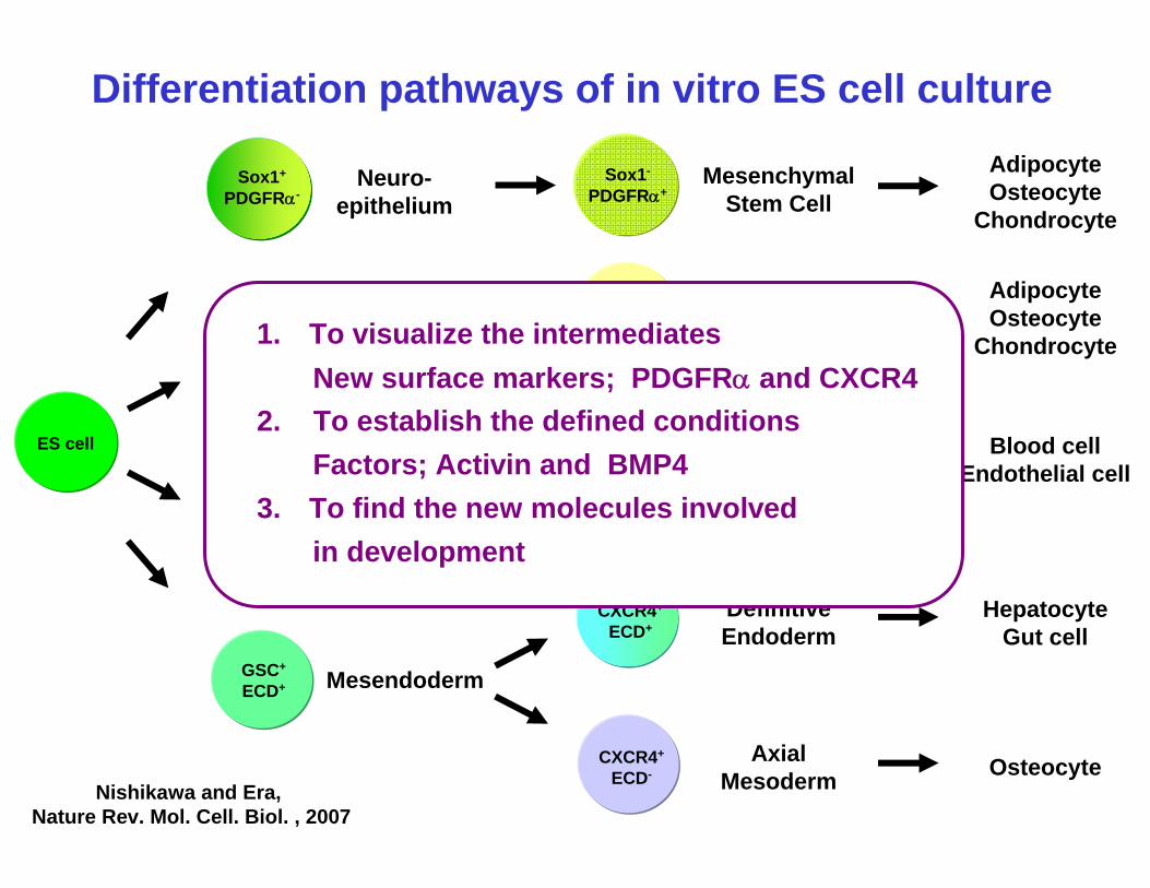
Differentiation pathways of in vitro ES cell culture
AdipocyteOsteocyte
Chondrocyte
Nishikawa and Era, Nature Rev. Mol. Cell. Biol. , 2007
1. To visualize the intermediatesNew surface markers; PDGFRα and CXCR4
2. To establish the defined conditionsFactors; Activin and BMP4
3. To find the new molecules involved in development

Gene expressions in the intermediatesGene 1Gene 2
Sox17Eomeso
Brachyury
ES cell Activin addedMesoderm
Day 1 Day 2 Day 3
foregut Midgut
E8.5 mouse embryo
Day 4Endoderm
cell lineGsc+,E-cad+
Day 5
Expr
essi
on In
tens
ity
Endoderm differentiation
PDGFRα+VEGFR2-
PDGFRα-VEGFR2+
PDGFRα+VEGFR2+
ES cell-derived samplesTada and Era, Development, 2005Takebe and Era, Dev. Biol. 2006

Mixl1 expression restore the defect of endoderm by Eomeso KD
Izumi and Era, Stem Cells, 2007
Mesendoderm marker, Goosecoid-GFP100 101 102 103 104
100
101
102
103
104
100 101 102 103 104100
101
102
103
104
100 101 102 103 104100
101
102
103
104
2 15
34
1 21
33
3 4
31
Control Eomesodermin Knock-downVector only Vector only Mixl1-overexpressed
Endoderm
Mixl1
Sox17
Eomesodermin
Endo
derm
mar
ker,
Sox1
7-hC
D25
AdipocyteOsteocyte
Chondrocyte
MesenchymalStem Cell
ParaxialMesoderm
Axial Mesoderm
Sox1+
PDGFRα-Sox1-
PDGFRα+Neuro-
epithelium
PDGFRα+
VEGFR2-
ImmatureMesoderm
PDGFRα-
VEGFR2+
Mesendoderm
PrimitiveEndoderm
LateralMesoderm
DefinitiveEndoderm
HepatocyteGut cell
Osteocyte
Blood cellEndothelial cell
GSC+
ECD+
CXCR4+
ECD+
CXCR4+
ECD-
PDGFRα+
VEGFR2+
PDGFRα+
VEGFR2+
PDGFRα+
VEGFR2-
PDGFRα-
VEGFR2+ES cell
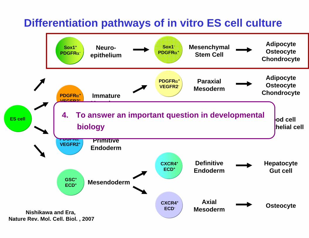
Differentiation pathways of in vitro ES cell culture
AdipocyteOsteocyte
Chondrocyte
Nishikawa and Era, Nature Rev. Mol. Cell. Biol. , 2007
4. To answer an important question in developmentalbiology

Marshak, D et al, Science, 1999
What is Mesenchymal stem cell (MSC) ?
Osteocytes
Chondrocytes
Adipocytes
Self-renewal
Fibroblastic morphology
Definition

Problems of MSCs ResearchUtilitiesMultipotency for differentiation Prevention from GVHD in transplantationSupport for regeneration of cardiac muscles
ProblemsDevelopemtal pathway is unclearSpecific surface markers are undefinedTransplantation methods are not established
ES cell study are useful for understanding what MSC is
Aims: To identify MSC progenitors and to define MSC differentiation pathway

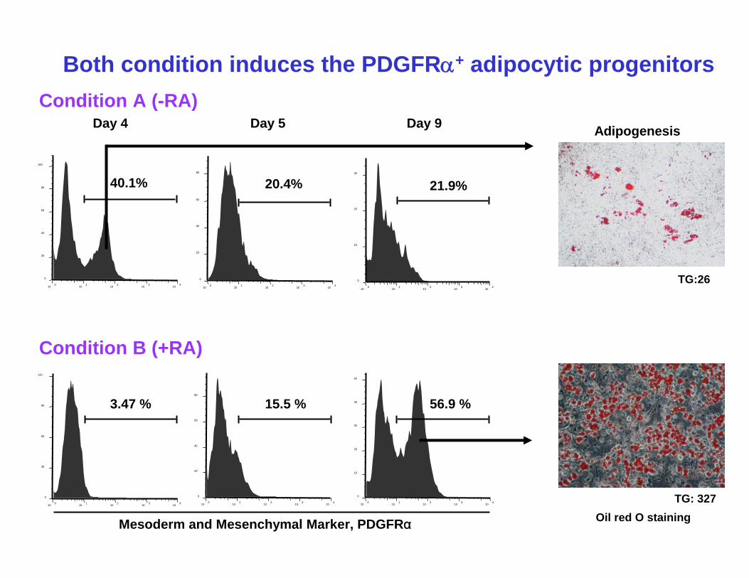
Day 0 Day 5 Day 9-RA
MonolayerCulture
Day 0 Day 5 Day 9+RA
On day 2 and 3
MonolayerCulture
Condition A (-RA)
Condition B (+RA)
Two distinct conditions induce adipocytes in in vitro ES cell culture
ES cells
Dani C. and Austin S. et al, Development, 1997
Sakurai, Era and Nishikawa et al, Stem Cells, 2006
Adipogenesis
Oil red O staining
+ Insulin, dexamethasone,
IBMX and troglitazone
+ Insulin, dexamethasone,
IBMX and troglitazone
Day 18
100
101
102
103
104
0
10
20
30
21.9%
100
101
102
103
104
0
20
40
60
80
100
40.1%
100
101
102
103
104
0
20
40
60
80
20.4%
Day 5Day 4 Day 9
100
101
102
103
104
0
10
20
30
40
50
56.9 %
Mesoderm and Mesenchymal Marker, PDGFRα
100
101
102
103
104
0
20
40
60
80
15.5 %
100
101
102
103
104
0
30
60
90
120
3.47 %
Condition A (-RA)
Both condition induces the PDGFRα+ adipocytic progenitors
Condition B (+RA)
TG:26
TG: 327
Adipogenesis
Oil red O staining

100
101
102
103
104
0
10
20
30
21.9%
100
101
102
103
104
0
20
40
60
80
100
40.1%
100
101
102
103
104
0
20
40
60
80
20.4%
Day 5Day 4 Day 9
100
101
102
103
104
0
10
20
30
40
50
56.9 %
Mesoderm and Mesenchymal Marker, PDGFRα
100
101
102
103
104
0
20
40
60
80
15.5 %
100
101
102
103
104
0
30
60
90
120
3.47 %
Brachyury
Mesp2
βActin
OB-CAD
Vimentin
PDGFRβ
Mesogenin
RT-PCR
MesodermMarkers
MesenchymalMarkers
Condition A (-RA)
Condition B induces PDGFRα+ mesenchymal cells
Condition B (+RA)
Mesodermal cells
Mesenchymal cells

Cell Growth
104
109
1014
1019
0 10 20
Cel
l num
ber
30
Passage
PDGFRα+ mesoderm cells
PDGFRα+ mesenchymal cells
ES cell-derived PDGFRα+ Mesenchymal stem cells
Osteopontin
ChondrocytesAlucian blue staining
PPARγ
Adiponectin
Osteocalcin
x100
Col2a1
Sox9
Markers
AdipocytesOil red O staining
OsteocytesAlizalin red staining
Fibroblastic morphology
Cloning
Condition B-derivedCondition A-derived

ES cells ?? PDGFRα+ adipocyteschondrocytes
osteocytes
Mesenchymalstem cells
What kinds of cells are progenitors for MSCs ?
+RA

Mesogenin
Day 3 4 5 3 4 5-RA +RA
Brachyury
Foxa2
GATA4
Mesoderm
Endoderm
β-actin
Otx2
Sox1
Neurogenin1
Ectoderm
RA treatment induces neuro-epithelial cells
RT-PCR
Condition A Condition B

Visualization of neuroepithelium as Sox1-GFP+ cells
Pevny et al. Development, 1998
Sox1 Expression
ISH
8.5dpc 9.5dpc
J. Aubert et al. PNAS, 2003
Sox1 Promoter GFP
Sox1-GFP Knock-in ES cells
Neuroepithelium
Neural Tube

Neuro-epithelium is a origin of ES cell-derived MSC
Sox1-GFPNeuroepithelium marker
Mes
ench
yme
mar
ker
PDG
FRα
0.8 0.5
48
Day 4
Sox1+ Purification
30 4.5
17
Sox1-GFP
PDG
FRα
6 days after purification Adipogenesis
Condition B (+RA)
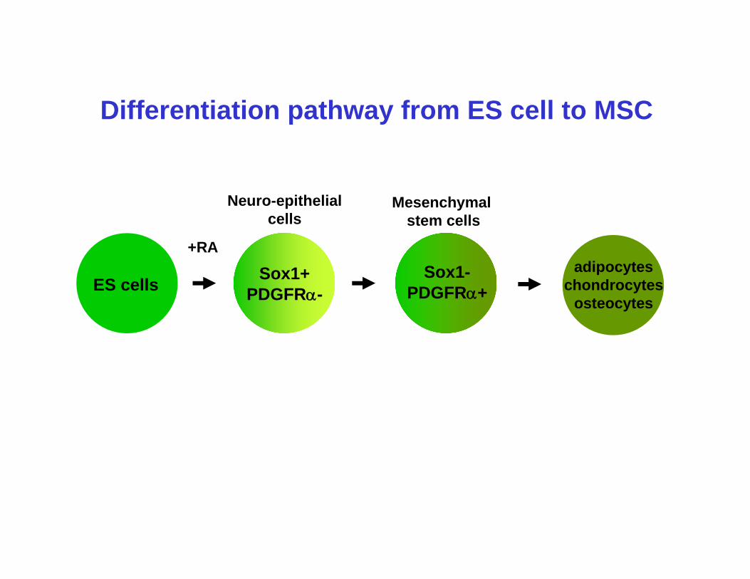
ES cells Sox1+PDGFRα-
Sox1-PDGFRα+
adipocyteschondrocytes
osteocytes
Mesenchymalstem cells
Differentiation pathway from ES cell to MSC
+RA
Neuro-epithelial cells

Tetraploid chimera
Review by Tam, PP. and Rossant, J. Development, 2003
4N tetraploid embryo
2N ES cell carrying GFP marker
Only ES cells can contribute to embryo.
E9.5 x100
Sox1/Nestin/TO-PRO
Sox1-GFPTetraploid
chimera

Sox1-GFP
PDG
FRα
41 4
31
Mesoderm
Neuroepithelium
E9.5 Mouse embryo
Sox1-GFP tetraploid chimera
Trunk region Osteogenesis
Chondrogenesis
Adipogenesis
Cloning
Visualization of Sox1+ cells in mouse embryos
Takashima, Era and Nishikawa Cell, 2007
Neuro-epithelium is the earliest origin of MSCs
FACS analysis

Gilbert, Developmental Biology
Development of Neural Crest CellsThey appear in the border between nuero-epithelium and epidermis and migrates from dorsal to ventral regions.
Bone, cartilage and neuron in head, and synpathetic neurons and pigment cells in trunk
Neural crest cells
Neuroepithelium
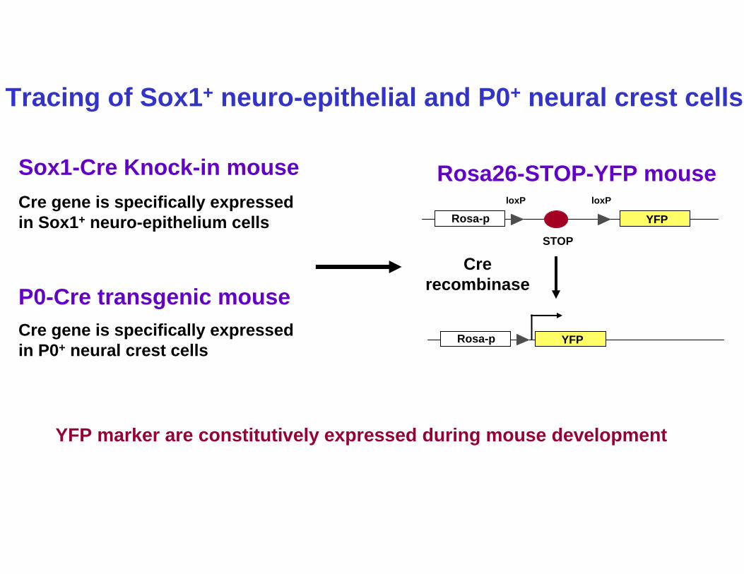
YFP marker are constitutively expressed during mouse development
Sox1-Cre Knock-in mouse
P0-Cre transgenic mouse
Tracing of Sox1+ neuro-epithelial and P0+ neural crest cells
Rosa26-STOP-YFP mouse
Rosa-p
STOP
loxP loxP
Crerecombinase
YFP
Rosa-p YFP
Cre gene is specifically expressed in Sox1+ neuro-epithelium cells
Cre gene is specifically expressed in P0+ neural crest cells

YFP expression in Sox1-Cre and P0-Cre embryos
Sox1-CreNeuroepithelium)
P0-Cre(Neural crest)
FACS Analysis
913
5
YFP
PDG
FRα
PDG
FRα
21 1
30
YFP
E14.5 embryos
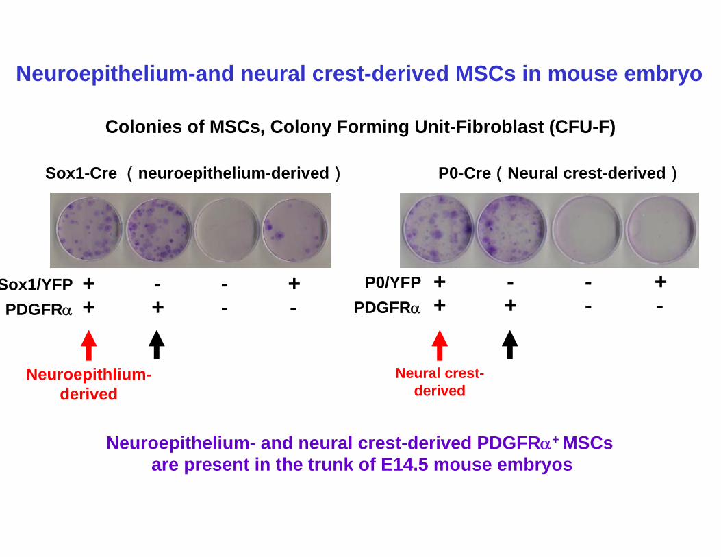
Neuroepithelium-and neural crest-derived MSCs in mouse embryo
Sox1-Cre (neuroepithelium-derived)
Neuroepithelium- and neural crest-derived PDGFRα+ MSCsare present in the trunk of E14.5 mouse embryos
Colonies of MSCs, Colony Forming Unit-Fibroblast (CFU-F)
+Sox1/YFPPDGFRα -
+++-
-- +P0/YFP
PDGFRα -+
++-
--
P0-Cre(Neural crest-derived)
Neuroepithlium-derived
Neural crest-derived
100 101 102 103 104100
101
102
103
10421.1 1.27
29.847.9
Sox1-Cre, Rosa-YFP
PD
GFR
α Single cell sorting
Cell Morphology:Fibroblastic
Passage 18
PDG
FRα
Vim
entin
PDG
FRβ
OBC
AD
βAct
in
Slug
Snai
l
Gene expression
RT-PCR
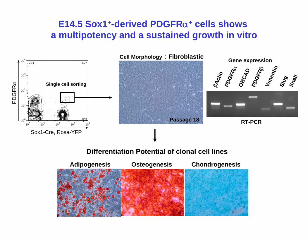
E14.5 Sox1+-derived PDGFRα+ cells shows a multipotency and a sustained growth in vitro
Adipogenesis Osteogenesis Chondrogenesis
Differentiation Potential of clonal cell lines
Sox1-Cre YFP
PDG
FRα
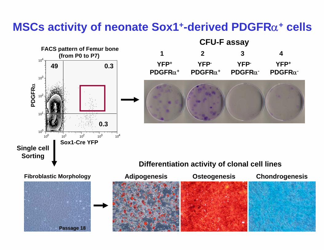
FACS pattern of Femur bone(from P0 to P7)
3100 101 102 103 104
100
101
102
103
104
0.3
0.3
49 YFP+
PDGFRα+YFP-
PDGFRα+YFP-
PDGFRα-YFP+
PDGFRα-
1 2 3 4CFU-F assay
MSCs activity of neonate Sox1+-derived PDGFRα+ cells
Adipogenesis Osteogenesis Chondrogenesis
Differentiation activity of clonal cell linesFibroblastic Morphology
Passage 18
Single cellSorting
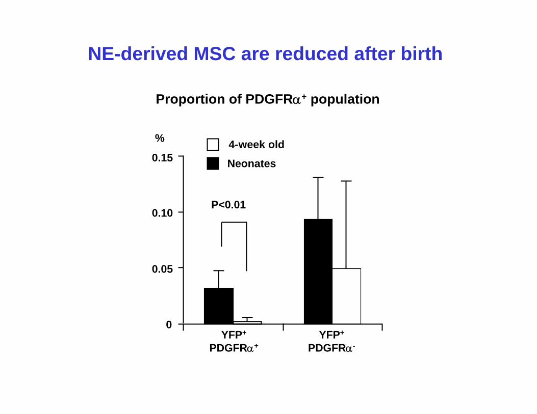
NE-derived MSC are reduced after birth
0.15
0.10
0.05
%
YFP+
PDGFRα+YFP+
PDGFRα-
P<0.01
0
Neonates
4-week old
Proportion of PDGFRα+ population
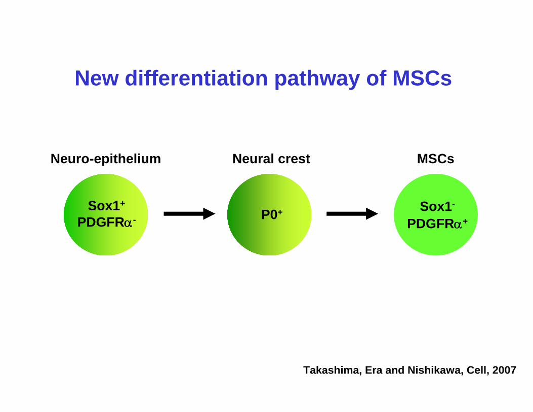
New differentiation pathway of MSCs
Takashima, Era and Nishikawa, Cell, 2007
MSCs
Sox1+
PDGFRα-
Neuro-epithelium
Sox1-
PDGFRα+P0+
Neural crest
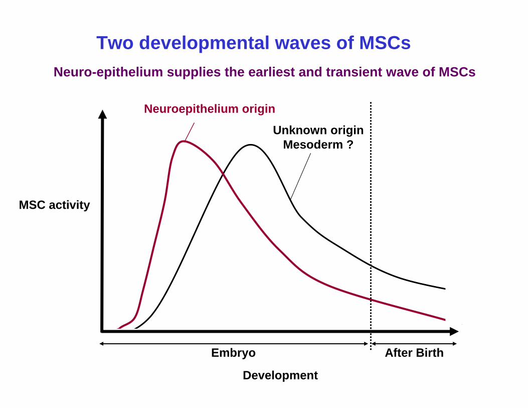
Neuroepithelium origin
Unknown originMesoderm ?
Development
MSC activity
Embryo After Birth
Two developmental waves of MSCsNeuro-epithelium supplies the earliest and transient wave of MSCs
RIKEN CDB
Yasuhiro TakashimaShin-Ichi Nishikawa
Acknowledgements
Cambridge UniversityAustin Smith Stem Cell Biology Group
Kazuki Nakao
Animal Resources and Genetic Engineering