molecular determinants of tooth formation formation is highly regulated at the molecular level •...
TRANSCRIPT

Tim Wright DDS, MSDepartment of Pediatric DentistryThe University of North Carolina
Tooth FormationTooth Formationandand
Developmental Developmental DefectsDefects
OdontogenesisOdontogenesis
Tooth formation is highly regulated at the molecular level• terminal differentiation of specific cell types• epithelial-mesenchymal interactions• secretion of specific extracellular matrices• controlled processing of those matrices• regulation of ion deposition• mineralization of the dental tissues
Tooth Formation is dependent Tooth Formation is dependent on:on:
• Genetic Factors– Hundreds to several thousand genes likely
involved (polygenic)• Environmental Factors
– Nutrition– Physical phenomenon– Infection
Molecular Molecular Determinants of Determinants of Tooth FormationTooth Formation
• Over 10,000 genes involved in making a tooth• Most genes involved in odontogenesis are
expressed in non-dental tissues• Some genes are relatively specific for
development of the dental tissues (e.g. amelogenin gene)
Molecular Control of Tooth Molecular Control of Tooth FormationFormation

Environmental Influences of Environmental Influences of AmelogenesisAmelogenesis
• Nutrition– Major and minor components
• Calcium, phosphorus, protein, fluoride etc…• Hypoxia• Hyperthermia• Infection
– Congenital rubella, syphillus, CMV, etc…• Physical Determinants
– Space– Trauma
Interactions Leading to Interactions Leading to Developmental Dental DefectsDevelopmental Dental Defects
Dental DefectsDental Defects
Host FactorsGenetic ConstitutionDisease, Age, Sex,
Environmental StressorsNutrition, DiseaseSocio-economic Status
DurationIntensityTiming
CellPhysiology
Secretion/Maturation
Tooth formation occurs over a Tooth formation occurs over a very long period of time!very long period of time! Stages Required for Tooth Stages Required for Tooth
FormationFormation
• Initiation• Histodifferentiation• Morphodifferentiation• Apposition
– Secretory Phase– Transition Phase– Maturation Phase
• Stages are not discrete for any given tooth.
• Teeth develop over years beginning with the coronal portion of the crown.
• Can be mineralizing at the cusp tips while cervically cells are differentiating.

Early stages of Early stages of cell cell
recruitment, recruitment, signaling, signaling,
differentiation, differentiation, and matrix and matrix production.production.
OOAA
Tooth Bud InitiationTooth Bud Initiation
Tooth DevelopmentTooth Development Dental Developmental FieldsDental Developmental Fields
Con Tis Res1995
MSX Expression During MSX Expression During OdontogenesisOdontogenesis
Con Tis Res1995
Abnormalities Abnormalities of of
Tooth NumberTooth Number
• Hypodontia – small or missing teeth• Anodontia – missing all teeth• Hyperdontia – increased number of teeth

Supernumerary teeth most often Supernumerary teeth most often occur in the maxillaoccur in the maxilla
• Mesiodens is most common supernumerary tooth.
• Rare in primary dentition – 0.5% of children
• Permanent dentition – 1 – 3% of children
Hereditary Conditions withHereditary Conditions withSupernumerary TeethSupernumerary Teeth
• Cleidocranial dysplasia
• Gardner Syndrome• Cleft lip/cleft palate
GeminationGemination Congenitally missing maxillary Congenitally missing maxillary lateral incisors, premolars, 3lateral incisors, premolars, 3rdrd molarsmolars
• MSX1 mutation causes autosomal dominant inheritance of congenitally missing teeth.
• Most common form of missing teeth.
Vastardis et al. Nature Genetics 1996
Severe HypodontiaSevere HypodontiaPAX9 Gene DeletionPAX9 Gene Deletion
Pax9 Deficient and Wild type MicePax9 Deficient and Wild type Mice
Peters, H. et al. Genes and Development 12:2735, 1998

Ectodermal DysplasiasEctodermal Dysplasias
• Diverse group of conditions that can affect normal development of teeth.
• There are over 130 different ectodermal dysplasias that are clinically and genetically heterogeneous.
Freire-Maia & Pinheiro, Ectodermal Dysplasia: A clinical and genetic study 1984
Ectodermal dysplasiasEctodermal dysplasias• Clinically and genetically diverse group of
conditions affecting development of tissues derived from ectoderm. Two affected tissues (e.g. hair, fingernails, teeth, skin).
• Two main types – Hypodidrotic (lack of sweat gland, hair, hypodontia)– Hidrotic – normal perspiration levels (variable
hair, teeth, nail abnormalities)
Ectodermal Dysplasia Molecular Ectodermal Dysplasia Molecular DefectsDefects
• First ED gene defect was reported in 1996• Molecular defects have now been identified
in 10 to 20 ectodermal dsyplasias– Hypohidrotic X linked ED– Reiger Syndrome– Tricho Dento Osseous Syndrome– Autosomal Dominant/Recessive ED– Clouston ED– Incontinentia Pigmenti
Ectodermal Dysplasias:Ectodermal Dysplasias:Defining the Molecular DefectsDefining the Molecular Defects
• X-linked hypohidrotic ED– novel transmembrane protein (ectodysplasin-A)
• Autosomal dominant and recessive hypohidrotic ED– novel tumor necrosis factor receptor (Downless
DL) • Rieger Syndrome
– homeobox gene (RIEG, PITX2)• Tricho-dento-osseous syndrome
– homeobox gene (DLX3)
Rieger SyndromeRieger SyndromePITX2 gene defect:Involved in anterior posteriorPatterning.
Formation of Tooth Mineralized Formation of Tooth Mineralized TissuesTissues
• The formation of dentin, enamel, and cementum required specialized cells that from unique extracellular matricies.
• These tissue perform highly unique functions that are dependent on the tissues composition and structure.

Dentin/OdontoblastsDentin/Odontoblasts• Most voluminous mineralized tissue
of teeth.• Largely determines morphology of teeth.• It is avascular• Functions to support rigid enamel outer tissue and
serves as interface between the dental crown and bone.
• Provides part of mechanism for neurosensory function of teeth.
• Provides mechanism for repair and tissue maintenance.
DentinDentin• Calcified tissue similar to bone but does not
remodel.• Collagen-rich organic matrix• Carbonate substituted hydroxyapatite
crystals• Number packing and density of crystals
largely determine stiffness of tissue.• Mechanical properties vary site to site.
Types of DentinTypes of Dentin•• Primary dentinPrimary dentin – rapidly produced dentin
formed up to completion of root formation and beginning of tooth function
•• Secondary dentinSecondary dentin – normal physiological dentin production that proceeds slowly throughout the life of the tooth
•• Tertiary dentinTertiary dentin – dentin produced in response to external stimulus (e.g. caries, restoration etc.)
Normal Circumpulpal Dentin Normal Circumpulpal Dentin
Odontoblast Life Odontoblast Life CycleCycle
• Differentiated from mesenchymal cells condensing adjacent to the inner enamel epithelium
• Are tall columnar cells during active formation of primary dentin
• Height decreases and less organelles are present as cells become less active
DentinogenesisDentinogenesis• Complex series of events including cell
interactions, differentiation, elaboration of a unique extracellular matrix and mineralization are required to produce dentin
• Numerous environmental and hereditary conditions can influence normal dentinogenesis

DentinogenesisDentinogenesis• Odontoblasts produce and
secret a collagen-rich extracellular matrix (predentin).
• Odontoblasts then control the deposition of inorganic calcium phosphate into this matrix to produce the final mineralized dentin tissue.
Dentin ECM ComponentsDentin ECM Components• Type I collagen – most abundant• Type III & type V – predentin, not normally
in mature dentin• Dentin sialophosphoprotein
– Dentin phosphophoryn– Dentin sialoprotein
• Dentin Matrix Protein 1• Proteoglycans – numerous species• Gla proteins – e.g. osteocalcin
Dentin CollagenDentin Collagen• Type I collagen accounts for 90% of dentin
organic matrix• Made of two α1(I) and one α2(1) to give
heterotrimer with triple helical structure• Rich in glycine (required for helix
formation) and proline and hydroxyproline• Collagen molecules are arranged
longitudinally creating alternating overlap zones and gap zones
Dentin PhosphoproteinsDentin Phosphoproteins• Historically the major dentin phosphoprotein
has been called phosphophoryn• Phosphoproteins make up half of the non-
collagenous dentin organic matrix• There are multiple species of dentin
phosphoproteins and the degree of phosphorylation varies.
Dentin PhosphoproteinDentin Phosphoprotein
• Serine and phosphoserine account for 50% of the amino acid residues
• Very acidic protein• Secreted at mineralization front and not in
predentin• Associated with insoluble collagen• Binds Ca++ with strong affinity
PredentinPredentin
• Unmineralized dentin matrix• Always present in normal healthy teeth• Usually 15 – 20 µm thick and is bounded
by odontoblasts on pulp side and dentin on outside
• Predentin exists in a closed compartment with components being determined and regulated by the odontoblasts

Mechanisms of Mineralization in Mechanisms of Mineralization in DentinDentin
• Matrix Vesicles – initiates mineralization in mantle dentin
• Collagen/phosphoprotein complex –required to maintain and continue normal dentin mineralization
Matrix vesicle mediated Matrix vesicle mediated mineralization in mineralization in mantle dentin layer.mantle dentin layer.
AA
DD
Linde & Goldberg Dentinogenesis.Critical Rev Oral Biol 4:679-728:1993)
Dentin Dentin MineralizationMineralization
• Begins near the DEJ• Predentin – dentin mineralization front
progress pulpally by mineralization and coalescence of the mineralizing calcospherites
Dentin Mineralization Front Dentin Mineralization Front MorphologyMorphology
Normal Dentin Tertiary Dentin
Collagen Collagen –– Dentin Dentin PhosphoproteinPhosphoprotein
• Dentin phosphoprotein is bound electrostatically to positively charged areas of type I collagen.
• Conventional dogma - Multiple phosphate esters located on the phosphoprotein are required for mineralization to occur.
Mouse Dentin Mouse Dentin Sialophosphoprotien (Dspp) Sialophosphoprotien (Dspp)
Gene StructureGene StructureExons 1Exons 1--55
1 2ATG
3 4 5TGA
= DSP= DSP
= DPP
Feng et al., J Biol Chem 273:9457-9464, 1998

DSPP DSPP KnockKnock--Out Out
MouseMouse
• Mantle dentin and initial circumpulpal dentin mineralization proceeds normally.
• Predentin zone increases in width and irregular dentin mineralization occurs.
Dentinogenesis Dentinogenesis Imperfecta Imperfecta
(Shields Type II)(Shields Type II)
DSPP Mutation in Dentinogenesis Imperfecta DSPP Mutation in Dentinogenesis Imperfecta Shields Type IIShields Type IINon-sense mutation (Bln45stop) in exon 3 of DSPP geneZhang et al., Nature Genetics 27:151-152, 2001
Dentinogenesis Dentinogenesis ImperfectaImperfecta
• Decreased dentin mineralization• Enamel fracturing• Blue-gray to yellow brown opalescent
discoloration • Altered crown and root morphology (increased
cervical constriction of crown)• Decreased number of highly branched dentinal
tubules• Pulp chamber obliteration
DI Radiographic FeaturesDI Radiographic Features
Dentinogenesis Dentinogenesis ImperfectaImperfecta
• Type I - Associated with osteogenesis imperfecta– COL1A1 and COL1A2 mutations
• Type II – Autosomal dominant condition– DSPP mutations
• Type III – Autosomal dominant variant with large pulp chambers– Allelic to type II (DSPP mutations)
Osteogenesis ImperfectaOsteogenesis Imperfecta• Genetically and clinically heterogeneous
group of hereditary disorders characterized by – Increased bone fragility– Blue sclera of eye– Hearing loss– Joint laxity– Dentinogenesis imperfecta (some families)

Osteogenesis ImperfectaOsteogenesis Imperfecta
Enamel Formation Enamel Formation and Structureand Structure
Ameloblast Ameloblast CellCell
LineageLineage
Secretory ameloblastSecretory ameloblast
Tall columnar cell Tall columnar cell with nucleus polarized with nucleus polarized away from the secretory away from the secretory end (Tomes process).end (Tomes process).
Enamelin

Amelogenin and Enamel Amelogenin and Enamel FormationFormation
Amelogenin Amelogenin
• Amelogenin is thought to control the direction and morphology of crystallite growth.
• Loss or abnormal enamel protein causes marked defects in enamel formation
Amelogenesis ImperfectaAmelogenesis Imperfecta
• Group of hereditary conditions caused by mutations in genes important in enamel formation.
• Phenotypes are highly variable depending on the mutation involved.
• Prevalence varies from 1:700 to 1:15,000 depending on population.
Amelogenin MutationsAmelogenin Mutations
• There are now 15 different mutations in the amelogenin gene (AMELX).
• The phenotypes vary from hypomaturation to hypoplastic defects depending on the type of mutation
XX--linked Hypomaturation AIlinked Hypomaturation AI
Pro70Thr Amelogenin MutationPro70Thr Amelogenin Mutation
XX--linked linked Amelogenesis Amelogenesis
ImperfectaImperfecta• Amelogenin gene
mutation with C deletion at nucleotides g4114delC.
• Frameshift introduces premature stop codon (L167fsX173).
• Truncates amelogenin protein deleting 18 c terminal amino acids.
FemaleFemale
MaleMale

The enamel extracellular The enamel extracellular matrix is a complex mix of matrix is a complex mix of multiple proteins, some of multiple proteins, some of which are derived from which are derived from amelobalsts (enamelin, amelobalsts (enamelin, ameloblastin) and others ameloblastin) and others that are not (albumin).that are not (albumin).
• Enamelin • Ameloblastin• Amelotin• Amelin• Tuft protein• Keratin• Albumin
NonNon--Amelogenin Enamel Proteins:Amelogenin Enamel Proteins: EnamelinEnamelin• Low abundance glycoprotein
immunolocalized to the secretory face of the ameloblast Tome’s process.
• Parent protein is a 186 kD glycoprotein• Cleaved into multiple smaller polypeptides• May interact with amelogenin
Hu et al., J Dent Res 76: 1720-1729, 1997
AD Smooth Hypoplastic AIAD Smooth Hypoplastic AIEnamelin MutationEnamelin Mutation
Human Molecular Genetics 10:1673-1677, 2001
Autosomal Dominant Local Autosomal Dominant Local Hypoplastic AI Hypoplastic AI –– Enamelin Enamelin
MutationMutation
• Exon 4 base substitution introducing stop codon
• Predicted protein is 52 amino acids vs 1142 in wildtype
Mardh et al., Human Molecular Genetics 11:1069-1074, 2002Picture from Sundell 1985
New Enamelin MutationNew Enamelin Mutation
• Single base deletion causes enamelin protein to be 270 AA vs wildtype 1142 AA in length.
Enamel Matrix ProcessingEnamel Matrix Processing• Enamel matrix must be removed for
crystallite growth to occur.• Controlled matrix processing allows
crystallites to grow in a highly ordered orientation.
• Multiple proteases are likely involved in enamel matrix processing.

Enamelysin (MMP20)Enamelysin (MMP20)
• Novel matrix metalloproteinases - 483 amino acids (54kD).
• Similar to stromelysins or collagenases.• Expressed by ameloblasts and odontoblasts.• Degrades amelogenin
Bartlett et al., Gene 183: 123-128, 1996
Autosomal Recessive Autosomal Recessive Hypomaturation AIHypomaturation AI
Autosomal Recessive Autosomal Recessive Hypomaturation AIHypomaturation AI
Autosomal Recessive Hypomaturation AIAutosomal Recessive Hypomaturation AI
Autosomal recessive Autosomal recessive hypomaturation AIhypomaturation AI
Autosomal Recessive Hypomaturation AIAutosomal Recessive Hypomaturation AI
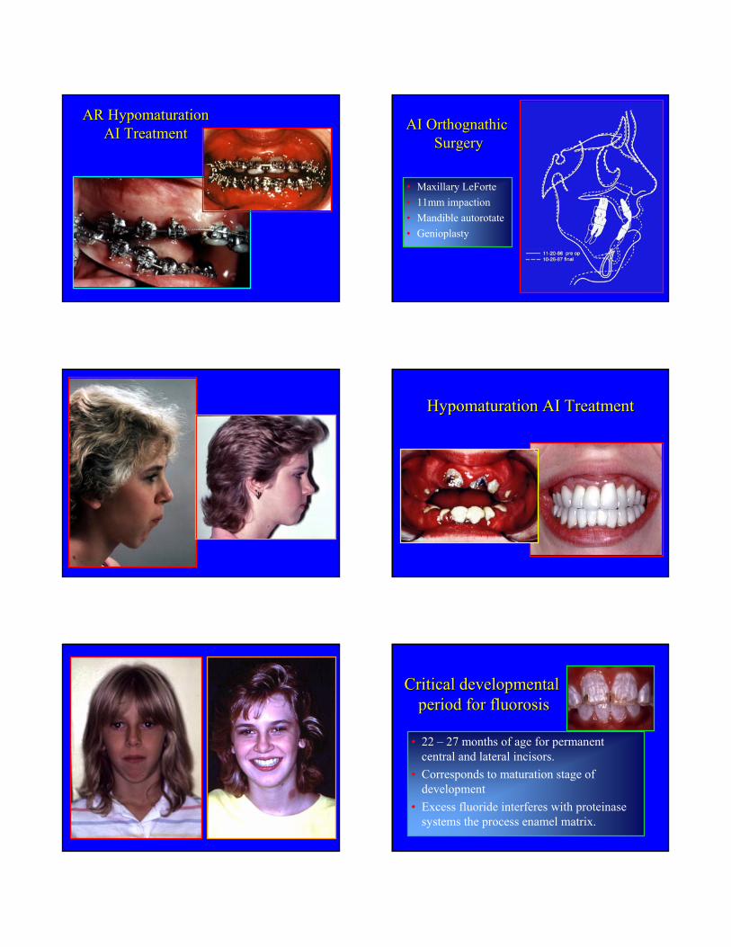
AR Hypomaturation AR Hypomaturation AI TreatmentAI Treatment
• Maxillary LeForte• 11mm impaction• Mandible autorotate• Genioplasty
AI Orthognathic AI Orthognathic SurgerySurgery
Hypomaturation AI TreatmentHypomaturation AI Treatment
Critical developmentalCritical developmentalperiod for fluorosisperiod for fluorosis
• 22 – 27 months of age for permanent central and lateral incisors.
• Corresponds to maturation stage of development
• Excess fluoride interferes with proteinase systems the process enamel matrix.

Fluorotic EnamelFluorotic Enamel
• Appears opaque. • Has a decreased mineral content
compared with normal enamel.• As severity of fluorosis increases the
enamel fluoride content increases and the mineral content decreases.
Bulk of enamel matrix:
AmelogeninAmelogeninLesser components
AmeloblastinAmeloblastinEnamelinEnamelin
Enamel matrix degradation:MMP20MMP20KLK4KLK4
DSPPDSPPDSPPDSPP
CrystallitesCrystallites
TuftelinTuftelinTuftelinTuftelin
Dentin-Enamel Junction
EnamelinEnamelin EnamelinEnamelin
EnamelinEnamelin
DSPPDSPP
AA
AA
AA
TuftelinTuftelin
Enamel Crystallite Enamel Crystallite Matrix ModelMatrix Model
Enamel Compsition by weight
Enamel Compositionby volume
Water4%
Protein1%
Water
13%
Protein2%
Enamel CompositionEnamel Composition Carbonate Substituted HydroxyapatiteCarbonate Substituted Hydroxyapatite
Enamel Crystallite Lattice Enamel Crystallite Lattice StructureStructure EnamelEnamel
crystallite growthcrystallite growth
Crystallites begin as Crystallites begin as wide thin needlewide thin needle--like like structures and growstructures and growin thickness during in thickness during
development.development.

Human Enamel Prism StructureHuman Enamel Prism Structure
Body
Tail
Etched Enamel Keyhole PatternEtched Enamel Keyhole Pattern
Crystallites
Enamel ArchitectureEnamel ArchitectureCrystallites
oriented into prism
Head – TailInterlocking
Prisms
Resin tags Resin tags penetrating etched penetrating etched enamel around enamel around crystallites and crystallites and between prisms.between prisms.
Resin
EnamelEnamel
Enamel Bonding via Resin Penetration into Etched EnamelEnamel Bonding via Resin Penetration into Etched Enamel
Diagnosis and ManagementDiagnosis and Management
Appropriate and optimal management of patients requires making an accurate diagnosis and understanding how the dental tissues can be affected by developmental defects.