guanine riboswitch variants from mesoplasma florum ...guanine riboswitch variants from mesoplasma...
TRANSCRIPT
Guanine riboswitch variants from Mesoplasma florumselectively recognize 2�-deoxyguanosineJane N. Kim†, Adam Roth‡, and Ronald R. Breaker†‡§¶
†Department of Molecular, Cellular, and Developmental Biology, §Department of Molecular Biophysics and Biochemistry, and ‡Howard Hughes MedicalInstitute, Yale University, P.O. Box 208103, New Haven, CT 06520-8103
Edited by Jeffrey W. Roberts, Cornell University, Ithaca, NY, and approved August 21, 2007 (received for review June 22, 2007)
Several mRNA aptamers have been identified in Mesoplasma florumthat have sequence and structural features resembling those ofguanine and adenine riboswitches. Two features distinguish theseRNAs from established purine-sensing riboswitches. All possess short-ened hairpin-loop sequences expected to alter tertiary contactsknown to be critical for aptamer folding. The RNAs also carry nucle-otide changes in the core of each aptamer that otherwise is strictlyconserved in guanine and adenine riboswitches. Some aptamersretain the ability to selectively bind guanine or adenine despite thesemutations. However, one variant type exhibits selective and high-affinity binding of 2�-deoxyguanosine, which is consistent with itsoccurrence in the 5� untranslated region of an operon containingribonucleotide reductase genes. The identification of riboswitch vari-ants that bind nucleosides and reject nucleobases reveals that naturalmetabolite-sensing RNA motifs can accrue mutations that expand thediversity of ligand detection in bacteria.
allosteric RNA � aptamer � metabolite � ribonucleotide reductase �transcription termination
G ene-control elements called riboswitches (1) are mRNAmotifs typically found in the 5� untranslated regions of
bacterial mRNAs. Riboswitches selectively bind small mole-cules, and structural changes within the 5� untranslated regionsare usually harnessed to control the expression of the adjoiningORF. The architectures of these RNAs are commonly formedfrom a metabolite-binding aptamer domain and an expressionplatform (2–5), although more diverse assemblies of aptamersand expression platforms have been found that yield morecomplex gene-control characteristics (6–9).
Each aptamer adopts a complex secondary- and tertiary-structured fold to form a conserved receptor for the ligand (10–17).This demand for precise structure formation and specific molecularrecognition causes the aptamer domains to be highly conservedeven among distantly related species. In contrast, the expressionplatform can adopt a variety of different structures provided itmaintains its responsiveness to the occupation state of the aptamerdomain.
Computer-aided searches based on conserved RNA sequencesand structures have been used to identify representatives of nu-merous riboswitch classes (18–24). However, these bioinformaticsalgorithms can fail to identify variants of known riboswitch classesthat differ substantially from the established aptamer consensus.Furthermore, there could be exceedingly rare classes of riboswitchaptamers or exceptionally small aptamers that will be missed byexisting bioinformatics algorithms because there are too few rep-resentatives for comparison or they have too few conserved fea-tures. Given these limitations, many new riboswitch classes mightremain undiscovered, and the true number of metabolite-sensingaptamer folds could greatly exceed the many types published todate (25).
Indications that there are many variant, small, or rare riboswitchclasses to be discovered come from several recent reports of newriboswitch classes. For example, a single C-to-U mutation withinthe core of guanine riboswitches can change the specificity of ligandbinding to adenine (11, 26–30). Also, two related types of ribos-
witch aptamers for the modified nucleobase 7-aminomethyl-7-deazaguanine have been identified that require as few as 34nucleotides to form a selective and high-affinity binding pocket(31). Moreover, there are three classes of aptamers for S-adenosylmethionine (SAM) (23, 24, 32) whose representatives aremore rare in bacteria than the SAM-I class of riboswitches com-monly found in Gram-positive bacteria (33–35). These findingsindicate that a far greater diversity of metabolite-sensing ribos-witches exists that might be difficult for existing search strategies todefinitively identify and classify.
One possibility is that some organisms will have recently evolvedriboswitch classes with aptamers that are unique in architecture orligand specificity. If such boutique riboswitches can easily emergethrough evolution, there could be far more riboswitches than havebeen discovered to date. In this report, we describe a series ofaptamers that are exceedingly rare among sequenced genomes andhave been identified only in the bacterial species Mesoplasmaflorum. One subclass of these RNAs is selective for 2�-deoxyguanosine (2�-dG). Our findings highlight the capacity formetabolite-binding RNAs to evolve specificities toward structurallyrelated derivatives and further demonstrate that exceptionally rareriboswitch classes are likely to be present in some organisms.
Results and DiscussionConsensus Sequences and Structures of Guanine- and Adenine-Sens-ing Riboswitches. Guanine-sensing riboswitches usually resideupstream of genes involved in purine biosynthesis, salvage, andtransport (26). The guanine riboswitch aptamer from the Bacil-lus subtilis xpt-pbuX mRNA exhibits a Kd for guanine of �5 nM.Ligand binding to this aptamer causes transcription termination,and a similar gene-control mechanism is predicted for mostother guanine riboswitches as well. X-ray crystallography hasbeen performed on this aptamer bound to either guanine or thefunctional analog hypoxanthine (10, 11). In both instances, theligands are almost completely enveloped by the RNA. Similarly,a related aptamer that binds adenine by using a similar archi-tecture also engulfs the ligand (11).
The tight ligand-binding pocket of this aptamer class is formedby conserved nucleotides at the junction of three stems termed P1,P2, and P3 (Fig. 1A). When the ligand is bound, the aptamer adoptsa conformation with the P2 and P3 stems extending parallel to oneanother. This structure is held in place by Watson–Crick base-pairing interactions and other hydrogen bonds formed between theloops of these stems, called L2 and L3. Most of the highly conservednucleotides forming the ligand-binding core of the aptamer are
Author contributions: J.N.K., A.R., and R.R.B. designed research; J.N.K. and A.R. performedresearch; J.N.K., A.R., and R.R.B. analyzed data; and J.N.K., A.R., and R.R.B. wrote the paper.
Conflict of interest statement: R.R.B. is a cofounder of BioRelix, a biotechnology companythat has licensed riboswitch technology from Yale University for antibiotics development.
This article is a PNAS Direct Submission.
Freely available online through the PNAS open access option.
Abbreviations: 2�-dA, 2�-deoxyadenosine; 2�-dG, 2�-deoxyguanosine; 2�-d-2,6-DAP, 2�-deoxy-2,6-diathinopurine nucleoside.
¶To whom correspondence should be addressed. E-mail: [email protected].
© 2007 by The National Academy of Sciences of the USA
16092–16097 � PNAS � October 9, 2007 � vol. 104 � no. 41 www.pnas.org�cgi�doi�10.1073�pnas.0705884104
Dow
nloa
ded
by g
uest
on
May
14,
202
0
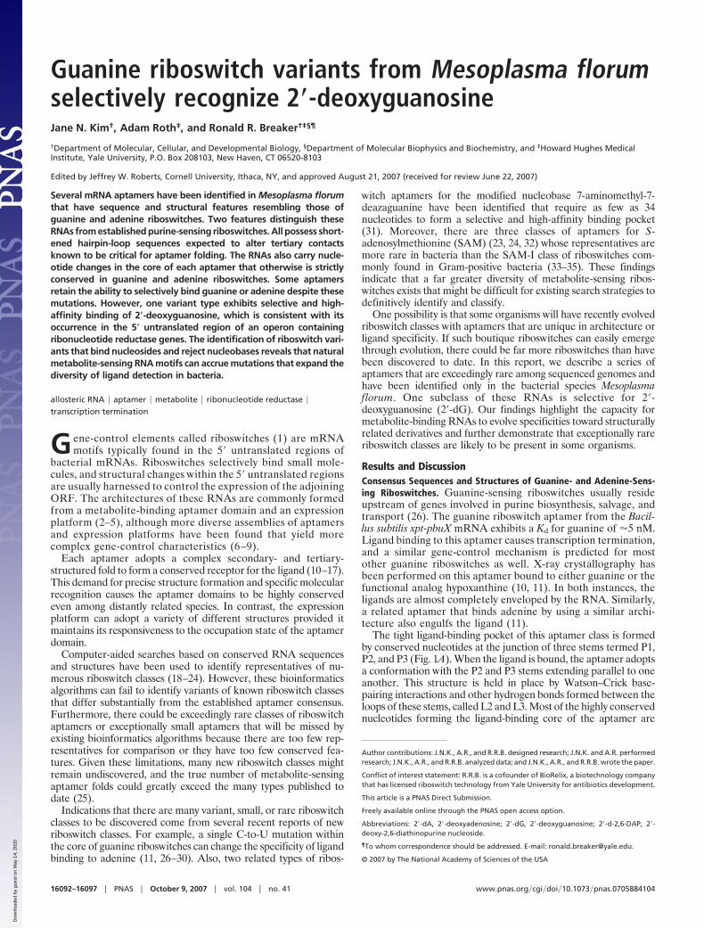
present in joining regions J1-2, J2-3, and J3-1, which link the threestems together (Fig. 1B).
The joining regions carry four nucleotides that form hydrogenbonds with functional groups of the purine ligand (Fig. 1C). Onekey interaction is made by nucleotide C74 of the xpt aptamer, whichforms a Watson–Crick base pair with guanine. Interestingly, severalvariants of this RNA motif were found that carry a C-to-U mutationat the equivalent position in the structure, and these RNAs rejectguanine and bind adenine with affinities measured in the midnanomolar range (27, 28). An atomic-resolution model of anadenine riboswitch aptamer from the add gene in B. subtilisconfirmed that adenine forms a Watson–Crick base-pairing inter-action with the variant U nucleotide, and that other features typicalof guanine riboswitches remained essentially identical (11). Indeed,this single-nucleotide change at position 74 is sufficient to changethe specificity of guanine aptamers to adenine and vice versa.
Discovering More Distant Homologs of Guanine and Adenine Ribos-witches. We used a bioinformatics search strategy to discovervariant purine riboswitch candidates. This process was achievedwith an algorithm that identifies sequences that closely correspondto the consensus sequence and secondary structure features ofknown purine-sensing aptamers (see Materials and Methods). Theparameters of this search were set to allow recovery of low-qualitymatches, and we focused the most attention on sequences thatdeviate substantially from the consensus, but that nonetheless existin genomic contexts consistent with riboswitch function.
We noticed one sequence in M. florum that could be threaded toconform reasonably well to the consensus structure, but thatdeviated in sequence at several key positions in the core and in loopsL2 and L3. Despite these significant differences, the location of thissequence in the apparent 5� untranslated region of the guaABoperon suggested that it might function as a purine-sensing ribos-witch. A BLAST search for related sequences uncovered severalmore examples of this motif in M. florum. This bacterium is anonparasitic member of the class Mollicutes, and organisms of thisclass are notable for their simplified cell structures and smallgenomes (36).
One of the M. florum sequences differed from characterizedpurine riboswitches only in the L2 and L3 regions, with the joiningregions otherwise adhering to the consensus. To determine whetherthere might be other variant purine aptamers analogous to thisRNA, we manually inspected sequences generated from the orig-inal search, scanning for sequence or structure irregularities. Threeadditional riboswitches were identified that contained shortened L3sequences relative to the consensus, in addition to the eight M.florum examples mentioned previously.
Subsequently, we performed automated searches by using algo-rithms trained on all known purine riboswitches, as well as algo-rithms trained more narrowly on the variant sequences, but noadditional variants were identified. In total, 12 new putative ribos-witch examples were found in bacteria (Fig. 1A), all of which bearclose similarity to the consensus sequence and structure establishedfor guanine aptamers. These RNAs were classified into five types(I–V) based on the mutations they carry relative to the guanineaptamer consensus. Interestingly, eight of the RNAs representingtypes I-IV are present in M. florum.
One of the eight RNAs (IV-A) carries a C-to-U mutation at theposition equivalent to nucleotide 74 of the xpt riboswitch in B.subtilis. Therefore, this RNA was predicted to sense adenine. Theseven remaining RNAs from M. florum carry mutations at two ormore positions that are highly conserved among known guanineand adenine riboswitch representatives (Fig. 1A). All seven RNAscarry mutations in otherwise conserved nucleotides in J1-2, J2-3,and L2 of the aptamer, and they also carry an L3 loop that is fournucleotides, rather than the seven or eight nucleotides normallypresent in known guanine-binding aptamers. The four remainingRNAs, classified as type V, are found in other bacterial species andcarry the distinctive nucleotide changes in L3 and, in some in-stances, L2. Specific interactions between the L2 and L3 regions areknown to be important for folding and function of guanine andadenine riboswitches (29), and therefore the loop mutations likelycause the variant RNAs to adopt a different structure for thistertiary interaction.
We speculated that the aptamer core mutations in the M. florumRNAs might substantially alter the ligand-binding pocket of each
J3-1
-
--
GAACACUCAUAUUAAUCGCGUGGAUA-UGGCACGCAAGUUUCUACCGGGUA CCGUAAAUGUCCGACUAUGGGUGAGCAAGAAACUUAUACAG-GGUAGC-AUAAUGGGCUACU-GACCCCGCCUUCAAACCUA--UUUGGAGACUAUAAGUGAAAAAGAAACUUAUACAG-GGUAGC-AUAAUGGGCUACU-GACCCCGCCAUGAAACCUA--UUUCAUGACUAUAGGUCUUUAAAAAACUUAUACAG-GGUAGC-AUAAUGGGCUACU-GAA-CCGCCCCGGGACCAA--UCUCGGGACUAUAAGUGUGUAAAAAACUUAUACAG-GGUAGC-AUAUUGGGCUACU-GUU-CCGCCUCAAGACCAA--UCUUGAGACUAUAAGUGUAAAAUUAACUUAUACAU-GAUAAC-AUAUCGGGUUGUC-GAC-CUGCCUUAAGACCGA--UCUUAAGACUAUAAGAAAAUAAAAAACUUAUACAU-GACAAC-AUAUUGGGUUGUC-GAC-CUGCCUCUGGACCUA--UCCUUAGACUAUAAGCGU---AAAAACUUAUACAU-GACAAC-AUAACGGGUUGUC-GAC-CUGCCUUAGGACCCA--UCCAAAGACUAUAAGCGCAGAGAAAACUUGUAUAA-UCCUUC-AUAUCGGGAAGGA-GUCUCUACCUAACA CCAA--UGUUAG-AUUAUGAGUUUUAUCCAAGCAGGUAUAUC-GUCGG-AUAAUGGCUGACA-GUUUCUACCCAACA CCAA--UGUUGG-ACUAUCUGUGGAUGUAAAUGCUGUAUAUAUCUAGUGAUA-UGGACUGGAUGUUUCUACUACCGAGCCUAC-CUUGGUGACUACAGUUUUUAG
(phosphoribosyltransferase)
(ribonucleotide reductase)(unknown lipoprotein)
xpt
I-AI-B
(glycerol-3-P transport)III-A
(phosphate transporter)II-A(unknown lipoprotein)II-B
(GMP synthase)III-B(xylulose transporter)III-C
(xanthine/uracil permease)IV-A
(IMP dehydrogenase)V-A
(xanthine/uracil permease)V-B1
-UUAGC-UGAUAUAGU-AUCGA-AUAAUGGUCGAUU-GUUUCUAGCCAGCA CCCA--UGCUGGAACUAUCAUAAACA-(xanthine/uracil permease)V-C
*P2’P1 P2 P3’ P1’P3A
B D
guanine aptamerconsensus
I-A aptamersequence
CAAUA
G G GA
C C C G
CC
A
C
UUAUA U
AUAA
C
U
C G
U
A
C
G
U
P1
P2
P3
A U
GGGC AG
AU G UC UC
UCC
AAA
UUUAG
AGUA
RAU
G G GU
U C UA
C C
A
C
YYRUA U
AYRR
C
A
Y R
Y
R
Y
Y
R
P1
P2
P3
5΄
47 51
22 74
L2 L3J1-2 J2-3
UAAAUGCUGUAUAUAUCUAGUGGUA-UGGACUAGAUGUUUCUACUACCGAGCCUAC-CUUGGUGACUACAGUUUUUAG(xanthine/uracil permease)V-B2
U22
U47
U51
C74
(B. subtilis xpt-pbuX)guanine riboswitch ligand binding site
C
40
60
80(74)
58 (51)(47)54
(22)31
Fig. 1. Sequence and structural features of guanineriboswitch aptamers and several newly found RNAs. (A)Sequence alignment comparing the xpt guanine ribos-witch aptamer sequence from the xpt-pbuX mRNA fromB. subtilis with related sequences from Mesoplasma flo-rum (types I, II, III, and IV) and from Oenococcus oeni,Vibrio sp., Vibrio splendidus, and Leuconostoc mesen-teroides (types V-A, V-B1, V-B2, and V-C, respectively).The known or putative functions of the genes immedi-ately downstream of each sequence are noted as pre-dicted elsewhere (54). Nucleotides corresponding topairing regions P1, P2, and P3 are shaded blue, green,and orange, respectively. Nucleotides corresponding toloop regions (L) and joining regions (J) also are identi-fied. The asterisk identifies the C nucleotide in xpt thatforms a Watson–Crick base pair with the guanine ligand.Nucleotides shaded gray are mutated relative to thehighly conserved nucleotides denoted in red that aretypically found in the J and L regions of guanine andadenine riboswitches or nucleotides that are inserted ordeleted in these regions. (B) Consensus sequence andsecondary structure of guanine riboswitch aptamers.Nucleotides in red are present in �90% of the knownrepresentatives. Circles identify nucleotides whose baseidentities are not conserved, and lines indicate Watson–Crick base pairing. Nucleotides that form hydrogen-bonding interactions with the guanine ligand areidentified according to the numbering system used previously for the xpt aptamer (10, 11, 26). (C) Structural model of the guanine-binding site formed by thexpt aptamer docked to guanine (10, 11). Dashed lines identify hydrogen-bonding contacts between the aptamer nucleotides (numbered as described in B) andthe ligand. The shaded area identifies the space that would be occupied by the sugar moiety of a guanine nucleoside. (D) Sequence and secondary structure ofthe type I-A aptamer from M. florum. Boxed nucleotides depicted in blue identify variations from the xpt aptamer that occur at otherwise highly conservedpositions. Dashed line represents a 3-nucleotide deletion compared with the L3 sequence of xpt. Nucleotide numbers are as described in Fig. 2, with the equivalentpositions for the xpt aptamer depicted in parentheses. Other notations are as defined for B.
Kim et al. PNAS � October 9, 2007 � vol. 104 � no. 41 � 16093
BIO
CHEM
ISTR
Y
Dow
nloa
ded
by g
uest
on
May
14,
202
0
riboswitch, allowing it to recognize a metabolite other than gua-nine. For example, RNA I-A carries 39 nucleotide changes (in-cluding insertions and deletions) relative to the xpt RNA (Fig. 1A),and 10 of these changes occur at positions with nucleotide identitiesthat are conserved in �90% of the known guanine riboswitchaptamers (Fig. 1D). In addition, three of the four nucleotides knownto contact guanine in the xpt aptamer (nucleotides 22, 47, and 51)(Fig. 1 B and C) are mutated in the I-A (nucleotides 31, 54, and 58)(Fig. 1D) and I-B aptamers. Although the nucleotide correspondingto C74 of xpt in the I-A and I-B aptamers could retain its recognitionof the base-pairing face of guanine, the other core mutations likelyrecognize other portions of a guanine-containing ligand. Moreover,these core mutations typically convert A and U residues to G andC residues, despite the fact that the M. florum genome has only 27%GC content. The acquisition of additional G and C residues in somevariant aptamers suggests adaptation to a new function.
A Guanine Riboswitch Variant Binds 2�-dG. Frequently, the metabo-lite that is sensed by a riboswitch can be discerned by noting thefunction of the protein product of the downstream gene. Some ofthe RNA motifs are located upstream of either unannotated genesor genes with functions that appear to be unrelated to purinemetabolism, and thus did not provide clues for possible ligands.However, some reside upstream of genes involved in purine bio-synthesis or transport (Fig. 1A), suggesting that the variant RNAsbind guanine or a ligand that includes this nucleobase. Of particularinterest was aptamer I-A, which resides upstream of genes encodingribonucleotide reductase subunits. Ribonucleotide reductase en-zymes convert ribonucleotides into their deoxyribonucleotide coun-terparts (37, 38). Given that three of the nucleotides mutated in theI-A aptamer are in the immediate vicinity of the N9 position ofguanine in known guanine riboswitches (Fig. 1C), we speculatedthat this variant riboswitch might respond to 2�-dG or one of its5�-phosphorylated derivatives.
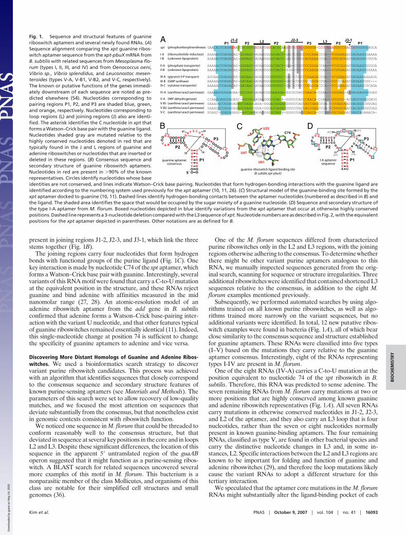
In-line probing (39) was performed by using a series of guanineand guanosine derivatives (see Materials and Methods for a com-plete list) to determine the ligand specificity for all variant ribos-witch types. In-line probing assays reveal shape changes in anaptamer that occur upon ligand binding. For example, I-A exhibitssubstantial structural modulation when 100 �M 2�-dG is present(Fig. 2A). Importantly, the pattern of spontaneous cleavage prod-ucts is consistent with the formation of a three-stem junction similarto guanine and adenine aptamers (26–28), and the majority of theinternucleotide linkages that become more structured upon 2�-dGaddition (Fig. 2B) are in the predicted ligand-binding core of I-ARNA. Furthermore, in-line probing data collected at various con-centrations of 2�-dG indicate changes in the extent of RNA cleavageat specific sites in the RNA that are consistent with a 1:1 bindingof ligand with an apparent Kd of �80 nM (Fig. 2C).
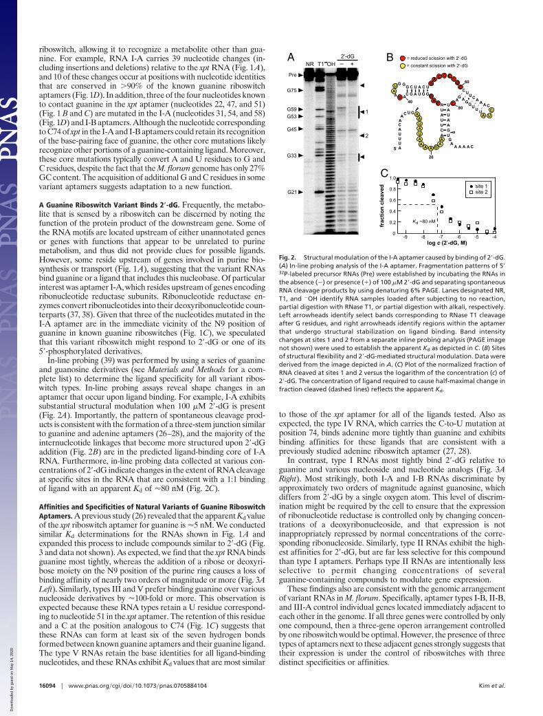
Affinities and Specificities of Natural Variants of Guanine RiboswitchAptamers. A previous study (26) revealed that the apparent Kd valueof the xpt riboswitch aptamer for guanine is �5 nM. We conductedsimilar Kd determinations for the RNAs shown in Fig. 1A andexpanded this process to include compounds similar to 2�-dG (Fig.3 and data not shown). As expected, we find that the xpt RNA bindsguanine most tightly, whereas the addition of a ribose or deoxyri-bose moiety on the N9 position of the purine ring causes a loss ofbinding affinity of nearly two orders of magnitude or more (Fig. 3ALeft). Similarly, types III and V prefer binding guanine over variousnucleoside derivatives by �100-fold or more. This observation isexpected because these RNA types retain a U residue correspond-ing to nucleotide 51 in the xpt aptamer. The retention of this residueand a C at the position analogous to C74 (Fig. 1C) suggests thatthese RNAs can form at least six of the seven hydrogen bondsformed between known guanine aptamers and their guanine ligand.The type V RNAs retain the base identities for all ligand-bindingnucleotides, and these RNAs exhibit Kd values that are most similar
to those of the xpt aptamer for all of the ligands tested. Also asexpected, the type IV RNA, which carries the C-to-U mutation atposition 74, binds adenine more tightly than guanine and exhibitsbinding affinities for these ligands that are consistent with apreviously studied adenine riboswitch aptamer (27, 28).
In contrast, type I RNAs most tightly bind 2�-dG relative toguanine and various nucleoside and nucleotide analogs (Fig. 3ARight). Most strikingly, both I-A and I-B RNAs discriminate byapproximately two orders of magnitude against guanosine, whichdiffers from 2�-dG by a single oxygen atom. This level of discrim-ination might be required by the cell to ensure that the expressionof ribonucleotide reductase is controlled only by changing concen-trations of a deoxyribonucleoside, and that expression is notinappropriately repressed by normal concentrations of the corre-sponding ribonucleoside. Similarly, type II RNAs exhibit the high-est affinities for 2�-dG, but are far less selective for this compoundthan type I aptamers. Perhaps type II RNAs are intentionally lessselective to permit changing concentrations of severalguanine-containing compounds to modulate gene expression.
These findings also are consistent with the genomic arrangementof variant RNAs in M. florum. Specifically, aptamer types I-B, II-B,and III-A control individual genes located immediately adjacent toeach other in the genome. If all three genes were controlled by onlyone compound, then a three-gene operon arrangement controlledby one riboswitch would be optimal. However, the presence of threetypes of aptamers next to these adjacent genes strongly suggests thattheir expression is under the control of riboswitches with threedistinct specificities or affinities.
G33
G45
G59G53
G21
G75
NR T1 OH2’-dG_ +
CAGGGUAGCAUA
AU G GG C U A C U
GA C C C
CG
CC
UA
AC
CUUAUA U
AUAAG
CUUC A A AUUUGGAG
A U
C
A
AA
A AAA
C
C
UUU
U
U
U
G
G
A
AA A
A
A AAAAA
G
C5‘
80
60
40
= reduced scission with 2’-dG= constant scission with 2’-dG
0.2
1.0
0.6
0.8
0.4
-6-9 -8 -7 -5 -4log c (2’-dG, M)
frac
tion
clea
ved
0
K ~80 nMd
A B
C
20
1
2
site 1site 2
Pre
Fig. 2. Structural modulation of the I-A aptamer caused by binding of 2�-dG.(A) In-line probing analysis of the I-A aptamer. Fragmentation patterns of 5�32P-labeled precursor RNAs (Pre) were established by incubating the RNAs inthe absence (�) or presence (�) of 100 �M 2�-dG and separating spontaneousRNA cleavage products by using denaturing 6% PAGE. Lanes designated NR,T1, and �OH identify RNA samples loaded after subjecting to no reaction,partial digestion with RNase T1, or partial digestion with alkali, respectively.Left arrowheads identify select bands corresponding to RNase T1 cleavageafter G residues, and right arrowheads identify regions within the aptamerthat undergo structural stabilization on ligand binding. Band intensitychanges at sites 1 and 2 from a separate inline probing analysis (PAGE imagenot shown) were used to establish the apparent Kd as depicted in C. (B) Sitesof structural flexibility and 2�-dG-mediated structural modulation. Data werederived from the image depicted in A. (C) Plot of the normalized fraction ofRNA cleaved at sites 1 and 2 versus the logarithm of the concentration (c) of2�-dG. The concentration of ligand required to cause half-maximal change infraction cleaved (dashed lines) reflects the apparent Kd.
16094 � www.pnas.org�cgi�doi�10.1073�pnas.0705884104 Kim et al.
Dow
nloa
ded
by g
uest
on
May
14,
202
0
A Single Mutation Swaps Ligand Specificity of the 2�-dG Aptamer I-A.A C-to-U mutation at nucleotide 74 of the xpt aptamer can changeits ligand specificity from guanine to adenine (27). This nucleotideforms a Watson–Crick base pair with the purine moiety, whereasother nucleotides make hydrogen-bonding interactions with otherpositions on the ligand (Fig. 1C) (10, 11, 40). Although mostpurine-sensing riboswitches respond to guanine, six natural exam-ples of adenine-sensing riboswitches carrying a U at the equivalentposition 74 have been identified (27, 41, 42), and a seventh exampleis represented by the type IV RNA reported in this study (Figs. 1Aand 3A).
If the I-A RNA binds 2�-dG by using a similar core structureadopted by guanine and adenine riboswitch aptamers, it is expectedthat a C-to-U mutation at the equivalent nucleotide 74 positionshould alter the specificity for the purine moiety of the ligand. We
conducted this test by using the ligand candidate 2�-dG and itsanalogs, 2�-deoxyadenosine (2�-dA) and 2�-deoxy-2,6-diaminopu-rine nucleoside (2�-d-2,6-DAP) (Fig. 3B). Although 2�-dA shouldcompensate for the aptamer C-to-U mutation, previous studieswith guanine- and nucleobase 7-aminomethyl-7-deazaguanine-sensing riboswitches have revealed that 2,6-DAP binds more tightlyto the mutant aptamers (27, 31). Furthermore, it has been shownthat an adenine riboswitch binds 2,6-DAP with an affinity �30-foldbetter than that for adenine (27). Aptamers carrying the C-to-Umutation likely exhibit preferences for ligands that carry 2,6-DAPbecause of the formation of hydrogen bonds between other nucle-otides in the aptamer core and the exocyclic amine at position 2 ofthe purine ring like those normally occurring in guanineriboswitches (Fig. 1C).
As expected, the unaltered I-A RNA tightly binds 2�-dG andrejects both 2�-dA and 2�-d-2,6-DAP (Fig. 3B). In contrast, the I-A*aptamer carrying the C-to-U mutation rejects both 2�-dG and 2�-dA(Kd values �1 mM), but binds 2�-d-2,6-DAP with a Kd of �8 �M.Thus, the 2,6-DAP analog also is preferred by the I-A* RNA asobserved for other aptamers carrying similar C-to-U mutations.With the common form of guanine- and adenine-sensing aptamers,a U residue at the position equivalent to nucleotide 51 forms ahydrogen bond with the exocyclic amine present in guanine and2,6-DAP (Fig. 1C). However, the I-A and I-A* aptamers carry adifferent nucleotide at the position equivalent to nucleotide 51, andtherefore they likely recognize this extra amine group differentlydespite similarities elsewhere in the aptamer structure.
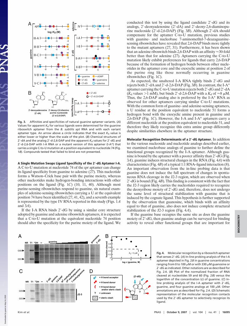
Molecular Recognition Determinants of a 2�-dG Aptamer. In additionto the various nucleoside and nucleotide analogs described earlier,we examined nucleobase analogs of guanine to further define thefunctional groups recognized by the I-A aptamer. Although gua-nine is bound by the aptamer with a poorer affinity than 2�-dG (Fig.3A), guanine induces structural changes in the RNA (Fig. 4A) withcharacteristics (Fig. 4B) of a typical 1:1 RNA–ligand interaction (8).An important observation from the in-line probing data is thatguanine does not induce the full spectrum of changes in sponta-neous RNA cleavage in the J2-3 region, which are observed when2�-dG is bound (Fig. 4B). This finding is consistent with the fact thatthe J2-3 region likely carries the nucleotides required to recognizethe deoxyribose moiety of 2�-dG and, therefore, does not undergothe same level of structural stabilization with guanine that isinduced by the cognate ligand. This hypothesis is further supportedby the observation that guanosine, which binds with an affinityequal to that of guanine, also does not induce complete structuralstabilization of the J2-3 region (Fig. 4A).
If the guanine base occupies the same site as does the guaninemoiety of 2�-dG, then guanine analogs can be surveyed for bindingactivity to reveal other functional groups that are important for
A xpt-8 -6 -4
III-C-8 -6 -4
III-B-8 -6 -4
III-A-8 -6 -4
V-C-8 -6 -4
V-B-8 -6 -4
-8 -6 -4V-A
guan
ine
B
-8 -6 -4
= 2‘-dG= 2‘-d-2,6-DAP
I-A
I-A*
spec
ifici
ty s
wap
-8 -6 -4
-8 -6 -4IV
II-A-8 -6 -4
II-B-8 -6 -4
I-B-8 -6 -4
I-A-8 -6 -4
2‘-d
Gad
enin
e= guanine= 2‘-dG= 3‘-dG
= dGMP= guanosine= adenine
2‘-deoxyadenosine(2‘-dA)
O
HO
HO
NH2
N
NN
N
2‘-deoxy-2,6-diamino
(2‘-d-2,6-DAP)purine nucleoside
NH2N
NN
N
HNH2
O
HO
HO
log K (M)D
Fig. 3. Affinities and specificities of natural guanine aptamer variants. (A)Values for apparent Kd for various ligands were determined for the guanineriboswitch aptamer from the B. subtilis xpt RNA and with each variantaptamer type. An arrow above a circle indicates that the exact Kd value iseither lower or higher than the scale of the plot. (B) Chemical structures of2�-dA and the analog 2�-d-2,6-DAP and the apparent Kd values for 2�-dG and2�-d-2,6-DAP with I-A RNA or a mutant version of this aptamer (I-A*) thatcarries a single C-to-U mutation at a position equivalent to nucleotide 74 (Fig.1B). Compounds tested that failed to bind are not presented.
NR T1 OH 2‘-dG
guanine
2,6-D
APN -m
ethylg
uanine
O -meth
ylguan
ine
7-meth
ylguan
ine
A B
C D
62
G53
G59J2-3
guanine
G53
G59
NR T1 OH 2‘-dG
guanosin
e
1
0.8
0.6
0.4
0.2
-4-5-6-70
K = 5 µMd
-8log c (guanine, M)
frac
tion
clea
ved
= H bond acceptor
= H bond donor
(2‘-dG)
O
NN
NN
N
H
H
H
O
HO
HO
?
= H bond donorand/or steric clash
? = unknown
= steric clash
Fig. 4. Molecular recognition by a riboswitch aptamerthat senses 2�-dG. (A) In-line probing analysis of the I-Aaptamer depicted in Fig. 2B in guanine concentrationsranging from 0 to 100 �M or with 330 �M guanosine or2�-dG as indicated. Other notations are as described forFig. 2A. (B) Plot of the normalized fraction of RNAcleaved at nucleotides 59 and 60 (Fig. 2B) versus thelogarithm of the concentration (c) of guanine. (C) In-line probing analysis of the I-A aptamer with 2�-dG,guanine, and four guanine analogs at 100 �M. Otherannotations are as described for Fig. 2A. (D) Schematicrepresentation of the molecular recognition contactsused by the 2�-dG aptamer to selectively recognize itsligand.
Kim et al. PNAS � October 9, 2007 � vol. 104 � no. 41 � 16095
BIO
CHEM
ISTR
Y
Dow
nloa
ded
by g
uest
on
May
14,
202
0
recognition by the aptamer. However, several guanine analogs thatcarry modifications of functional groups on the purine ring are notbound by the I-A aptamer when present at 100 �M (Fig. 4C). Thesefindings, and those presented earlier, reveal that the I-A aptamerrecognizes nearly every available functional group to form a precisebinding pocket (Fig. 4D).
Transcription of RNAs Carrying I-A and III-B Aptamers Reveals Me-tabolite-Mediated Termination. The majority of guanine and ade-nine riboswitches are predicted to control gene expression byregulating transcription termination (J. Barrick and R.R.B., un-published data). In these instances, the aptamer resides a shortdistance upstream of a predicted intrinsic transcription terminator(43, 44), which forms a strong base-paired stem followed by a runof U residues. All of the newly found aptamer variants in M. florumlie immediately upstream of putative intrinsic terminator stems(data not shown), indicating that they are components of ribos-witches that control transcription termination.
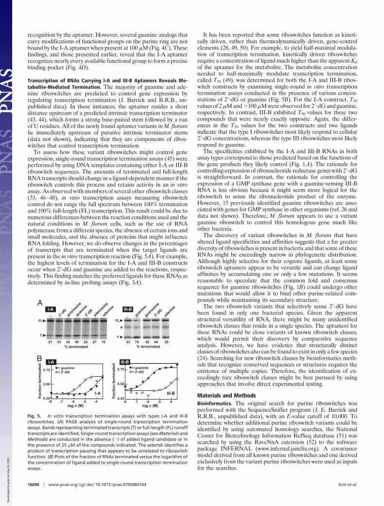
To assess how these variant riboswitches might control geneexpression, single-round transcription termination assays (45) wereperformed by using DNA templates containing either I-A or III-Briboswitch sequences. The amounts of terminated and full-lengthRNA transcripts should change in a ligand-dependent manner if theriboswitch controls this process and retains activity in an in vitroassay. As observed with members of several other riboswitch classes(33, 46–48), in vitro transcription assays measuring riboswitchcontrol do not range the full spectrum between 100% terminationand 100% full-length (FL) transcription. This result could be due tonumerous differences between the reaction conditions used and thenatural conditions in M. florum cells, such as the use of RNApolymerase from a different species, the absence of certain ions andsmall molecules, and the absence of proteins that might influenceRNA folding. However, we do observe changes in the percentagesof transcripts that are terminated when the target ligands arepresent in the in vitro transcription reaction (Fig. 5A). For example,the highest levels of termination for the I-A and III-B constructsoccur when 2�-dG and guanine are added to the reactions, respec-tively. This finding matches the preferred ligands for these RNAs asdetermined by in-line probing assays (Fig. 3A).
It has been reported that some riboswitches function as kineti-cally driven, rather than thermodynamically driven, gene-controlelements (28, 49, 50). For example, to yield half-maximal modula-tion of transcription termination, kinetically driven riboswitchesrequire a concentration of ligand much higher than the apparent Kdof the aptamer for the metabolite. The metabolite concentrationneeded to half-maximally modulate transcription termination,called T50 (49), was determined for both the I-A and III-B ribos-witch constructs by examining single-round in vitro transcriptiontermination assays conducted in the presence of various concen-trations of 2�-dG or guanine (Fig. 5B). For the I-A construct, T50values of 2 �M and �100 �M were observed for 2�-dG and guanine,respectively. In contrast, III-B exhibited T50 values for these twocompounds that were nearly exactly opposite. Again, the differ-ences in the T50 values for the two constructs and two ligandsindicate that the type I riboswitches most likely respond to cellular2�-dG concentrations, whereas the type III riboswitches most likelyrespond to guanine.
The specificities exhibited by the I-A and III-B RNAs in bothassay types correspond to those predicted based on the functions ofthe gene products they likely control (Fig. 1A). The rationale forcontrolling expression of ribonucleotide reductase genes with 2�-dGis straightforward. In contrast, the rationale for controlling theexpression of a GMP synthase gene with a guanine-sensing III-BRNA is less obvious because it might seem more logical for theriboswitch to sense the ribonucleotide product of the enzyme.However, 15 previously identified guanine riboswitches are asso-ciated with genes for GMP synthase in other organisms (ref. 26 anddata not shown). Therefore, M. florum appears to use a variantguanine riboswitch to control this homologous gene much likeother bacteria.
The discovery of variant riboswitches in M. florum that havealtered ligand specificities and affinities suggests that a far greaterdiversity of riboswitches is present in bacteria and that some of theseRNAs might be exceedingly narrow in phylogenetic distribution.Although highly selective for their cognate ligands, at least someriboswitch aptamers appear to be versatile and can change ligandaffinities by accumulating one or only a few mutations. It seemsreasonable to speculate that the common fold and consensussequence for guanine riboswitches (Fig. 1B) could undergo othermutations that would allow it to bind other purine-related com-pounds while maintaining its secondary structure.
The two riboswitch variants that selectively sense 2�-dG havebeen found in only one bacterial species. Given the apparentstructural versatility of RNA, there might be many unidentifiedriboswitch classes that reside in a single species. The aptamers forthese RNAs could be close variants of known riboswitch classes,which would permit their discovery by comparative sequenceanalysis. However, we have evidence that structurally distinctclasses of riboswitches also can be found to exist in only a few species(24). Searching for new riboswitch classes by bioinformatics meth-ods that recognize conserved sequences or structures requires theexistence of multiple copies. Therefore, the identification of ex-ceedingly rare riboswitch classes might be best pursued by usingapproaches that involve direct experimental testing.
Materials and MethodsBioinformatics. The original search for purine riboswitches wasperformed with the SequenceSniffer program (J. E. Barrick andR.R.B., unpublished data), with an E-value cutoff of 10,000. Todetermine whether additional purine riboswitch variants could beidentified by using automated homology searches, the NationalCenter for Biotechnology Information RefSeq database (51) wassearched by using the RaveNnA extension (52) to the softwarepackage INFERNAL (www.infernal.janelia.org). A covariancemodel derived from all known purine riboswitches and one derivedexclusively from the variant purine riboswitches were used as inputsfor the searches.
T
2′-dG
guanosin
e
guanine
2′-dA
aden
osine
aden
ine
21 4053 16272234% terminated
FL
_ guanine
_ aden
ine
2′-dG
33 42 44 3574% terminated
T
FL
A
B
-7 -5-6-8 -4
0.75
0.50
0.25
log c (M)
frac
tion
term
inat
ed = guanine= 2′-dG
-7 -5-6-8 -4
0.75
0.50
0.25
log c (M)
frac
tion
term
inat
ed = guanine= 2′-dG
I-A III-B
*
I-A III-B
I-A III-B
guanosin
e
Fig. 5. In vitro transcription termination assays with types I-A and III-Briboswitches. (A) PAGE analysis of single-round transcription terminationassays. Bands representing terminated transcripts (T) or full-length (FL) runofftranscripts are identified. Single-round transcription assays (see Materials andMethods) are conducted in the absence (�) of added ligand candidate or inthe presence of 25 �M of the compounds indicated. The asterisk identifies aproduct of transcription pausing that appears to be unrelated to riboswitchfunction. (B) Plots of the fraction of RNAs terminated versus the logarithm ofthe concentration of ligand added to single-round transcription terminationassays.
16096 � www.pnas.org�cgi�doi�10.1073�pnas.0705884104 Kim et al.
Dow
nloa
ded
by g
uest
on
May
14,
202
0
Chemicals and Oligonucleotides. 2�-deoxyguanosine, 3�-deox-yguanosine, 2�-deoxyadenosine, 2�-deoxyguanosine-5�-phosphate,2�-deoxyadenosine-5�-phosphate, 2�-deoxyguanosine-5�-diphos-phate, 2�-deoxyadenosine-5�-diphosphate, 2�-deoxyguanosine-5�-triphosphate, 2�-deoxyadenosine-5�-triphosphate, guanosine-5�-phosphate, guanosine-2�-phosphate, guanosine-5�-diphosphate,guanosine-5�-triphosphate, adenosine-5�-triphosphate, 2�-deoxy-2,6-diaminopurine nucleoside (2,6-diaminopurine 2�-deoxyriboside),guanine, adenine, 2,6-diaminopurine, N2-methylguanine, O6-methylguanine, and 7-methylguanine were purchased from Sigma–Aldrich (St. Louis, MO). DNA oligonucleotides were synthesizedby the Howard Hughes Medical Institute Keck Foundation Bio-technology Resource Center at Yale University; purified by dena-turing PAGE; eluted from the gel by crush-soaking in 10 mMTris�HCl (pH 7.5 at 23°C), 200 mM NaCl, and 1 mM EDTA; andprecipitated with ethanol.
In-Line Probing Assays. RNA constructs were prepared from syn-thetic double-stranded DNA templates by in vitro transcription byusing methods similar to those described previously (53). Theresulting RNAs were dephosphorylated by using alkaline phospha-tase (Roche Diagnostics, Indianapolis, IN) and subsequently la-beled with 32P by using T4 polynucleotide kinase (New EnglandBiolabs, Ipswich, MA) following the manufacturer’s instructions.Radiolabeled RNAs (�2 nM) were subjected to in-line probing byincubation with or without various ligands for 40 h in 10-�l reactionscontaining 50 mM Tris�HCl (pH 8.5 at 23°C), 20 mM MgCl2, and100 mM KCl. Denaturing 10% PAGE was used to separatespontaneously cleaved products, which were visualized by using aMolecular Dynamics PhosphorImager (Sunnyvale, CA). Image-QuaNT software was used to quantitate spontaneous cleavageamounts.
In Vitro Transcription Termination Assays. The protocol for single-round transcription assays was adapted from that described previ-
ously (45). The lysC promoter of B. subtilis was used to facilitategreater transcription yield with the I-A construct. Transcriptionreactions contained 100 nM DNA template in 20 mM Tris�HCl (pH8.0 at 23°C)/20 mM NaCl/14 mM MgCl2/0.1 mM EDTA/1 mg/mlBSA/50% glycerol 2.2 �M E. coli RNA polymerase holoenzyme(Epicenter Technologies, Madison, WI). Transcription was initi-ated by adding 2.5 �M GTP and UTP, 1 �M ATP, 4 �Ci[�-32P]ATP, and 1.35 �M ApA dinucleotide. After incubating for10 min at 37°C, 0.075 mM GTP, ATP, and CTP; 0.025 mM UTP;and 0.1 mg/ml heparin were added, and the resulting mixture wasallowed to incubate for 20 min at 37°C. Products were separated bydenaturing 6% PAGE and imaged and quantitated by using aPhosphorImager and ImageQuaNT software.
The FL and T transcript amounts were established by correctingfor the differences in the number of A residues in the molecules.The percentage of [�-32P]ATP compared with total ATP concen-tration in the initiation and elongation reactions (7% and 0.4%,respectively) was established, and the relative amount of radioac-tivity per T (UT) and FL (UFL) transcripts was calculated for eachtranscript size by using the following equation: [(Number of Aresidues in initiation region)(7%) � (Number of A residues inelongation region)(0.4%)] � U.
UT/UFL is equal to the correction factor (X%) that accountsfor the increased number of radiolabeled adenosine residues inthe FL transcript. The equation used to establish the percent-age of transcription termination was: [T/(T � FL)(X%)] �percentage termination.
We thank Beth Grove for performing automated homology searches, Dr.Tom Knight (Massachusetts Institute of Technology, Cambridge, MA)for providing Mesoplasma florum genomic DNA, members of the R.R.B.laboratory for helpful discussions, and Dr. Narasimhan Sudarsan forcomments on experiments. This work was supported by the HowardHughes Medical Institute, National Institutes of Health GrantsGM068819 and DK070270, and a National Science Foundation predoc-toral fellowship (to J.N.K.).
1. Nahvi A, Sudarsan N, Ebert MS, Zou X, Brown KL, Breaker RR (2002) Chem Biol9:1043–1049.
2. Mandal M, Breaker RR (2004) Nat Rev Mol Cell Biol 5:451–463.3. Soukup JK, Soukup GA (2004) Curr Opin Struct Biol 14:344–349.4. Winkler WC (2005) Curr Opin Chem Biol 9:594–602.5. Winkler WC, Breaker RR (2005) Annu Rev Microbiol 59:487–517.6. Mandal M, Lee M, Barrick JE, Weinberg Z, Emilsson GM, Ruzzo WL, Breaker RR
(2004) Science 306:275–279.7. Sudarsan N, Hammond MC, Block KF, Welz R, Barrick JE, Roth A, Breaker RR
(2006) Science 314:300–304.8. Welz R, Breaker RR (2007) RNA 13:573–582.9. Stoddard CD, Batey RT (2006) ACS Chem Biol 1:751–754.
10. Batey RT, Gilbert SD, Montange RK (2004) Nature 432:411–415.11. Serganov A, Yuan YR, Pikovskaya O, Polonskaia A, Malinina L, Phan AT,
Hobartner C, Micura R, Breaker RR, Patel DJ (2004) Chem Biol 11:1729–1741.12. Thore S, Leibundgut M, Ban N (2006) Science 312:1208–1211.13. Serganov A, Polonskaia A, Phan AT, Breaker RR, Patel DJ (2006) Nature 441:1167–
1171.14. Montange RK, Batey RT (2006) Nature 441:1172–1175.15. Kline DJ, Ferre-D’Amare AR (2006) Science 313:1752–1756.16. Edwards TE, Ferre-D’Amare AR (2006) Structure (London) 14:1459–1468.17. Cochrane J, Lipchock S, Strobel S (2007) Chem Biol 14:97–105.18. Rodionov DA, Vitreschak AG, Mironov AA, Gelfand MS (2002) J Biol Chem
277:48949–48959.19. Barrick JE, Corbino KA, Winkler WC, Nahvi A, Mandal M, Collins J, Lee M, Roth
A, Sudarsan N, Jona I, et al. (2004) Proc Natl Acad Sci USA 101:6421–6426.20. Rodionov DA, Vitreschak AG, Mironov AA, Gelfand MS (2003) Nucleic Acids Res
31:6748–6757.21. Rodionov DA, Vitreschak AG, Mironov AA, Gelfand MS (2003) J Biol Chem
278:41148–41159.22. Nahvi A, Barrick JE, Breaker RR (2004) Nucleic Acids Res 32:143–150.23. Corbino KA, Barrick JE, Lim J, Welz R, Tucker BJ, Puskarz I, Mandal M, Rudnick
ND, Breaker RR (2005) Genome Biol 6:R70.24. Weinberg Z, Barrick JE, Yao Z, Roth A, Kim JN, Gore J, Wang JX, Lee ER, Block
KF, Sudarsan N, et al. (2007) Nucleic Acids Res 4809–4819.25. Breaker RR (2006) In The RNA World, eds Gesteland RF, Cech TR, Atkins JF (Cold
Spring Harbor Lab Press, Cold Spring Harbor, NY), 3rd Ed, pp 89–107.26. Mandal M, Breaker RR (2003) Cell 113:577–586.27. Mandal M, Breaker RR (2004) Nat Struct Mol Biol 11:29–35.
28. Wickiser JK, Cheah MT, Breaker RR, Crothers DM (2005) Biochemistry 44:13404–13414.
29. Lemay J-F, Penedo JC, Tremblay R, Lilley DMJ, Lafontaine DA (2006) Chem Biol13:857–868.
30. Rieder R, Lang K, Graber D, Micrua R (2007) ChemBioChem 8:896–902.31. Roth A, Winkler WC, Regulski EE, Lim J, Jona I, Barrick JE, Ritwik A, Kim J,
Iwata-Reuyl D, Breaker RR (2007) Nat Struct Mol Biol 14:308–317.32. Fuchs RT, Grundy FJ, Henkin TM (2006) Nat Struct Mol Biol 13:226–233.33. McDaniel BA, Grundy FJ, Artsimovitch I, Henkin TM (2003) Proc Natl Acad Sci USA
100:3083–3088.34. Winkler WC, Nahvi A, Sudarsan N, Barrick JE, Breaker RR (2003) Nat Struct Biol
10:701–707.35. Epshtein V, Mironov AS, Nudler E (2003) Proc Natl Acad Sci USA 100:5052–5056.36. Hutchison CA III, Montague MG (2002) In Molecular Biology and Pathogenicity of
Mycoplasmas, eds Razin S, Herrmann R (Kluwer Academic/Plenum, New York), pp221–253.
37. Jordan A, Reichard P (1998) Annu Rev Biochem 67:71–98.38. Nordlund P, Reichard P (2006) Annu Rev Biochem 75:681–706.39. Soukup GA, Breaker RR (1999) RNA 5:1308–1325.40. Noeske J, Richter C, Grundl MA, Nasiri HR, Schwalbe H, Wohnert J (2005) Proc
Natl Acad Sci USA 102:1372–1377.41. Bengert P, Dandekar T (2004) Nucleic Acids Res 32:W154–W159.42. Lemay J-F, Lafontaine DA (2007) RNA 13:339–350.43. Gusarov I, Nudler E (1999) Mol Cell 3:495–504.44. Yarnell WS, Roberts JW (1999) Science 284:611–615.45. Landick R, Wang, D, Chan CL (1996) Methods Enzymol 274:334–353.46. Sudarsan N, Wickiser JK, Nakamura S, Ebert MS, Breaker RR (2003) Genes Dev
17:2688–2697.47. Winkler WC, Nahvi A, Sudarsan N, Barrick JE, Breaker RR (2003) Nat Struct Biol
10:701–707.48. Blount KF, Wang JX, Lim J, Sudarsan N, Breaker RR (2007) Nat Chem Biol 3:44–49.49. Wickiser JK, Winkler WC, Breaker RR, Crothers DM (2005) Mol Cell 18:49–60.50. Gilbert SD, Stoddard CD, Wise SJ, Batey RT (2006) J Mol Biol 359:754–768.51. Pruitt KD, Tatusova T, Maglott DR (2005) Nucleic Acids Res 35:D61–D65.52. Weinberg Z, Ruzzo WL (2006) Bioinformatics 22:35–39.53. Roth A, Nahvi A, Lee M, Jona I, Breaker RR (2006) RNA 12:607–619.54. Tatusov RL, Natale DA, Garkavtsev IV, Tatusova TA, Shankavaram UT, Rao BS,
Kiryutin B, Galperin MY, Fedorova ND, Koonin EV (2001) Nucleic Acids Res29:22–28.
Kim et al. PNAS � October 9, 2007 � vol. 104 � no. 41 � 16097
BIO
CHEM
ISTR
Y
Dow
nloa
ded
by g
uest
on
May
14,
202
0