gms 6644 apoptosis introduction (2-15-06) weboge.med.ufl.edu/courses/syllabus/gms 6644_lecture...
TRANSCRIPT
GMS 6644: Apoptosis
Introduction(Feb. 15, 2006)
Lei Xiao, Ph.D.Department of Anatomy & Cell Biology
UF Shands Cancer CenterARB Rm R4-250, 846-1199, [email protected]
Outline of the LectureDifferent types of cell death• Apoptosis versus Necrosis• Other types of cell death
Molecular mechanisms of apoptosis• Apoptotic signaling• Caspases• Bcl-2 family proteins
Physiological roles and significance of apoptosis• Embryonic development• Viral and microbial infections• ER stress response• Cancer

Two distinct modes of cell death
1. Bioenergetic catastrophe-disordered
2. Plasma membrane integrity lost
3. Random DNA degradation4. Immuno-stimulating5. Cell demise; Initiation of
cell growth and tissue repair6. Cell: swell and burst
1. Energy-dependent-organized
2. Plasma membrane integrity maintained
3. Ordered DNA degradation4. Immuno-suppressive5. Cell elimination via
phagocytosis6. Cell: shrinking and nuclear
condensed; PS exposure
NecrosisApoptosis
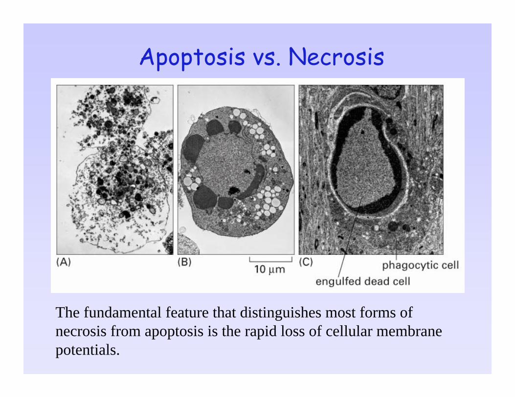
Apoptosis vs. Necrosis
The fundamental feature that distinguishes most forms of necrosis from apoptosis is the rapid loss of cellular membrane potentials.
Apoptosis and necrosis may be inter-connected.
Many insults induce apoptosis at lower doses and necrosis at higher doses.Apoptosis and necrosis may coexist in the same cell. Apoptosis-induced dysfunction of mitochondria may ultimately lead to cellular energy depletion, therefore necrosis. (delayed necrosis or slow cell death)In the absence of phagocytosis, dead cells in the late stages of apoptosis may present necrotic features. (apoptotic necrosis or secondary necrosis)
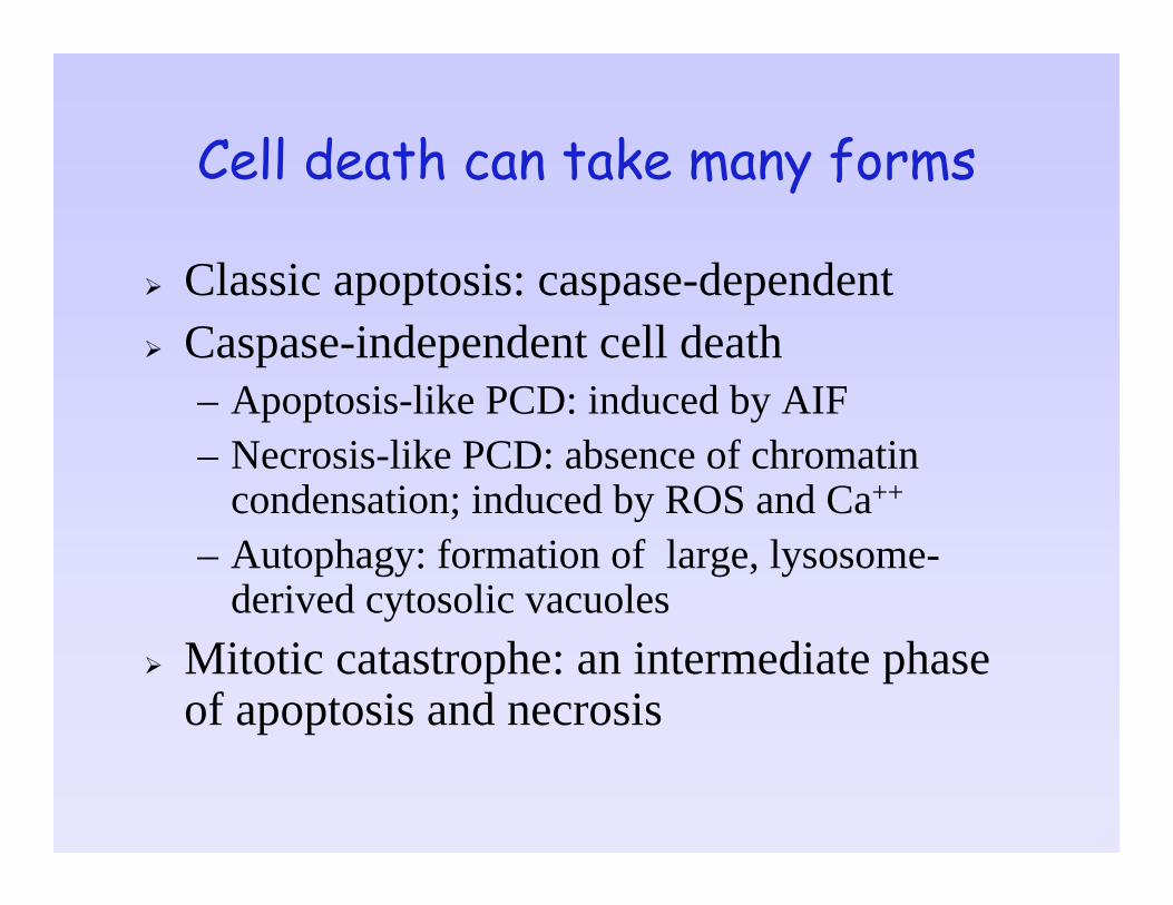
Cell death can take many forms
Classic apoptosis: caspase-dependentCaspase-independent cell death– Apoptosis-like PCD: induced by AIF – Necrosis-like PCD: absence of chromatin
condensation; induced by ROS and Ca++
– Autophagy: formation of large, lysosome-derived cytosolic vacuoles
Mitotic catastrophe: an intermediate phase of apoptosis and necrosis
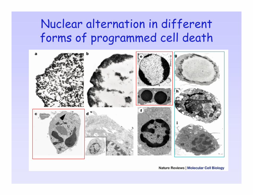
Nuclear alternation in different forms of programmed cell death
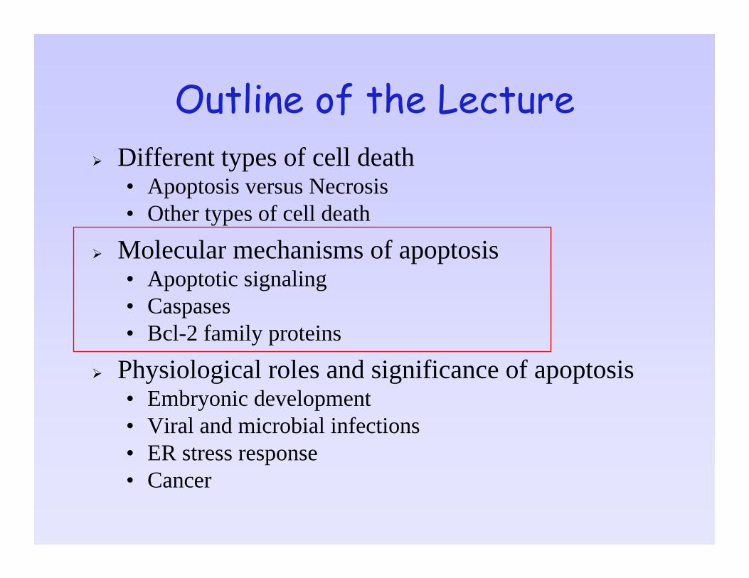
Outline of the LectureDifferent types of cell death• Apoptosis versus Necrosis• Other types of cell death
Molecular mechanisms of apoptosis• Apoptotic signaling• Caspases• Bcl-2 family proteins
Physiological roles and significance of apoptosis• Embryonic development• Viral and microbial infections• ER stress response• Cancer
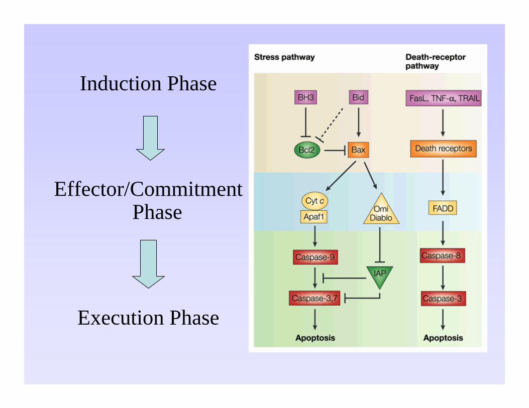
Induction Phase
Effector/Commitment Phase
Execution Phase
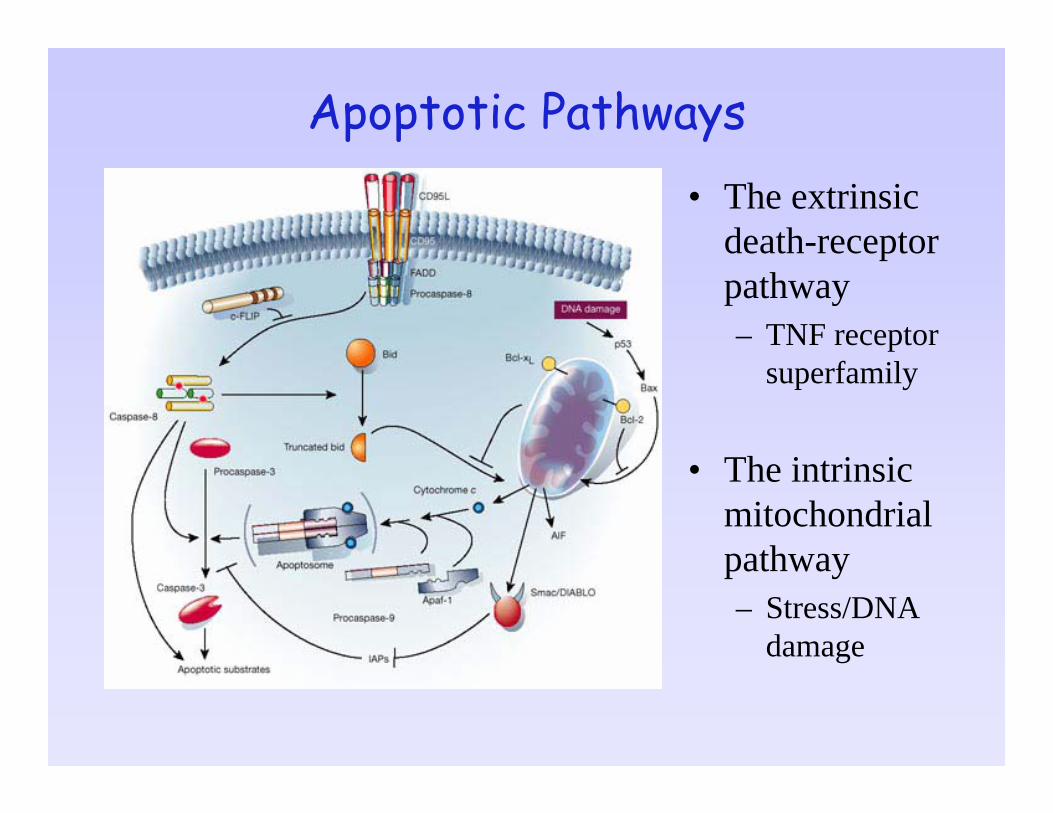
Apoptotic Pathways• The extrinsic
death-receptor pathway – TNF receptor
superfamily
• The intrinsic mitochondrial pathway– Stress/DNA
damage
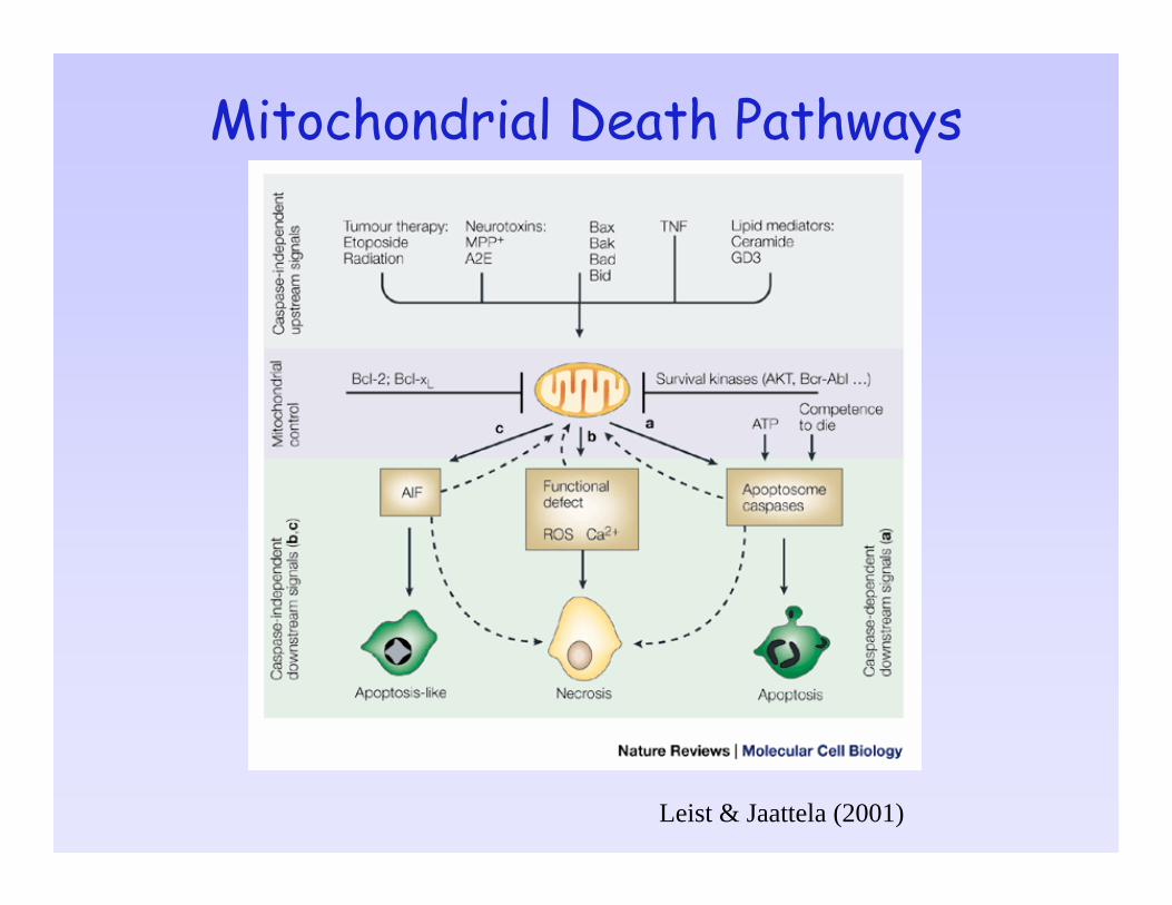
Mitochondrial Death Pathways
Leist & Jaattela (2001)
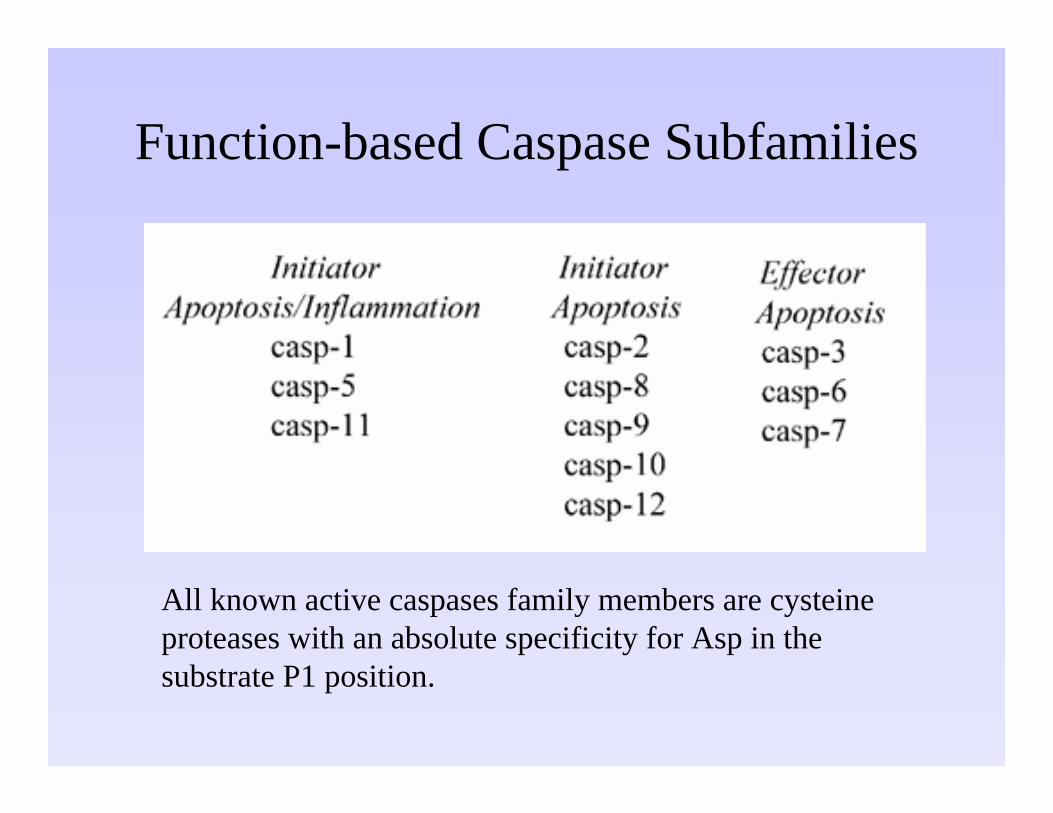
Function-based Caspase Subfamilies
All known active caspases family members are cysteine proteases with an absolute specificity for Asp in the substrate P1 position.
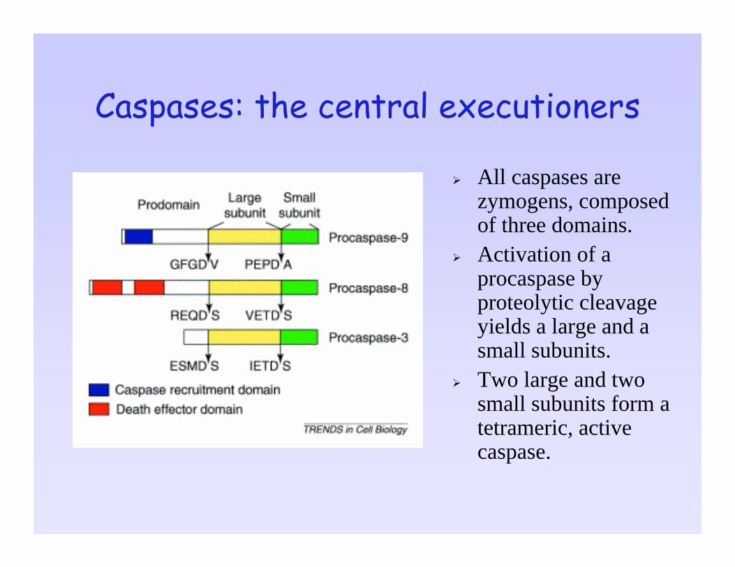
Caspases: the central executioners
All caspases are zymogens, composed of three domains.Activation of a procaspase by proteolytic cleavage yields a large and a small subunits.Two large and two small subunits form a tetrameric, active caspase.
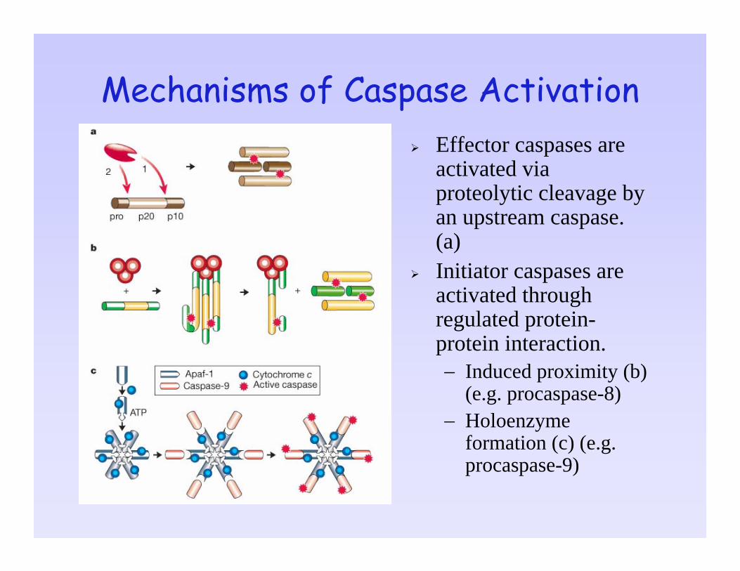
Mechanisms of Caspase ActivationEffector caspases are activated via proteolytic cleavage by an upstream caspase. (a)Initiator caspases are activated through regulated protein-protein interaction.− Induced proximity (b)
(e.g. procaspase-8)− Holoenzyme
formation (c) (e.g. procaspase-9)
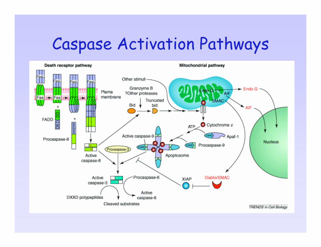
Caspase Activation Pathways

Inhibitors of Apoptosis Proteins (IAPs)
Consisting of NAIP, XIAP, cIAP1, cIAP2, and survivnSuppress apoptosis by preventing procaspaseactivation and inhibiting the activity of mature caspases (caspase-3, -7, and -9) by directly binding to caspases.Expression of cIAP1/2 is stimulated by NF-κB-mediated survival signals.Negative regulators of IAPs: Smac/DIABLO, XAF1, and OMI/HTRA2
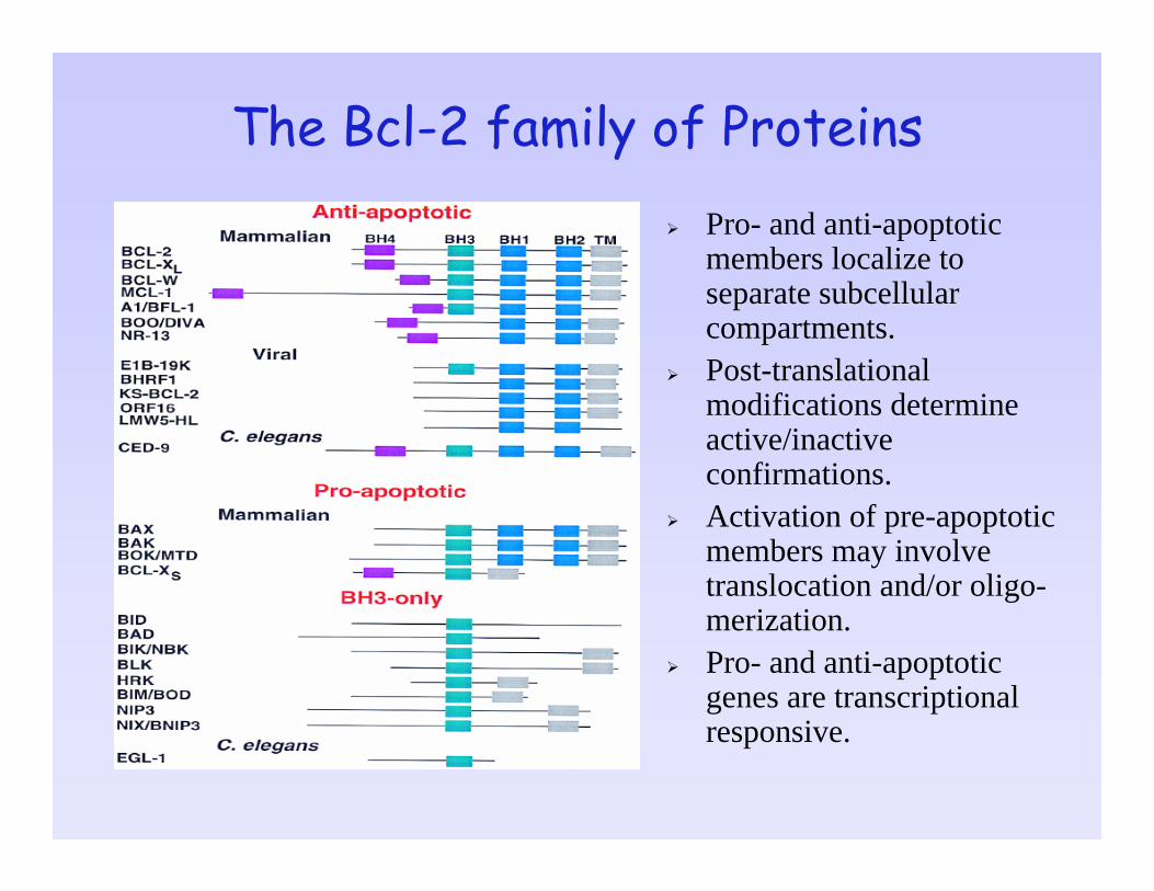
The Bcl-2 family of ProteinsPro- and anti-apoptotic members localize to separate subcellularcompartments.Post-translational modifications determine active/inactive confirmations.Activation of pre-apoptotic members may involve translocation and/or oligo-merization.Pro- and anti-apoptotic genes are transcriptional responsive.
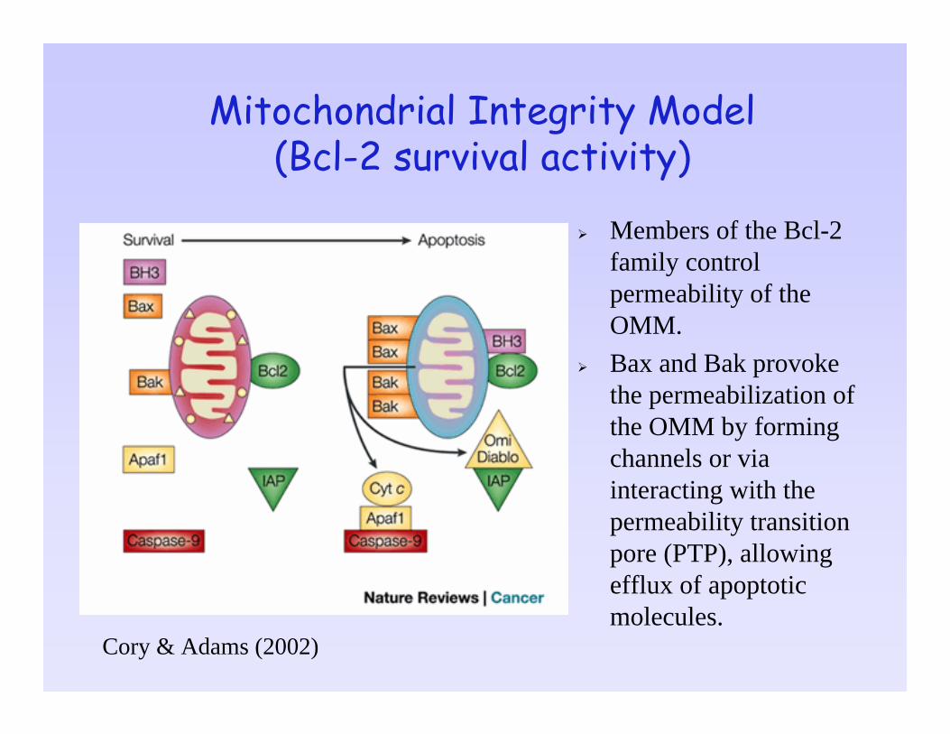
Mitochondrial Integrity Model (Bcl-2 survival activity)
Members of the Bcl-2 family control permeability of the OMM. Bax and Bak provoke the permeabilization of the OMM by forming channels or via interacting with the permeability transition pore (PTP), allowing efflux of apoptotic molecules.
Cory & Adams (2002)
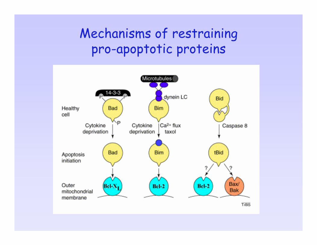
Mechanisms of restraining pro-apoptotic proteins
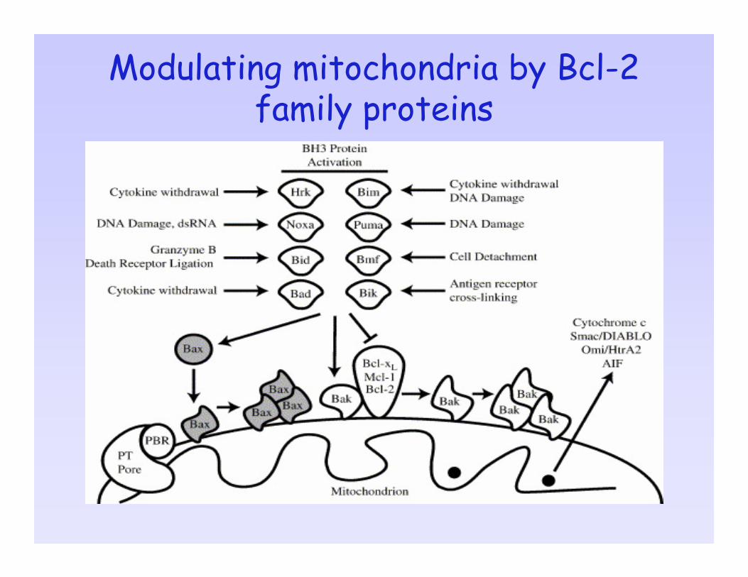
Modulating mitochondria by Bcl-2 family proteins

Apoptotic and Survival Pathways Involving Bcl-2 Members
Gross et al, Genes Dev. (1999)
Outline of the LectureDifferent types of cell death• Apoptosis versus Necrosis• Other types of cell death
Molecular mechanisms of apoptosis• Apoptotic signaling• Caspases • Bcl-2 family proteins
Physiological roles and significance of apoptosis• Embryonic development• Viral and microbial infections• ER stress response• Cancer

Apoptosis: essential for a health lifeA major form of cell death to remove excess, damaged or infected cells throughout life.− A fundamental process in the development of an
organism and in control of self-renewing tissues − A balance of cell division (e.g. regulating cell numbers
in the developing nervous system)− A self-defense mechanism against viral and pathogen
infectionLoss of control of the apoptotic program contributes to many diseases– Accumulation of unwanted cells through inefficient
apoptosis (e.g. cancer)– Cell loss as a result of excessive apoptosis (e.g.
neurodegeneration, stroke and heart failure)
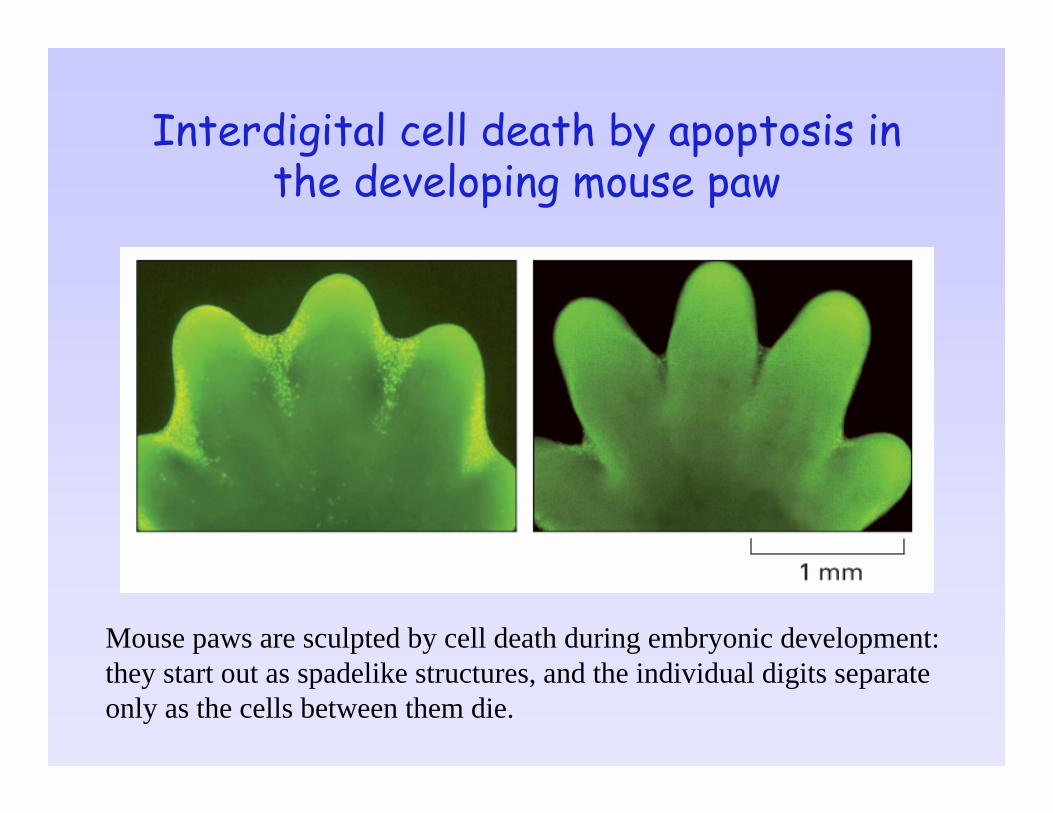
Interdigital cell death by apoptosis in the developing mouse paw
Mouse paws are sculpted by cell death during embryonic development: they start out as spadelike structures, and the individual digits separate only as the cells between them die.
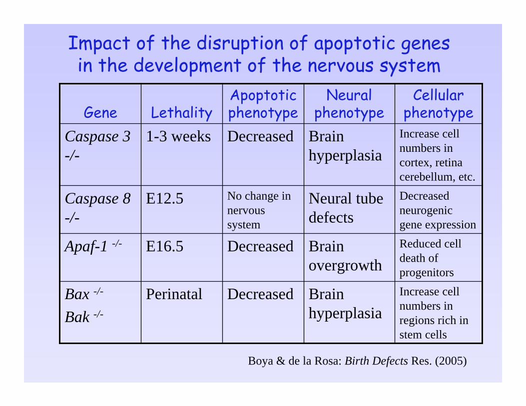
Impact of the disruption of apoptotic genes in the development of the nervous system
Increase cell numbers in regions rich in stem cells
Brain hyperplasia
DecreasedPerinatalBax -/-
Bak -/-
Reduced cell death of progenitors
Brain overgrowth
DecreasedE16.5Apaf-1 -/-
Decreased neurogenicgene expression
Neural tube defects
No change in nervous system
E12.5Caspase 8 -/-
Increase cell numbers in cortex, retina cerebellum, etc.
Brain hyperplasia
Decreased1-3 weeksCaspase 3 -/-
Cellular phenotype
Neural phenotype
Apoptotic phenotypeLethalityGene
Boya & de la Rosa: Birth Defects Res. (2005)
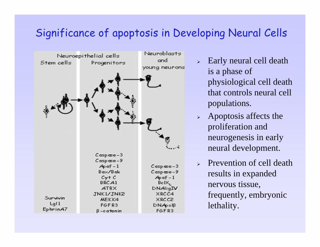
Significance of apoptosis in Developing Neural Cells
Early neural cell death is a phase of physiological cell death that controls neural cell populations. Apoptosis affects the proliferation and neurogenesis in early neural development.
Prevention of cell death results in expanded nervous tissue, frequently, embryonic lethality.

Deregulation of apoptosis leads to embryonic malformations
Hernandez-Sanchez et al. EMBO J (2003)
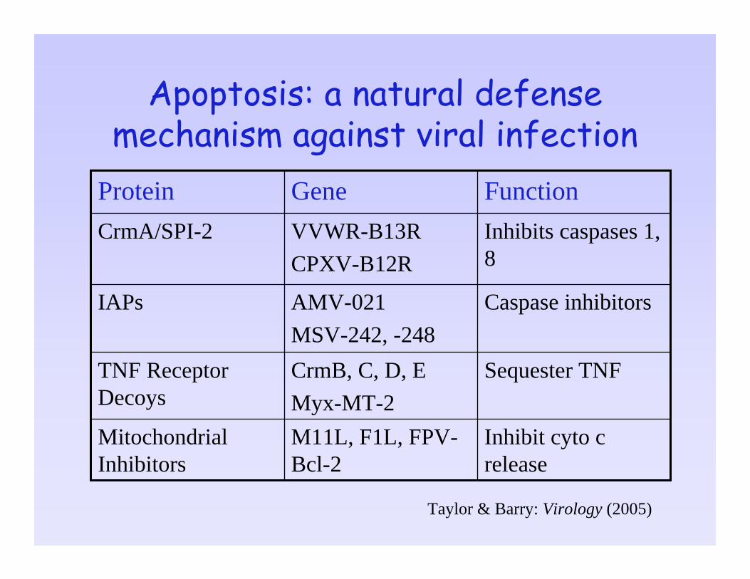
Apoptosis: a natural defense mechanism against viral infection
Inhibit cyto c release
M11L, F1L, FPV-Bcl-2
Mitochondrial Inhibitors
Sequester TNFCrmB, C, D, EMyx-MT-2
TNF Receptor Decoys
Caspase inhibitorsAMV-021MSV-242, -248
IAPs
Inhibits caspases 1, 8
VVWR-B13RCPXV-B12R
CrmA/SPI-2
FunctionGeneProtein
Taylor & Barry: Virology (2005)
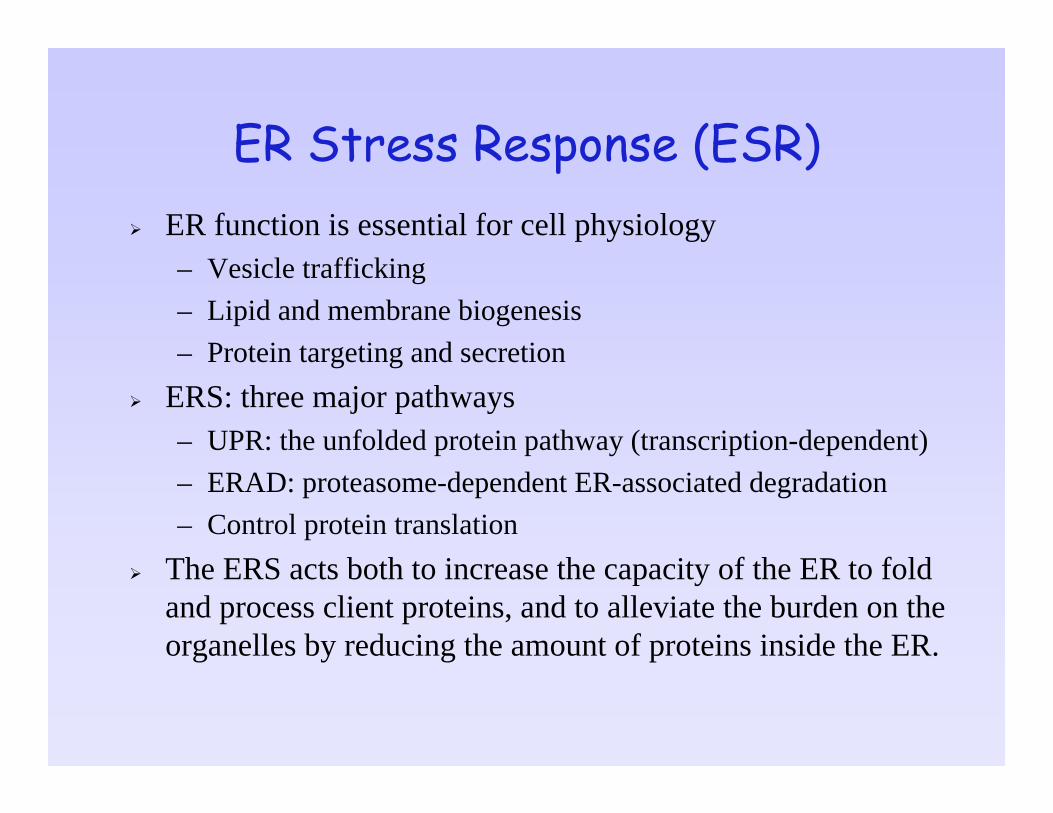
ER Stress Response (ESR)ER function is essential for cell physiology– Vesicle trafficking– Lipid and membrane biogenesis– Protein targeting and secretion
ERS: three major pathways– UPR: the unfolded protein pathway (transcription-dependent)– ERAD: proteasome-dependent ER-associated degradation– Control protein translation
The ERS acts both to increase the capacity of the ER to fold and process client proteins, and to alleviate the burden on the organelles by reducing the amount of proteins inside the ER.

ER Stress–induced apoptotic pathways
Boyce & Yuan: Cell Death Differ. (2006)
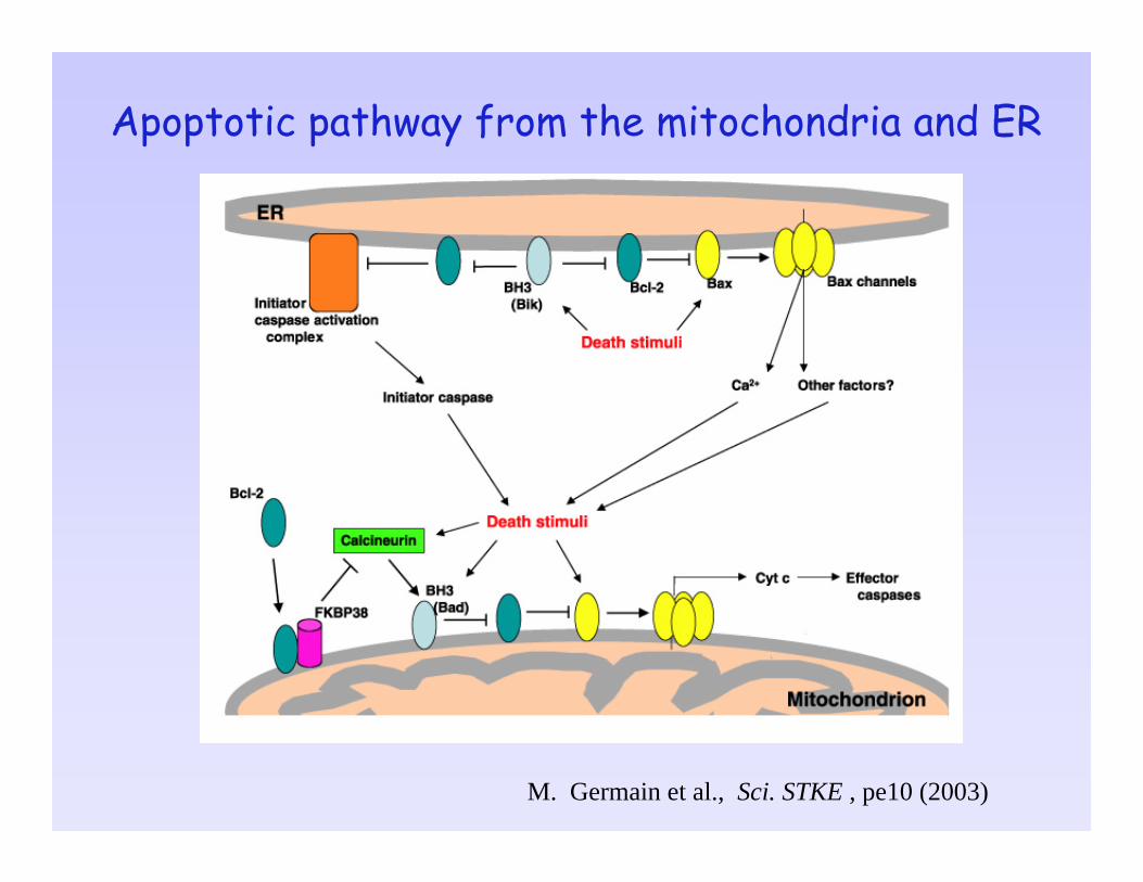
Apoptotic pathway from the mitochondria and ER
M. Germain et al., Sci. STKE , pe10 (2003)
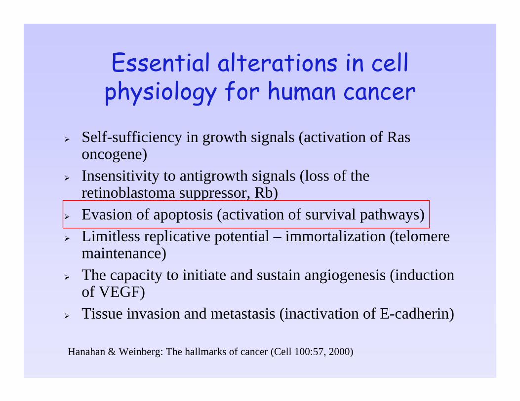
Essential alterations in cell physiology for human cancer
Self-sufficiency in growth signals (activation of Ras oncogene)Insensitivity to antigrowth signals (loss of the retinoblastoma suppressor, Rb)Evasion of apoptosis (activation of survival pathways)Limitless replicative potential – immortalization (telomere maintenance)The capacity to initiate and sustain angiogenesis (induction of VEGF)Tissue invasion and metastasis (inactivation of E-cadherin)
Hanahan & Weinberg: The hallmarks of cancer (Cell 100:57, 2000)

Damages/Stress
p53
Cell cyclearrest
Apoptosis DNA Repair Angiogenesisinhibition
Tumor InhibitionChemo- and radio-sensitivity
Loss of p53 pathway function can contribute not only to aggressive tumor behavior but also to therapeutic resistance.
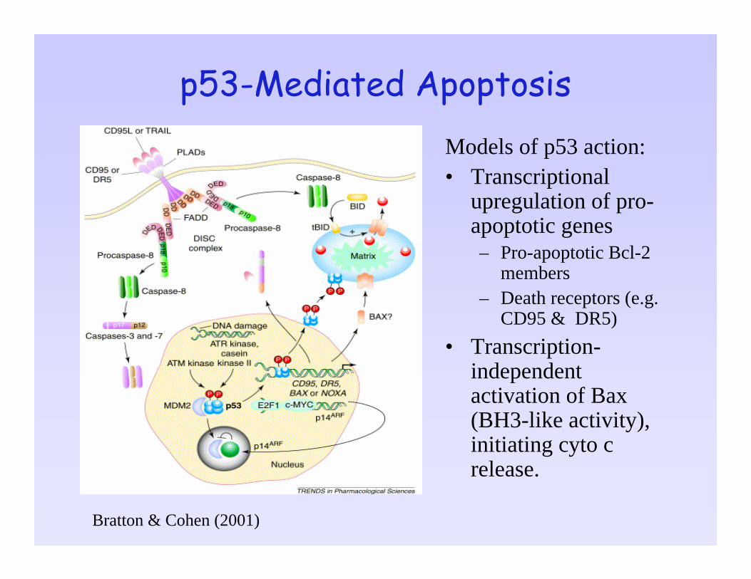
p53-Mediated ApoptosisModels of p53 action:• Transcriptional
upregulation of pro-apoptotic genes– Pro-apoptotic Bcl-2
members– Death receptors (e.g.
CD95 & DR5)• Transcription-
independent activation of Bax(BH3-like activity), initiating cyto c release.
Bratton & Cohen (2001)

Dysregulation of the Intrinsic Apoptotic Pathway in Cancer Cells
Upstream from the mitochondriaMutations on those targeting upstream components of the apoptotic program (p53, PTEN, Akt, Ras)
At the MitochondriaBcl-2 family members (pro- and anti-apoptotic)
Downstream from the mitochondriaInhibitors of apoptosis proteins (IAPs) and heat shock proteins (Hsp70/90)Epigenetic silencing of Apaf-1, caspase-3 deletion, etc.
Caspase-independent mechanismsAIF (apoptosis inducing factor)

The mitochondrial pathway plays the central role in chemotherapy-induced apoptosis
Chemotherapeutic agents induce mitochondrial membrane disruption and mitochondrial release of cytochrome c that is inhabitable by Bcl-2 and Bcl-xL.Apaf-1 overexpression sensitizes cancer cells to chemotherapeutic agents, accompanied with increased caspase-9 and -3 activation.Cells deficient in Apaf-1 or caspase-9 are protected from apoptosis induced by anticancer drugs, whereas cells deficient in caspase-8 and -2 show no protecting effect against anticancer drugs.
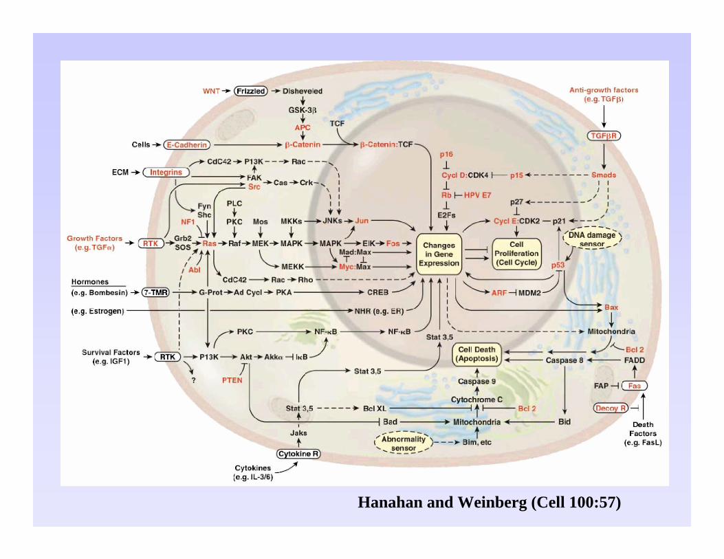
Hanahan and Weinberg (Cell 100:57)