gluteal region - ju medicine · 1-superior gluteal artery a branch from the internal iliac artery...
TRANSCRIPT
Gluteal region
Dr. Heba Kalbouneh
Associate Professor of Anatomy and Histology
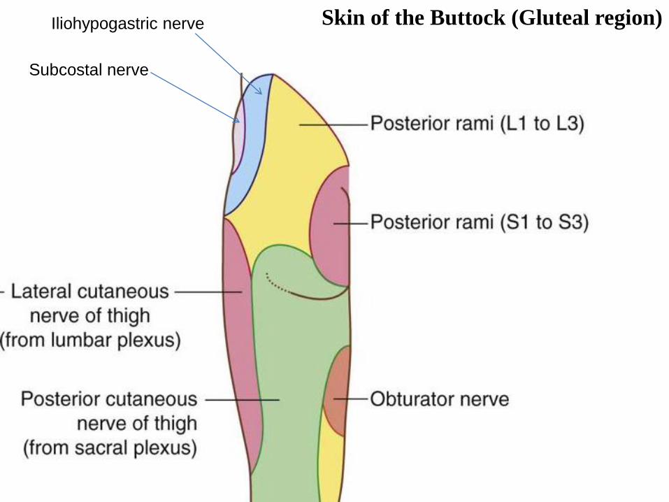
Skin of the Buttock (Gluteal region) Iliohypogastric nerve
Subcostal nerve

L5
L4
L3
L2
L1
Ilio-inguinal nerve
Ilio-hypogastric nerve
L1
Subcostal nerve
T12

1-Superficial fascia; is thick especially in women.
It contributes to the prominence of the buttock.
2-Deep fascia; continuous with the deep fascia of the
thigh (fascia lata).
Fascia of the Buttock (Gluteal region)
Glu
teu
s M
axim
us
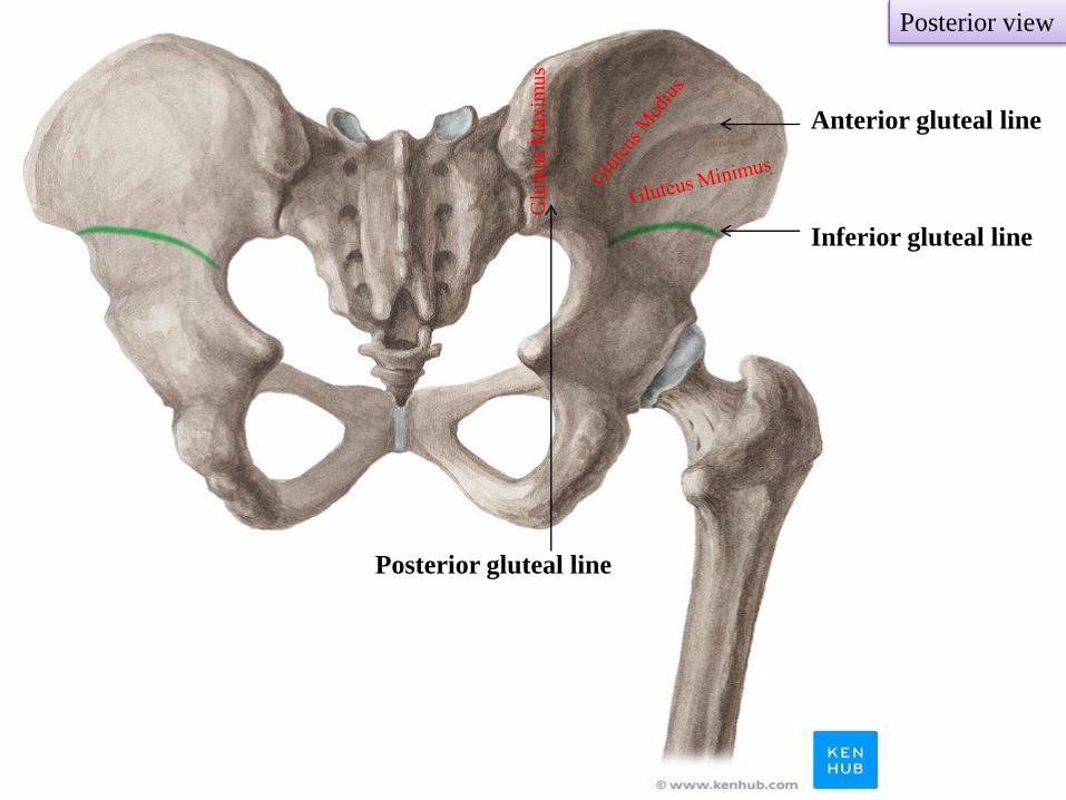
Posterior view
Posterior gluteal line
Inferior gluteal line
Anterior gluteal line
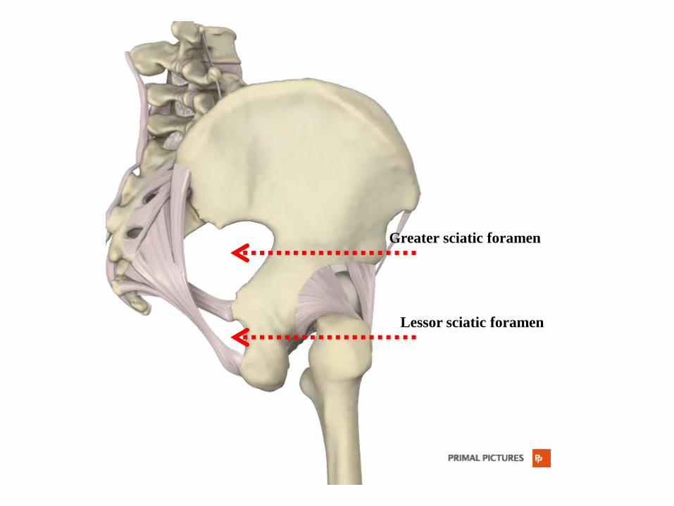
Greater sciatic foramen
Lessor sciatic foramen

Posterior view
Gluteus
maximus
Insertion:
1-The superficial three –fourths are
inserted into the iliotibial tract
2-The deep one fourth is inserted into the
gluteal tuberosity of femur
Origin:
1-Ilium (area behind the
posterior gluteal line)
2-Back of sacrum and coccyx
3-Back of sacrotuberous
ligament
Gluteus maximus
Iliotibial
tract

Action:
1-Extends thigh, some lateral
rotation
(Main extensor of hip joint)
2-Plays an important role in
climbing upstairs and cycling
Powerful extensor of flexed
femur at hip joint
3-Supports the extended knee
joint through iliotibial tract
Nerve supply:
Inferior gluteal nerve (L5;S1,2)
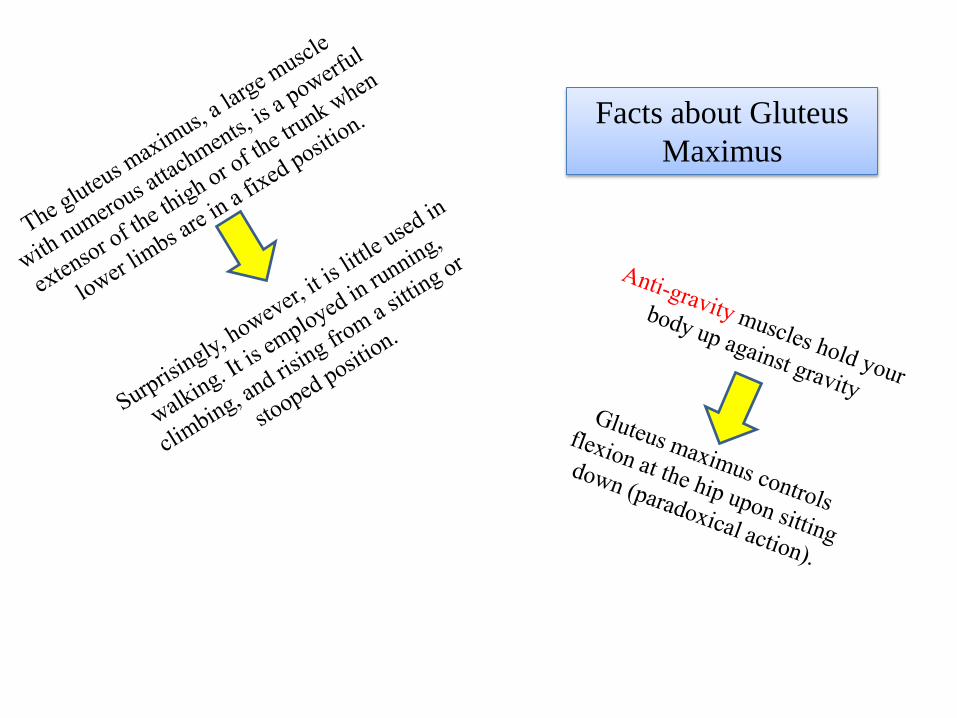
Facts about Gluteus
Maximus
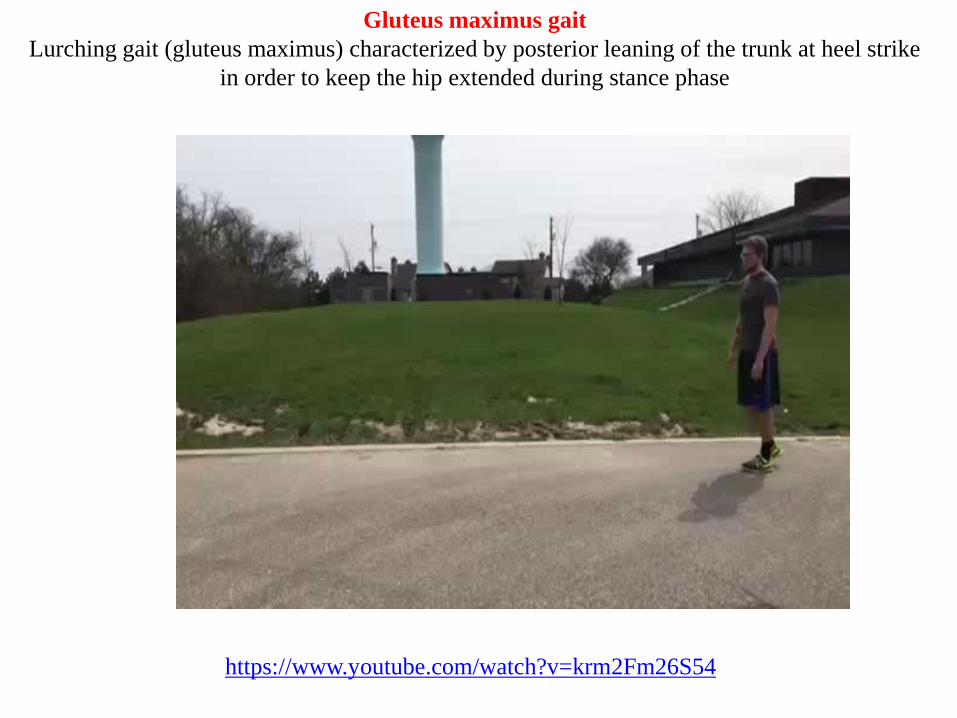
Gluteus maximus gait
Lurching gait (gluteus maximus) characterized by posterior leaning of the trunk at heel strike
in order to keep the hip extended during stance phase
https://www.youtube.com/watch?v=krm2Fm26S54

Gluteus medius
Gluteus minimus
Origin: Ilium ?
Insertion: Greater trochanter of femur
Action:
1-Abduction (main abductors of the hip joint)
2-Medial rotation (anterior fibers)
3-Both muscle contract reflexly on each side alternatively during
walking to prevent tilting of the pelvis to the unsupported side
Nerve supply: Superior gluteal nerve (L4, L5, S1)
Medius
Minimus
Posterior view


During walking, the glutei medius and minimus of the stance limb tilt the pelvis so
that the swinging limb can clear the ground
Stance Swing


During walking, the glutei medius and minimus of the stance limb tilt the pelvis so
that the swinging limb can clear the ground

When standing on one leg, the abductors of the hip on this side (gluteus medius and minimus
and tensor fascia lata) maintain fixation at the hip joint
If, however, there is any defect in these muscles or lever mechanism
of the hip joint, the weight of the body in these circumstances forces
the pelvis to tilt downwards on the opposite side.
Trendelenburg’s test
The normal pelvis (right) tilts
downwards
Left Right
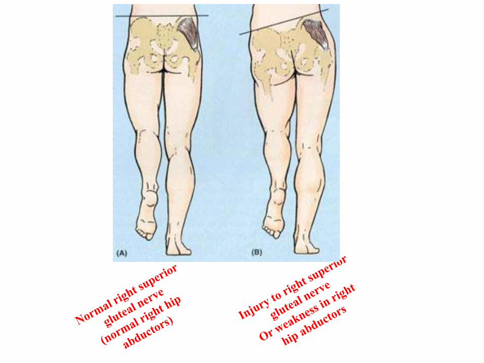

Injury to the superior gluteal nerve
On one side causes Lurching gait
Both sides Waddling gait
Positive Trendelenburg’s test
Note
Other conditions also my cause lurching and waddling gates such as:
Clinical Notes

During the stance phase, the weakened abductor muscles allow the pelvis to tilt down on the
opposite side. The pelvis sags on the opposite side of the lesioned superior gluteal nerve.
https://www.youtube.com/watch?v=Rz7V1i8kYGU

Lurching gait (gluteus medius and minimus)
To compensate, the trunk lurches to the weakened side to
attempt to maintain a level pelvis throughout the gait
cycle
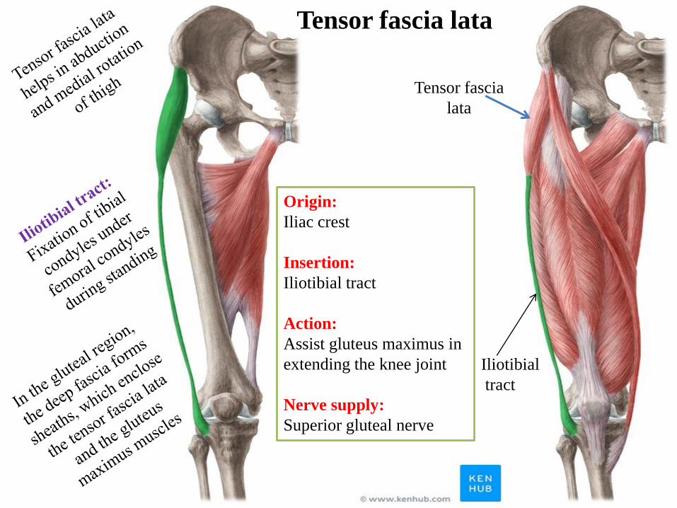
Tensor fascia
lata
Origin:
Iliac crest
Insertion:
Iliotibial tract
Action:
Assist gluteus maximus in
extending the knee joint
Nerve supply:
Superior gluteal nerve
Tensor fascia lata
Iliotibial
tract
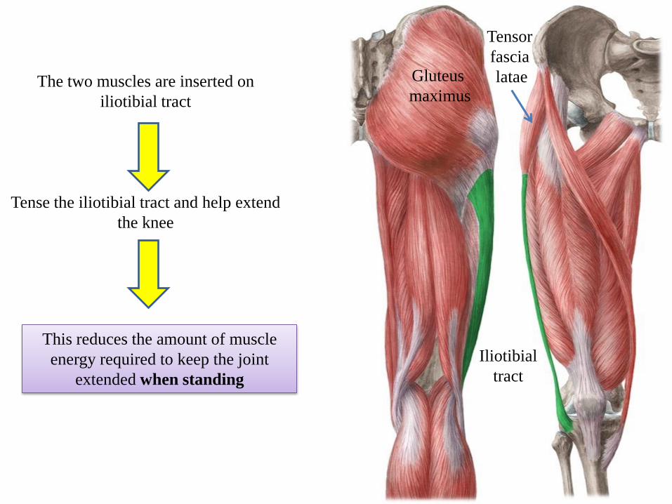
Tensor
fascia
latae Gluteus
maximus
Iliotibial
tract
The two muscles are inserted on
iliotibial tract
Tense the iliotibial tract and help extend
the knee
This reduces the amount of muscle
energy required to keep the joint
extended when standing

Short Lateral rotator muscles
1- Piriformis
2- Obturator internus
3- Superior gemellus
4- Inferior gemellus
5-Quadratus femoris
6- Obturator externus
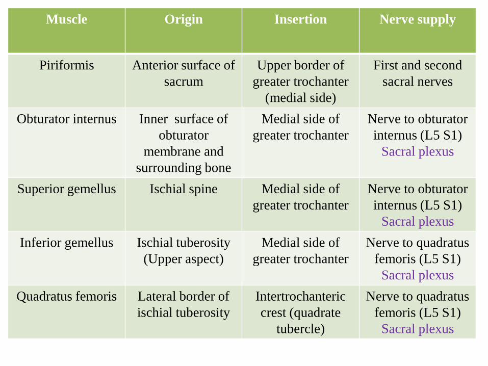
Muscle Origin Insertion Nerve supply
Piriformis Anterior surface of
sacrum
Upper border of
greater trochanter
(medial side)
First and second
sacral nerves
Obturator internus Inner surface of
obturator
membrane and
surrounding bone
Medial side of
greater trochanter
Nerve to obturator
internus (L5 S1)
Sacral plexus
Superior gemellus Ischial spine Medial side of
greater trochanter
Nerve to obturator
internus (L5 S1)
Sacral plexus
Inferior gemellus Ischial tuberosity
(Upper aspect)
Medial side of
greater trochanter
Nerve to quadratus
femoris (L5 S1)
Sacral plexus
Quadratus femoris Lateral border of
ischial tuberosity
Intertrochanteric
crest (quadrate
tubercle)
Nerve to quadratus
femoris (L5 S1)
Sacral plexus

Obturator internus
Piriformis
Inferior gemellus
Quadratus femoris
Superior gemellus
Posterior view

Obturator internus
Piriformis
Inferior gemellus
Quadratus femoris
Superior gemellus
Gluteus Medius
Gluteus Minimus
Posterior view

Obturator internus
Posterior view

Posterior view
Origin: Outer surface of obturator membrane and
surrounding bone
Insertion: Medial surface of greater trochanter
Nerve supply: Obturator nerve
Action: Laterally rotates thigh at hip joint
Obturator externus
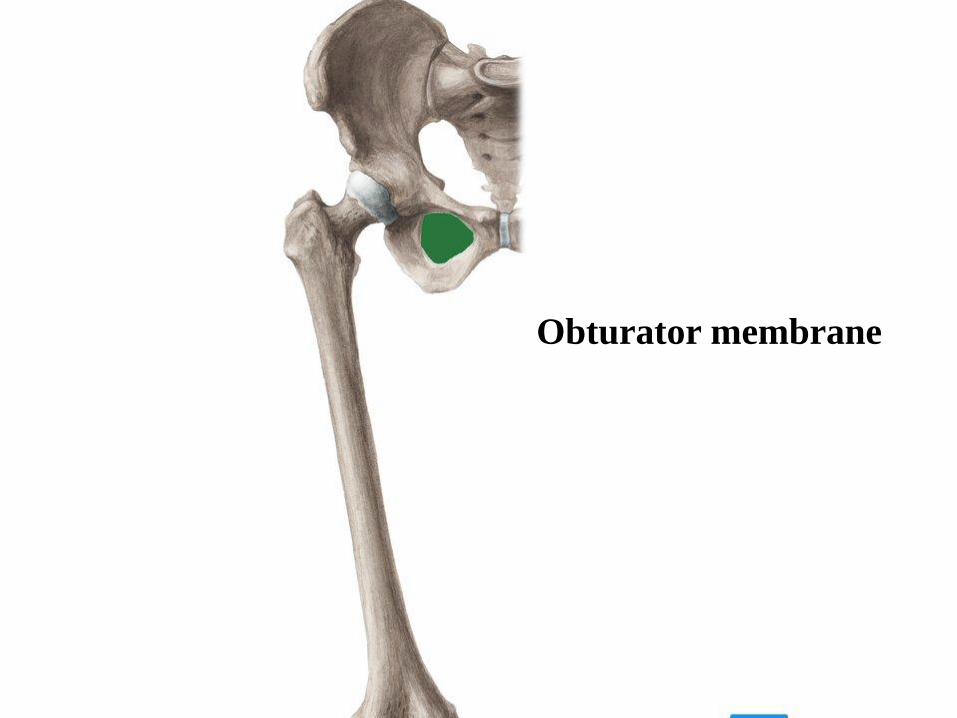
Obturator membrane

o Piriformis: fills the foramen almost
completely leaving some structures to pass
either above or below it.
Structures passing above Piriformis
muscle:
o Superior gluteal nerve and vessels
Structures passing below Piriformis
muscle:
o Inferior gluteal nerve and vessels
o Sciatic nerve
o Posterior cutaneous nerve of the thigh
o Nerve to quadratus femoris
o Pudendal nerve
o Internal pudendal vessels
o Nerve to obturator internus
o Tendon of obturator internus
o Nerve to obturator internus
o Pudendal nerve
o Internal pudendal vessels
Structures passing through the greater sciatic foramen:
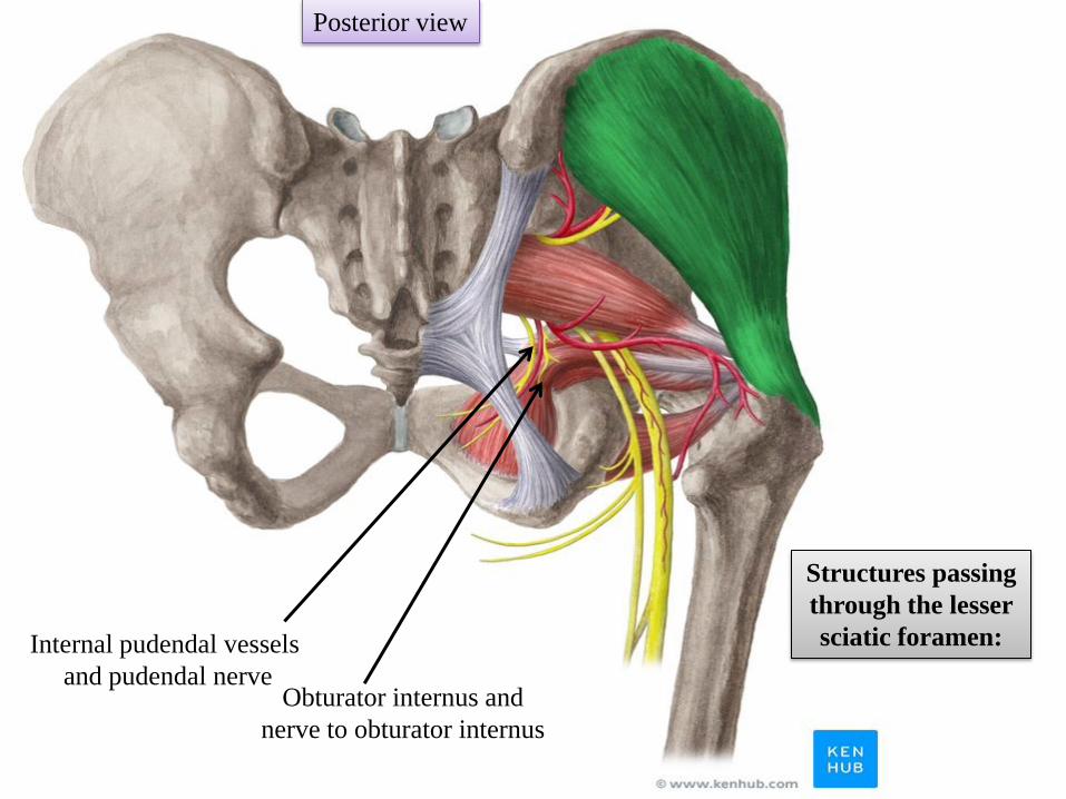
Structures passing through the lesser sciatic foramen:

Piriformis fills the greater
sciatic foramen almost
completely leaving some
structures to pass either
above or below it.
Piriformis passes through
the greater sciatic foramen.

Obturator internus passes
through the lesser sciatic
foramen.
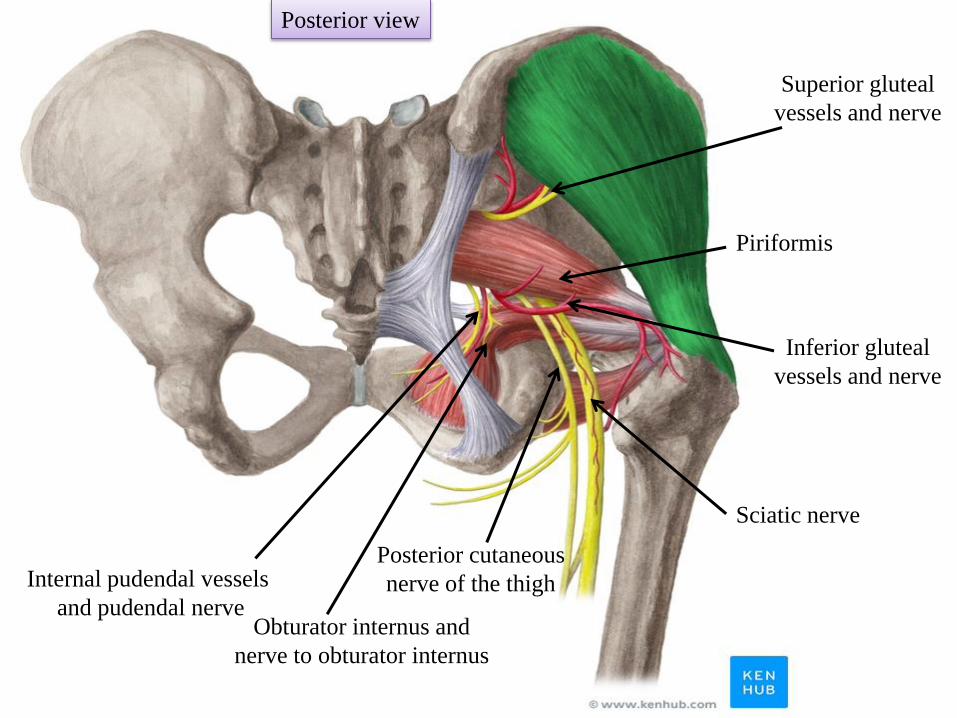
Posterior view
Piriformis
Inferior gluteal
vessels and nerve
Superior gluteal
vessels and nerve
Obturator internus and
nerve to obturator internus
Internal pudendal vessels
and pudendal nerve
Sciatic nerve
Posterior cutaneous
nerve of the thigh
Posterior view
Obturator internus and
nerve to obturator internus
Internal pudendal vessels
and pudendal nerve
Structures passing
through the lesser
sciatic foramen:

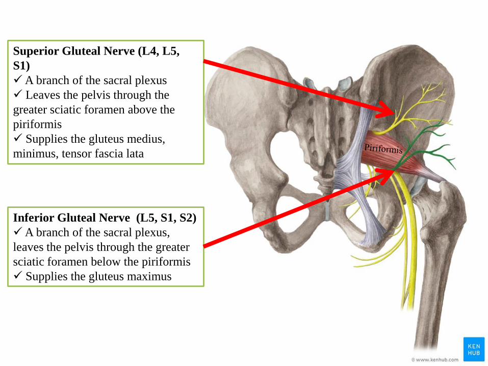
Superior Gluteal Nerve (L4, L5,
S1)
A branch of the sacral plexus
Leaves the pelvis through the
greater sciatic foramen above the
piriformis
Supplies the gluteus medius,
minimus, tensor fascia lata
Inferior Gluteal Nerve (L5, S1, S2)
A branch of the sacral plexus,
leaves the pelvis through the greater
sciatic foramen below the piriformis
Supplies the gluteus maximus
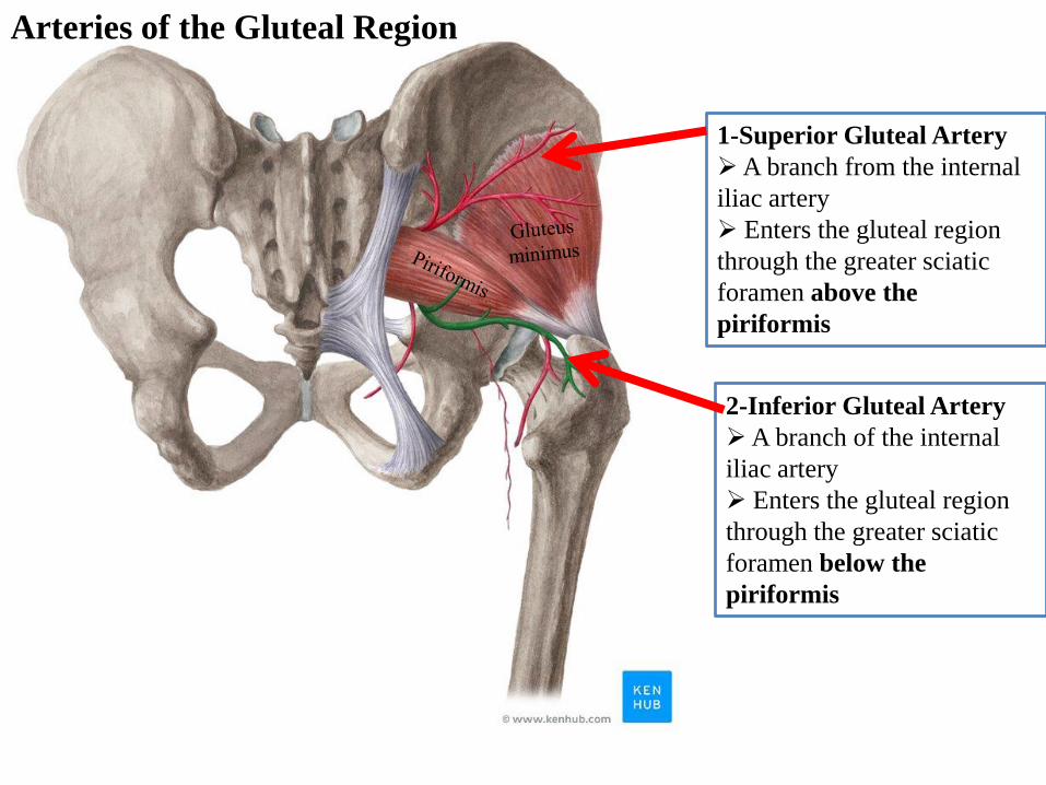
1-Superior Gluteal Artery
A branch from the internal
iliac artery
Enters the gluteal region
through the greater sciatic
foramen above the
piriformis
2-Inferior Gluteal Artery
A branch of the internal
iliac artery
Enters the gluteal region
through the greater sciatic
foramen below the
piriformis
Arteries of the Gluteal Region
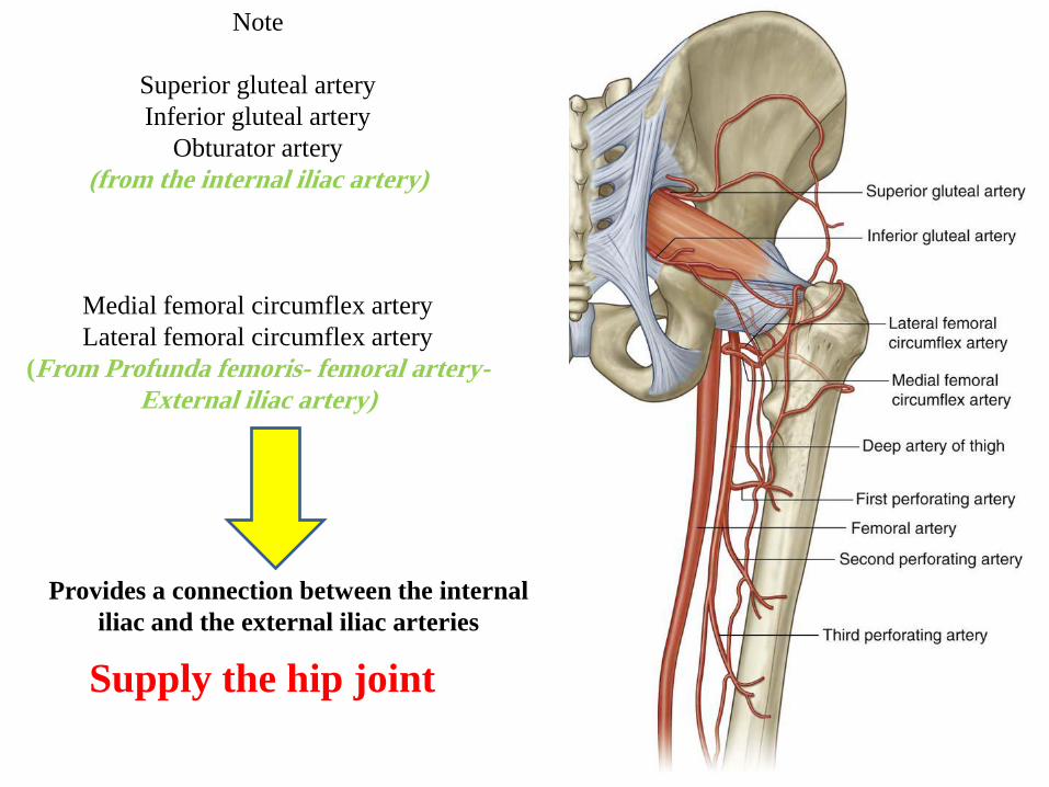
Note
Superior gluteal artery
Inferior gluteal artery
Obturator artery
(from the internal iliac artery)
Medial femoral circumflex artery
Lateral femoral circumflex artery
(From Profunda femoris- femoral artery-
External iliac artery)
Provides a connection between the internal
iliac and the external iliac arteries
Supply the hip joint
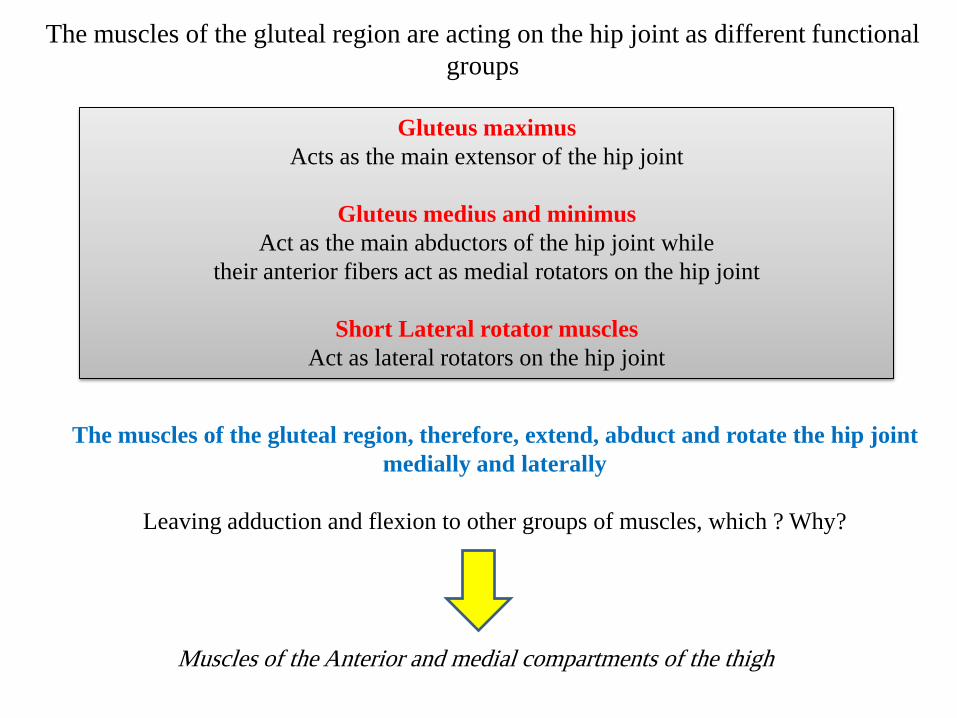
The muscles of the gluteal region are acting on the hip joint as different functional
groups
Gluteus maximus
Acts as the main extensor of the hip joint
Gluteus medius and minimus
Act as the main abductors of the hip joint while
their anterior fibers act as medial rotators on the hip joint
Short Lateral rotator muscles
Act as lateral rotators on the hip joint
Muscles of the Anterior and medial compartments of the thigh
The muscles of the gluteal region, therefore, extend, abduct and rotate the hip joint
medially and laterally
Leaving adduction and flexion to other groups of muscles, which ? Why?

Intramuscular
(IM) injection

Clinical Notes
The great thickness of
gluteus maximus muscle
makes it ideal for
intramuscular injections.
To avoid injury to the
underlying sciatic nerve,
the injection should be
given well forward on
The upper outer quadrant
of the buttock
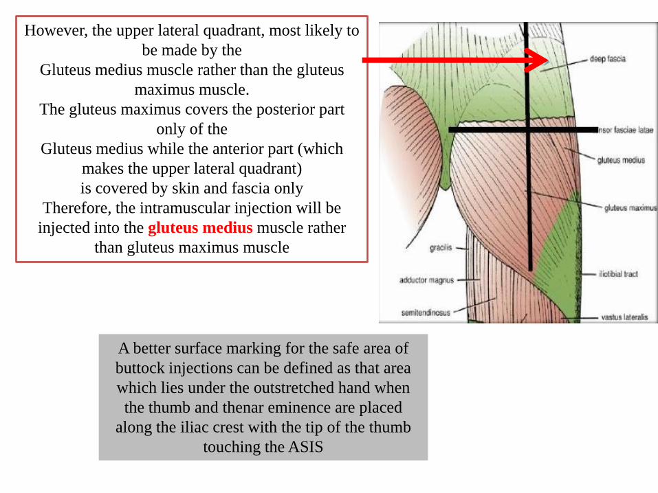
However, the upper lateral quadrant, most likely to
be made by the
Gluteus medius muscle rather than the gluteus
maximus muscle.
The gluteus maximus covers the posterior part
only of the
Gluteus medius while the anterior part (which
makes the upper lateral quadrant)
is covered by skin and fascia only
Therefore, the intramuscular injection will be
injected into the gluteus medius muscle rather
than gluteus maximus muscle
A better surface marking for the safe area of
buttock injections can be defined as that area
which lies under the outstretched hand when
the thumb and thenar eminence are placed
along the iliac crest with the tip of the thumb
touching the ASIS
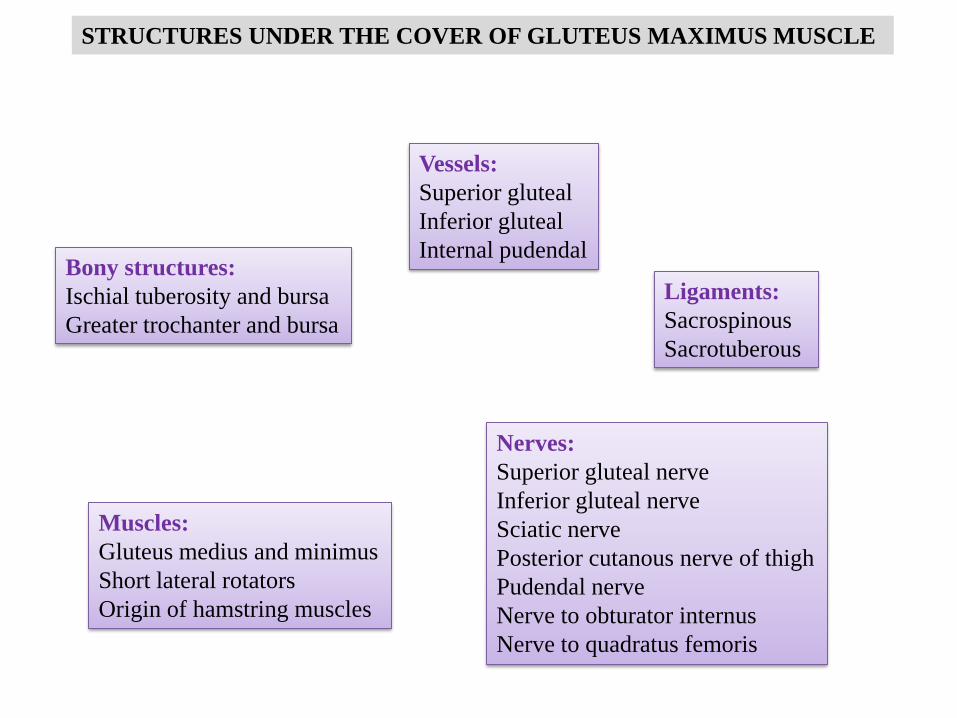
STRUCTURES UNDER THE COVER OF GLUTEUS MAXIMUS MUSCLE
Bony structures:
Ischial tuberosity and bursa
Greater trochanter and bursa
Ligaments:
Sacrospinous
Sacrotuberous
Muscles:
Gluteus medius and minimus
Short lateral rotators
Origin of hamstring muscles
Vessels:
Superior gluteal
Inferior gluteal
Internal pudendal
Nerves:
Superior gluteal nerve
Inferior gluteal nerve
Sciatic nerve
Posterior cutanous nerve of thigh
Pudendal nerve
Nerve to obturator internus
Nerve to quadratus femoris


Bursa
Bursa
Sacrotuberous
ligament
Sacrospinous
ligament

Ischial tuberosity
Greater trochanter

