glucagonoma syndrome: a case report
TRANSCRIPT
CASE REPORT Open Access
Glucagonoma syndrome: a case reportPablo Granero Castro*, Alberto Miyar de León, Jose Granero Trancón, Paloma Álvarez Martínez*,Jose A Álvarez Pérez, Jose C Fernández Fernández, Carmen M García Bernardo, Luis Barneo Serra andJuan J González González
Abstract
Introduction: Glucagonoma syndrome is a rare paraneoplastic phenomenon, with an estimated incidence of onein 20 million, characterized by necrolytic migratory erythema, hyperglucagonemia, diabetes mellitus, anemia,weight loss, glossitis, cheilitis, steatorrhea, diarrhea, venous thrombosis and neuropsychiatric disturbances in thesetting of a glucagon-producing alpha-cell tumor of the pancreas. Necrolytic migratory erythema is the presentingmanifestation in the majority of cases, so its early suspicion and correct diagnosis is a key factor in themanagement of the patient.
Case presentation: We present the case of a 70-year-old Caucasian woman with glucagonoma syndrome due toan alpha-cell tumor located in the tail of the pancreas, successfully treated with surgical resection.
Conclusion: Clinicians should be aware of the unusual initial manifestations of glucagonoma. Early diagnosisallows complete surgical resection of the neoplasm and provides the only chance of a cure.
IntroductionA glucagonoma is a slow-growing alpha-cell tumor ofthe pancreatic islets of Langerhans. It may appear as abenign and localized alpha-cell adenoma but at least50% of cases will have metastatic disease when diag-nosed [1]. Glucagonomas can be associated with othertumors in Multiple Endocrine Neoplasia syndrome 1(MEN 1), but this association is rare and comprises nomore than 3% of glucagonomas. Even though glucago-nomas related to MEN 1 syndrome probably carry abetter prognosis due to early recognition through peri-odic screening visits, 80% are malignant and frequentlyspread to the liver [2]. Glucagonoma syndrome is a rareparaneoplastic phenomenon, with an estimated inci-dence of one in 20 million, characterized by necrolyticmigratory erythema (NME), hyperglucagonemia, dia-betes mellitus, anemia, weight loss, glossitis, cheilitis,steatorrhea, diarrhea, venous thrombosis and neuropsy-chiatric disturbances in the setting of a glucagon-produ-cing alpha-cell tumor of the pancreas [3]. The mostcommon features of this syndrome are weight loss,NME and diabetes mellitus [4]. Of these, NME presentsas the hallmark clinical sign of glucagonoma syndrome
[3]. Its early recognition allows a prompt diagnosis ofthe tumor and leads to a better prognosis. Surgery is theoptimal treatment for a glucagonoma. We present apatient with glucagonoma syndrome due to a well cir-cumscribed alpha-cell tumor of the pancreas, in whichsurgical removal of the tumor by distal pancreatectomywith splenectomy led to resolution of the cutaneous andsystemic features.
Case presentationA 70-year-old Caucasian woman was referred to ourDepartment of Dermatology with a persistent subacuteeczema affecting her lower extremities and groin areathat had been present for 12 months. She was treatedwith topical and oral steroids with no improvement. Hermedical history revealed a long-standing type 2 diabetesmellitus and recurrent episodes of deep-vein thrombosisin her right leg despite anticoagulant therapy. The skineruption initially appeared in her lower extremities butthere was a rapid progression with involvement of hertrunk, upper extremities and perioral area. These skinlesions were associated with weight loss (15 kg in oneyear), anorexia, weakness, glossitis and angular stomati-tis. A physical examination revealed itching cutaneouseruptions of erythematous polycyclic migratory lesionswith scaling advancing borders and central resolution.
* Correspondence: [email protected]; [email protected] of General Surgery and Gastroenterology, Hospital UniversitarioCentral de Asturias, Oviedo, Spain
Castro et al. Journal of Medical Case Reports 2011, 5:402http://www.jmedicalcasereports.com/content/5/1/402 JOURNAL OF MEDICAL
CASE REPORTS
© 2011 Granero Castro et al; licensee BioMed Central Ltd. This is an Open Access article distributed under the terms of the CreativeCommons Attribution License (http://creativecommons.org/licenses/by/2.0), which permits unrestricted use, distribution, andreproduction in any medium, provided the original work is properly cited.
The entire course of the local skin lesion healed withintwo weeks while new cutaneous eruptions occurred inother locations. Chronic lesions often evolved into liche-nification. Laboratory data showed a low hemoglobinlevel (10.5 g/dL), hyperglycemia (176 mg/dL), hypoalbu-minemia (22 g/L) and hypoproteinemia (49 g/L). Herwhite cell count (6100/μL) and platelet level (26.2 ×10⁴/μL) were also within normal limits, and an abnorm-ality of cell form was not found in her peripheral blood.Her levels of serum iron, vitamin B12 and erythropoie-tin, and the number of reticulocytes were found to benormal. Electrophoresis of her serum protein was per-formed because of the possibility of multiple myeloma;however, no abnormal protein was found. Although thepossibility of gastrointestinal (GI) bleeding was consid-ered, no abnormality was detected on an upper GIendoscopy, barium enema, or barium examination ofher small bowel. A skin biopsy performed on a peritibiallesion showed a spongiotic epidermis with vacuolizationof the granular layer and presence of necrotic keratino-cytes in the horny layer. A mild infiltrate of lymphocyteswas present in the papillary dermis (Figure 1). Histologi-cal findings were compatible with the diagnosis of NME.Ultrasonography was performed as a screening examina-tion, and revealed a hypoechoic tumor in her distal pan-creas. An abdominal computed tomography (CT) scanshowed a hypervascularized tumor measuring 5 to 7 cmin the tail of her pancreas without evidence of meta-static disease (Figure 2). Carcinoembryonic antigen andcarbohydrate antigen 19-9 levels were normal. The exo-crine function of her pancreas was normal. However,her level of serum glucagon was elevated to 2340 pg/mL(normal range, 55-177 pg/mL), while her levels of otherhormones, such as somatostatin or gastrin, were withinnormal limits, and insulin was low. Glucagonoma of thepancreas was diagnosed and distal pancreatectomy withsplenectomy was performed. This resection involved
dissection of her regional lymph nodes (D1). Histo-pathological examination revealed a 6 cm alpha-cellpancreatic tumor with vascular and perineural tumorinvasion. Dissection of the regional lymph nodes showedthat a total of 16 lymph nodes were isolated, of whichthree were affected. Inmunohistochemical staining waspositive for glucagon, chromogranin and synaptophysin,but negative for other hormones, such as insulin, gastrinand somatostatin. On the basis of these findings, a diag-nosis of malignant glucagonoma of the pancreas wasmade. Five days after surgery, the skin lesions disap-peared and postoperative plasma glucagon levelsdecreased to 197 pg/dL. Our patient has been withoutrecurrence for one and a half years since the surgeryand remains asymptomatic.
DiscussionStacpoole [5] reported that all of the following criteriashould be satisfied to diagnose a glucagonoma: demon-stration of a tumor mass by direct visualization or radio-graphic techniques; proof that the tumor shows apreponderance of glucagon-containing cells on appropri-ate staining and/or proof of increased tissue levels ofimmunoreactive glucagon; elevation of basal circulatingimmunoreactive glucagon; and at least one of the fol-lowing coincidental findings; (a) skin rash, (b) glucoseintolerance, or (c) hypoaminoacidemia. The patientreported here fulfilled these criteria.The high level of glucagon secreted by the tumor may
promote glycogenolysis, gluconeogenesis, ketogenesisand lipodieresis by activating phosphorylase in the liver,stimulating the secretion of insulin, and inhibiting theexternal secretion of the pancreas, thus resulting in theincreased level of blood glucose. In persistent hyperglu-cagonemia, diabetes mellitus develops at the expense oftissue glycogen stores, muscle and fat mass. Impairedfasting glycemia or diabetes mellitus is found in 80% of
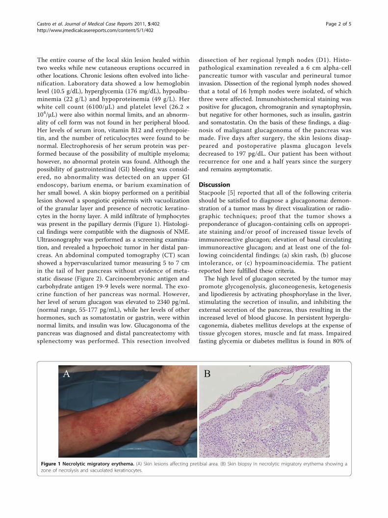
A B
Figure 1 Necrolytic migratory erythema. (A) Skin lesions affecting pretibial area. (B) Skin biopsy in necrolytic migratory erythema showing azone of necrolysis and vacuolated keratinocytes.
Castro et al. Journal of Medical Case Reports 2011, 5:402http://www.jmedicalcasereports.com/content/5/1/402
Page 2 of 5
patients with glucagonoma syndrome [6]. Hyperglucago-nemia in healthy patients and in glucagonoma syndromereduces plasma amino acid concentrations and enhancesessential amino acid catabolism.NME is the presenting manifestation in 70% of
patients with glucagonoma syndrome [3]. The lesionsconsist of erythematous scaling and crusting patchesmost frequently observed in the groin, intergluteal andgenital areas. Central healing may occur giving an annu-lar appearance. The most specific feature on skin histo-logical examination is necrolysis of the upper epidermiswith vacuolated keratinocytes, leading to focal or conflu-ent necrosis, but this histopathologic feature may beseen in other deficiency states like pellagra, necrolyticacral erythema or zinc deficiency [4]. Normalization ofglucagon concentrations by surgery results in a rapiddisappearance of the skin rash. However, abnormal glu-cagon levels alone cannot explain all of the skin find-ings. Hypoaminoacidemia, nutritional lack of zinc andfatty acids or hepatocellular dysfunctions are all consid-ered to be possible triggering factors of NME. Hyperglu-cagonemia provokes multiple nutrient and vitamin Bdeficiencies, which in turn are the probable cause of thistypical skin disorder [7]. Other systemic pathologies,
such as chronic liver disease, inflammatory bowel dis-ease, malabsorptive state, pancreatitis, various malignantneoplasms and heroin abuse have been associated withNME without glucagonoma. The early recognition ofNME is therefore important because it will prevent thecatabolic clinical features and reduce the risk of metas-tasis with obvious quality of life improvements. Eventhough NME has traditionally been considered an earlymanifestation of disease, it is more likely a late manifes-tation of years of tumor growth. The relatively lowoccurrence rate of NME in patients with MEN-asso-ciated glucagonoma diagnosed early in the clinicalcourse by screening efforts supports this contention[3,8].As seen in our patient, glucagonoma syndrome can be
associated with a high incidence of thromboembolism.A thromboembolic phenomenon, such as deep-veinthrombosis or pulmonary embolism, may be present in10-30% of patients, often resulting in death. In fact,thromboembolic events may account for over 50% of alldeaths directly attributed to the glucagonoma syndrome.The mechanism for this coagulopathy is poorly under-stood and seems to be related to an increased factor ×production by the pancreatic alpha-cells [9].
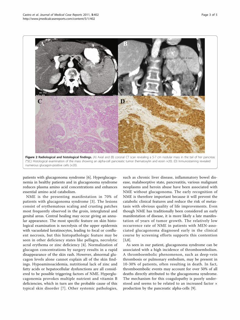
C D
A B
Figure 2 Radiological and histological findings. (A) Axial and (B) coronal CT scan revealing a 5-7 cm nodular mass in the tail of her pancreas(*)(C) Histological examination of the mass showing an alpha-cell pancreatic tumor (hematoxylin and eosin ×20). (D) Inmunostaining revealednumerous glucagon-positive cells (×20).
Castro et al. Journal of Medical Case Reports 2011, 5:402http://www.jmedicalcasereports.com/content/5/1/402
Page 3 of 5

Although the optimal treatment for glucagonoma issurgery, 50% of the tumors have metastasized by thetime of diagnosis [1]. Transabdominal ultrasound hasbeen used to demonstrate tumor localization but haslimited utility because of its inferior sensitivity. Endo-scopic ultrasound visualization of the tumor is report-edly highly sensitive but has not gained widespreadacceptance. A contrast-enhanced CT scan is very usefulin attempting to demonstrate the presence of a pancrea-tic tumor and it is usually the initial radiographic testbecause of its non-invasiveness. However, selective visc-eral angiography is considered the gold standard in diag-nosis and localization of glucagonomas. Its superiorsensitivity is related to the hypervascularity of these neo-plasms. Although it is an invasive test, it has the advan-tage of being able to demonstrate hepatic metastasiseven in cases with normal liver scans. The role of mag-netic resonance imaging (MRI) in the diagnosis of gluca-gonoma has not been clearly defined. The utility ofpancreatic venous sampling for glucagon levels to diag-nose smaller tumors has also been reported [5]. Thediagnosis is made by the finding of a pancreatic alpha-cell tumor. Although the average size of a glucagonomamay be large, diagnostic confirmation and localizationby needle biopsy is not usually performed due to theease and precision of alternative methods. These othermethods to localize the tumor in suspected casesinclude ultrasound, CT and selective visceral angiogra-phy. Tumors and metastases can be visualized by CT.Neuroendocrine tumors, in contrast to pancreatic exo-crine adenocarcinoma, are hypervascular lesions, andthis characteristic is often useful when reviewing ima-ging studies. Somatostatin receptor scintigraphy is fre-quently used as a complementary method toconventional imaging such as CT and MRI as it is usefulfor consistent supervision of somatostatin receptorexpression and dissemination of the tumor metastases[4,10]. The liver is the most frequent site of metastasis,followed by the peripancreatic lymph nodes, bone adre-nal gland, kidney and lung.Pancreatic endocrine tumors represent a heteroge-
neous group with varying tumor biology and prognosis.These neoplasms are classified as functional if they areassociated with a hormone-related clinical syndromecaused by hormone release from the tumor, or non-func-tional if the tumor is not associated with a hormone-related clinical syndrome [11]. The differential diagnosisis based on histopathology demonstrating neuroendo-crine features such as positive staining for chromograninA and specific hormones such as gastrin, proinsulin andglucagon. The differential diagnosis of glucagonomaincludes other primary pancreatic neoplasms, intrapan-creatic malignant mesothelioma, and solid pseudopapil-lary neoplasms [12]. A close relationship between
glucagon expression in pancreatic endocrine tumors andcystic formation is also reported in the literature, but cys-tic glucagonomas are not associated with a glucagonomasyndrome in the majority of cases as they are non-func-tioning glucagon-producing neuroendocrine tumors [13].Treatment of glucagonoma syndrome should be direc-
ted at the underlying etiology. Removal of the primarytumor with a distal pancreatectomy brought evidentrelief of all clinical symptoms for 1 to 2 year periods.Pancreatic fistula and delayed gastric emptying are themost prevalent complications of distal pancreatectomybut they can be managed by conservative measures inthe majority of cases. The tumor is resistant to che-motherapy and metastatic disease is often not amenableto surgical resection. The prognosis of this disease variesgreatly according to the stage at which the disease isdiagnosed. Estimations of mean survival after diagnosishave ranged from three to seven years or more [14].Long-acting somatostatin analogues, which are potentinhibitors of glucagon release, have been proven effec-tive in suppressing glucagon secretion from glucagono-mas and controlling the metastatic growth. Since thetumor is slow growing, prolonged survival is possibleand, in metastatic disease, most causes of death appearto be unrelated to the tumor. Control of liver metastasesby metastasectomy, cryoablation, radiofrequency abla-tion or chemoembolization has been reported [4,6].Recent studies suggest that conventional contraindica-tions to surgical resection, such as superior mesentericvein invasion and nodal or distant metastases, should beredefined in patients with advanced neuroendocrinetumors. These patients will benefit from extensive surgi-cal debulking in combination with adjuvant medicaltreatments, such as somatostatin analogues. This combi-nation may result in enhanced survival rates comparedwith either procedure alone [10].
ConclusionClinicians should be aware of the unusual initial mani-festations of glucagonoma. Early diagnosis allows com-plete surgical resection of the neoplasm and providesthe only chance of a cure.
ConsentWritten informed consent was obtained from the patientfor publication of this case report and any accompany-ing images. A copy of the written consent is availablefor review by the Editor-in-Chief of this journal.
Authors’ contributionsAML, JGT, PAM, JAP, JFF, CGB, LBS and JGG were involved in the direct careof this patient. In addition, PGC was responsible for drafting the manuscriptand JAP, JGT and PAM helped to draft the manuscript. All authors have readand approved the final manuscript.
Castro et al. Journal of Medical Case Reports 2011, 5:402http://www.jmedicalcasereports.com/content/5/1/402
Page 4 of 5
Competing interestsThe authors declare that they have no competing interests.
Received: 24 February 2011 Accepted: 22 August 2011Published: 22 August 2011
References1. Hellman P, Andersson M, Rastad J, Juhlin C, Karacagil S, Eriksson B,
Skogseid B, Akerström G: Surgical strategy for large or malignantendocrine pancreatic tumors. World J Surg 2000, 24:1353-1360.
2. Okauchi Y, Nammo T, Iwahashi H, Kizu T, Hayashi I, Okita K, Yamagata K,Uno S, Katsube F, Matsuhisa M, Kato K, Aozasa K, Kim T, Osuga K,Nakamori S, Tamaki Y, Funahashi T, Miyagawa J, Shimomura I:Glucagonoma diagnosed by arterial stimulation and venous sampling(ASVS). Inter Med 2009, 48:1025-1030.
3. Wermers RA, Fatourechi V, Wynne AG, Kvols LK, Lloyd RV: Theglucagonoma syndrome. Clinical and pathologic features in 21 patients.Medicine 1996, 75:53-63.
4. Chastain MA: The glucagonoma syndrome: a review of its features anddiscussion of new perspectives. Am J Med Sci 2001, 321:306-320.
5. Stacpoole PW: The glucagonoma syndrome: clinical features, diagnosis,and treatment. Endocr Rev 1981, 2:347-361.
6. O’Grady HL, Conlon KC: Pancreatic neuroendocrine tumours. Eur J SurgOncol 2008, 34:324-332.
7. van Beek AP, de Haas ER, van Vloten WA, Lips CJ, Roijers JF, Canninga-vanDijk MR: The glucagonoma syndrome and necrolytic migratoryerythema: a clinical review. Eur J Endocrinol 2004, 151:531-537.
8. Lobo I, Carvalho A, Amaral C, Machado S, Carvalho R: Glucagonomasyndrome and necrolytic migratory erythema. Int J Dermatol 2010,49:24-29.
9. Teixeira RC, Nico MM, Ghideti AC: Necrolytic migratory erythemaassociated with glucagonoma: a report of 2 cases. Clinics (Sao Paulo)2008, 63:267-270.
10. Norton JA, Kivlen M, Li M, Schneider D, Chuter T, Jensen RT: Morbidity andmortality of aggressive resection in patients with advancedneuroendocrine tumors. Arch Surg 2003, 138:859-866.
11. Öberg K: Pancreatic endocrine tumors. Semin Oncol 2010, 37:594-618.12. Papavramidis T, Papavramidis S: Solid pseudopapillary tumors of the
pancreas: review of 718 patients reported in English literature. J Am CollSurg 2005, 100:965-972.
13. Konukiewitz B, Enosawa T, Klöppel G: Glucagon expression in cysticpancreatic neuroendocrine neoplasms: an immunohistochemicalanalysis. Virchows Arch 2011, 458:47-53.
14. Schwartz RA: Glucagonoma and pseudoglucagonoma syndromes. Int JDermatol 1997, 36:81-89.
doi:10.1186/1752-1947-5-402Cite this article as: Castro et al.: Glucagonoma syndrome: a case report.Journal of Medical Case Reports 2011 5:402.
Submit your next manuscript to BioMed Centraland take full advantage of:
• Convenient online submission
• Thorough peer review
• No space constraints or color figure charges
• Immediate publication on acceptance
• Inclusion in PubMed, CAS, Scopus and Google Scholar
• Research which is freely available for redistribution
Submit your manuscript at www.biomedcentral.com/submit
Castro et al. Journal of Medical Case Reports 2011, 5:402http://www.jmedicalcasereports.com/content/5/1/402
Page 5 of 5