glaucoma · 2019-07-18 · of glaucoma? 1. primary open-angle glaucoma this is by far the most...
TRANSCRIPT

www.aucklandeye.co.nz
GLAUCOMA
phone (64) 09 529 2480fax (64) 09 529 2481email [email protected] www.aucklandeye.co.nz

GLAUCOMA
ABOUT GLAUCOMA
Glaucoma is an eye disease that affects 2-3% of people over the age of 60. It is the second most common cause of blindness in the western world and the second leading cause of blindness worldwide after cataracts.
There may be no symptoms to glaucoma as it is often a silent disease slowly developing over time. It is often detected during a glasses assessment by a vigilant optometrist, or because a patient is aware of the risk and has chosen to have their eyes formally checked.
Early detection, through regular and complete eye exams, is the key to protecting your vision from damage caused by glaucoma.
HOW DOES GLAUCOMA OCCUR?
Glaucoma occurs when the intraocular pressure (IOP) within the eye is high enough to cause progressive damage to the optic nerve, resulting in vision loss.
A normal, healthy optic nerve has about a million nerve fibres which come from the retina (the seeing membrane at the back of the eye). When glaucoma develops, these nerve fibres start to die off and if enough nerve tissue is lost, then the vision starts to be affected.
In the initial stages of glaucoma there is usually no noticeable change in vision but it has been
estimated that during this time a patient can lose as many as half the nerve fibres in the eye and these cannot be regenerated.
Even when glaucoma deterioration does become more advanced, the loss of sight starts out in the peripheral part of vision and people are often unaware of it. Once it reaches the central vision and does become noticeable, glaucoma has already reached an advanced stage and cannot be reversed. While there is currently no cure for glaucoma, the condition can be controlled. Auckland Eye are specialists at supporting and treating those affected.
NORMAL VISION
VISION WITH GLAUCOMA
WHAT CAUSES GLAUCOMA?
The causes of glaucoma include:
1. Elevated intraocular pressure (IOP)
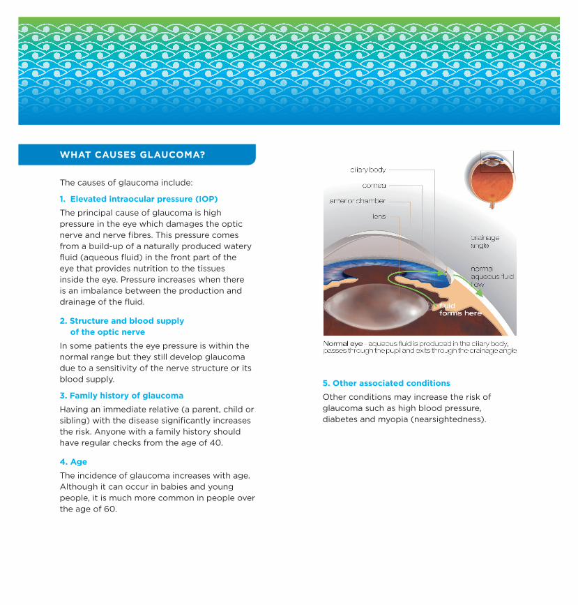
The principal cause of glaucoma is high pressure in the eye which damages the optic nerve and nerve fibres. This pressure comes from a build-up of a naturally produced watery fluid (aqueous fluid) in the front part of the eye that provides nutrition to the tissues inside the eye. Pressure increases when there is an imbalance between the production and drainage of the fluid.
2. Structure and blood supply of the optic nerve
In some patients the eye pressure is within the normal range but they still develop glaucoma due to a sensitivity of the nerve structure or its blood supply.
3. Family history of glaucoma
Having an immediate relative (a parent, child or sibling) with the disease significantly increases the risk. Anyone with a family history should have regular checks from the age of 40.
4. Age
The incidence of glaucoma increases with age.Although it can occur in babies and young people, it is much more common in people over the age of 60.
5. Other associated conditions
Other conditions may increase the risk ofglaucoma such as high blood pressure, diabetes and myopia (nearsightedness).

HOW IS GLAUCOMA DIAGNOSED?
There are a number of tests that can be performed to determine whether someone has glaucoma. These generally involve measuring the IOP, a test to measure peripheral (side) vision and assessments of changes in the optic nerve.
1. Tonometry
Tonometry measures the inner pressure of the eye providing an IOP reading. Anaesthetic eye drops are used to numb the eye and then the doctor or technician uses a special device (tonometer) to measure the eye’s pressure.
2. Gonioscopy
Gonioscopy is a painless eye test using a contact lens that checks to see if the drainage angle of the eye is normal.
3. Pachymetry
This is a simple, quick and painless test to measure the thickness of the cornea. It is important to assess this as it can have a bearing on the accuracy of eye pressure measurements.
_ = less risk
+ = higher risk
Average corneal thickness = 555 microns
High eye pressure = over 21 mmHg
Normal eye pressure = 6 - 21 mmHg
4. Visual field testing
Computerised measurements of a patient’s peripheral vision are helpful to diagnose and to monitor glaucoma. During this test, the patient is asked to look straight ahead and indicate when they see a light appear in their peripheral vision. This test helps to draw a “map” of a patient’s vision and any related gaps.
Glaucoma Risk
Thick Cornea + Normal eye pressure
_ _
Thick Cornea + High eye pressure
+
Thin Cornea + Normal eye pressure
+/_
Thin Cornea + High eye pressure
++++
Average Cornea + Normal eye pressure
_ _
Average Cornea + High eye pressure
++

5. Nerve fibre layer measurements
The thickness of the nerve fibre layer can be measured using a non-invasive imaging technique known as Optical Coherence Tomography (OCT). Like visual field testing, this can help in diagnosing glaucoma and also monitor its progression.
6. Optic nerve imaging
Obtaining baseline readings of the optic nerve, and repeating the tests regularly, is important as the doctor can then see if there are any changes over time. Photos and laser scanners are used to provide this information.
WHAT ARE THE TYPES OF GLAUCOMA?
1. Primary open-angle glaucoma
This is by far the most common form of glaucoma and appears in two-thirds of all cases. In open-angle glaucoma the drainage area in the eye is open but not working properly and pressure slowly builds up, causing gradual loss of peripheral vision.
2. Normal tension glaucoma
This is where the pressure inside the eye is within the normal range but nerve damage still occurs in spite of this. This accounts for about 25% of glaucoma cases and is more common in Asian races and people who have migraine or blood circulation problems.
3. Angle closure glaucoma
This uncommon type of glaucoma is caused by a blockage in the pressure drainage system. It is the only type of glaucoma that can be painful and develops rapidly. Patients may notice a severe aching in the eye along with eye redness and blurring of vision. It can damage vision within a day or two so an urgent assessment is needed.
4. Secondary glaucoma
Secondary glaucoma can arise due to other eye problems such as inflammation, blood vessel blockages and trauma.

5. Congenital and juvenile glaucoma
Uncommon, this type of glaucoma can occur in babies, children or young adults and the treatment may be different from adult glaucoma.
SHOULD I BE SCREENED FOR GLAUCOMA?
People should be tested and screened for glaucoma in keeping with the following recommendations:
People aged 40 to 49 years should be tested every five years
People aged 50 to 59 years should be tested every three years
People over the age of 60 should be tested every two years
Anyone with risk factors should be tested every year or two after the age of 40. Community optometrists are in the best position to offer this sort of screening.
HOW DO I MANAGE GLAUCOMA?
The aim of treatment is to reduce the IOP, which will reduce stress on the optic nerve and slow down or stop any further nerve damage. If the pressure is brought down to a satisfactory level, then the risk of visual field loss is reduced.
1. Medical treatment
Eye drops are usually the first treatment for glaucoma. There are various eye drops that can be used and an eye specialist can recommend the one that is the most appropriate. It may be necessary to try several eye drops to find the one drop, or combination, that is best for you.
2. Laser for primary open angle glaucoma
A new form of laser surgery called Selective Laser Trabeculoplasty (SLT) is a very safe and straightforward treatment for POAG. For some people it will avoid the need for ongoing eye drops while in others it will provide extra pressure lowering in combination with drops. SLT is better than older laser treatments because there is no/minimal discomfort and it can be repeated if the effect wears off over time. Overall it is effective in about 75% of people and carries no risk of side effects.
3. Laser for narrow angle glaucoma
Another form of laser surgery called YAG Peripheral Iridotomy (PI) creates an opening through the iris which allows the eye fluid to bypass the normal drainage pathway. This can reverse closed angle glaucoma and prevent

its development in those who have narrow drainage systems and are at risk of acute loss of vision.
4. Surgery
Surgery can be used to control pressure inside the eye but is generally used only when eye drops have proven unsatisfactory.
ONGOING MANAGEMENT
Once glaucoma is diagnosed, ongoing management and treatment is required throughout life. Like a lot of other conditions, such as blood pressure and diabetes, treatment is about controlling the disease but not curing it. If treatment is discontinued, then the glaucoma will progress again. Fluctuations in eye pressure are damaging, so it is very important to ensure that eye drops are taken regularly as missed doses can be harmful.
GLAUCOMA NEW ZEALAND
Glaucoma New Zealand is a non-profit organisation that provides support for New Zealanders with glaucoma. If you would like further information then please contact Glaucoma New Zealand:
www.glaucoma.org.nz email: [email protected]
Tel: +64 9 373 8779 Fax: + 64 9 373 7947
Auckland Eye, New Zealand’s leading private sub-specialty eye centre, is dedicated to providing the highest quality service in a caring environment. Auckland Eye’s team of leading experts are each highly trained in their particular area of expertise, providing assessment and management of a comprehensive range of ophthalmic problems.
They now provide surgical services in their brand new state-of-the-art facility, Oasis Surgical, the largest purpose built ophthalmic day-stay facility in New Zealand.
Auckland Eye and Oasis Surgical are the only procedure and consulting group in Auckland accredited by the DAA group for quality of patient care.
Auckland Eye and Oasis Surgical are centrally located in Remuera with easy motorway access, plentiful off street parking and wheelchair access.
Auckland Eye also has dedicated consulting facilities in Albany and New Lynn as well as providing appointments at a wide range of other locations across the Auckland region.
Auckland Eye is an affiliated provider to Southern Cross Health Society.
For more information on glaucoma please contact our friendly specialist team.
AUCKLAND EYE LIFE-CHANGING OPHTHALMIC CARE

AUCKLAND EYESURGEONS
AUCKLAND EYESURGEONS
Dr Stephen BestBSc, MBChB, FRANZCORemuera, Botany
Dr Taras PapchenkoBHB, MBChB, PhD, FRANZCORemuera, Takapuna, New Lynn
Dr Alison PereiraMBChB, FRCOphth, FRANZCORemuera, Albany
Dr Justin MoraMBChB, FRANZCO
Remuera, Papakura, Pukekohe, New Lynn
Dr David PendergrastMBChB, FRANZCORemuera, Papakura, Pukekohe
Dr Archie McGeorgeMBChB, PhD, FRANZCO
Remuera, Albany, Takapuna, Orewa
Dr Chi ChouMBChB, FRANZCO
Remuera, Takapuna, New Lynn
Assoc. Prof. Philip PolkinghorneMBChB, MD, FRANZCO, FRCOphthRemuera, Papatoetoe, Whangarei
Dr Yvonne NgMBChB, FRANZCO
Remuera, Botany, Henderson, Albany
Dr Sarah WelchBSc, BMedSci, MBChB, FRANZCORemuera, New Lynn, Pukekohe
Dr Dean CorbettBSc, MBChB, FRANZCORemuera, Albany, Orewa
Dr Stuart CarrollMBChB, FRANZCORemuera, Silverdale
Dr Sue OrmondeMBChB, MD, FRCOphth, FRANZCORemuera, Albany, Westgate
Dr Shenton ChewBHB, MBChB, MD, PGDipOphthBS, FRANZCORemuera, Takapuna, New Lynn