university of groningen population based glaucoma … · 2016-03-09 · the additional yield of a...
TRANSCRIPT

University of Groningen
Population based glaucoma screeningStoutenbeek, Remco
IMPORTANT NOTE: You are advised to consult the publisher's version (publisher's PDF) if you wish to cite fromit. Please check the document version below.
Document VersionPublisher's PDF, also known as Version of record
Publication date:2010
Link to publication in University of Groningen/UMCG research database
Citation for published version (APA):Stoutenbeek, R. (2010). Population based glaucoma screening Groningen: s.n.
CopyrightOther than for strictly personal use, it is not permitted to download or to forward/distribute the text or part of it without the consent of theauthor(s) and/or copyright holder(s), unless the work is under an open content license (like Creative Commons).
Take-down policyIf you believe that this document breaches copyright please contact us providing details, and we will remove access to the work immediatelyand investigate your claim.
Downloaded from the University of Groningen/UMCG research database (Pure): http://www.rug.nl/research/portal. For technical reasons thenumber of authors shown on this cover page is limited to 10 maximum.
Download date: 30-07-2018

Chapter 6 The additional yield of a periodic screening programme for open-angle glaucoma: a population-based comparison of incident glaucoma cases detected in regular ophthalmic care with cases detected during screening
95

Chapter 6
THE ADDITIONAL YIELD OF A PERIODIC SCREENING PROGRAMME FOR OPEN-ANGLE GLAUCOMA: A POPULATION-BASED COMPARISON OF INCI-DENT GLAUCOMA CASES DETECTED IN REGULAR OPHTHALMIC CARE WITH CASES DETECTED DURING SCREENING R Stoutenbeek,1,2 S de Voogd,2 R C W Wolfs,2,3 A Hofman,2 P T V M de Jong,2,4,5 N M Jansonius1,2 1University Medical Center Groningen, University of Groningen, Groningen, The Netherlands; 2 Department of Epidemiology & Biostatistics, Erasmus Medical Center, Rotterdam, The Netherlands; 3 Department of Ophthalmology, Erasmus Medical Center, Rotterdam, The Netherlands; 4 The Netherlands Institute for Neuroscience, RAAS, Amsterdam, The Netherlands; 5 Department of Ophthalmology, Academic Medical Center, Amsterdam, The Netherlands Abstract Aim: To study the additional yield of a periodic screening programme for open-angle glaucoma (OAG) by comparing, in a population-based setting, incident OAG (iOAG) cases detected in regular ophthalmic care with those detected during screening. Methods: Participants aged 55 and over from the population-based Rotterdam Study underwent the same ophthalmic examination at baseline (1991–3) and follow-up (1997–9), including visual field testing and simultaneous stereo optic disc photography. Of 3842 participants, 87 (2.3%) developed iOAG during a mean follow-up time of 6.5 years. Of these 87 iOAG cases, 78 (90%) were included in this study. Results: Of the 78 iOAG cases detected at follow-up, 23 (29%) had already been detected before during regular ophthalmic care. The remaining 55 (71%) undetected iOAG cases more often showed glaucomatous optic neuropathy without glaucomatous visual field loss (29 of 55 (53%)) as compared with the detected cases (four of 23 (17%); p = 0.009). Of the undetected iOAG cases, only four had developed significant visual field loss in their better eye. Conclusion: The additional yield of a periodic OAG screening programme is lower than expected from published prevalence data. In the discussion, the authors estimate that—in a white population with a low prevalence of pseudoexfoliation—about one in 1000 screened persons could be saved from bilateral end-stage OAG.
96

The additional yield of a periodic screening programme for open-angle glaucoma
6.1 Introduction Open-angle glaucoma (OAG) is a chronic disease leading to glaucomatous optic neuropathy (GON) and causing irreversible blindness. Due to its insidious nature, the early stages of OAG do not cause any symptoms. Treatment of OAG can arrest or slow its progress.1,2 Therefore, screening is often noted as an option to reduce the OAG burden. Patients detected by a periodic screening programme can differ from those detected in another way. One reason for this is that cases with a severe and progressive variant of a disease are more likely to be detected before their next periodic screening test takes place. This phenomenon is described as ‘length bias’ and is a major source of ongoing debate on the benefit of screening programmes.3-5 Many OAG cases are currently detected by case finding during a visit to an optician or ophthalmologist (regular ophthalmic care). However, only about half of all OAG cases are detected in this way.6,7 This discovery is often used as an argument in favour of starting a periodic OAG screening programme. Whether such a programme in addition to the current practice of case finding is indeed an efficient approach, however, depends— inter alia—on length bias. If the currently undetected cases are mainly elderly patients with a slowly progressive form of the disease at its early stages, a periodic screening programme is unlikely to be efficient for blindness prevention. The aim of the present study was to investigate the additional yield of a periodic OAG screening programme. For this purpose, we used data from the population-based Rotterdam Study.8 In the ophthalmic part of this study, participants were examined on two different occasions in order to determine the incidence of OAG.9 Since the average interval between the baseline and follow-up examinations was 6.5 years (range 5.0–9.4 years), our study applies to a hypothetical situation in which a population is screened periodically at 6.5- year intervals. We compared iOAG cases (cases with OAG at follow-up but not at baseline) that had already been detected in regular ophthalmic care before the follow-up examination with those who remained undetected until the follow-up examination. From these data, we estimated the number of persons that could be saved from bilateral end-stage OAG by screening. 6.2 Methods Study population In the ophthalmic section of the Rotterdam Study— a prospective, population-based cohort study of all residents aged 55 and older living in a district of
97

Chapter 6
Rotterdam—home interviews and ophthalmic examinations at an examination centre were conducted after approval by the Medical Ethics Committee of the Erasmus University Rotterdam.7,10 All participants gave written informed consent. After the baseline examination between 1991 and 1993, the first follow-up examination for OAG was performed from 1997 to 1999. The average time to follow-up was 6.5 years. The ophthalmic part of the Rotterdam Study included 6773 participants (response rate 78%). Baseline examination identified 221 cases (3.2%) with OAG in at least one eye.9 Of the 6552 (6773 – 221) participants at risk of developing OAG during follow-up, 3842 (59%) completed the follow-up examination. Of the 2710 (6552 – 3842; 41%) non-participants, 1244 (19%) had died, and 1466 (22%) were unable or unwilling to participate (see Discussion). There were 87 cases (2.3%) with iOAG in at least one eye at follow-up (for definitions of OAG and iOAG, see below). 9 Of these 87 cases, six had already been referred to an ophthalmologist at baseline because of an elevated IOP without further signs of OAG at that moment. These six cases were excluded from this analysis (see Discussion), as were three cases with missing visual-field (VF) data (see Discussion), leaving 78 iOAG cases. Data collection The ophthalmic examination, identical at baseline and follow-up, included Goldmann applanation tonometry, direct and indirect ophthalmoscopy, and simultaneous stereoscopic fundus photography in pharmacological mydriasis. The VF of each eye was screened using a 52-point supra-threshold test that covered the central field with a radius of 24° (Humphrey Field Analyzer 640; Carl Zeiss Meditec, Dublin, CA). When the participant did not respond to the light stimulus (6 dB above a threshold-related estimate of the hill of vision) in at least three contiguous test points (or four including the blind spot), visual field loss (VFL) was considered as present. If the first VF test was unreliable (>33% false positive or false-negative catch trials) or a reliable test showed VFL in at least one eye, a second supra-threshold test was performed on that eye. When VFL remained present on the second supra-threshold test, or if the test was unreliable again, Goldmann kinetic perimetry (Haag Streit, Bern, Switzerland) was performed on both eyes by a skilled perimetrist. VFL on Goldmann perimetry was considered to be glaucomatous visual-field loss (GVFL) only after excluding all other possible causes.10 Simultaneous stereo colour transparencies (20°) of the optic disc were digitised and analysed with a semiautomatic image analyser (ImageNet; Topcon Optical Company, Tokyo) to obtain vertical cup-to-disc ratio (VCDR) and minimal neural rim widths. Cut-off values for GON were based on the 99.5 percentile (probable GON) and 97.5 percentile (possible GON) of the distribution in this population, preferentially on ImageNet data.7 Probable GON was defined as a VCDR ≥0.8, or asymmetry between both eyes ≥0.3, or minimal neural rim width <0.05 with
98

The additional yield of a periodic screening programme for open-angle glaucoma
ImageNet. Where no ImageNet data were available (six of 78), ophthalmoscopically determined probable GON was defined as a VCDR ≥0.9, or asymmetry between both eyes ≥0.3. Minimal rim widths were not quantified by ophthalmoscopy. Likewise, possible GON was defined as VCDR ≥0.7, or asymmetry ≥0.2 for both ImageNet and ophthalmoscopy, or minimal neural rim width <0.10 (ImageNet criterion only). OAG cases were classified into four subgroups—(1) definite OAG (both GVFL and any GON), (2) probable OAG based on GVFL only, (3) probable OAG based on probable GON only, and (4) possible OAG based on possible GON only.7 iOAG was defined as no or possible OAG at baseline and probable or definite OAG at follow-up. Excluded from this incidence definition were participants with possible GON without GVFL at baseline and probable GON without GVFL at follow-up because they were considered not to have changed sufficiently to count as iOAG cases.9 At follow-up, iOAG cases were classified as detected cases if they were using intraocular pressure (IOP) lowering medication and/or stated that they were visiting an ophthalmologist regularly for glaucoma-related reasons. Medication use was verified through an automated computer database of all medical prescriptions from the seven pharmacies located in the district where the study was conducted, from 1991 onwards. Patient records were reviewed for verification of glaucoma-related visits. The remaining iOAG cases were classified as undetected cases. Analysis We used the chi-squared test for proportions, with Yates correction where appropriate. The Student t test and Mann– Whitney U test were used for continuous variables (SPSS 12.0.2 for Windows, SPSS, Chicago). 6.3 Results Of the 78 iOAG cases, 23 (29%) had already been detected during regular ophthalmic care before their follow-up examination (detected cases), whereas 55 (71%) had remained undetected (undetected cases). Table 6.3.1 shows the characteristics of the detected and undetected iOAG cases, with univariate comparisons. Of the iOAG cases detected prior to the follow-up examination, 12 of 23 (52%) had IOP-lowering treatment at follow-up. This is reflected in the tendency towards a lower mean IOP at follow-up as compared with baseline in this group (table 6.1; 15.8 vs 14.4 mm Hg; p=0.083).
99

Chapter 6
Tabel 6.3.1 Characteristics (at baseline, unless otherwise indicated) of detected and undetected cases with incident open-angle glaucoma.
characteristics detected cases
(n = 23) undetected cases
(n = 55) p value
age at follow-up, years* 77.0 (6.0) 74.0 (7.4) 0.091
male gender† 43.5 49.1 0.841
IOP,‡ mm HG* 15.8 (3.1) 15.6 (3.4) 0.854
IOP‡ at follow-up, mmHg* 14.4 (3.0) 15.5 (3.9) 0.235
emmetropia†,§ 26.1 25.5 0.823
family history of OAG† 39.1 1.8 <0.001
diabetes mellitus† 4.3 7.5 1.000
educational level*,¶ 3.0 (2.0) 3.8 (2.0) 0.100
vertical cup-disc ratio *, ** 0.66 (0.09) 0.56 (0.14) 0.003
possible GON 8/23 10/55 0.345
*Mean (SD) †Percentage ‡Mean of both eyes §Between -0.99 and +0.99 D spherical equivalent ¶Ordinal scale from 1 (primary school) to 8 (university) **Larger value of both eyes IOP = intraocular pressure; OAG = open-angle glaucoma; GON = glaucomatous optic neuropathy. Table 6.3.2 presents a comparison of detected and undetected cases with respect to the severity of iOAG. The distributions are significantly different (p=0.001). Of the 55 undetected cases, only 26 (47%) had GVFL (definite OAG or probable OAG based on GVFL only); the remaining 29 undetected cases had GON only. Of the 23 detected cases, 19 (83%) had GVFL (p=0.009). Of all cases with GVFL, 19 of 45 (42%) were detected in regular ophthalmic care before their follow-up examination.
100

The additional yield of a periodic screening programme for open-angle glaucoma
Tabel 6.3.2 Distribution of detected and undetected cases with incident open-angle glaucoma according to severity of disease
severity of glaucoma detected
cases (n = 23)
undetected cases
(n = 55)
P value
definite OAG 13 13
probable OAG based on GVFL only 6 13
based on GON only 4 29
0.001
GON = glaucomatous optic neuropathy; GVFL = glaucomatous visual-field loss (for definitions, see Methods); OAG = open-angle glaucoma. We performed a more detailed comparison within the subgroup of iOAG cases with GVFL (n=45) between detected (n=19) and undetected (n=26) cases, based on their second supra-threshold VF test score (table 6.3.3; number of missed points). Calculations were performed both for the better eye (the eye with the lower number of missed points) and for the worse eye. There was no significant difference between detected and undetected cases in the number of missed points for either the better (p=0.65) or the worse eye (p=0.98).
101

Chapter 6
Tabel 6.3.3 Number of missed points on supra-threshold visual field test for detected and undetected cases with incident glaucomatous visual-field loss (n=45).
eye percentile detected
cases (n = 19)
undetected cases
(n = 26) p value
p25 1 1
p50 4 3 better eye
p75 7 8
0.65
p25 8 9
p50 14 14 worse eye
p75 20 19
0.98
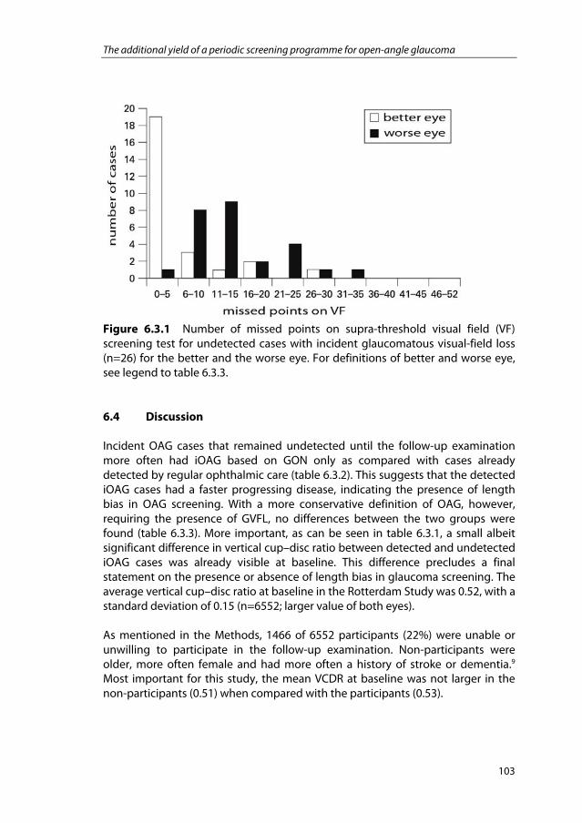
Better eye = eye with the lower number of missed points on VF test; p25 = 25th percentile (25% of cases have a number of missed points lower than or equal to this value); p50 = median; p75 = 75th percentile; worse eye, = eye with the higher number of missed points on VF test. From the perspective of blindness prevention, the severity of GVFL in the undetected cases is the most important measure of the additional screening yield. Figure 6.3.1 shows the distributions of VF scores for the undetected cases with GVFL (n=26) for the better and the worse eyes. As can be seen in this figure, only four of these cases had >10 missed points in their better eyes.
102
The additional yield of a periodic screening programme for open-angle glaucoma
Figure 6.3.1 Number of missed points on supra-threshold visual field (VF) screening test for undetected cases with incident glaucomatous visual-field loss (n=26) for the better and the worse eye. For definitions of better and worse eye, see legend to table 6.3.3. 6.4 Discussion Incident OAG cases that remained undetected until the follow-up examination more often had iOAG based on GON only as compared with cases already detected by regular ophthalmic care (table 6.3.2). This suggests that the detected iOAG cases had a faster progressing disease, indicating the presence of length bias in OAG screening. With a more conservative definition of OAG, however, requiring the presence of GVFL, no differences between the two groups were found (table 6.3.3). More important, as can be seen in table 6.3.1, a small albeit significant difference in vertical cup–disc ratio between detected and undetected iOAG cases was already visible at baseline. This difference precludes a final statement on the presence or absence of length bias in glaucoma screening. The average vertical cup–disc ratio at baseline in the Rotterdam Study was 0.52, with a standard deviation of 0.15 (n=6552; larger value of both eyes). As mentioned in the Methods, 1466 of 6552 participants (22%) were unable or unwilling to participate in the follow-up examination. Non-participants were older, more often female and had more often a history of stroke or dementia.9 Most important for this study, the mean VCDR at baseline was not larger in the non-participants (0.51) when compared with the participants (0.53).
103

Chapter 6
The most obvious difference between the detected and undetected iOAG cases is the presence of a positive family history of glaucoma (table 6.3.1). A possible explanation of this difference is an elevated visit rate by relatives of OAG patients to opticians or ophthalmologists, as is widely recommended. Of all participants at baseline, 8.5% had a positive family history of glaucoma. Of the 3842 participants at risk of OAG at baseline, 87 developed iOAG during a follow-up period of 6.5 years. Of the 78 iOAG cases included in this study, 23 had already been diagnosed by an ophthalmologist before the follow-up examination took place. Only four of the remaining 55 undetected cases had >10 missed points (arbitrarily chosen) in their better eye on VF testing (fig 6.3.1). If we tentatively assume that these four iOAG cases would become blind without treatment while the other undetected cases would retain useful vision (see also next paragraph), it can be calculated that about 1000 OAG screening tests would have to be performed in order to prevent one OAG case from becoming severely visually impaired or blind (about 200 tests if we would start this discussion with >10 missed points in the worse eye, aiming to prevent unilateral loss). Assuming a test specificity of 95% for a suitable OAG screening test, 1000 screening tests will produce 50 false-positive test results, all needing further investigation. At first glance, incorporating prior selection criteria like myopia or family history might improve the feasibility of screening.11–17 However, only a minority of the undetected cases (18 of 55) had an OAG risk factor (17 of 55 myopia; 1 of 55 positive family history; see also table 6.3.1). The use of supra-threshold testing, as performed in our study, might have resulted in missing cases with early glaucoma. It is unlikely, however, that cases with moderate or severe glaucoma, the most important ones for the estimate as presented here, have been overlooked.18 We re-estimated the number of undetected iOAG cases that would develop end-stage OAG before death, now using data from the study by Wilson et al.19 They analysed progression of VFL in untreated black OAG cases in the West Indies. Following the AGIS scoring system,20 7% of their cases had developed end-stage OAG in both eyes after a follow-up of 10 years. The average life expectancy at follow-up of our 55 undetected cases was 11.4 years.21 From this it can tentatively be estimated that, given the higher prevalence of OAG in blacks, 22 four at most (7% of 55) of 3842, that is 0.1% of the white participants, might have become blind before dying if they had remained undetected. Obviously, this is only a rough estimate. For example, we assumed that the disease severity of our 55 undetected iOAG cases at follow-up could be compared with the baseline findings of Wilson et al,1,9 and we based our estimate on average life expectancy, assuming that the lower incidence of glaucoma blindness in those dying earlier than average balances the higher incidence in those living longer than average. Both our estimates suggest, however, that the real yield of a periodic OAG screening programme in terms of preventing severe visual impairment or
104

The additional yield of a periodic screening programme for open-angle glaucoma
blindness (estimated to be 0.1%) is much lower than the yield as estimated from the prevalence of undetected OAG (typically 1%).6,7
As mentioned in the Methods section, six iOAG cases were excluded because at baseline, they were referred to an ophthalmologist because of an elevated IOP without further signs of OAG at that stage. From a methodological point of view, it might have been better not to report any baseline abnormality to these participants. However, ethically, and in accordance with the guidelines for unexpected findings in the Rotterdam Study, this would have been unacceptable. Of inhabitants aged 40 years and older and living in The Netherlands, 84% visit an optician or an ophthalmologist at least once every 5 years,23 and most opticians perform noncontact tonometry in clients in this age group. Hence, at least some if not most of these six iOAG cases with elevated IOP at baseline would have been detected before the follow-up measurement. Likewise (see Methods), three iOAG cases had to be excluded because of missing VF data. These three cases were all undetected at follow-up. Based on disc data, all three cases presumably had unilateral disease. Within the remaining 78 cases, only one had unreliable perimetric results according to the criteria as listed in the Methods section. This case was not excluded from the analyses. Grodum et al. compared 402 OAG cases identified through a large population screening among elderly citizens of Malmo, Sweden, with 354 cases identified through retrospective patient record analysis at the Eye Department of Malmo University Hospital.24 They found that the latter group had considerably more VFL than the former group. This cross-sectional finding provides information on the screening yield if a population is screened for the first time. Our data are complementary to their findings—based on incident cases only, our data provide information on the ongoing yield of screening if a population is screened periodically. Most of the participants of the Rotterdam Study were white. As the prevalence of OAG depends on ethnicity,25 our findings should not automatically be applied to non-white populations. We did not exclude cases with pseudoexfoliation (hence OAG rather than POAG), but none of the iOAG cases found at follow-up suffered from pseudoexfoliation. The latter finding suggests that pseudoexfoliation is of little importance to the OAG burden in The Netherlands. This might be different elsewhere.26,27 Due to the design of the Rotterdam Study, we were unable to address the additional yield of screening in a younger population or in a rural area directly. A recent study from Finland suggested that screening is unlikely to be efficient under 55 years of age.28 In an earlier study, we did not find any difference in optician/ophthalmologist visit frequency between urban and rural area.23 That study reported that of inhabitants aged 40 years and older and living in The Netherlands, 84% visit an optician or an ophthalmologist at least once every five
105

Chapter 6
years. This apparently high percentage may have contributed to the poor additional yield as found in this study. In summary, the additional yield of periodic OAG screening is less than expected from published prevalence data because (1) many cases had already been detected at early disease stages in regular ophthalmic care and (2) only a minority of the undetected cases had severe enough disease to be seriously at risk of reaching end-stage OAG in both eyes during life—were they to remain undetected. Funding The Netherlands Organization for Health Research and Development (ZonMw) grant 2200.0035, The Hague. Foundations: Stichting Nederlands Oogheelkundig Onderzoek, Nijmegen/Rotterdam; Optimix, Amsterdam; Netherlands Organisation for Scientific Research (NWO), The Hague; Physico Therapeutic Institute, Rotterdam; Blindenpenning, Amsterdam; Sint Laurens Institute, Rotterdam; Bevordering van Volkskracht, Rotterdam; Blindenhulp, The Hague; Algemene Nederlandse Vereniging ter Voorkoming van Blindheid, Doorn; Rotterdamse Blindenbelangen Association, Rotterdam; OOG, The Hague; kfHein, Utrecht; Prins Bernhard Cultuurfonds, Amsterdam; Van Leeuwen Van Lignac, Rotterdam. All in The Netherlands. Unrestricted grants were obtained from Topcon Europe BV, Capelle aan de IJssel, The Netherlands, and from Heidelberg Engineering, Dosselheim, Germany.
106

The additional yield of a periodic screening programme for open-angle glaucoma
References for chapter six
1. Heijl A, Leske MC, Bengtsson B, et al. Early Manifest Glaucoma Trial Group. Reduction of intraocular pressure and glaucoma progression: results from the Early Manifest Glaucoma Trial. Arch Ophthalmol 2002;120:1268–79.
2. Maier PC, Funk J, Schwarzer G, et al. Treatment of ocular hypertension
and open angle glaucoma: meta-analysis of randomised controlled trials. BMJ 2005;331:134–9.
3. Wilson JM, Jungner G. Principles and practice of screening for disease.
34th edn. Geneva: World Health Organization, 1968:1–163.
4. Johnson GJ, Minassian DC, Weale RA, et al. The epidemiology of eye disease. Second edn. London: Hodder Arnold, 2003.
5. Baum M. Breast cancer screening comes full circle. J Natl Cancer Inst
2004;96:1490–1.
6. Hollows FC, Graham PA. Intra-ocular pressure, glaucoma, and glaucoma suspects in a defined population. Br J Ophthalmol 1966;50:570–86.
7. Wolfs RC, Borger PH, Ramrattan RS, et al. Changing views on open-angle
glaucoma: definitions and prevalences—The Rotterdam Study. Invest Ophthalmol Vis Sci 2000;41:3309–21.
8. Hofman A, Grobbee DE, de Jong PT, et al. Determinants of disease and
disability in the elderly: the Rotterdam Elderly Study. Eur J Epidemiol 1991;7:403–22.
9. De Voogd S, Ikram MK, Wolfs RC, et al. Incidence of open-angle glaucoma
in a general elderly population: the Rotterdam Study. Ophthalmology 2005;112:1487–93.
10. Ramrattan RS, Wolfs RC, Panda-Jonas S, et al. Prevalence and causes of
visual field loss in the elderly and associations with impairment in daily functioning: the Rotterdam Study. Arch Ophthalmol 2001;119:1788–94. [Erratum in: Arch Ophthalmol 2002;120:525]
107
Chapter 6
11. Mitchell P, Hourihan F, Sandbach J, et al. The relationship between glaucoma and myopia: the Blue Mountains Eye Study. Ophthalmology 1999;106:2010–15.
12. Grodum K, Heijl A, Bengtsson B. Refractive error and glaucoma. Acta
Ophthalmol Scand 2001;79:560–6.
13. Wong TY, Klein BE, Klein R, et al. Refractive errors, intraocular pressure, and glaucoma in a white population. Ophthalmology 2003;110:211–17.
14. Tielsch JM, Katz J, Sommer A, et al. Family history and risk of primary
open angle glaucoma. The Baltimore Eye Survey. Arch Ophthalmol 1994;112:69–73.
15. Wolfs RC, Klaver CC, Ramrattan RS, et al. Genetic risk of primary open-
angle glaucoma. Population-based familial aggregation study. Arch Ophthalmol 1998;116:1640–5.
16. Mitchell P, Rochtchina E, Lee AJ, et al. Bias in self-reported family history
and relationship to glaucoma: the Blue Mountains Eye Study. Ophthalmic Epidemiol 2002;9:333–45.
17. Le A, Mukesh BN, McCarty CA, et al. Risk factors associated with the
incidence of open-angle glaucoma: the visual impairment project. Invest Ophthalmol Vis Sci 2003;44:3783–9.
18. Topouzis F, Coleman AL, Yu F, et al. Sensitivity and specificity of the 76-
suprathreshold visual field test to detect eyes with visual field defect by Humphrey threshold testing in a population-based setting: the Thessaloniki eye study. Am J Ophthalmol 2004;137:420–5.
19. Wilson MR, Kosoko O, Cowan CL Jr, et al. Progression of visual field loss in
untreated glaucoma patients and glaucoma suspects in St. Lucia, West Indies. Am J Ophthalmol 2002;134:399–405.
20. AGIS Investigators. Advanced Glaucoma Intervention Study. 2. Visual
field test scoring and reliability. Ophthalmology 1994;101:1445–55.
21. Statistics Netherlands. Life expectancy 1996–2000. http://www.cbs.nl (accessed 14 Nov 2006).
22. Leske MC, Connell AM, Schachat AP, et al. The Barbados Eye Study.
Prevalence of open angle glaucoma. Arch Ophthalmol 1994;112:821–9.
108

The additional yield of a periodic screening programme for open-angle glaucoma
109
23. Stoutenbeek R, Jansonius NM. Glaucoma screening during regular optician visits: can the population at risk of developing glaucoma be reached? Br J Ophthalmol 2006;90:1242–4.
24. Grodum K, Heijl A, Bengtsson B. A comparison of glaucoma patients
identified through mass screening and in routine clinical practice. Acta Ophthalmol Scand 2002;80:627–31.
25. Tielsch JM, Sommer A, Katz J, et al. Racial variations in the prevalence of
primary open-angle glaucoma. The Baltimore Eye Survey. JAMA 1991;266:369–74.
26. Ball SF. Exfoliation syndrome prevalence in the glaucoma population of
South Louisiana. Acta Ophthalmol Suppl 1988;184:93–8.
27. Lindblom B, Thorburn W. Observed incidence of glaucoma in Halsingland, Sweden. Acta Ophthalmol (Copenh) 1984;62:217–22.
28. Vaahtoranta-Lehtonen H, Tuulonen A, Aronen P, et al. Cost effectiveness
and cost utility of an organized screening programme for glaucoma. Acta Ophthalmol Scand 2007;85:508–18.