gestational - core.ac.uk filecross sectional study: a five year review of gestational trophoblastic...
TRANSCRIPT

CROSS SECTIONAL STUDY:
A FIVE YEAR REVIEW OF
GESTATIONAL TROPHOBLASTIC DISEASE
IN KUANTAN GENERAL HOSPITAL (JAN 1995- DEC 1999)
-by-
DR MOHAMAD FAIZ B. MOHAMED JAMLI
DISSERTATION SUBMITTED IN PARTIAL FULFILMENT QF.;J,..I;CJJ.:!_®lREMENT FOR
THE DEGREE OF MASTER OF MEDICINE
(OBSTETRICS & GYNAECOLOGY)
UNIVERSITY SAINS MALAYSIA 2001

Q};aU~wk~~~
Iff~ QT~ 19ZJ~
II

CKNOWLEDGEMENTS
By the name of Allah, 1 wish to express greatest
appreciation and special thanks for the exceptional effort of my
supervisor Dr Awang Nila Ismail, Head of department, senior
consultant and lecturer Department of Obstetrics and
Gynaecology Hospital USM , in preparation of this
dissertation. Completion of this dissertation would not have
been possible without his guidance and assistant. Thank you
also to Dr Mohd Shukri Othman, senior consultant and
lecturer Department of Obstetrics and Gynaecology Hospital
USM , Dr Ghazali Ismail , head of department, senior
consultant Department of Obstetrics and Gynaecology
Hospital Tengku Ampuan Afzan Kuantan Pahang Darul Makmur
(HTAA), all lecturers, consultants Dr Mokhtar Awang (HTAA)
and Dr Lee Say Fat (HTAA), medical officers ({ISM), medical
III

officers (HTAA) and medical staff (HTAA) for their assistance
and guidance through out the course of my training and
preparation of this book.
I would also like to express my gratitude to my parent Abah &
Umi and family especially my wife Noora=ah bte Zakaria, my
daughter Fatehah Ezzatunnur, and my son Muhammad Furqaan
whose endless pray, understanding, sacrifices, support,
encouragement and patience have guided me through my
career.
Wassallam
IV
All II Ill! lT I 1l T I 0 11 ~
&hCG
CNS
CT
D&C
DM
EMACO
EGF
GTD
GTT
HLA
HM
HPL
1M
n.. HTAA
MRI
OCP
POA
PTD
RM
TVS
UK
us USA
PSTI
PHM
HPE
WHO
Beml-luman Ghorionic gonadotrophin
Central Nervous Systems
Computed tomography
Dilatation and curretage
Darul Makmur
Etoposide, Methotrexate, ,Actinomycin. Cyclophosphamide. Vmcristine(Oncovin)
Endothelial growth Factors
Gestation~! Trophoblastic Disease
Gestational Trophoblastic Tumour
Human leukocyte antigen
Hydatidiform mole
Human Placenta Lactogen
Intramuscular
lnterleukins
Hospita Tengku Ampuan Afzan Kuantan Pahang
Magnetic resonance imaging
Oral contraceptive pills
Period of amenorrhoea
Persistent Trophoblastic Disease
Ringgit Malaysia
Transvaginal scan
United Kingdom
Ultrasound
United State of America
Placental site trophoblastic tumour
Partial Hydatidifom Mole
Histopathological examination
World health Organizations
v
CONTENTS
PAGE
I ACKNOWLEDGEMENTS.......................................................... m 2 ABBREVIATIONS.................................................................. V
3 THE STATE OF PAHANG......................................................... VII
5 INTRODUCTION TO HOSPITAL TENGKU AMPUAN AFZAN KUANTAN, XI
PAHANG & DEPARTMENT OF OBSTETRICS AND GYNAECOLOGY ...... .
6 ABSTRACT OF DISSERTATION
A) BAHASA MALAYSIA VERSION........................ XVI
B) ENGLISH VERSION...................................... XVID
7 INTRODUCTION . . . . . . . . . . . . . . . . . . . . . . . . . . . . . . . . . . . . . . . . . . . . . . . . . . . . . . . . . . . . . . . . . . 1
8 OBJECTIVES........................................................................ 46
9 MATERIALS AND METHODOLOGY............................................. 47
10 RESULTS........................................................................... 58
11 DISCUSSIONS..................................................................... 120
12 SUMMARY . . . . . . . . . . . . . . . . . . . . . . . . . . . . . . . . . . . . . . . . . . . . . . . . . . . . . . . . . . . . . . . . . . . . . . ... 155
13 CONCLUSIONS..................................................................... 159
14 RECOMMENDATIONS............................................................ 160
15 LIMITATIONS...................................................................... 162
16 BIBLIOGRAPHY..................................................................... 163
VI

STATE OF
p AHAN G
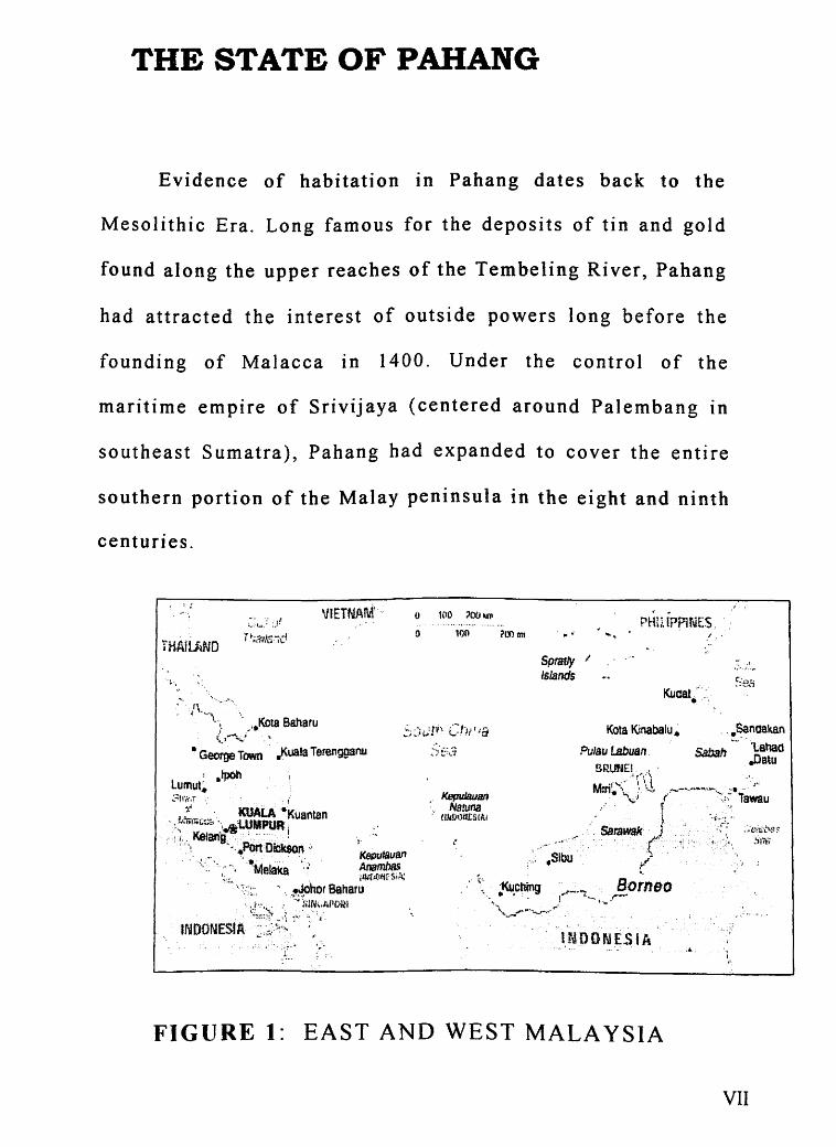
THE STATE OF PAHANG
Evidence of habitation in Pahang dates back to the
Mesolithic Era. Long famous for the deposits of tin and gold
found along the upper reaches of the Tembeling River, Pahang
had attracted the interest of outside powers long before the
founding of Malacca in 1400. Under the control of the
maritime empire of Srivijaya (centered around Palembang in
southeast Sumatra), Pahang had expanded to cover the entire
southern portion of the Malay peninsula in the eight and ninth
centuries.
~ 1 :
VIETNA1\r(-
H!AJ!..AND
'f\ ~ ·--....... ·. ~·•Kota Baharu
.. . (, ... ~:.;.." ·'
• George Town j<uala Terengganu
{' :. :·
0 1£10 :?OOkrP ........
0 100 ?L~ m1 rrm !P?U~ES .
Spratly I
Islands
....
Kuaat ·C· ..
• Kota Kinabalu ~
l.NOONESIA .•.
.1,.
FIGURE 1: EAST AND WEST MALAYSIA
.)-.. ,-".\_
VII
•
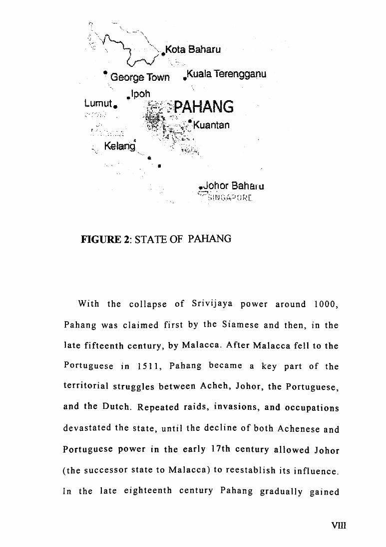
FIGURE 2: STATE OF PAHANG
With the collapse of Srivijaya power around 1000,
Pahang was claimed first by the Siamese and then, in the
late fifteenth century, by Malacca. After Malacca fell to the
Portuguese in 1511, Pahang became a key part of the
territorial struggles between Acheh, Johor, the Portuguese,
and the Dutch. Repeated raids, invasions, and occupations
devastated the state, until the decline of both Achenese and
Portuguese power in the early 17th century allowed Johor
(the successor state to Malacca) to reestablish its influence.
In the I ate eighteenth century Pahang gradually gained
vm

autonomy, and in the middle of the nineteenth century it had
become an independent state.
Mention Pahang to a Malaysian, and he will probably
conjure up visions of lush tropical forests, cool mountain
air, beaches, lakes and waterfalls nestling in the arms of
mountain crevices. The largest state in peninsular Malaysia
ts, tn many ways, one of its most wild and Edenic. Two
thirds of it is covered by unspoiled rain forest.
Visitors to Pahang are usually there to visit the state's
famous hill resorts, its internationally-known islands and
beaches, or Taman Negara, the Peninsula's finest park. Each
of these attractions is substantial enough to merit special
attention in our pages. Pahang's other attractions, though
less well-known, should not be missed by anyone visiting
the state
The natural heart of Pahang is unquestionably Taman
Negara, Malaysia's oldest national park, lovingly referred to
as "The Green Heart. n Within the park IS also the
peninsula's highest point, the forest encrusted Gunung
IX
Tahan. Also in the interior are many of Malaysia~s hill
stations., mountain resorts where the tropical heat is kept at
bay by the altitude. On the coast of Pahang, the jungle gives
way to clean., palm-lined beaches, fishing villages, and the
multicultural state capital, Kuantan.
Pahang is the largest state in Peninsular Malaysia and is
situated in the eastern coastal region. The state'"s 35,964 sq. km
encompasses a remarkable range of Malaysiats many different
environments, from the majestic peaks and cool hill regions of
the state~s western region to the miles of soft sand beach along
the South China Sea. Pahang•s nearly one million people
constitute a representative mix of Malaysiats three main ethnic
communities Malay, Chinese, Indian and Orang-asli. The total
population of Malaysia in Census 2000, about 21.,890 thousand
or 94.1% were Malaysian citizens. Of the total Malaysian
citizens, Bumiputera comprised 65.1 %, Chinese 26.0% and
Indians 7 _7%., the ethnic composition being 60.6o/o., 28.1% and
7. 9% respectively in 1991. Non-Malaysian citizens totalled
1,385 thousand (or 5.9%) in Census 2000 as against 805
thousand (or 4.4%) in 1991.
X
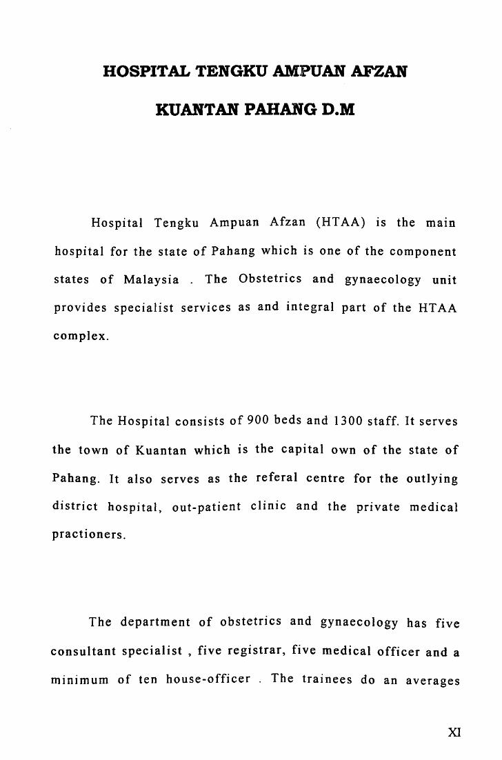
HOSPITAL TENGKU AMPUAN AFZAN
KUANTAN PAHANG D.M
Hospital Tengku Ampuan Afzan (HT AA) is the matn
hospital for the state of Pahang which is one of the component
states of Malaysia . The Obstetrics and gynaecology unit
provides specialist services as and integral part of the HT AA
complex.
The Hospital consists of 900 beds and 1300 staff. It serves
the town of Kuantan which is the capital own of the state of
Pahang. It also serves as the referal centre for the outlying
district hospital, out-patient clinic and the private medical
practioners.
The department of obstetrics and gynaecology has five
consultant specialist , five registrar, five medical officer and a
minimum of ten house-officer . The trainees do an averages
XI

number of eight calls a month . They involve actively in the
management of emergency cases during on call under close
supervision of the specialist consultants.
There are 36 bed in the antenatal \Yard cases which are
admitted to the antenatal ward will usually be assessed in the
screening I admission room . The Obstetrics unit also provides
day care services to patients who come for observation for
maternal and fetal well being or blood investigations such as
blood sugar profile .
The Labour room has 10 beds of which 2 beds are used for
monitoring of pre-eclampsia cases . Being a husband friendly
ward , there are 3 beds whereby the patient,s husbands or
companion are allowed to be with the patients throughout
process as well as during deliveries of the baby _ For the year
199 5-1999 , there was a total number 39011 deliveries . In year
1999 total deliveries was 8647. The statistics are as follows :
xu
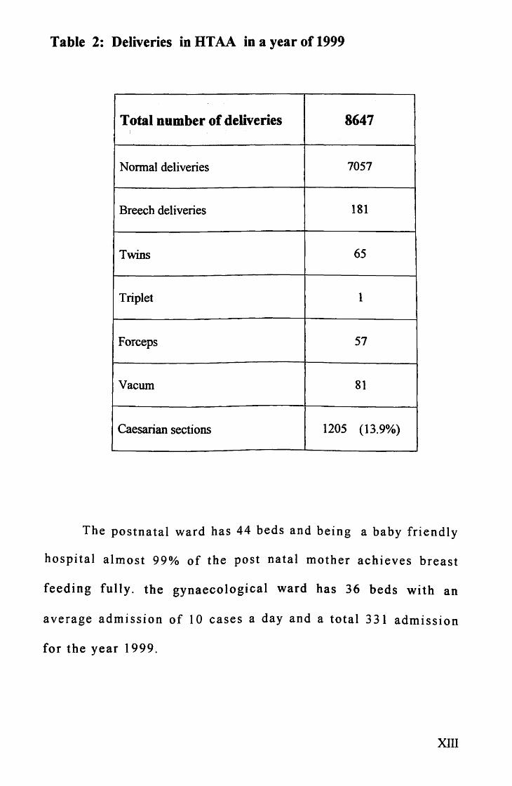
Table 2: Deliveries in HTAA in a year of 1999
Total number of deliveries 8647
Normal deliveries 7057
Breech deliveries 181
Twins 65
Triplet 1
Forceps 57
Vacurn 81
Caesarian sections 1205 (13.9%)
The postnatal ward has 44 beds and being a baby friendly
hospital almost 99% of the post natal mother achieves breast
feeding fully. the gynaecological ward has 36 beds with an
average admission of 10 cases a day and a total 331 admission
for the year 1999.
xrn

There are two operative sesstons per week for elective cases.
The facility for caesarean sections is shared between the general
operating theatre and the maternal operating theatre which is
located adjacent to the labour ward.
We perform a wide variety of the gynecological operation
which include total abdominal hysterectomy, vaginal
hysterectomy, wertheim hysterectomy, oophorectomy,
cystectomy, myomectomy, laparoscopic and hysteroscopy
procedure, sterilization, culposuspension and minor procedure
such as evacuation of product of conception and marsupilazation
and so on.
The department also conduct regular teaching sesstons
such as the weekly registrar presentation, viva sessions, and
journal club. We also have a regular caesarean audit and
perinatal mortality and morbidity audit every fortnightly and a
combined meeting with the paediatric department monthly.
Since 1999 HT AA also become the teaching hospital for
the medical faculty of University Islam Antarabangsa (
International Islamic University). With the Input of this
university , the hospital is expected to be even better equipped
XIV
with medical facilities and have more academic programmes in
the near future.
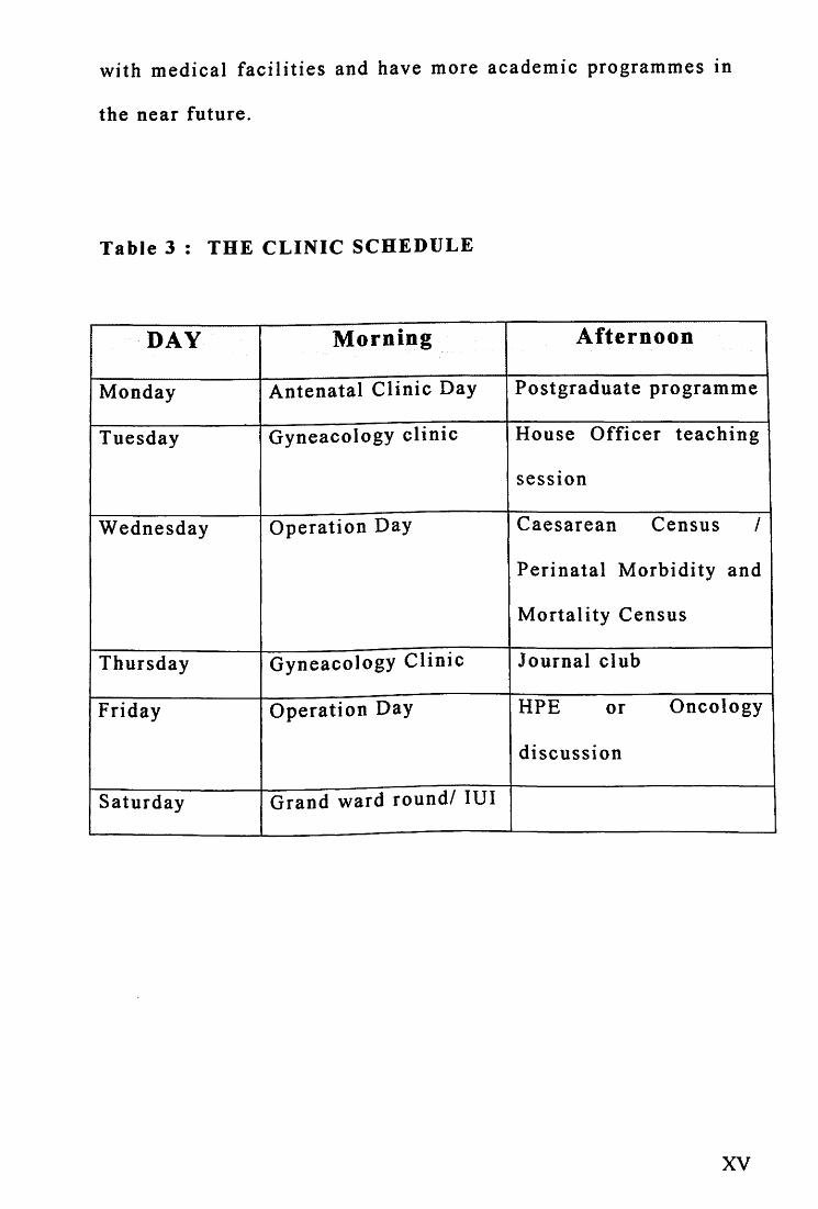
Table 3 : THE CLINIC SCHEDULE
DAY Morning Afternoon
Monday Antenatal Clinic Day Postgraduate programme
Tuesday Gyneacology clinic House Officer teaching
sesston
Wednesday Operation Day Caesarean Census I
Perinatal Morbidity and
Mortality Census
Thursday Gyneacology Clinic Journal club
Friday Operation Day HPE or Oncology
discussion
Saturday Grand ward round/ lUI
XV
ABSTRACTS ( Malay version )
ABSTRAK
OBJECTIF :Mengkaji epidemologi dan keadaan penyakit, rawatan GTD
dan melihat corak penurunan B-hCG penyakit H.M Mengesan faktor yang
boleh menambah risiko pembentukan GTT dan H.M..Membandingkan
kesan rawatan yang diberi kepada GTT yang diketahui anteseden
kandungan (H.M) dan GTT yang tidak diketahui anteseden kandungan.
METODOLOGI: /a adalah kajian Cross-Sectional ke atas 96 pesakit
GTD di Hospital Besar Tengku Ampuan Afzan Kuantan, selama 5 tahun(
Januari /995- Disember 1999).
KEPUTUSAN: Dalam kelahiran sebanyak 39,011 da/am tempoh kajian di
HTAA terdapat 96 kes GTD, kadar GTD di Hospital 1:410 kelahiran.
Kadar penyakit dilihat lebih kerap da/am golongan minorit1: umur 41
tahun dan ke atas , sosio-ekonomi yang rendah dan pariti lebih dari 5 .
Gejala yang kerap dilihai ialah pendarahan haid yang tidak menentu,
(87%). "'Snow storm u dapat dilihat dengan ultrasound pada 74% pesakit.
Pada pesakit Hydatidiform Mole didapati 64% mempunyai rahim
besar dari tarikh amenorhea. Ujian bersiri serum bHCG (n=71) . 79%
XVI
(n=56) beransur susut dengan spontan da/am tempoh 14 minggu dan 21%
(n= 15) pesakit mempunyai susutan yang tidak normal dan hertukar ke
GTT~ Faktor yang dapat mengesan risiko pembentukan GTT adalah umur
40 tahun ke alas, pariti lebih dari 5, rahim hesar dari tarikh haid,
bacaan awal BHCG > 100,000 miulml selepas evakuasi dan darah tinggi.
Jenis HA,f, Theca lutein cyst, jenis darah tidak mempunyai risiko yang
signifilcan. Lapan puluh lima kes dirawat dengan 'cuci,. (suction and
curretages) dan 8 kes histerektomi (termasuk 7 pesakit di 'cuci,. sebelum
histerektomi). 62.5% dari pesakit GTT menerima regim kemoierapi risiko
rendah. 17.4% menerima regim risiko sederhana dan 17.4% menerima
regim risiko tinggi. Kita dapati tiada hubungan yang signifikan di antara
dua status antesedan kandungan dalam lconteks histerektomi, jenis
kemoterapi dan respon terhadap kemoterapi dan kematian.
KESIMPULAN: GTD berlaku pada kadar 1 dalam 410 kelahiran dan
faktor risiko untuk bertulcar ke GTT boleh dilcenal pasti. Perhatian yang
.~erius adalh penting lantas dapat merawat dengan betuL Harapan untuk
sembuh dari penyakit GTD adalah besar walaupun te/ah menular ke GTT.
xvn

ABSTRACTS
OBJECTIVES: To study the epidemiology, features, treatment of
Gestational Trophoblastic Disease (GTD) and regression of serum B
hCG in Hydatidiform mole. To identify factors that predict the risk of
developing Gestational Trophoblastic Tumour (GTT) from Hydatidiform
mole and to compare the outcome of treatment between Gestational
Trophoblastic Tumour (GTT) from known antecedent pregnancy
(Hydatidiform mole) and unknown antecedent pregnancy.
METHODOLOGY: A Cross Sectional study of 96 cases of
Gestational Trophoblastic Disease in General Hospital ofTengku Ampuan
Afzan Kuantan for a period of 5 years (January 1995-December 1999).
RESULTS: The prevalence rate of GTD in this hospital was 1 : 410
deliveries. There were 96 cases of GTD out of 39,011 deliveries. The
prevalence rate was higher in the minority ethnic groups, women of
lower socio-economic status, age ~ 41 years and parity more than 5.
The commonest presentations was irregular vaginal bleeding (87%).
Typical snow storm appearance was seen in 74.4% of the patients.
In patient with hydatidiform mole , 64% has uterus larger than
date. In patient with available serial serum B-hCG (n= 71), 79% (n=56)
X VITI
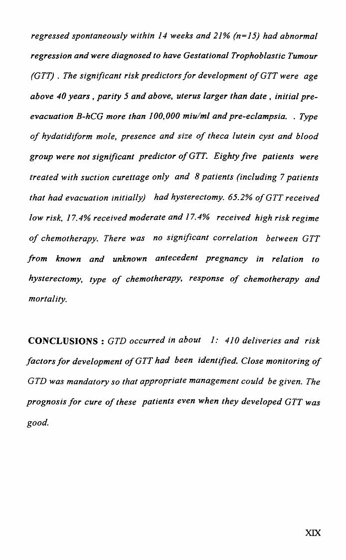
regressed spontaneously within 14 weeks and 21% (n=l5) had abnormal
regression and were diagnosed to have Gestational Trophoblastic Tumour
(GTT) . The significant risk predictors for development of GTT were age
above 40 years , parity 5 and above, uterus larger than date , initial pre
evacuation B-hCG more than 100,000 miulml and pre-eclampsia . . Type
of hydatidiform mole, presence and size of theca lutein cyst and blood
group were not significant predictor ofGTT. Eighty five patients were
treated with suction curettage only and 8 patients (including 7 patients
that had evacuation initially) had hysterectomy. 65.2% ofGTT received
low risk, 17.4% received moderate and 17.4% received high risk regime
of chemotherapy. There was no significant correlation between GTT
from known and unknown antecedent pregnancy in relation to
hysterectomy, type of chemotherapy, response of chemotherapy and
mortality.
CONCLUSIONS : GTD occurred in about 1: 410 deliveries and risk
factors for development ofGTT had been identified. Close monitoring of
GTD was mandatory so that appropriate management could be given. The
prognosis for cure of these patients even when they developed GTT was
good.
XIX


INTRODUCTION
Gestational trophoblastic disease ts a spectrum of
heterogenous conditions which arise from products of conception
and which may threatened the livehood and health of the young
women . Although uncommon, its importance lies in the need for
early recognition if the treatment is to be translated into cure and
continued fertility. It is a health hazard which must be included
in the differential diagnosis of a wide variety of clinical
conditions.
GESTATIONAL TROPHOBLASTIC 01S£AS£ 1995-1999 HTAA KUANTAN 1
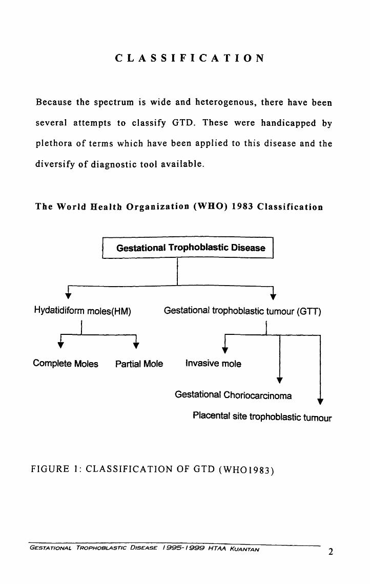
CLASSIFICATION
Because the spectrum is wide and heterogenous, there have been
several attempts to classify GTD. These were handicapped by
plethora of terms which have been applied to this disease and the
diversify of diagnostic tool available.
The World Health Organization (WHO) 1983 Classification
Gestational Trophoblastic Disease
Hydatidiform moles(HM) Gestational trophoblastic tumour (GTT)
I
Complete Moles Partial Mole Invasive mole
Gestational Choriocarcinoma
Placental site trophoblastic tumour
FIGURE 1: CLASSIFICATION OF GTD (WH01983)
GESTATIONAL TROPHOBLASTIC DISEASE I 995- I 999 HTAA KUANTAN 2

Gestational trophoblastic diseases ( GTD ) refer to both the
benign and malignant permutations of proliferative trophoblastic
allograft: Hydatidiform moles, invasive mole, choriocarcinoma,
and placenta-site trophoblastic tumour (PSTT)
Hydatidiform mole IS a general term that includes two
distinct entities, complete Hydatidiform mole and partial moles.
(i) Complete mole : is an abnormal conceptus without the embryo
or fetus characterized by loss of villous vascularity leading to
gross hydropic swelling and central cistern formation and to
pronounced cytotrophoblastic and syncytiotrophoblastic
hyperplasia. (ii) Partial mole: IS an abnormal conceptus with
persistent embryonic and fetal elements and a placenta with a
mosaic of normal -appearing villi alternating with areas of focal
villous swelling and trophoblastic hyperplasia.
GESTATIONAL TROPHOBLASTIC DISEASE 1995-1999 HTAA KUANTAN 3
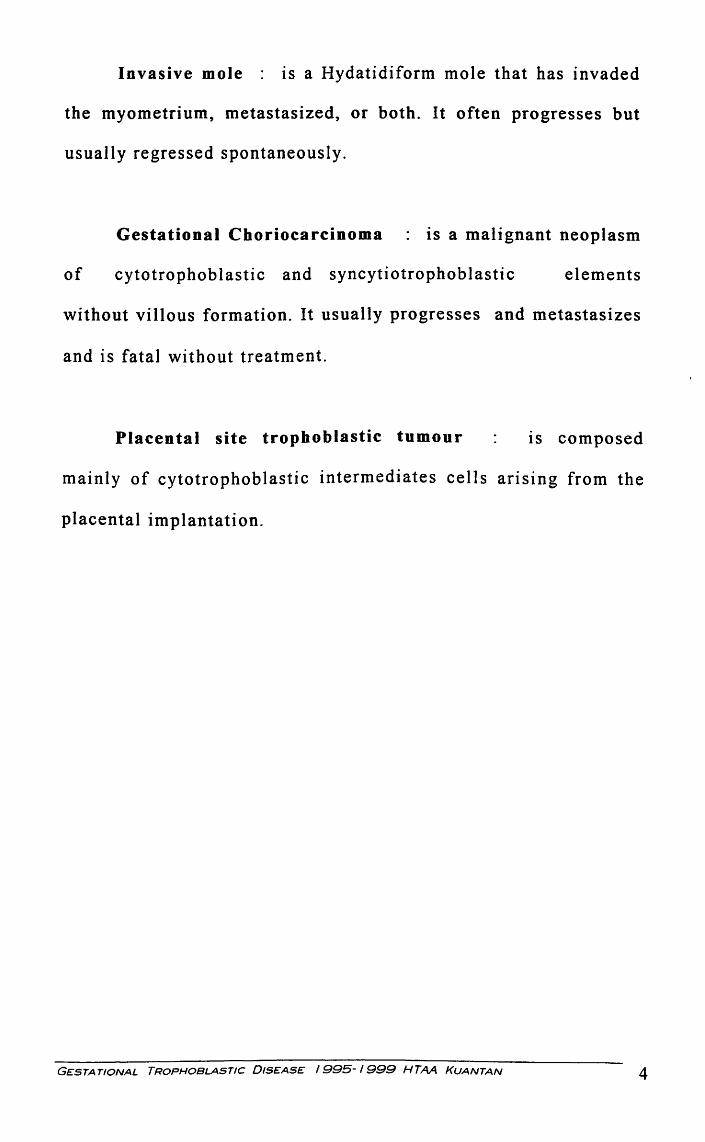
Invasive mole : ts a Hydatidiform mole that has invaded
the myometrium, metastasized, or both. It often progresses but
usually regressed spontaneously.
Gestational Choriocarcinoma : is a malignant neoplasm
of cytotrophoblastic and syncytiotrophoblastic elements
without villous formation. It usually progresses and metastasizes
and is fatal without treatment.
Placental site trophoblastic tumour ts composed
mainly of cytotrophoblastic intermediates cells arising from the
placental implantation.
GESTATIONAL TROPHOBLASTIC DISEASE I 995- 1999 HTAA KUANTAN 4
CELLULAR CLASSIFICATION
Gestational trophoblastic tumors may be classified as
follows: hydatidiform mole, chorioadenoma destruens (invasive
mole), choriocarcinoma, and placental-site trophoblastic tumor.
Hydatidiform mole is defined as products of conception that lack
an intact fetus and show gross cyst-like swellings of the
chorionic villi due to accumulation of fluid. There ts
disintegration and loss of blood vessels in the villous core.
Invasive mole (chorioadenoma destruens) is a locally invasive,
rarely metastatic lesion characterized microscopically by
trophoblastic invasion of the myometrium with identifiable
villous structures. Microscopically, this lesion is characterized by
hyperplasia of cytotrophoblastic and syncytial elements and
persistence of villous structures.
Choriocarcinoma is a malignant tumor of the trophoblastic
epithelium. Uterine muscle and blood vessels are invaded with
areas of hemorrhage and necrosis. Columns and sheets of
trophoblastic tissue invade normal tissues and spread to distant
GESTATIONAL TROPHOBLASTIC DISEASE 1995-1999 HTAA KUANTAN 5

sites, the most common of which are lungs, brain, liver, pelvis,
vagina, spleen, intestines, and kidney.
Placental-site trophoblastic disease is an extremely rare
tumor arising from the placental implantation site and resembles
an exaggerated form of syncytial endometritis. Trophoblastic
cells infiltrate the myometrium, and there is vascular invasion.
Human placental lactogen is present in the tumor cells, while
immunoperoxidase staining for HCG is positive in only scattered
cells, and serum HCG is relatively low
GESTATIONAL TROPHOBLASTIC DISEASE 1995-1999 HTM KUANTAN 6

GENERAL COMPARISON BETWEEN NORMAL PREGNANCY & GTD
Table 1: Comparison Between Normal Pregnancy And GTD
D NORMAL ITRANsmoNALI PARTIAL I COMPLETE GESTATION . MOLE _ MOLE . MOLE
Lynonymsi[Gerber Baby" lrligbted Owm" I[InoompletelClassic"
IJ ltvilli with fetal I Some swelling. Focal IAU villi swollen ~essels. Most nonnal vtlli swelling of and empty (no
I!Histofogy I with nonnal villi without vessels).
vessels. vessels.
I ~ara:d I f!:. j
f Single layer of jHypoplasia of Focal Circumferential All atypical overgrowth ~ cytotrophoblast fYlotrophoblast syncytial . atypical of cytotrophoblast and ~d Ed -.J r ·r lasm pvergrowth or ~cytiotrophoblast syncytiotrophoblastJ~tiotropboblast ~sealloping). p;iotrophoblast.
II ~ ~.:mr • .u ,
~ hCG S0-100,000 <50,000 >SO,OOO usually
I hCG Low I HCG HCG I hCG>50!'000 ~·
-~75%)
~~~==4ro=·c~~~~=1 =xv======~~:=:=:=~=~====~c~~~~7~o~id~y~:~~~:o~9=5~%====~~==========~~~·
1 2:1 n II 1 n ~ $mall for I Small for Hl Baby
Weight [Appropriate. for [Gestaional Ag~
rl
~ Gestational ~e85% I ~estational I Gesational Age
~ 65% ~3%
I Data from Kate 0 Hanlan lviD Gynaecology Oncology ,Surgey, endoscopy http://www.ohanian.com/tneoplasia.htm
GES7:4TIONAL TROPHOBLASTIC DISEASE /995-1999 HTAA KUANTAN 7
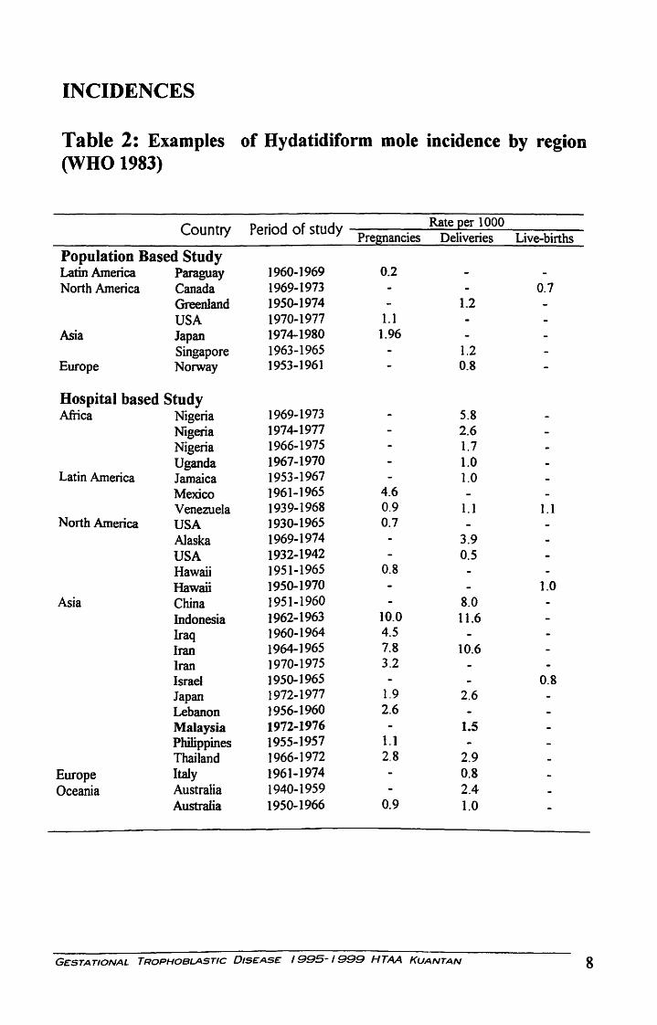
INCIDENCES
Table 2: Examples of Hydatidiform mole incidence by region (WHO 1983)
Country Period of study Rate Qer 1 000 Pregnancies Deliveries Live-births
Population Based Study Latin America Paraguay 1960-1969 0.2 North America Canada 1969-1973 0.7
Greenland 1950-1974 1.2 USA 1970-1977 1.1
Asia Japan 1974-1980 1.96
Singapore 1963-1965 1.2 Europe Norway 1953-1961 0.8
Hospital based Study Afiica Nigeria 1969-1973 5.8
Nigeria 1974-1977 2.6 Nigeria 1966-1975 1.7 Uganda 1967-1970 1.0
Latin America Jamaica 1953-1967 1.0 Mexico 1961-1965 4.6 Venezuela 1939-1968 0.9 1.1 1.1
North America USA 1930-1965 0.7
Alaska 1969-1974 3.9 USA 1932-1942 0.5 Hawaii 1951-1965 0.8
Hawaii 1950-1970 1.0 Asia China 1951-1960 8.0
Indonesia 1962-1963 10.0 11.6 Iraq 1960-1964 4.5
Iran 1964-1965 7.8 10.6 Iran 1970-1975 3.2
Israel 1950-1965 0.8 Japan 1972-1977 1.9 2.6 Lebanon 1956-1960 2.6
Malaysia 1972-1976 1.5 Philippines 1955-1957 1.1 Thailand 1966-1972 2.8 2.9
Europe Italy 1961-1974 0.8 Oceania Australia 1940-1959 2.4
Australia 1950-1966 0.9 1.0
G~STATIONAL TROPHOBLASTIC 0tS£AS~ /995- 1999 HTAA KUANTAN 8
(I) CD
N ---0 -m
,..
~
r-
u COf-c: ca c 01 Q) L. a. ....... 0 0 0 ,.... L.
[ (0
f/) Q)
0 ~
E .... 0 -:0
·.;:::; nJ -o ~ J:
M
- ~ -~ I 5 1 . . . .
. ~ ... -c 11:1 J ~
f-
~ ... -
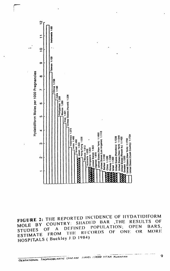
FIGURE 2: THE REPORTED INCIDENCE OF HYDATIDIFORM MOLE BY COUNTRY: SHADED BAR ,THE RESULTS OF STUDIES OF A DEFINED POPULATION: OPEN BARS ESTIMATE FROM TilE RECORDS OF ONE OR MORl~ HOSPITALS ( Buckley J. D 1984)
G£STATIONAL TROPHOBLASTIC 01SfASf I p~~; 1999 HTAA KUANTAN

PREGNANCY IMMUNOLOGY
The uterus is not an immunologically privileged site. Fetal tissue,
as well as trophoblastic neoplasia, does demonstrate antigenic
immaturity with respect to ABO and HLA. There is, importantly,
a nonantigenic barrier formed by the syncytotrophoblastic cells
which produce a mucoprotein layer of sialomucin, which is
protective. Furthermore, maternal immunoglobulins recognizing
any foreign antigens will attach to this basement membrane and
function as "blocking antibodies" covering the antigens. Despite
this, due to significant normal vascular invasion by the
trophoblast, some of the epithelial cells of the villi will "deport",
and travel through the venous return through the heart, imbedding
in the lung parenchyma.
GESTATIONAL TROPHOBLASTIC DISEASE 1995-1999 HTAA KUANTAN 10
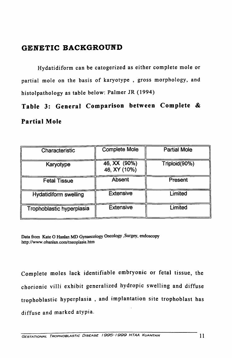
GENETIC BACKGROUND
Hydatidiform can be catogerized as either complete mole or
partial mole on the basis of karyotype , gross morphology, and
histolpathology as table below: Palmer JR ( 1994)
Table 3: General Comparison between Complete &
Partial Mole
I Characteristic
II Complete Mole
II
I
Karyotype 46, XX (90o/o)
I 46, XY (10o/o)
I Fetal Tissue Absent
II
I Hydatidiform swelling
I Extensive
II Trophoblastic hyperplasia
I Extensive
II
Data from Kate 0 Hanlan MD Gynaecology Oncology ,Surgey, endoscopy http://www.ohanlan.com/tneoplasia.htm
Partial Mole
Triploid(90o/o)
Present
Limited
Limited
Complete moles lack identifiable embryonic or fetal tissue, the
chorionic villi exhibit generalized hydropic swelling and diffuse
trophoblastic hyperplasia , and implantation site trophoblast has
diffuse and marked atypia.
GESTATIONAL TROPHOBLASTIC DISEASE 1995-1999 HTAA KUANTAN 11
I
I
I
I
I
GROWTH FACTORS & ONCOGENES
The excess of paternal chromosomes in moles probably
contributes to the induction of trophoblastic hyperplasia . The
genomic imbalance may cause changes in the gene expression of
growth factors located on the paternal allele (Berkowitz RS, et al
19 85). An in sui in growth factors (IGF2 ) specifically located on
the paternal allele may be inappropriately expressed in molar
pregnancies, thus stimulating uncontrolled growth.
Both normal placentas and molar pregnancies contain
paternal antigens; therefore, upon implantation , an immunologic
response ts initiated with infiltration of lymphocytes and
macrophages and secretion of cytokines. The growth of
choriocarcinoma may be related to the abundant expression of
epidermal growth factor receptor. Macrophage derived cytokines
interleukin (IL-l alpha, 11-1 beta), and tumour necrosis factor can
suppress cell growth and increase EGF receptor expression in
choriocarcinoma cell lines, thus acting as paracrine mediators of
cell growth.
GESTATIONAL TROPHOBLASTIC 01S£AS£ I 995- 1999 HTAA KUANTAN 12
The contribution of several oncogenes to the malignant
transformation of GTD also has been examined. Growth
regulation in the trophoblast recently has been found to be
associated with expression of the transciption factor Mash-2.
Cheung et al ( 1993) have demonstrated increase expression of c
fm RNA in complete mole compare with that in normal placenta .
In choriocarcinoma, increase expression of c-myc and ras RNA
has been observed. At present, the significance of these findings
is uncertain. Because trophoblast are, by nature, rapidly dividing
and invasive, increase expression of these oncogenes may be
essential for normal cell function. Further studies are needed to
elucidate these findings. Recently, expression of the c-erb B-2
oncogene product in persistent GTD was examined and found to
have a significant contribution. Thus far, no gene mutation or
rearrangement in GTD have been reported.
Progression of the tumour has been associated with the
inactivation of tumour suppressor gene. The inactivation of p53
by mutation of the p53 gene has been observed in nearly 50% of
patient with ovarian cancer. Expression of p53 in hydatidiform
moles has recently been studied. Expression of p53 1n
GESTATIONAL TROPHOBLASTIC DISEASE I 995-1999 HTAA KUANTAN 13

hydatidiform mole was observed to be increased over that normal
trophoblast. No p53 mutation were found and further noted an
over accumulation of p53 protein in 50% choriocarcinoma and
78% of hydatidiform moles but none in partial mole and normal
placenta. Increase p53 expression may thus be an attempt to
abrogate excessive trophoblast proliferation in hydatidiform
moles.
GESTATIONAL TROPHOBLASTIC 01S£AS£ 1995-1999 HTAA KUANTAN 14
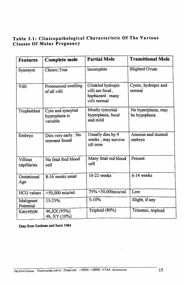
Table 3.1: Clinicopathological Characteristic Of The Various Classes Of Molar Pregnancy
Features Complete mole Partial Mole Transitional Mole
Synonym Classic; True Incomplete Blighted Ovum
Villi Pronounced S\velling Crinkled hydropic Cystic, hydropic and of all villi villi are focal ., normal
I haphazard : many villi normal
Trophoblast Cyto and syncytial Mostly syncytial No hyperplasia, may hyperplasia is hyperplasia; focal I be hypoplasia variable and mild
r
Embryo Dies very early : No Usually dies by 9 Amnion and slunted
remnant found · weeks ; may survive embryo
I till term
Villous l No fetal Red blood Many fetal red blood Present
capillaries cell cell
Gestational 8-16 weeks usual 10-22 weeks ~6-14 weeks Age
HCG values >50,000 miulml 75o/<> <50,000miu!ml Lo\V
Malignant 1. 15-25% 5-10% Slight, if any
Potential Karyotype 46)0((95%) Triploid (80°/o) Trisomic, triploid
46, XY (10~/o)
Data from Szulman and Surti 1984
GESTATIONAL TROPHOBLASTIC DISEASE 1995-1999 HTAA KUANTAN 15
I
STAGE INFORMATION
Hydatidiform mole (molar pregnancy) is disease limited to
the uterine cavity. Invasive mole (chorioadenoma destruens) is a
locally invasive, rarely metastatic lesion.
The FIGO staging system is as follows:
Stage 1: Disease confined to the uterus Stage lA: Disease confined to the uterus with no risk factors
Stage m: Disease confined to the uterus with one risk factor
Stage IC: Disease confined to the uterus with two risk factors
Stage II: GTT extends outside of the uterus but is
limited to the genital structures (ovary, tube, vagina,
broad ligament)
Stage llA: GTT involving genitaJ structures without risk factors
Stage DB:GTT extends outside of the uterus but limited to genital structures with one
risk factor
Stage DC:GTT extends outside of the uterus but limited to the genital structures with two
risk factors
G£STA TIONAL TROPHOBLASTIC DISE:AS£ I 995-1999 HTAA KUANTAN 16
Stage ill: GTT extends to the lungs, with or without
known genital tract involvement Stage IDA: GTI extends to the lungs, with or without genital tract involvement and with
no risk factors
Stage DIB: GTI extends to the lungs, with or without genital tract involvement and with
one risk factor
Stage illC: GTI extends to the lungs, with or without genital tract involvement and with
two risk factors
Stage IV: All other metastatic sites Stage IV A: All other metastatic sites, without risk factors
Stage IVB: All other metastatic sites, with one risk factor
Stage IVC: All other metastatic sites, with two risk factors
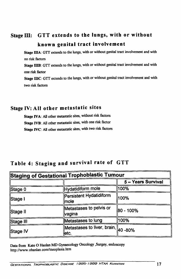
Table 4: Staging and survival rate of GTT
jStaging of Gestational Trophoblastic Tumour
I II II 5 -Years Survival
jstage 0 IIHydatidiform mole 11100%
ftage I I Persistent Hydatidiform
1100% mole
ftage II I Met~stases to pelvis or ro- 100% vag ana
!Stage Ill !!Metastases to lung ll100o/o
Jstage IV I Metastases to liver, brain, ~0 -80% etc.
Data from Kate 0 Hanlan MD Gynaecology Oncology ,Surgey, endoscopy http://www. ohanlan.com/tneoplasia.htm
GESTATIONAL TROPHOBLASTIC DISEASE 1995-1999 HTAA KUANTAN 17
I I I
l l I
I
HUMAN CHORIONIC GONADOTROPIN ASSAY
Successs in the treatment of GTD has been achieved tn
part because of the technical ability to quantitate serum or
urtnary concentrations of human chorionic gonadotrophin
(hCG ). Regular monitoring of hCG concentrations during
treatment provides information regarding disease status and the
response to therapy that permits appropriate intervention on an
individualized basis. The hCG assay thus has become essential
to the clinical management of trophoblastic disease , and basic
understanding of the assay methods and their limitations is the
important for the clinician involved in the care of these patients.
It has recently been recognized that hCG ts present in
patients in multiple forms : intact hCG, nicked hCG where the
binding site of the tumour has been split, hCG which has lost the
carboxy terminal part of the beta subunit, free beta and beta core
fragment . Early in a normal pregnancy the majority of hCG is in
intact form and nicking occurs increasingly through the
GESTATIONAL TROPHOBLASTIC 01S£AS£ I 995- I 999 HTAA KUANTAN 18
pregnancy. In GTT there is a much higher proportion of hCG
degradation products present and not all pregnancy test detect
these efficiently. The ideal hCG test for monitoring GTT is one
which detects all the beta epitopes present in intact hCG through
the beta core fragment. (Cole et al 1994).
Human chorionic gonadotrophin (hCG ) is a sensitive and
specific marker for the diagnosis and treatment of gestational
trophoblastic tumour. Bagshawe ( 1976) defined the risk more
specifically, noting the fatality rate of 4 % if the hCG was less
than 10,000 IU, 15o/o at 10,000- 100,000 IU, 27% at 100,000-
1,000,000 IU and 61 'Yo when the level was greater than
1,000,000 IU. Lurain et al (1982) reported that hCG level
greater than 100,000 IU /L were significantly associated with
fewer remission in patients with metatastatic GTT.
The usefulness of the HCG subunit assays in the prediction
of the development of malignant disease has not been settled.
Recent reports suggest that secretion of free b-hCG subunit in
comparison to whole HCG secretion showed an increasing trend
from pregnancy to mole to choriocarcinoma, and the free b-hCG
subunit level may be used in the prediction of GTD (Patiilo &
Houssa 1984, Khazaeli 1986, Fan 1987). Free b-hCg subunit
secretion appeared to be related to the maturity or degree of
GESTATIONAL TROPHOBlASTIC DISEASE 1995-1999 HTAA KUANTAN 19

differentiation of the trophoblast. Nigerian pregnant women
were found to secrete a persistently higher percentage of alfa -
hCG free subunit when compared with American pregnant
women (Eiegbe 1984 ). During the IVth World Congress of
Gestational Trophoblastic Disease held in Beijing in October
1988, preliminary results of b-hCG core fragment estimation
reported that this may be useful for the early diagnosis of
malignant disease during the follow up of mole patients. Ma
H.K. et al ( 1990).
GESTATIONAL TROPHOBLASTIC DISEASE 1995-1999 HTAA KUANTAN 20
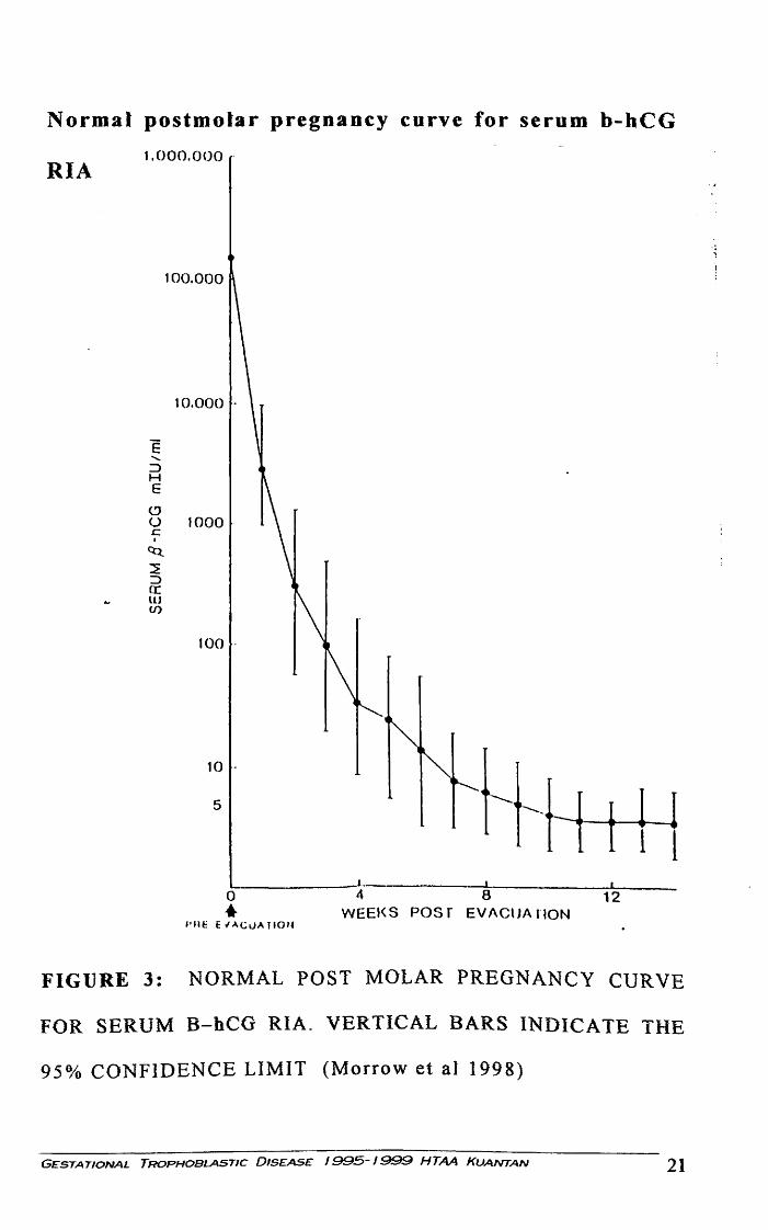
Normal postmolar pregnancy curve for serum b-hCG
RIA 1.000.000 .
E ....... :J H E
~ u c I
~
~ ::> (( LU U)
100.000
10.000 .
1000
100 .
5
0 • I'Jtt: t l.l.CuA Jl0'4
!-!-l
.L 4 8 12
WEEl<S POSf EVAClJA liON
FIGURE 3: NORMAL POST MOLAR PREGNANCY CURVE
FOR SERUM B-hCG RIA. VERTICAL BARS INDICATE THE
95% CONFIDENCE LIMIT (Morrow et al 1998)
GESTATIONAL TROPHOBLASTIC DISEASE 1995-1.9.9.9 HTAA KUANTAN 21

Abnormal Regression of serum hCG
Brewer et al ( 1971 ), Bagshawe et al ( 1973) provide a basis
for the interpretation of post molar gonadotrophin values
relative to the diagnosis of GTT and the need of therapeutic
intervention. Brewer et al ( 1971) reported that by 60 days after
molar evacuation, approximately 70% of their patients achieved
a normal hCG level, that is, less than 25miu/ml. An additional
15% demonstrates continuous drop in their values, although the
values were still higher than normal. In the remaining 15% of
the group with an elevated bCG at 60 days after evacuation, the
values were either plateu or rising.
GESTATIONAL TROPHOBLASTIC 01S£AS£ 1995-1999 HTAA KUANTAN 22

COMPLETE HYDATIDIFORM MOLE
This entity is characterized by the absence of a fetus and a
uterus filled by grossly swollen avascular villi made up of
oedematous stroma surrounded by hyperplastic
syncyctiotrophoblast. The incidence~ which 1s difficult to
determine and varies geographically, is approximately I per
1000 live births. Genetically it is usually diploid.
Symptomatology
Patients with Hydatidiform mole present as a complication
of an existing pregnancy and a positive pregnancy test. The
presenting symptoms can be bleeding, passage of grapes like
product or hyperemesis gravidarum (30%). Pain is not a
dominant symptom.
Clinical sign .
These patients are usually well with no overt distress or
haemodynamic comprise but they may be pale. If hyperemesis is
present, they may be signs of dehydration and ketosis.
GESTATIONAL TROPHOBLASTIC DISEASE 1995-1999 HTAA KUANTAN 23

In 65% of patients, uterine examination will reveal a fundal
height discordant with gestational age. The consistency of the
uterus is doughy rather than cystic.
Vaginal examination may confirm bleeding or a dilated cervtx
with vesicles products of conception in the vagina.
Twenty percent of patients will show features of pre-eclampsia.
Proteinuric hypertension before 20 weeks of pregnancy should
raise the possibility of GTD. Hyperthyroidism may manifest as.
clinical thyrotoxicosisin 10% of cases.
Diagnosis and investigation
The ultrasound appearance of an enlarged uterus with
echolucent black hole surrounded by a rim of white is absolutely
specific. Abdominal x ray may provide useful information (an
absent fetal skeleton) and excessive opacification in the region
of the uterine shadow. But if ultrasonography not available,
patients with suspected molar disease, if at all possible, should
be referred to an institution with this facility. Pelvic
angiography and amniography are specific but excessively
invasive. Extensive investigation for metastatic disease is not
GESTATIONAL TROPHOBLASTIC DISEASE 1995-1999 HTAA KUANTAN 24