gestational trophoblastic neoplasm
DESCRIPTION
GTTTRANSCRIPT

Gestational Trophoblastic Gestational Trophoblastic Neoplasm:Neoplasm:
Invasive MoleInvasive Mole&&
ChoriocarcinomaChoriocarcinomaBy: ParmitasariBy: Parmitasari

Invasive MoleInvasive Mole
Biologically, invasive mole is the Biologically, invasive mole is the intermediate formintermediate form of hydatidiform mole (complete and partial) and of hydatidiform mole (complete and partial) and choriocarcinomachoriocarcinoma
Locally destructiveLocally destructive but very unlikely to metastasize but very unlikely to metastasize 10% of complete moles develop into invasive mole10% of complete moles develop into invasive mole Overall, “invasive moles” occur at an estimated rate Overall, “invasive moles” occur at an estimated rate
of 1 pregnancy in 15,000.of 1 pregnancy in 15,000.

Invasive moleInvasive mole
Defined asDefined as mole that penetrates and may mole that penetrates and may even perforate the uterine walleven perforate the uterine wall
MacroscopicallyMacroscopically: :
presents as hydropic chorionic villi that presents as hydropic chorionic villi that invade myometrium invade myometrium uterine rupture uterine rupture

Figure 22-66 A, Invasive mole presenting as a hemorrhagic mass adherent to the uterine wall. (Courtesy of Dr. David R. Genest,
Brigham and Women's Hospital, Boston, MA.)Downloaded from: Robbins & Cotran Pathologic Basis of Disease (on 12 February 2007 02:26 AM)
© 2007 Elsevier

Invasive moleInvasive mole
MicroscopicallyMicroscopically:: Proliferation of both cytotrophoblast and Proliferation of both cytotrophoblast and
syncytiotrophoblastsyncytiotrophoblast May invade parametrial tissue and blood May invade parametrial tissue and blood
vesselsvessels

Invasive moleInvasive mole
Clinical manifestationClinical manifestation:: Vaginal bleeding Vaginal bleeding Irregular uterine enlargementIrregular uterine enlargement Persistent elevated Persistent elevated ß-HCGß-HCG
MetastasesMetastases No distant metastasesNo distant metastases Hydropic villi may embolize to distant sites Hydropic villi may embolize to distant sites
( e.g lungs, brains) ( e.g lungs, brains) do not grow as true do not grow as true metastasesmetastases

Invasive moleInvasive mole
PrognosisPrognosis The tumor responds well to The tumor responds well to
chemotherapychemotherapy May result in uterine rupture May result in uterine rupture need need
hysterectomyhysterectomy

ChoriocarcinomaChoriocarcinoma
Gestational choriocarcinoma is an Gestational choriocarcinoma is an epithelial epithelial malignant neoplasmmalignant neoplasm of trophoblastic cells of trophoblastic cells
derived from any form of previously normal or derived from any form of previously normal or abnormal pregnancyabnormal pregnancy
Rapidly invasive, widely metastasizing Rapidly invasive, widely metastasizing malignant neoplasmmalignant neoplasm

choriocarcinomachoriocarcinoma
Incidence Incidence
choriocarcinoma is an uncommon conditionchoriocarcinoma is an uncommon condition:: U.S. U.S. 1:20,000-1:30,000 1:20,000-1:30,000 Ibadan, Nigeria & Asian coutriesIbadan, Nigeria & Asian coutries 1:2500 1:2500
EtiologyEtiology
50%50% arise in hydatidiform moles arise in hydatidiform moles
25%25% in previous abortions in previous abortions
22%22% in normal pregnancies in normal pregnancies

choriocarcinomachoriocarcinoma
choriocarcinomachoriocarcinoma
1:40 hydatidiform moles1:40 hydatidiform moles
1:150,000 normal pregnancies1:150,000 normal pregnancies
MacroscopicallyMacroscopically soft, fleshy, yellow-white tumorsoft, fleshy, yellow-white tumor Large pale areas of ischemic necrosis, foci of Large pale areas of ischemic necrosis, foci of
cystic softening, and extensive hemorrhagecystic softening, and extensive hemorrhage
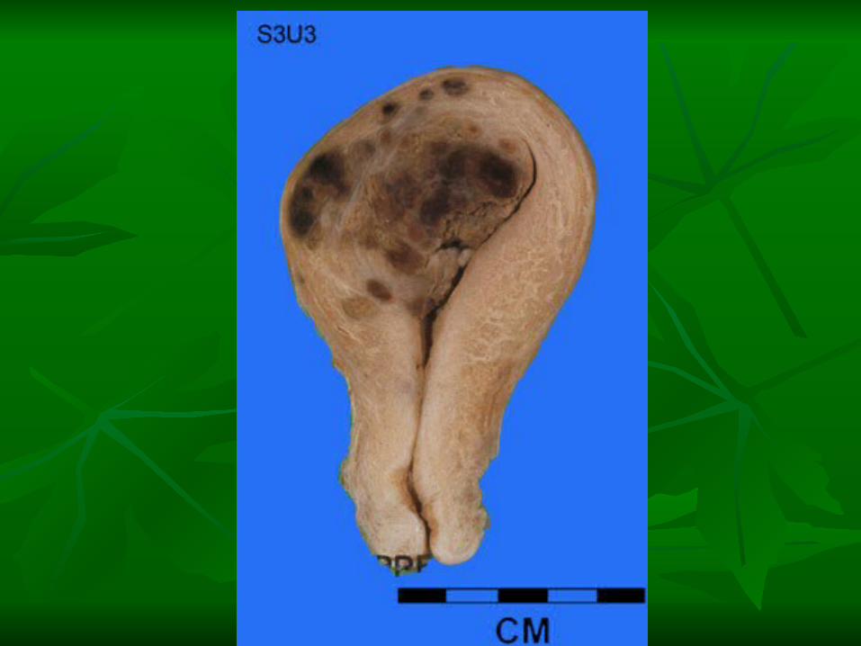

Figure 22-67 A, Choriocarcinoma presenting as a bulky hemorrhagic mass invading the uterine wall. (Courtesy of Dr. David R. Genest, Brigham and Women's Hospital,
Boston, MA.)
Downloaded from: Robbins & Cotran Pathologic Basis of Disease (on 13 February 2007 03:20 AM)
© 2007 Elsevier

choriocarcinomachoriocarcinoma
MicroscopicallyMicroscopically Purely ephitelial tumorPurely ephitelial tumor Does not produce chorionic villiDoes not produce chorionic villi Abnormal proliferation of both Abnormal proliferation of both
cytotrophoblast and cytotrophoblast and syncytiotrophoblastsyncytiotrophoblast

ChoriocarcinomaChoriocarcinoma, characterized by proliferation of cytotophoblast (blue , characterized by proliferation of cytotophoblast (blue circle) and syncytiontrophoblast (green circle), but no villi are present.circle) and syncytiontrophoblast (green circle), but no villi are present.
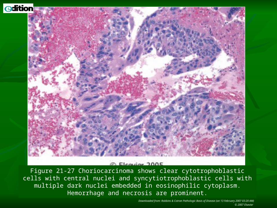
Figure 21-27 Choriocarcinoma shows clear cytotrophoblastic cells with central nuclei and syncytiotrophoblastic cells with multiple dark nuclei embedded in eosinophilic
cytoplasm. Hemorrhage and necrosis are prominent.
Downloaded from: Robbins & Cotran Pathologic Basis of Disease (on 13 February 2007 03:20 AM)
© 2007 Elsevier

choriocarcinomachoriocarcinoma
Clinical courseClinical course Manifest by Manifest by irregular spotting of a bloodyirregular spotting of a bloody, brown, , brown,
sometimes foul-smelling fluidsometimes foul-smelling fluid Usually, by the time the tumor is discovered Usually, by the time the tumor is discovered
locally, radiographs of the chest and bones already locally, radiographs of the chest and bones already disclose the disclose the presence of metastatic lesions presence of metastatic lesions
The titers of The titers of ß-HCG elevatedß-HCG elevated > in hydatidiform > in hydatidiform molesmoles
The tumor The tumor invades myometriuminvades myometrium uterine serosa, uterine serosa, penetrates blood vessels and lymphatics penetrates blood vessels and lymphatics

MetastasesMetastases Widespread metastasesWidespread metastases
characteristic!!characteristic!! Lungs (50%), vagina (30-40%), brain, Lungs (50%), vagina (30-40%), brain,
liver and kidneyliver and kidney

choriocarcinomachoriocarcinoma
PrognosisPrognosis Respond well to chemotherapyRespond well to chemotherapy
Up to 100 % cure or remissionUp to 100 % cure or remission

Invasive MoleInvasive Mole
Chorionic villi (+) Locally destructive, no
metastases Proliferation of cytotrophoblast and syncytiotrophoblast
Respond well to chemotherapy
ChoriocarcinomaChoriocarcinoma
Chorionic villi (-) Widespread metastases
Proliferation of cytotrophoblast and syncytiotrophoblast
Respond well to chemotherapy

Reference:Reference:Robbins and Cotran Pathologic Basis of Robbins and Cotran Pathologic Basis of
Disease. 7th Edition. (V Kumar, A K Disease. 7th Edition. (V Kumar, A K Abbas, and N Fausto). Philadelphia. Abbas, and N Fausto). Philadelphia. Elsevier Saunders. Elsevier Saunders.
