genotypic mapping ofhpv assessment ebv prevalence...
TRANSCRIPT

97 Clin Pathol 1997;50:904-910
Genotypic mapping of HPV and assessment ofEBV prevalence in endocervical lesions
J J O'Leary, R J Landers, M Crowley, I Healy, W F Kealy, J Hogan, C Cullinane,P Kelehan, C T Doyle
Nuffield Departmentof Pathology andBacteriology,University of Oxford,UKJ J O'Leary
Departmnent ofPathology, Universityof Sheffield, UKR J Landers
Department ofPathology, UniversityCollege Cork, IrelandM CrowleyI HealyW F KealyJ HoganC T Doyle
Department ofPathology, St James'University TeachingHospital, Leeds, UKC Cullinane
Department ofPathology, TheNational MaternityHospital, Dublin,IrelandP Kelehan
Correspondence to:Dr O'Leary, Department ofPathology, Cornell UniversityMedical College, CornellMedical Center, The NewYork Hospital, New York,NY 10021, USA.
Accepted for publication2 September 1997
AbstractAins-To examine the prevalence ofhuman papillomavirus (HPV) andEpstein-Barr virus (EBV) in low gradeglandular intraepithelial lesions of thecervix, adenocarcinoma with high gradeglandular intraepithelial lesions com-
bined, and adenocarcinomas; and to per-form a genotyping mapping analysis ofendocervical carcinomas to determine theextent ofHPV infections in such lesions.Material-Archival paraffin wax embed-ded material from the files of the depart-ments of pathology, National MaternityHospital, Dublin, and University CollegeCork, Ireland.Methods-HPV prevalence was examinedusing type specific HPV PCR, generalprimer HPV PCR (pan HPV screen), non-isotopic in situ hybridisation (NISH), andPCR in situ hybridisation (PCR-ISH). Insitu hybridisation was performed usingfluorescein labelled oligonucleotide cock-tail for eber transcripts ofEBV. Genotypicanalysis was performed, in all cases wherepossible, using a grid systemResults-HPV 16 and 18 were predomi-nantly identified in low grade glandularintraepithelial lesions, high grade glandu-lar intraepithelial lesions, and adenocar-cinomas, with HPV prevalence increasingwith grade of dysplasia. EBV was onlyidentified in subepithelial lymphocytes ina minority of cases. No link could beshown between HPV and EBV in endocer-vical lesions. HPV infection was not clonalin endocervical cancer and coexistentadjacent cervical intraepithelial neopla-sia, where present, tended to show a simi-lar HPV type.Conclusions-The restriction of HPVtypes 16 and 18 to endocervical lesionssuggests that their effect is restricted andspecific to endocervical mucosa, but themechanism of interaction is currentlyunknown.(7 Clin Pathol 1997;50:904-9 0)
Keywords: human papillomavirus; Epstein-Barr virus;endocervical lesions
The relative frequency of adenocarcinoma ofthe cervix has increased to between 12.7% and18.5% in recent years.1-3 In contrast, theincidence of invasive squamous cell carcinomahas decreased, largely because of cervical
screening, which has little apparent effect onendocervical lesions.'
Criteria for the diagnosis of glandularintraepithelial neoplasia and -adenocarcinomain situ of the cervix are controversial. Suchlesions are often focal and quite superficial andtherefore are not commonly diagnosed. Moreextensive multicentric lesions, although moreeasy to see, are often quite difficult to differen-tiate from adenocarcinoma of the cervix.
Endocervical glandular intraepithelial neo-plasia is diagnosed by the presence of endocer-vical glands, the epithelium of which showsintraluminary papillary projections with focalcribriform or bridging patterns. Basophilia ofthe cytoplasm with nuclear atypia and pseu-dostratification are usually prominent. Catego-risation into low and high grade lesions hasbeen suggested, but there is considerable over-lap between the high grade designation andadenocarcinoma in situ (see below). In thispaper, the low and high grade categorisationformula is adopted, taking into account thevirtual similarity between high grade glandularintraepithelial lesions and adenocarcinoma insitu.Adenocarcinoma in situ again shows baso-
philic cytoplasm, significant nuclear atypia,frequent mitotic figures, and nuclear pseudo-stratification over several layers. Papillaryprojections are prominent and the architecturecomplex, but stromal invasion is not seen.The precise biological significance of less
severe endocervical lesions, termed atypicalendocervical hyperplasia of the columnarendocervix, has not been well established.Although the clinical and histological featuresof microinvasive squamous cell carcinoma arewell defined, those of its endocervical counter-part are not.The potential for undifferentiated cells in
Mullerian tissues to differentiate along severallines leads ultimately to a wide range of tumourtypes, among which are endometrioid or papil-lary serous adenocarcinoma of the cervix.The most frequent member of this group is
represented by endocervical adenocarcinoma,the most well differentiated form called"adenoma malignum."4 Papillary serouscarcinoma of the cervix typically grows in apapillary fashion, with a proportion differenti-ating along tubal lines resembling serouscarcinoma of the ovary. Endometrioid carci-noma can arise in foci of cervical endometrio-sis but more commonly arises from undiffer-entiated endocervical reserve cells. Clear cellcarcinoma is morphologically identical to clear
904
on 1 June 2018 by guest. Protected by copyright.
http://jcp.bmj.com
/J C
lin Pathol: first published as 10.1136/jcp.50.11.904 on 1 N
ovember 1997. D
ownloaded from

HPV and EBV in endocervical lesions
cell carcinoma of the endometrium; these aretumours of Muillerian duct origin, some ofwhich occur in young women exposed prena-tally to diethylstilboestrol.6 Mesonephric ad-enocarcinomas are extremely rare neoplasmsand are histologically similar to clear celladenocarcinomas of Mullerian type.7 Entericadenocarcinomas of intestinal type occur veryrarely in the cervix.8 Adenosquamous carcino-mas are also found, some of which having a"glassy cell" morphology.9Over 70 types of human papillomavirus
(HPV) are now described, and there is a strongassociation between "low" and "high" riskHPV types and the development of premalig-nant and malignant squamous epithelial le-sions of the cervix. HPV 6 and 11 areconsidered to be low risk types, associated withcondylomas and cervical intraepithelial neo-plasia (CIN) grade 1. HPV types 16, 18, 31,33, 35, 39, 51 are high risk types related to thedevelopment of high grade CIN (CIN 2 and 3)and invasive squamous cell carcinoma. HPVhas also been detected in glandular intraepithe-lial neoplasia, adenocarcinoma in situ, andadenocarcinoma of the endocervix. Prevalencerates vary but approximately 79% of adenocar-cinoma in situ lesions and 43% of adenocarci-nomas contain HPV, predominantly of types16 and 18.10-12 It seems reasonable therefore topostulate that similar cellular events are opera-tional in endocervical lesions as in CIN andinvasive squamous cell carcinoma.The idea of a sexually transmittable infec-
tious carcinogen as the dominant factor in thepathogenesis of cervical neoplasia has receivedwidespread attention.12 13 Possible agents in-clude herpes virus 2 (HSV 2), Chlamydiatrachomatis, HPV, cytomegalovirus, and morerecently, Epstein-Barr virus (EBV) . Theassociation between human papillomavirusand squamous cell cervical neoplasia is wellknown, although its role in endocervicalneoplasia is still inconclusive."3 The role ofsynergistic/carcinogenic factors such as two ormore viruses interacting, possibly at differentstages of cervical carcinogenesis, has previouslybeen considered by zur Hausen. 15 EBV, anoncogenic herpes virus, has been suggested as apossible viral cofactor. EBV has a ubiquitousdistribution, causing widespread and largelyasymptomatic infection worldwide. The virusis known to be associated with three lympho-proliferative disorders of B cell origin-infectious mononucleosis, Burkitt's lymphoma,and lymphomas-in immunocompromised pa-tients. An association between EBV and undif-ferentiated nasopharyngeal carcinoma hasbeen documented previously.'6 '7 Indeed othercarcinomas, for example, tonsil, tongue, larynx,and thymus, have also shown an associationwith EBV.'8-22
Sixbey et al have produced in vitro and invivo evidence of EBV viral replication in thecervix.23 In particular, they have shown thatcultured ectocervical epithelial cells can beinfected with EBV.23 Using cultures of ectocer-vical epithelium, they also have demonstratedthat EBV can binding to epithelial cells.Cultured ectocervical epithelial cells react with
several monoclonal antibodies specific for theEBV receptor on B lymphocytes, strongly sug-gesting expression of a functional EBV recep-tor on cervical epithelial cells. To our knowl-edge, no such data exist in relation toendocervical mucosa. Similar observationshave been seen in other stratified squamousepithelia, for example, pharynx and tongue.24In addition, late EBV DNA viral antigens havebeen identified in exfoliating cells, but not inattached epithelial monolayer culture cells,suggesting an association between viral replica-tion and the stage of epithelial differentiation.Infectious EBV capable of transforming Blymphocytes have been isolated in cervicalwashings from women recovering from infec-tious mononucleosis, and from EBV positiveindividuals who have had no clinical orserological evidence of acute EBV infection.25It appears that the cervix may be a site ofchronic viral shedding in a manner similar tothe nasopharynx.
In this study, we examine the prevalence ofHPV and EBV in three cohorts of endocervicallesions- low grade glandular intraepitheliallesions, adenocarcinoma with adjacent highgrade glandular intraepithelial lesions/adeno-carcinoma in situ, and pure adeno-carcinomas-to investigate any synergistic rela-tion between HPV and EBV in thepathogenesis of endocervical carcinoma. Inaddition, we performed an HPV genotypicmapping analysis of the adenocarcinoma co-hort, to establish the extent ofHPV infection insuch lesions. In genotypic mapping, selectivefields are analysed within a tumour in order todelineate the extent and clonality of a particu-lar genetic marker (that is, the presence ofHPV, ras, p53 mutations, and so on).
MethodsMATERIALSArchival paraffin wax embedded material wasretrieved from the files of the departments ofpathology, National Maternity Hospital, Dub-lin, and University College Cork, Ireland. Tenlow grade glandular intraepithelial lesions,eight high grade glandular intraepithelial le-sions and adenocarcinoma combined, and 11adenocarcinomas of the cervix were examined.Coexistent areas of CIN were also assessed.HPV prevalence was examined using type spe-cific HPV polymerase chain reaction (PCR),general primer HPV PCR (pan HPV screen),non-isotopic in situ hybridisation (NISH), andPCR in situ hybridisation (PCR-ISH). Geno-typic analysis was performed (in all cases wherepossible) using a grid system (1-4), asillustrated in fig 1. Tissue was microdissectedfrom each of the areas designated and analysedby solution phase PCR.
HPV IN SITU HYBRIDISATION
Three 5 gm sections (selected from differentareas of the tumour) were cut onto APEScoated glass slides (PH 106, C A Hendley,Essex, UK).
Tissues were dewaxed in xylene and rehy-drated through a graded alcohol series. Proteo-lytic digestion was carried out using 0.5 mg/ml
905
on 1 June 2018 by guest. Protected by copyright.
http://jcp.bmj.com
/J C
lin Pathol: first published as 10.1136/jcp.50.11.904 on 1 N
ovember 1997. D
ownloaded from

O'Leary, Landers, Crowley, Healy, Kealy, Hogan, et al
B C
i..
Figure 1 (A) Genotypic mapping experiment in an endocervical adenocarcinoma, based on a grid system, divided intoareas 1-4. (B) Adenocarcinoma in situ in an endocervical gland. (C) HPV 16 NISH in adenocarcinoma in situ showinga discrete nuclear signal.
proteinase K in proteinase K buffer (50 mMTris HCl pH 7.6, 5 mM EDTA) at 370C for 10minutes. Slides were immersed in phosphatebuffered saline (PBS) containing 2 mg/ml gly-cine for five minutes and washed in PBS for afurther five minutes. For alkaline phosphatasedetection, slides were immersed in 20%(vol/vol) aqueous acetic acid at 4°C for 15 sec-onds. For peroxidase detection, slides wereimmersed in 3% sodium azide/hydrogen per-oxide to abolish endogenous peroxidase activ-ity. Slides were then washed in PBS for fiveminutes. A postfixation step was not carriedout. Dehydration of the sections then tookplace through graded alcohols to water.
Cloned HPV probes (6, 11, 16, 18, 31, 33)(genital types; gifts of Harald Zur Hausen)were labelled with biotin or digoxigenin using astandard nick translation protocol.26 Probeswere prepared at 200 ng/ml.The hybridisation buffer contained 2x SSC
(sodium chloride/sodium citrate), 5% dextransulphate, 0.2% dried milk powder containingno vegetable extracts, and 50% formamide.Approximately 10-20 ng of the appropriateprobe in hybridisation buffer was applied to thecentre of each PH106 slide well. Gel bond(FMC Bioproducts, Rockland, Maine, USA)cut to cover slip size, was placed hydrophobicside down over each tissue section and sealed inplace with nail varnish. Sections were dena-tured at 90°C for 10 minutes. After denatura-tion, slides were transferred to a humidifiedbox and incubated at 37-42°C overnight. Fol-lowing hybridisation, the gel bond was re-moved with a scalpel blade and the slidesimmersed in 2x SSC at room temperature.Medium stringency posthybridisation wash-
ings were initially applied using 2x SSC at60°C for 20 minutes, followed by 0.2x SSC for42°C at 20 minutes, then O.lx SSC at roomtemperature for five minutes, and 2x SSC atroom temperature for five minutes. Higherstringency washes included 0.2x SSC at 55°Cand 60°C for 10 minutes and O.lx SSC atroom temperature, 42°C and 55°C.
The hybridisation signal was detected usingone step, two step, or three step techniques asdescribed previously."6 Colorimetric detectionwas achieved using a NBT/BCIP substrate foralkaline phosphatase or aminoethylcarbazole(Zymed kit for peroxidase, California, USA).`Sections were counterstained with 2% methylgreen for alkaline phosphatase detection orhaematoxylin for the peroxidase detection sys-tem.
Tissue controls for NISH included HPVpositive cervical wart, and myocardium (nega-tive for HPV). Reaction controls includedhybridisation buffer on its own, biotin/digoxigenin labelled plasmid sequences(pBR322), and irrelevant probe (herpes zostervirus, HZV). Labelled human placental DNAwas used to check hybridisation efficiency.
Detection controls included omitting pri-mary or secondary antibody steps and additionof the colorimetric substrate only.
EBER-ISHTissue dewaxing was carried out as above. Insitu hybridisation was performed using a fluo-rescein labelled oligonucleotide cocktail foreber transcripts of EBV: 20 gl of probehybridisation solution were incubated on thetissue sections for two hours at 37°C; slideswere then washed in Tris buffered salinecontaining 0. 1% Triton X- 1 00 for threeminutes three times. Detection was achievedusing the Novocastra in situ hybridisationdetection kit, following the recommendedmethod for hybrid detection.
SOLUTION PHASE PCRHPV DNA sequences were derived from theEMBL genetic sequence database. HPV E6sequences which remain intact following viralDNA integration were chosen."' Oligonucle-otide primers were synthesised on a PerkinElmer Applied Biosystems DNA synthesiser(Perkin Elmer, Cheshire, UK), deprotected,and stored in liquid ammonia at -200C.During oligo synthesis, a biotin reporter
906
. 1* I .... .I
" ., .7
t. -I'
#. .: f,
on 1 June 2018 by guest. Protected by copyright.
http://jcp.bmj.com
/J C
lin Pathol: first published as 10.1136/jcp.50.11.904 on 1 N
ovember 1997. D
ownloaded from

HPV and EBV in endocervical lesions
molecule with a 15 carbon atom linker arm wasadded to the 5' end of the oligonucleotideprobe, which was subsequently used as aninternal probe to confirm product specificity.The nucleotide sequence of the primers wereas previously described.27
General HPV primers (which identifysequenced and unsequenced human papil-lomaviruses) were also used for a panHPV screen, unrestricted by the type specificviruses chosen. These were as previouslydescribed.28For EBV amplification EBNA-1 primers
were used-primer 1: ATCGTGGTCAAG-GAGGTTCC; primer 2: ACTCAATGGTG-TAAGACGAC-and probed with a 30 mer 5'biotin labelled probe: AGAGCTCTCCT-GGCTAGGAGTCACGTAGAA, using South-ern analysis.Ten 5 gm sections were cut and placed in
sterile Eppendorf tubes. Strict anticontamina-tion protocols were adopted. Nucleic acidextraction was carried out using proteinase K(0.1-0.5 mg/ml) in proteinase K buffer(100 mM NaCl, 10 mM Tris HC1, 25 mMEDTA, 0.5% sodium dodecyl sulphate,pH 8.4). Proteinase K incubation was carriedout for three to five days at 37°C with adequatemixing of samples. Proteinase K inactivationwas then carried out at 94°C for 10 minutes.DNA was purified using a standard phenolchloroform isoamyl alcohol technique. Nucleicacid was precipitated using 3 M sodium acetateand ice cold ethanol.For genotypic mapping areas were selected
as in fig 1 and microdissected using a sterileblade. DNA was extracted as above.For type specific HPV PCR, the PCR
solution consisted of PCR buffer (50 mMKC1, 10 mM Tris HCI pH 8.3, 1.5 mMMgCl2, 0.01% gelatine, 200,uM of eachdNTP, 1.0,M of each primer, 2.5 units ofAmpliTaq DNA polymerase and 100 ng ofDNA template). Samples were then subjectedto 40 cycles of PCR in a Perkin Elmer 480DNA thermocycler. Cycling parameters wereas follows: 94°C for one minute, followed by94°C for one minute, 55°C for two minutes,72°C for three minutes x 40 cycles, with afinal extension set for 72°C for five minutes.Similar conditions were applied for EBNA-1PCR.For general primer PCR, amplification con-
ditions were similar except 3.5 mM MgCl, wasused and an annealing temperature of 400Cwas applied.Type specific HPV PCR products were con-
firmed by dot blot hybridisation, as previouslydescribed.27
HPV AND EBV PCR-ISHSections of 3-5 gm thickness (again selectedfrom different areas of the tumour) were cutonto APES coated single well slides and placedon a hot plate for 12 to 16 hours. Sectiondewaxing was carried out as for normal in situhybridisation. Proteinase digestion was carriedout using 0.5 mg/ml proteinase K in proteinaseK buffer as for in situ hybridisation at 37°C for10 to 15 minutes. Blocking of endogenous
alkaline phosphatase and peroxidase was car-ried out as above. Slides were then washed inPBS for five minutes. Tissues were amplifiedusing the GeneAmp in situ PCR system 1000(Perkin Elmer), which ensures maximal ther-mal kinetics.29 30The PCR solution consisted of PCR buffer
(50 mM KC1, 10 mM tris HC1 pH 8.3,4.5-5.0 mM magnesium chloride, 0.01% gela-tine, 200 ,uM of each dNTP, 2 ,uM each primer,5 units of TaqIS/50 ,ul, and 0.2% bovine serumalbumin). HPV primers were as previouslyshown.27 EBNA-1 primers were as detailedabove. A control reference gene, 13 globin, wasused to assess the efficiency of amplification. [Globin primers were as follows: primer 1(5'-ACACAACTGTGTTCACTAGC-3') andprimer 2 (5'-CAACTTCATCCACG-TTCA-CC-3') and a biotinylated [3globin oligoprobe(5'-GACTCCTGAGGAGAAGTCTGCCGT-TACTGC-3'). Slide assembly was achievedusing Amplicover clips and discs and the slideassembly tool.The following cycling parameters were
applied: 94°C for six minutes followed by 40cycles of 94°C x one minute, 55°C x two min-utes. In addition, a separate extension step wasincluded in some protocols but did not appearto yield better results. Following amplification,disassembly of the Amplicover clip and discwas performed. Slides were then carefullydipped in 100% alcohol to dehydrate and post-fix the amplified product. In addition someslides underwent 2% paraformaldehyde post-fixation for three to five minutes.The amplified product was then detected
using standard in situ hybridisation as de-scribed in the in situ hybridisation section,using an internal oligoprobe at 5-10 pmol/100 jl of hybridisation mix containing (2xSSC, 5% dextran sulphate, and 10% forma-mide).
Colorimetric detection was achieved usingNBT/BCIP as for in situ hybridisation.
Appropriate controls used included fixedSiHa cells containing one to two copies ofHPV16, fixed CaSki cells containing 200 copies ofHPV 16, and fixed P3HR1 cells containingEBV genome. For each assay the followingPCR in situ hybridisation controls wereincluded: (1) reference control gene ([Bglobin)31; (2) no Taq DNA polymerase; (3) noprimers; (4) primer 1 only; (5) primer 2 only;(6) target primers with an irrelevant probe(HHV 8); (7) irrelevant primers (that is,HHV8) with the target probe; (8) referencecontrol gene primers ([ globin/PDH) with thetarget probe.
ResultsLOW GRADE GLANDULAR INTRAEPITHELIALLESIONSThe age range of patients with glandularintraepithelial neoplasia examined was 26 to 40years (mean 33.5 years). Results of the HPVanalysis are given in table 1.By PCR, three of 10 cases were HPV 16
positive. NISH analysis confirmed these twopositive cases, but extensive NISH analysiswas not possible because of lack of adequate
907
on 1 June 2018 by guest. Protected by copyright.
http://jcp.bmj.com
/J C
lin Pathol: first published as 10.1136/jcp.50.11.904 on 1 N
ovember 1997. D
ownloaded from

O'Leary, Landers, Crowley, Healy, Kealy, Hogan, et al
Table 1 Prevalence ofhuman papillomavirus in endocervical lesions
Non-isotopic in situ hybridisation Polymerase chain reaction
6 11 16 18 31 33 6 11 16 18 31 33
Low grade GINNT NT 2/2* NT 0/10 NT 0/10 0/10 3/10 0/10 N/A 0/10
Adenocarcinoma and high grade GINIACIS combined0/8 0/8 2/8 1/8 0/8 0.8 0/8 0/8 5/8 2/8 N/A 0/8
Adenocarcinoma0/11 0/11 2/11 1/11 0/11 0/11 0/11 0/11 3/11 1/11I N/A 0/1 1
*Two residual samples showed evidence of GIN.ACIS, adenocarcinoma in situ; GIN, glandular intraepithelial neoplasia; N/A, sequence notavailable for primer design; NT, no tissue available.
A B /
Wl...
Figure 2 (A) NISH in adenocarcinoma in situ of the cervix showing HPV 16 in one cellnucleus. (B) HPV 16 PCR-ISH ofa parallel section, showing two cells positive with astronger intranuclear signal visible as compared to NISH. Notice also leakage ofamplification product into the cell cytoplasm focally, which indicates minimaloverpermeabilisation.
ADENOCARCINOMA AND HIGH GRADE GLANDULARINTRAEPITHELIAL NEOPLASIA/ADENOCARCINOMAIN SITU COMBINEDThe age range ofpatients with adenocarcinomaand adenocarcinoma in situ combined was 38to 48 years (mean 41.8 years). Results of theHPV analysis are given in table 1.By PCR analysis, five of eight and two of
eight cases were HPV 16 and 18 positive,respectively. Coexistent CIN 2 was seen inthree cases, all of which were HPV 16 positive.By NISH, two of eight and one of eight cases
were HPV 16 and 18 positive, respectively. Thein situ hybridisation signal in HPV positivecases was punctate in all cases. In three of thecases (with high grade glandular intraepitheliallesions/adenocarcinoma in situ in relation toadenocarcinoma) both the intraepithelial le-sion and adenocarcinoma were positive.EBV was only identified by PCR in two
cases, again subepithelial in location with nointraepithelial localisation seen.
The genotypic mapping experiment per-formed on six cases showed monotypic HPVtypes (that is, either 16 or 18) in each respectivecase. In three cases all four areas were HPVpositive, in two cases two areas were HPVpositive, and in one case only one area waspositive by PCR analysis. NISH and PCR-ISHconfirmed this geographical segregation. Lowrisk HPV types 6 and 11 were not identified inany of the cases.
histological material. Two HPV 16 positivecases had coexistent HPV 16 positive CIN 2.EBV was identified in one case only, insubepithelial lymphocytes. No intraepithelialEBV signals were seen, either on NISH or
PCR-ISH.Of the two cases positive on NISH for HPV
16, the in situ signal was punctate but tissuepreservation was poor. PCR-ISH increased thesignal intensity, and the number of cells positivein an individual dysplastic gland (fig 2).
ADENOCARCINOMAThe age range ofpatients with adenocarcinomaof the cervix was 36 to 63 years (mean 47.37years). Results are illustrated in table 1.By PCR analysis, three of 11 and one of 11
cases were positive for HPV 16 and 18, respec-tively. Of the four cases positive by PCR, onlytwo of four areas in each case were HPV posi-tive on genotypic mapping, suggesting that thetumour was not clonal for HPV. NISH andPCR-ISH confirmed solution phase PCR dataexcept in one HPV 16 case. No unsequenced
Table 2 Epidemiological data for adenocarcinoma of the cervix, adenocarcinoma in situ (ACIS), glandular intraepithelialneoplasia (GIN), and human papillomavirus (HPV) prevalence
HPV type (number of caseslnumber tested)
Country Method 6 11 16 18 31 33 Reference No
Ireland PCR Adca 0/11 0/11 3/11 1/11 0/11 0/11Adca + ACIS 0/8 0/8 5/8 2/8 0/8 0/8Low grade GIN 0/10 0/10 3/10 0/10 0/10 0/10
UK In situ Adca + ACIS 0/16 0/16 7/16 4/16 0/16 0/16 32
S Africa In situ Adca + ACIS 0/22 0/22 0/22 9/22 0/22 0/22 32(6 of the virus positive carcinomas had adjacent carcinoma in situ)
Denmark In situ Adca - 4/11* - 33ACIS - - 4/4* - 33
UK In situ (No HPV association)
USA In situ Adca 0/17 - 1/17 16/17 - - 10ACIS 0/7 - 0/18 0/18 - - 10GIN 0/18 - 0/18 0/18 - - 10
USA In situ ACIS - - 4/13 7/13 - - 37(mRNA)
*Combined data (HPV 16 and 18).ACIS, adenocarcinoma in situ; Adca, adenocarcinoma; GIN, glandular intraepithelial neoplasia.
908
on 1 June 2018 by guest. Protected by copyright.
http://jcp.bmj.com
/J C
lin Pathol: first published as 10.1136/jcp.50.11.904 on 1 N
ovember 1997. D
ownloaded from
HPVand EBV in endocervical lesions
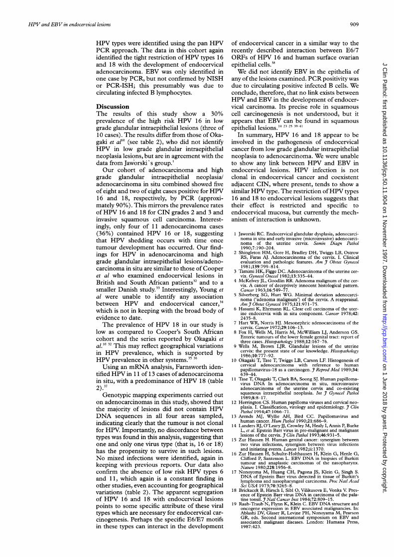
HPV types were identified using the pan HPVPCR approach. The data in this cohort againidentified the tight restriction ofHPV types 16and 18 with the development of endocervicaladenocarcinoma. EBV was only identified inone case by PCR, but not confirmed by NISHor PCR-ISH; this presumably was due tocirculating infected B lymphocytes.
DiscussionThe results of this study show a 30%prevalence of the high risk HPV 16 in lowgrade glandular intraepithelial lesions (three of10 cases). The results differ from those of Oka-gaki et all' (see table 2), who did not identifyHPV in low grade glandular intraepithelialneoplasia lesions, but are in agreement with thedata from Jaw;orski' s group.'Our cohort of adenocarcinoma and high
grade glandular intraepithelial neoplasia/adenocarcinoma in situ combined showed fiveof eight and two of eight cases positive for HPV16 and 18, respectively, by PCR (approxi-mately 90%). This mirrors the prevalence ratesofHPV 16 and 18 for CIN grades 2 and 3 andinvasive squamous cell carcinoma. Interest-ingly, only four of 11 adenocarcinoma cases(36%) contained HPV 16 or 18, suggestingthat HPV shedding occurs with time oncetumour development has occurred. Our find-ings for HPV in adenocarcinoma and highgrade glandular intraepithelial lesions/adeno-carcinoma in situ are similar to those of Cooperet al who examined endocervical lesions inBritish and South African patients32 and to asmaller Danish study.33 Interestingly, Young etal were unable to identify any associationbetween HPV and endocervical cancer,34which is not in keeping with the broad body ofevidence to date.The prevalence of HPV 18 in our study is
low as compared to Cooper's South Africancohort and the series reported by Okagaki etal.'0 32 This may reflect geographical variationsin HPV prevalence, which is supported byHPV prevalence in other systems.3536Using an mRNA analysis, Farnsworth iden-
tified HPV in 1 1 of 13 cases ofadenocarcinomain situ, with a predominance of HPV 18 (table
2.37Genotypic mapping experiments carried out
on adenocarcinomas in this study, showed thatthe majority of lesions did not contain HPVDNA sequences in all four areas sampled,indicating clearly that the tumour is not clonalfor HPV. Importantly, no discordance betweentypes was found in this analysis, suggesting thatone and only one virus type (that is, 16 or 18)has the propensity to survive in such lesions.No mixed infections were identified, again inkeeping with previous reports. Our data alsoconfirm the absence of low risk HPV types 6and 11, which again is a constant finding inother studies, even accounting for geographicalvariations (table 2). The apparent segregationof HPV 16 and 18 with endocervical lesionspoints to some specific attribute of these viraltypes which are necessary for endocervical car-cinogenesis. Perhaps the specific E6/E7 motifsin these types can interact in the development
of endocervical cancer in a similar way to therecently described interaction between E6/7ORFs of HPV 16 and human surface ovarianepithelial cells.38We did not identify EBV in the epithelia of
any ofthe lesions examined. PCR positivity wasdue to circulating positive infected B cells. Weconclude, therefore, that no link exists betweenHPV and EBV in the development of endocer-vical carcinoma. Its precise role in squamouscell carcinogenesis is not understood, but itappears that EBV can be found in squamousepithelial lesions."4 23 25 39-41
In summary, HPV 16 and 18 appear to beinvolved in the pathogenesis of endocervicalcancer from low grade glandular intraepithelialneoplasia to adenocarcinoma. We were unableto show any link between HPV and EBV inendocervical lesions. HPV infection is notclonal in endocervical cancer and coexistentadjacent CIN, where present, tends to show asimilar HPV type. The restriction ofHPV types16 and 18 to endocervical lesions suggests thattheir effect is restricted and specific toendocervical mucosa, but currently the mech-anism of interaction is unknown.
1 Jaworski RC. Endocervical glandular dysplasia, adenocarci-noma in situ and early invasive (microinvasive) adenocarci-noma of the uterine cervix. Semin Diagn Pathol1990;7:190-204.
2 Shingleton HM, Gore H, Bradley DH, Twiggs LB, OstrowRS, Faras AJ. Adenocarcinoma of the cervix. I. Clinicalevaluation and pathologic features. Am _7 Obstet Gynecol1981;139:799-814.
3 Tamimi HK, Figge DC. Adenocarcinoma of the uterine cer-vix. Gynecol Onccol 1982;13:335-44.
4 McKelvey JL, Goodlin RR. Adenoma malignum of the cer-vix. A cancer of deceptively innocent histological pattern.Cancer 1963;16:549-77.
5 Silverberg SG, Hurt WG. Minimal deviation adenocarci-noma ('adenoma malignum') of the cervix. A reappraisal.AmJ Obstet Gynecol 1975;121:971-75.
6 Hasumi K, Ehrmann RL. Clear cell carcinoma of the uter-ine endocervix with in situ component. Cancer 1978;42:2435-8.
7 Hart WR, Norris HJ. Mesonephric adenocarcinoma of thecervix. Cancer 1972;29:106-13.
8 Fox H, Wells M, Harris M, McWilliam LJ, Anderson GS.Enteric tumours of the lower female genital tract: report ofthree cases. Histopathology 1988;12:167-76.
9 Wells M, Brown LJR. Glandular lesions of the uterinecervix: the present state of our knowledge. Histopathology1986;10:777-92.
10 Okagaki T, Tase T, Twiggs LB, Carson LF. Histogenesis ofcervical adenocarcinoma with reference to humanpapillomavirus-18 as a carcinogen. J Reprod Med 1989;34:639-44.
11 Tase T, Okagaki T, Clark BA, Soong SJ. Human papilloma-virus DNA In adenocarcinoma in situ, microinvasiveadenocarcinoma of the uterine cervix and co-existingsquamous intraepithelial neoplasia. Int J Gynecol Pathol1989;8:8-17.
12 Herrington CS. Human papilloma viruses and cervical neo-plasia. I. Classification, virology and epidemiology. J ClinPathol 1994;47:1066-71.
13 Arends MJ, Wyllie AH, Bird CC. Papillomavirus andhuman cancer. Hum Pathol 1990;21:686-9.
14 Landers RJ, O'LearyJJ, Crowley M, Healy I, Annis P, BurkeL, et al. Epstein Barr virus in pre-malignant and malignantlesions of the cervix. J Clin Pathol 1993;46:931-5.
15 Zur Hausen H. Human genital cancer: synergism betweentwo virus infections, synergism between virus infectionsand initiating events. Lancet 1982;ii: 1370.
16 Zur Hausen H, Schulte-Holthausen H, Klein G, Henle G,Clifford P, Santesson L. EBV DNA in biopsies of Burkitttumour and anaplastic carcinomas of the nasopharynx.Nature 1980;228:1956-8.
17 Nonoyoma M, Huang CH, Pagona JS, Klein G, Singh S.DNA of Epstein Barr virus detected in tissue of Burkitt'slymphoma and nasopharyngeal carcinoma. Proc Nati AcadSci USA 1973;70:3265-8.
18 Brickacek B, Hirsch I, Sibl 0, Vilikusova E, Vonka V. Pres-ence of Epstein Barr virus DNA in carcinoma of the pala-tine tonsil. J Natl Cancer Inst 1984;72:809-15.
19 Raab-Traub N, Flynn K, Klein C. EBV DNA structure andoncogene expression in EBV associated malignancies. In:Ablashi DV, Glaser R, Levine PH, Nonoyama M, PearsonGR, eds. Second international symposium on EBV andassociated malignant diseases. London: Humana Press,1987:423.
909
on 1 June 2018 by guest. Protected by copyright.
http://jcp.bmj.com
/J C
lin Pathol: first published as 10.1136/jcp.50.11.904 on 1 N
ovember 1997. D
ownloaded from
O'Leary, Landers, Crowley, Healy, Kealy, Hogan, et al
20 Brickacek B, Hirsch I, Sibl 0, Vilikosova E, Vonka VAssociation of some supra glottic laryngeal carcinomaswith EB virus. Int3' Cancer 1983;32:193-7.
21 Saemundsen AK, Albeck H, Hansen JP, Neilsen NH,Anuret M, Henle W, et al. Epstein Barr virus innasopharyngeal and salivary gland carcinomas in Green-land Eskimos. BrJ Cancer 1982;46:721-8.
22 Leyvraz S, Henle W, Chahinian AP, Perlman C, Klein G,Gordon RC, et al. Association of Epstein Barr virus withthymic carcinoma. NEnglJ7Med 1985;312:1296-9.
23 Sixbey JW, Vesterinen EH, Nedrud JG, Raab-Traub N,Walton LA, Pagano JS. Replication ofEpstein-Barr virus inhuman epithelial cells infected in vitro. Nature 1983;306:480-3.
24 Young LS, Clark D, Sixbey JW, Rickinson AB. Epstein-Barrvirus receptors on human pharyngeal epithelia. Lancet1986;i:240-2.
25 Sixbey JW, Lemon SM, Pagano JS. A second site forEpstein-Barr virus shedding: the uterine cervix. Lancet1986;ii:1 122-4.
26 O'Leary JJ, Browne G, Crowley M, Healy I, Bashir MS,Lewis FA, et al. Non -isotopic detection ofDNA in tissues.In: Levy ER, Herrington CS, eds. In-situ hybridisation. Apractical approach. Oxford: Oxford University Press, 1994:51-83.
27 Arends MJ, Donaldson YK, Duvall E, Wyllie AH, Bird CC.HPV in full thickness cervical biopsies: high prevalence inCIN 2 and CIN 3 detected by a sensitive PCR method. JPathol 1991;165:301-9.
28 Van den Brule AJC, Snijders PJF, Gordijn RLJ, Bleker OP,Meijer CJLM, Walboomers JMM. General primer-mediated polymerase chain reaction permits the detectionof sequenced and still unsequenced human papillomavirusgenotypes in cervical scrapes and carcinomas. Int J Cancer1990;45:644-9.
29 O'Leary JJ, Browne G, Johnson MI, Landers RJ, CrowleyM, Healy IB, et al. PCR in-situ hybridisation detection ofHPV 16 in fixed CaSki and fixed SiHa cells-anexperimental model system. J Clin Pathol 1994;47:933-8.
30 Boshoff C, Schultz TF, Kennedy MM, Graham AK, FisherC, Thomas A, et al. Kaposi's sarcoma associated herpesvirus (KSHV) infects endothelial and spindle cells. NatureMed 1995; 1: 1274-8.
31 O'Leary JJ, Chetty R, Graham AK, McGee J O'D. In situPCR: pathologists dream or nightmare? JPathol 1996;178:11-20.
32 Cooper K, Herrington CS, Lo ESF, Evans MF, McGeeJO'D. Integration of human papillomavirus types 16 and18 in cervical adenocarcinoma. J Clin Pathol 1992;45:382-4.
33 Nielsen AL. Human papillomavirus type 16/18 in uterinecervical adenocarinoma in situ and adenocarcinoma. Astudy by in situ hybridisation with biotinylated DNAprobes. Cancer 1990;65:2588-93.
34 Young Fl, Ward LM, Brown LJR. Absence of human papil-lomavirus in cervical adenocarcinoma determined by insitu hybridisation. J Clin Pathol 1991;44:340-1.
35 Anwar K, Nakakuki K, Shiraishi T, Naiki H, Yatani R, Inu-zuka M. Presence of ras oncogene mutations and humanpapillomavirus DNA in human prostate carcinomas.Cancer Res 1992;52:5991-6.
36 Efferet PJ, Frye RA, Neubauer A, Liu ET, Walther PJ.Human papillomavirus types 16 and 18 are not involved inhuman prostate carcinogenesis: analysis of archival humanprostate cancer specimens by differential polymerase chainreaction. J Urol 1992;147:192-6.
37 Farnsworth A, Laverty C, Stoler MH. Human papillomavi-rus messenger RNA expression in adenocarcinoma in situof the uterine cervix. IntJ7 Gynecol Pathol 1989;8:321-30.
38 Tsao SW, Mok SC, Fey EG, Fletcher JA, Wan TS, ChewEC, et al. Characterisation of human ovarian surfaceepithelial cells immortalised by human papilloma viraloncogenes (HPV-E6E7 ORFs). Exp Cell Res 1995;218:499-507.
39 van den Brule AJC, Walboomers JMM, Meijer CJLM.Epstein Barr viral infection as a cofactor in cervicalcarcinogenesis. J Pathol 1995;176:219-20.
40 Se-Thoe SY, Wong KK, Pathmanathan R, Sam CK, ChengHM, Prasad U. Elevated secretory IgA antibodies toEpstein Barr Virus (EBV) and presence of EBV DNA andEBV receptors in patients with cervical carcinoma. GynecolOncol 1993;50:168-72.
41 Wong KY, Collins RJ, Srivastava G, Pittaluga S, CheungAN, Wong LC. Epstein Barr virus in carcinoma of the cer-vix. Int3' Gynecol Pathol 1993;12:224-7.
910
on 1 June 2018 by guest. Protected by copyright.
http://jcp.bmj.com
/J C
lin Pathol: first published as 10.1136/jcp.50.11.904 on 1 N
ovember 1997. D
ownloaded from