general complications following … 14 neonatal cardiac conditions: surgical and medical management...
TRANSCRIPT

Section: 14 Neonatal Cardiac Conditions: Surgical and Medical Management
Neonatology Clinical Guidelines
General complications following cardiac surgery King Edward Memorial/Princess Margaret Hospitals Date Revised: May 2015 Perth Western Australia This document should be read in conjunction with the NCCU Disclaimer Page 1 of 28
WOMEN AND NEWBORN HEALTH SERVICE King Edward Memorial Hospital
Section 14: Neonatal Cardiac Conditions Neonatology Clinical Guidelines General complications following cardiac surgery and management
King Edward Memorial/Princess Margaret Hospitals
Date created: May 2015 Perth Western Australia
GENERAL COMPLICATIONS FOLLOWING CARDIAC SURGERY AND
MANAGEMENT
ARRHYTHMIAS
BLEEDING
CAPILLARY LEAK SYNDROME
CARDIAC ARREST
CARDIAC TAMPONADE
CHYLOTHORAX
DIAPHRAGMATIC PALSY
HYPERKALAEMIA
HYPERTENSION
HYPOCALCAEMIA
HYPOKALAEMIA
METABOLIC ACIDOSIS AND LACTIC ACIDOSIS
NEUROLOGICAL PROBLEMS
PULMONARY HYPERTENSION
RENAL DYSFUNCTION
RENAL REPLACEMENT THERAPY
SEPSIS
STRIDOR
NEONATAL CARDIAC CONDITIONS:
MEDICAL AND SURGICAL MANAGEMENT
NCCU CLINICAL GUIDELINES
SECTION: 14
Section: 14 Neonatal Cardiac Conditions: Surgical and Medical Management
Neonatology Clinical Guidelines
General complications following cardiac surgery King Edward Memorial/Princess Margaret Hospitals Date Revised: May 2015 Perth Western Australia This document should be read in conjunction with the NCCU Disclaimer Page 2 of 28
Arrhythmias Post-operative arrhythmias usually occur in those that have had open cardiac surgery. They are rare in babies who have had closed cardiac surgery and have not been on bypass. If an arrhythmia is suspected, an immediate rhythm strip should be obtained, and this should be followed by a 12-lead ECG as soon as possible. For more details please refer to PMH PICU cardiac guidelines. Unless there is an immediate threat to life, all arrhythmias should be discussed with the NICU consultant, cardiologist and cardiac surgeon.
Narrow Complex Tachyarrhythmias
By far most common arrhythmia in our population and may reflect global disturbances such as hypoxia, pyrexia, electrolyte disturbance.
a) Sinus Tachycardia
Cardiac - Low cardiac output due to hypovolaemia/cardiac tamponade
Respiratory
CNS - Catecholamine release due to pain/ agitation - Seizures
Fever and/or sepsis
Drugs
Treatment – Correct the underlying cause.
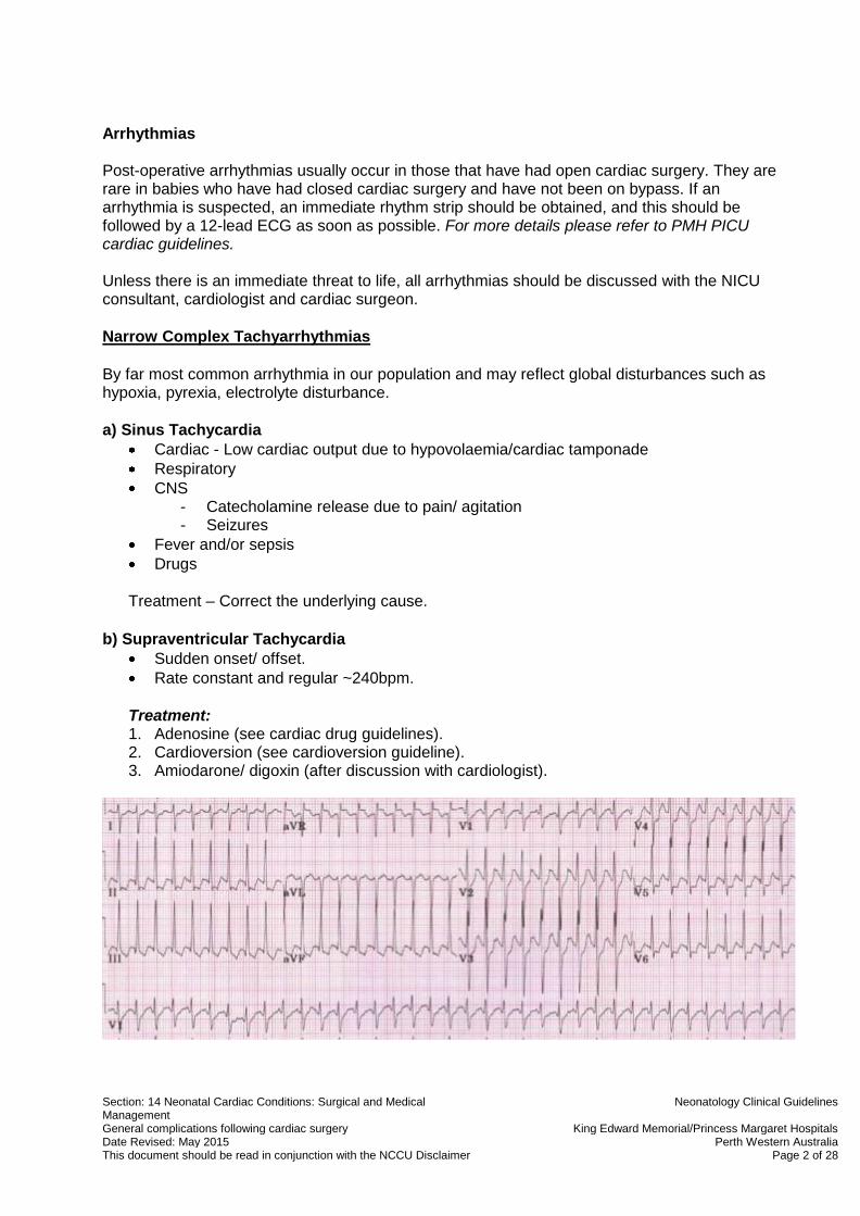
b) Supraventricular Tachycardia
Sudden onset/ offset.
Rate constant and regular ~240bpm.
Treatment: 1. Adenosine (see cardiac drug guidelines). 2. Cardioversion (see cardioversion guideline). 3. Amiodarone/ digoxin (after discussion with cardiologist).

Section: 14 Neonatal Cardiac Conditions: Surgical and Medical Management
Neonatology Clinical Guidelines
General complications following cardiac surgery King Edward Memorial/Princess Margaret Hospitals Date Revised: May 2015 Perth Western Australia This document should be read in conjunction with the NCCU Disclaimer Page 3 of 28
c) JET (Junctional Ectopic Tachycardia)
Very unusual in our patient group.
More common in those following open cardiac surgical repair of Tetralogy of Fallot, VSD, AVSD and TAPVD.
Narrow complex, rate usually regular at 180-250bpm. Beat to beat variability in blood pressure.
AV dissociation, ventricular rate > atrial rate.
Haemodynamic instability due to loss of AV synchrony.
Usually within 72hrs of operation, more likely with fever.
Treatment: 1. Options consist of cooling, avoidance of adrenergic (eg. catecholamines) or vagolytic
(eg. pancuronium) drugs, correction of any electrolyte imbalance, magnesium, anti-arrhythmic drugs, pacing and ECMO.
2. If suspected, consult PICU consultant/ cardiologist immediately.
d) Atrial Flutter/ Fibrillation
Variable AV block.
Saw tooth/ irregular baseline.
Adenosine may unmask the block.
Treatment:
1. Cardioversion (see cardioversion guideline). 2. Digoxin may be useful in slowing ventricular response. 3. Amiodarone occasionally terminates the atrial re-entry.
Section: 14 Neonatal Cardiac Conditions: Surgical and Medical Management
Neonatology Clinical Guidelines
General complications following cardiac surgery King Edward Memorial/Princess Margaret Hospitals Date Revised: May 2015 Perth Western Australia This document should be read in conjunction with the NCCU Disclaimer Page 4 of 28
Broad Complex Tachycardias
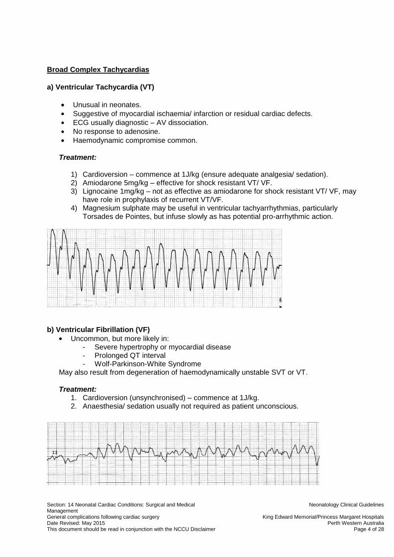
a) Ventricular Tachycardia (VT)
Unusual in neonates.
Suggestive of myocardial ischaemia/ infarction or residual cardiac defects.
ECG usually diagnostic – AV dissociation.
No response to adenosine.
Haemodynamic compromise common.
Treatment:
1) Cardioversion – commence at 1J/kg (ensure adequate analgesia/ sedation). 2) Amiodarone 5mg/kg – effective for shock resistant VT/ VF. 3) Lignocaine 1mg/kg – not as effective as amiodarone for shock resistant VT/ VF, may
have role in prophylaxis of recurrent VT/VF. 4) Magnesium sulphate may be useful in ventricular tachyarrhythmias, particularly
Torsades de Pointes, but infuse slowly as has potential pro-arrhythmic action.
b) Ventricular Fibrillation (VF)
Uncommon, but more likely in: - Severe hypertrophy or myocardial disease - Prolonged QT interval - Wolf-Parkinson-White Syndrome
May also result from degeneration of haemodynamically unstable SVT or VT.
Treatment: 1. Cardioversion (unsynchronised) – commence at 1J/kg. 2. Anaesthesia/ sedation usually not required as patient unconscious.
Section: 14 Neonatal Cardiac Conditions: Surgical and Medical Management
Neonatology Clinical Guidelines
General complications following cardiac surgery King Edward Memorial/Princess Margaret Hospitals Date Revised: May 2015 Perth Western Australia This document should be read in conjunction with the NCCU Disclaimer Page 5 of 28
Bradyarrhythmias
a) Sinus Bradycardia
HR <100
Slow rate disadvantageous in immediate post-op period.
Treatment: 1. Atropine may be useful for vagal induced bradycardia. 2. May be atrial paced if wires in situ (in PICU).
b) Others including AV block, slow junctional rhythm and sinus node dysfunction - Usually occur post open cardiac surgery and may require pacing if intracardiac wires
are in situ post-op in PICU. Can also use external pacing.

Section: 14 Neonatal Cardiac Conditions: Surgical and Medical Management
Neonatology Clinical Guidelines
General complications following cardiac surgery King Edward Memorial/Princess Margaret Hospitals Date Revised: May 2015 Perth Western Australia This document should be read in conjunction with the NCCU Disclaimer Page 6 of 28
BLEEDING
Surgical Bleeding
Early and relentless, can be catastrophic.
Coagulation studies may be near normal.
Definitive management is re-exploration.
Medical Bleeding
Mainly happens post cardiac bypass due to platelet dysfunction +/- thrombocytopaenia and/ or inadequate reversal of heparin effect.
Coagulopathy is common after massive blood loss +/- transfusion and more common in polycythaemic patients (those with cyanotic CHD).
Guidelines for Excessive Bleeding
The decision to re-explore is ultimately made by the surgeon, but a useful algorithm when deciding is as follows:
Loss in any 1 hour 10% of blood volume/hr = 8 mL/kg
Loss in any 2 hours 8% of blood volume/hr = 6 mL/kg
Loss in any 3 hours 6% of blood volume/hr = 5 mL/kg
Total blood volume in the neonate is ~85 mL/kg (95 mL/kg in premature neonate).
Management: (NICU patients will not have had cardiopulmonary bypass, so consideration of pump blood/ giving protamine is not relevant and not included.)
The NICU consultant and cardiac surgeon should be notified of any bleeding >3 mL/kg/hr.
Check FBC/ coagulation studies.
Maintain patency of drains. If drain loss stops abruptly beware of cardiac tamponade.
Replace blood loss with cross-matched packed red blood cells.
If bleeding persists and there is laboratory evidence of coagulopathy:
o Give FFP 10 mL/kg over 30 mins. o If fibrinogen <1.0 give cryoprecipitate 5 mL/kg over 30 mins. o Give vitamin K 1mg iv.
If bleeding persists and platelet count <80 give platelets 10 mL/kg over 30 mins.
Repeat FBC/ coags and repeat blood products as necessary.

Section: 14 Neonatal Cardiac Conditions: Surgical and Medical Management
Neonatology Clinical Guidelines
General complications following cardiac surgery King Edward Memorial/Princess Margaret Hospitals Date Revised: May 2015 Perth Western Australia This document should be read in conjunction with the NCCU Disclaimer Page 7 of 28
If massively transfused (>1x blood volume):
- Give FFP 10-20 mL/kg and consider platelet transfusion and cryoprecipitate in conjunction with FBC/ coagulation study results.
If bleeding persists after all surgical causes have been addressed consider use of Novoseven rfVIIa after discussion with haematologist.
Section: 14 Neonatal Cardiac Conditions: Surgical and Medical Management
Neonatology Clinical Guidelines
General complications following cardiac surgery King Edward Memorial/Princess Margaret Hospitals Date Revised: May 2015 Perth Western Australia This document should be read in conjunction with the NCCU Disclaimer Page 8 of 28
CAPILLARY LEAK SYNDROME
Capillary leak syndrome typically develops in neonates and infants who have undergone complex cardiac surgery under periods of prolonged cardiopulmonary bypass (CPB) or circulatory arrest. The syndrome is thought to result from systemic inflammatory responses resulting in damage to the capillary endothelium. The ‘leaky’ capillaries disrupt the normal balance of oncotic and hydrostatic forces as albumin and other large molecules are no longer reliably retained within the capillary. Proteins and fluid leak into the interstitium. A similar syndrome is seen in severe sepsis, again due to systemic inflammatory responses.
Features:
Unstable circulation with falling BP.
Low filling pressures.
Increased fluid requirement to maintain filling pressures.
Absence of bleeding to explain above.
Increasing systemic oedema/ pleural effusions/ ascites. It usually presents within 24 hrs of CPB.
Management:
There is no specific treatment. Management is aimed at supporting compromised systems.
Keep the filling pressures as low as is compatible with a good cardiac output.
Higher ventilatory pressures, particularly PEEP are required as interstitial oedema and pleural effusions develop to maintain good oxygenation.
Drainage of pleural effusion/s may be required to improve ventilation.
Optimise haemodynamics. Maintaining a high-normal haematocrit may help.
Consider peritoneal drainage of ascites +/- peritoneal dialysis if renal replacement therapy required.
As the fluid lost is protein rich and includes clotting factors and immunoglobulins, fluid replacement may include albumin and FFP if bleeding is apparent. If losses are over days, monitoring of Ig levels may be required.
Section: 14 Neonatal Cardiac Conditions: Surgical and Medical Management
Neonatology Clinical Guidelines
General complications following cardiac surgery King Edward Memorial/Princess Margaret Hospitals Date Revised: May 2015 Perth Western Australia This document should be read in conjunction with the NCCU Disclaimer Page 9 of 28
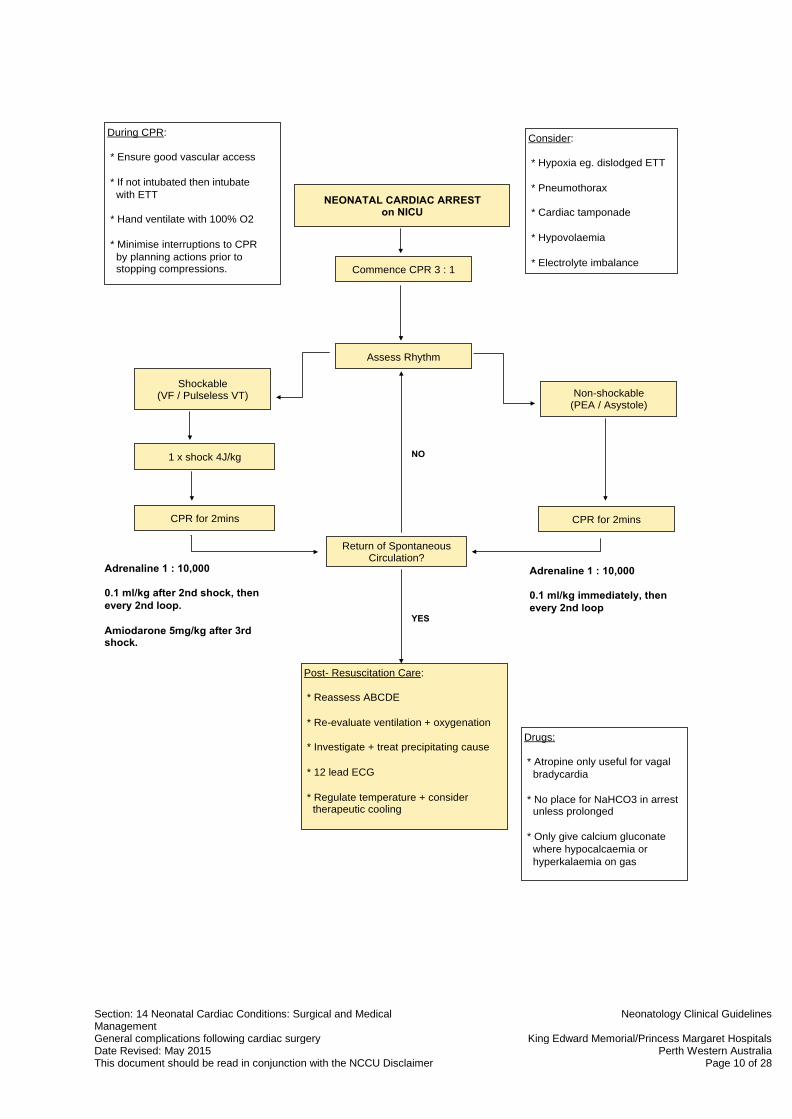
CARDIAC ARREST
Causes:
Most commonly due to hypoxia.
Cardiac tamponade a rare but important cause.
Electrolyte disturbance such as hyperkalaemia common cause.
May result from pulmonary hypertensive crisis.
Tension pneumothorax.
Ventricular arrhythmia rare compared with adults.
Management: (Based upon Australian Resuscitation Council Guidelines 2010, UK Resuscitation Council Guidelines 2010, American Heart Association Guidelines 2010)
Section: 14 Neonatal Cardiac Conditions: Surgical and Medical Management
Neonatology Clinical Guidelines
General complications following cardiac surgery King Edward Memorial/Princess Margaret Hospitals Date Revised: May 2015 Perth Western Australia This document should be read in conjunction with the NCCU Disclaimer Page 10 of 28
NEONATAL CARDIAC ARREST on NICU
Commence CPR 3 : 1
Assess Rhythm
During CPR:
* Ensure good vascular access
* If not intubated then intubate
with ETT
* Hand ventilate with 100% O2
* Minimise interruptions to CPR
by planning actions prior to stopping compressions.
Consider:
* Hypoxia eg. dislodged ETT
* Pneumothorax
* Cardiac tamponade
* Hypovolaemia
* Electrolyte imbalance
Shockable (VF / Pulseless VT)
1 x shock 4J/kg
CPR for 2mins
Non-shockable (PEA / Asystole)
CPR for 2mins
Return of Spontaneous Circulation?
Post- Resuscitation Care:
* Reassess ABCDE
* Re-evaluate ventilation + oxygenation
* Investigate + treat precipitating cause
* 12 lead ECG
* Regulate temperature + consider therapeutic cooling
Drugs:
* Atropine only useful for vagal
bradycardia
* No place for NaHCO3 in arrest unless prolonged
* Only give calcium gluconate
where hypocalcaemia or
hyperkalaemia on gas
Adrenaline 1 : 10,000
0.1 ml/kg after 2nd shock, then
every 2nd loop.
Amiodarone 5mg/kg after 3rd shock.
YES
Adrenaline 1 : 10,000
0.1 ml/kg immediately, then
every 2nd loop
NO
Section: 14 Neonatal Cardiac Conditions: Surgical and Medical Management
Neonatology Clinical Guidelines
General complications following cardiac surgery King Edward Memorial/Princess Margaret Hospitals Date Revised: May 2015 Perth Western Australia This document should be read in conjunction with the NCCU Disclaimer Page 11 of 28
CARDIAC TAMPONADE
Rare in closed cardiac cases. Caused by a collection of fluid (usually blood) around the heart, usually within the pericardial sac. It causes compression of the heart chambers and haemodynamic embarrassment. Cardiac tamponade may follow removal of transthoracic lines and be heralded by a sudden change in chest drainage, either an increase or sudden cessation.
Clinical signs:
Symptoms may occur abruptly or insidiously over a few hours. Features of low cardiac output:
Hypotension
Low pulse pressure.
Tachycardia
Poor peripheral perfusion and increased core-peripheral temperature gap.
Oliguria
Metabolic acidosis.
Examination may reveal muffled heart sounds and large liver.
CXR may show cardiomegaly (insensitive sign).
Management:
If suspected, the cardiac surgeon and PICU consultant should be contacted immediately.
The cardiologist should also be contacted as an urgent echocardiogram (time permitting) with possible needle aspiration may be required.
Blood should be X-matched urgently and the thoracotomy/ sternotomy set-up should be collected from PICU but not opened.
Section: 14 Neonatal Cardiac Conditions: Surgical and Medical Management
Neonatology Clinical Guidelines
General complications following cardiac surgery King Edward Memorial/Princess Margaret Hospitals Date Revised: May 2015 Perth Western Australia This document should be read in conjunction with the NCCU Disclaimer Page 12 of 28
CHYLOTHORAX
Diagnosis:
It presents as a milky pleural effusion.
A triglyceride level on the fluid is >1.1 mmol/L (if fed) and white cell count >1000/ L (>80% lymphocytes).
Lipoprotein electrophoresis looking for chylomicrons may be diagnostic.
Treatment:
1. Drainage with an intercostal catheter improves lung function and is useful to monitor drainage amounts.
2. Conservative treatment is with dietary manipulation by excluding long-chain fatty acids in the diet and using medium-chain fatty acids in the form of ‘Monagen’ feeds for a minimum of 2 weeks. These get absorbed directly into the portal system and bypass the thoracic duct.
3. If necessary, flow through the thoracic duct can be further reduced by the use of TPN for a 1-2 weeks followed by MCFA feeds.
4. Sometimes surgical ligation of the duct is required when conservative measures fail (continued drainage of >10mL/kg/d despite above treatment).
NB Persistent prolonged loss of chyle may cause hypoproteinaemia, lymphopaenia and hypogammaglobulinaemia.
Section: 14 Neonatal Cardiac Conditions: Surgical and Medical Management
Neonatology Clinical Guidelines
General complications following cardiac surgery King Edward Memorial/Princess Margaret Hospitals Date Revised: May 2015 Perth Western Australia This document should be read in conjunction with the NCCU Disclaimer Page 13 of 28
DIAPHRAGMATIC PALSY
Diaphragmatic palsy is relatively common and is due to phrenic nerve damage. A paralysed diaphragm may prevent weaning from mechanical ventilation.
Diagnosis:
May initially be suspected on CXR where a raised hemidiagrapm is seen. Usually not appreciated until off positive pressure ventilation.
Diagnosis is made by video fluoroscopy or ultrasound.
Treatment: 1. If the phrenic nerve has only been damaged and not severed then it may recover function
over a period of 7-10 days. Supportive measures should be taken to allow time for healing.
2. If function does not recover and the diaphragmatic palsy is preventing coming off ventilatory support, then surgical plication of the diaphragm should be considered. The timing of surgery should be decided by consensus decision by the cardiac surgeon and neonatologists.

Section: 14 Neonatal Cardiac Conditions: Surgical and Medical Management
Neonatology Clinical Guidelines
General complications following cardiac surgery King Edward Memorial/Princess Margaret Hospitals Date Revised: May 2015 Perth Western Australia This document should be read in conjunction with the NCCU Disclaimer Page 14 of 28
HYPERKALAEMIA
Neonates are generally tolerant of relatively high levels of potassium, however, an arterial K >6.0 mmol/L in a neonate post-cardiac surgery (>7.0 in non-cardiac patients) should be treated as a medical emergency. It can cause ECG disturbance in the way of peaked T waves, then broadened QRS complexes and this may lead to VT and cardiac arrest. A rising K may also be a marker of declining cardiac output.
Causes:
Exogenous K administration eg, K infusion, blood products.
Renal failure.
Acidosis – for every 0.1 fall in pH, K increases by 0.2-0.4 mmol/L.
Excessive cell breakdown.
K sparing diuretics eg. spironolactone.
Immediate Actions:
- STOP all exogenous K administration including feeds. - Urgently repeat K on ABG. - Seek cause. - If confirmed on repeat sample/ ECG evidence of peaked T waves or broadened QRS
then consider urgent treatment. - Notify consultant.
Treatment:
1. Calcium gluconate 10% 0.5 mL/kg (0.11 mmol/kg) iv slowly to antagonise effects of K.
2. Glucose 50% 1-2 mL/kg then infusion of 1-2 mL/kg/hr (the concomitant administration of insulin can be considered in resistant hyperkalaemia but very careful monitoring of PGL is required).
3. Bicarbonate 8.4% to fully correct acidosis = wt (kg) x BE x 0.3.
4. Frusemide 1mg/kg iv.
5. Salbutamol 4 mcg/kg slow iv. Can be repeated once after 2 hours. (nebulised salbutamol may also be effective – give 400mcg/kg/dose 2 hourly).
6. Calcium resonium 250 mg/kg/dose PR/ NG 6hrly up to a maximum dose 1g/kg/dose 6 hrly.
7. Consider urgent dialysis.
Repeat an arterial blood gas potassium level every hour until <5.5 mmol/L.

Section: 14 Neonatal Cardiac Conditions: Surgical and Medical Management
Neonatology Clinical Guidelines
General complications following cardiac surgery King Edward Memorial/Princess Margaret Hospitals Date Revised: May 2015 Perth Western Australia This document should be read in conjunction with the NCCU Disclaimer Page 15 of 28
HYPERTENSION
Normal blood pressure values in a neonate depend upon gestation, weight and age of the baby. Blood pressure values increase with gestation and weight. Following birth an individual’s blood pressure increases gradually over 3-4 days until reaching a plateau. (See graphs of normal blood pressure values in appendices). As a guide, the normal range of values for a term neonate >3days age are:
- Normal systolic BP is 55-80 mmHg
- Normal diastolic BP is 30-50 mmHg
- Normal mean BP is 40-60 mmHg Sometimes, hypertension occurs following surgery, which may require treatment, for instance, following post coarctation of the aorta repair where repair may require protecting from the harmful effects of high blood pressure (see guideline on ‘management of the neonate following surgical repair of CoA’).
Diagnosis:
BP measurement should occur via an indwelling arterial line preferably in the right arm. Cuff sphygmomanometry gives a useful indication especially in the other limbs post CoA repair as to adequacy of the repair.
As a general rule, antihypertensive therapy may be indicated where the BP is 20% above pre-op level or >90
th centile for age.
BP limits should be agreed by NICU consultant and surgeon for each individual case and are also dependent upon pre-op LV status and associated lesions.
Causes:
Pain is a common cause. Hence, pay attention to pain scores.
Fluid overload.
Pre-operative elevated levels of circulating catecholamines and renin.
Increased endogenous noradrenaline release during aortic cross-clamping and increased renin release after revascularisation.
Complications:
Acute left heart failure due to high ventricular afterload.
Acute haemorrhagic complications – Surgical anastomosis/ CNS.
Post-coarctectomy syndrome (see guideline on ‘management of the neonate following surgical repair of CoA’).
Section: 14 Neonatal Cardiac Conditions: Surgical and Medical Management
Neonatology Clinical Guidelines
General complications following cardiac surgery King Edward Memorial/Princess Margaret Hospitals Date Revised: May 2015 Perth Western Australia This document should be read in conjunction with the NCCU Disclaimer Page 16 of 28
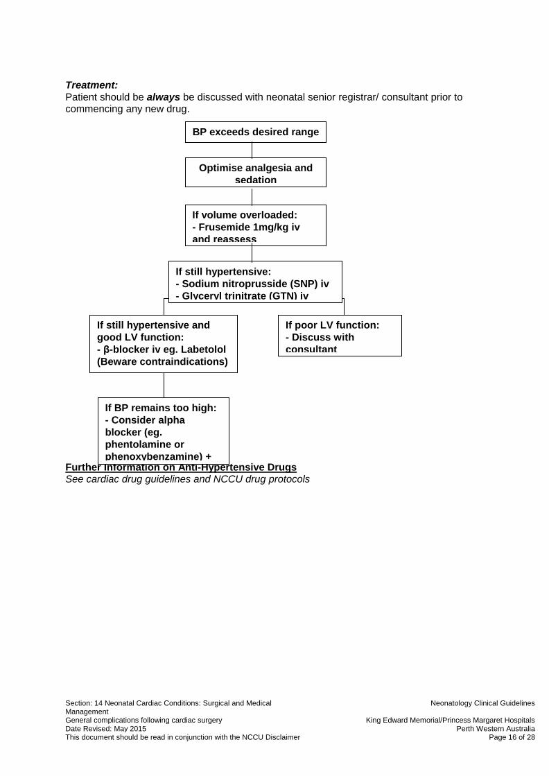
Treatment:
Patient should be always be discussed with neonatal senior registrar/ consultant prior to commencing any new drug.
Further Information on Anti-Hypertensive Drugs See cardiac drug guidelines and NCCU drug protocols
If poor LV function:
- Discuss with
consultant
If still hypertensive and
good LV function:
- β-blocker iv eg. Labetolol
(Beware contraindications)
If BP remains too high:
- Consider alpha
blocker (eg.
phentolamine or
phenoxybenzamine) +
beta blocker
BP exceeds desired range
Optimise analgesia and
sedation
If volume overloaded:
- Frusemide 1mg/kg iv
and reassess
If still hypertensive:
- Sodium nitroprusside (SNP) iv
- Glyceryl trinitrate (GTN) iv

Section: 14 Neonatal Cardiac Conditions: Surgical and Medical Management
Neonatology Clinical Guidelines
General complications following cardiac surgery King Edward Memorial/Princess Margaret Hospitals Date Revised: May 2015 Perth Western Australia This document should be read in conjunction with the NCCU Disclaimer Page 17 of 28
HYPOCALCAEMIA
Maintenance of normal ionised serum Ca levels 1.0-1.3 mmol/L improves cardiac contractility.
A very high ionised Ca level does not add benefit and may increase systemic vascular resistance.
Ionised Ca level may be low due to blood product administration (citrate) and bypass.
Beware of hypocalcaemia in those with known 22q11 del or those with arch abnormalities who may have it.
NB Ca is lowered by excess heparin in the sample.
Treatment:
1. Indicated when ionised Ca <1.0 mmol/L 2. Calcium gluconate 10% (0.22mmol/mL Ca) by infusion at 0.1-0.4 mL/kg/hr preferably
through a central line. 3. Rapid administration may cause bradycardia.
Remember to check and correct magnesium level also. Recheck level after 4 hrs of infusion.

Section: 14 Neonatal Cardiac Conditions: Surgical and Medical Management
Neonatology Clinical Guidelines
General complications following cardiac surgery King Edward Memorial/Princess Margaret Hospitals Date Revised: May 2015 Perth Western Australia This document should be read in conjunction with the NCCU Disclaimer Page 18 of 28
HYPOKALAEMIA
- Very common post-op especially post-bypass and following diuretic administration.
- Arrhythmias may occur especially in the presence of digoxin administration. Digoxin should
not be administered if K <3.0 mmol/L.
To correct a low K see cardiac drug protocols. Consideration to the oral route should be given if on feeds. Serum K should then be monitored every 1-2 hrs by arterial blood gas.

Section: 14 Neonatal Cardiac Conditions: Surgical and Medical Management
Neonatology Clinical Guidelines
General complications following cardiac surgery King Edward Memorial/Princess Margaret Hospitals Date Revised: May 2015 Perth Western Australia This document should be read in conjunction with the NCCU Disclaimer Page 19 of 28
METABOLIC AND LACTIC ACIDOSIS
Metabolic Acidosis:
Aetiology:
Low cardiac output state
‘Wash-out’ acidosis as vascular beds re-open eg. post CPB or post- CoA
Renal dysfunction (associated with low cardiac output/ acute tubular necrosis (ATN))
Sepsis (rare in early post-op period)
Management:
Seek and treat cause of acidosis
If BE -5 (or more negative) for 2 consecutive hours, notify consultant
Bicarbonate replacement may be indicated if the acidosis is thought to be impairing cardiac output. Use ½ correction initially and review.
Lactic Acidosis:
Blood lactate is a marker of adequacy of tissue perfusion and an early predictor of poor
outcome in critically ill infants.
Lactate is a dead-end product formed from pyruvate during glycolysis (anaerobic metabolism). It is produced when there is inadequate cellular oxygen delivery to meet cellular energy demands by the more efficient oxygen dependent metabolic pathways.
ie.Lactate production implies energy production by oxygen dependent mechanisms are saturated and that energy can only be produced by anaerobic or non-oxygen dependent metabolism. This is the escape hatch for the body to continue producing energy in the absence of oxygen. The price paid is lactic acidosis. The lactate level is therefore a measure of the degree of anaerobic glycolysis.
Glucose
Pyruvate Lactate + H+
Kreb’s Cycle (Oxygen dependent energy) pathway)

Section: 14 Neonatal Cardiac Conditions: Surgical and Medical Management
Neonatology Clinical Guidelines
General complications following cardiac surgery King Edward Memorial/Princess Margaret Hospitals Date Revised: May 2015 Perth Western Australia This document should be read in conjunction with the NCCU Disclaimer Page 20 of 28
Lactate production and excretion:
Produced in all cells, but major source is those dependent upon glycolysis – skeletal muscle, skin, erythrocytes and leucocytes.
Predominantly excreted by liver (therefore also an early marker of liver failure), but also kidneys, heart and skeletal muscle.
Normal lactate level is less than 2 mmol/L.
Any level >4.0 mmol/L (or rising level) must be reported to consultant immediately.
Causes of a Raised Lactate Level:
Circulatory failure
Severe hypoxaemia
Sepsis
High dose adrenaline infusion (decreases pyruvate dehydrogenase activity)
Severe anaemia (rare)
Prolonged seizures
Malnutrition (thiamine and biotin deficiency)
Inborn errors of the metabolism
Liver dysfunction
Lactate Measurement in Critically Ill: - Easy to measure, venous or arterial (little difference).
- High level associated with increased mortality and said to correlate with development of multi-organ systemic failure.
- Serial levels most useful – falling levels with improved survival and rising levels with poor outcome.
- In CHD, Duke et al have shown that, in infants, lactate levels >5 mmol/L immediately post-op and >4mmol/L 4 and 8 hours after surgery were reliable predictors of major adverse events (cardiac arrest).

Section: 14 Neonatal Cardiac Conditions: Surgical and Medical Management
Neonatology Clinical Guidelines
General complications following cardiac surgery King Edward Memorial/Princess Margaret Hospitals Date Revised: May 2015 Perth Western Australia This document should be read in conjunction with the NCCU Disclaimer Page 21 of 28
NEUROLOGICAL PROBLEMS
The main symptom (if any) of brain injury is seizures (see NCCU guidelines for seizure management). Many injuries are ‘silent’ and will only be realised months or years later.
Background Risk Factors:
There are already background risk factors for developmental/ neurological problems in those with congenital heart disease prior to birth:
Patient has a genetic syndrome/ chromosomal abnormality which has associations with developmental delay/ learning difficulties.
Fetal circulation in some cardiac conditions differs from the normal fetal circulation:
- Normally the most highly saturated blood is directed from the ductus venosus and left hepatic vein through the foramen ovale to the left ventricle, and subsequently the cerebral circulation. In contrast, in d-TGA, the aorta arises from the right ventricle, and so receives relatively desaturated blood from the SVC, lower body and coronary sinus and the left ventricle delivers the more highly saturated blood to the lungs, lower body and placenta.
- In hypoplastic left heart syndrome, the fetal circulation is characterised by a mixture of all venous streams in the right atrium and ventricle. The ascending aorta is only small, delivering blood in a retrograde direction to the coronary arteries. The aortic arch is hypoplastic and shows flow reversal to supply blood to the brain and upper body. The hypoplastic aorta may also function as a resistor, decreasing the pulsatility and perfusion pressure to the cerebral circulation. It has been shown that the ascending aorta diameter predicts the degree of microcephaly in newborns with HLHS.
Acquired Brain Injury: Neonates with congenital heart disease are at risk of discrete acquired brain injury in the perioperative period which may be exacerbated by delayed brain development.

Section: 14 Neonatal Cardiac Conditions: Surgical and Medical Management
Neonatology Clinical Guidelines
General complications following cardiac surgery King Edward Memorial/Princess Margaret Hospitals Date Revised: May 2015 Perth Western Australia This document should be read in conjunction with the NCCU Disclaimer Page 22 of 28
Risk factors for Acquired Brain Injury:
Preoperative Intraoperative Postoperative
o Low arterial Hb saturation o Length of time to surgery o Catheter-based procedure
eg. balloon atrial septostomy o Preoperative base deficit o Preoperative cardiac arrest o Morphologically immature
brain
o Prolonged total circulatory arrest (>40 min)
o Decreased cerebral oxygenation o CPB strategy (regional cerebral
perfusion) o Air or particulate emboli o Inflammation
o Low blood pressure o Low arterial PaO2 o Morphologically immature
brain o Single ventricle physiology
Preoperative Brain Injury Pre-operative brain injury in the form of white matter injury or stroke as seen on MRI is present in 28-39% of babies prior to neonatal cardiac surgery. For risk factors see table above. Intraoperative Brain Injury Most patients who acquire brain injury intra-operatively do so on cardiopulmonary bypass (CPB). Risk factors relate predominantly to the method of CPB used – circulatory arrest vs. low flow bypass and the length of time. Circulatory arrest is felt to be associated with an increased risk of brain injury especially when prolonged, however, low flow bypass prolongs the exposure to pump-related sources of injury including embolism and inflammation. Postoperative Brain Injury Low cardiac output syndrome (tachycardia, oliguria, cold extremities or cardiac arrest) related to hypotension, hypoxaemia and lactic acidosis (>4mmol/L) is the biggest risk factor for postoperative brain injury.
Section: 14 Neonatal Cardiac Conditions: Surgical and Medical Management
Neonatology Clinical Guidelines
General complications following cardiac surgery King Edward Memorial/Princess Margaret Hospitals Date Revised: May 2015 Perth Western Australia This document should be read in conjunction with the NCCU Disclaimer Page 23 of 28
PULMONARY HYPERTENSION
Labile pulmonary hypertension, although uncommon, is a potentially fatal complication seen in babies and children following cardiac surgery.
Causes:
Post-operative pulmonary hypertension is seen in certain conditions where there has been pre-operative conditioning of the pulmonary vasculature with either large pulmonary re-circulationary (L→R) shunts or pulmonary venous obstruction:
- Large VSD - AVSD - Truncus arteriosus - Anomalous pulmonary venous drainage (TAPVD)
Pulmonary hypertensive crises may be triggered by a number of mechanisms such as hypoxia, hypercarbia, acidosis and handling (suctioning, cares and noxious stimuli such as cold, noise,pain), but may occur without stimulus or warning. Intercurrent sepsis may dramatically aggravate PHT.
Symptoms: - Arterial desaturation. - Systemic hypotension. - Decreased lung compliance and wheeze/ air trapping.
Pulmonary hypertension induces right heart failure and, through ventricular interdependence, LV failure and circulatory collapse.
Treatment:
Standard Preventative Measures in those at Risk of PHT:
Adequate sedation - Adequate to suppress haemodynamic response to handling and to permit standard
post-op care.
Ventilation - Normoventilation or mild hyperventilation - Aim for pH 7.35-7.45/ PaCO2 35-45mmHg - Maintain normoxaemia, aiming for SaO2 94-97% (if non-cyanotic heart lesion).
Hyperoxygenation is unnecessary and may actually be harmful due to the release of free radicals.
- Consider muscle relaxant (paralysis).
Cardiovascular support - Inotropes as required, consider dobutamine.
Suctioning - Pre-oxygenate on ventilator to achieve SaO2 >95%.

Section: 14 Neonatal Cardiac Conditions: Surgical and Medical Management
Neonatology Clinical Guidelines
General complications following cardiac surgery King Edward Memorial/Princess Margaret Hospitals Date Revised: May 2015 Perth Western Australia This document should be read in conjunction with the NCCU Disclaimer Page 24 of 28
- Give bolus of morphine (1mL of standard infusion =10microg/kg) and midazolam
(0.5mL of standard infusion=30microg/kg) and allow time to work. Repeat as necessary.
Acute Unstable Pulmonary Hypertension:
Those with hypotension, arterial desaturation and decreased lung compliance can be considered to have unstable PHT.
1. Increase FiO2. 2. Increase sedation and consider muscle relaxant. 3. Commence inhaled NO 10-20ppm. 4. Maintain systemic arterial pressure in desired range with inotropic support if necessary.
Consider dobutamine. 5. Consider vasodilator:
- Prostaglandin 5-25 nanogram/kg/min - Milrinone - Magnesium sulphate (see cardiac drug guidelines – ‘drugs to correct electrolyte
imbalance’ for dosing). - Sildenafil orally 0.25-2 mg/kg 4-6hrly. - Intravenous sildenafil is available, but use cautiously as can cause systemic
hypotension. One study has found it to be well tolerated and effective in neonates when used for PPHN. (Pediatr. 2009 Dec;155(6):841-847.e1. Intravenous sildenafil in the treatment of neonates with persistent pulmonary hypertension. Steinhorn RH, Kinsella JP, Pierce C, Butrous G, Dilleen M, Oakes M, Wessel DL).
- Discuss with PICU regarding ECMO.
Section: 14 Neonatal Cardiac Conditions: Surgical and Medical Management
Neonatology Clinical Guidelines
General complications following cardiac surgery King Edward Memorial/Princess Margaret Hospitals Date Revised: May 2015 Perth Western Australia This document should be read in conjunction with the NCCU Disclaimer Page 25 of 28
RENAL DYSFUNCTION AND RENAL REPLACEMENT THERAPY
Renal dysfunction is relatively common post cardiac surgery due to:
Low cardiac output/ hypotension.
Hypoxia.
Pre-op condition of the patient – particularly in coarctation of the aorta.
Nephrotoxic drugs eg. aminoglygosides.
Inflammatory response post CPB.
Oliguria (urine output <0.5mL/kg/hr) – is when the urine output has dropped below the minimum level for adequate solute and fluid removal and is the level that will almost certainly result in fluid overload (intake>output).
A minimum urine output of 0.5-1mL/kg/hr should be the aim.
Management:
Consider:
Catheter blockage/ leakage or if no catheter, urinary retention (then go on and catheterise).
Urine biochemistry - Pre-renal: High specific gravity 1.020/ low sodium levels <20mmol/L - Intrinsic: Fixed specific gravity1.010-1.020/ High sodium levels >40mmol/L
Hypovolaemia – give small aliquot of volume and reassess.
Low cardiac output/ hypotension – commence/ increase support (usually with inotrope).
If oliguria persists despite above measures give: - Frusemide 1mg/kg - If no response, repeat above frusemide dose
If no response to above, further trials of diuretics are futile and may delay definitive treatment ie. urgent renal support.
If urine flow established following diuretic therapy, then regular boluses of frusemide may be required or a frusemide infusion 0.1-0.5 mg/kg/hr commenced (avoids intermittent intravascular depletion, but prolonged t½ in neonates).
Monitor gas electrolytes 4hrly and formal U&E daily.

Section: 14 Neonatal Cardiac Conditions: Surgical and Medical Management
Neonatology Clinical Guidelines
General complications following cardiac surgery King Edward Memorial/Princess Margaret Hospitals Date Revised: May 2015 Perth Western Australia This document should be read in conjunction with the NCCU Disclaimer Page 26 of 28
Renal Replacement Therapy
Indications:
Anuria resistant to diuretics.
Fluid overload with oliguria + low cardiac output + instability.
Hyperkalaemia >6.5 mmol/L with metabolic acidosis pH <7.2 resistant to treatment.
Peritoneal Dialysis (PD): - Best method of renal replacement therapy in neonates. - Rapid and simple to commence especially in high risk patients who have already had a
catheter placed in theatre in anticipation. If no catheter in situ call surgeons urgently. - May not be effective at high vasoconstrictor doses. - Catheter dysfunction may limit use.
Initial Prescription: - Depends upon patient stability and degree of overload. - 1.5-2.5% PD solution to commence, no added KCl for first cycle, heparin 500 units/L. - Commence with 10-20mL/kg/cycle, with 30min dwell time (about 60 mins total cycle time –
15min/30min/15min for in/ dwell/ out respectively). - For patients receiving gentamicin, it may be added to the PD fluid in a concentration of 8-
10 mg/L and cease parenteral gentamicin. - Lactate free solutions may be of benefit with circulatory instability.
Subsequent Prescription: - 1.5-4.25% PD solution depending upon net fluid loss achieved and desired. - Add KCl (4 mmol/L) to dialysate unless contraindicated. - Aim for 20 mL/kg/cycle if tolerated. If ineffective consider increasing to 30 mL/kg/cycle but
beware effects on haemodynamics and chest compliance.
Catheter Non-function: - Check catheter tip position on AXR – may not be lying in pelvis. - Try repositioning patient. - Flush the catheter (complete sterile technique). - Persistent non-function will require replacement of catheter (consult cardiac surgeon
before any operatively placed catheter is removed).
Monitoring: - Gas electrolytes/ glucose 4 hrly - U&E/ Ca/ Mg /PO4/ LFT/ Albumin daily - PD fluid M/C/S daily
Haemofiltration is rarely required or used in neonates.
Section: 14 Neonatal Cardiac Conditions: Surgical and Medical Management
Neonatology Clinical Guidelines
General complications following cardiac surgery King Edward Memorial/Princess Margaret Hospitals Date Revised: May 2015 Perth Western Australia This document should be read in conjunction with the NCCU Disclaimer Page 27 of 28
SEPSIS
Sepsis is unusual in the first 24hrs post-op, but fever is not.
Causes of fever in first 24hrs post-op:
Stress response to surgery, particularly following cardiopulmonary bypass (CPB).
Low cardiac output leads to central hyperthermia and peripheral coolness (↑ toe-core gap).
Sepsis uncommon.
Malignant hyperthermia (rare).
If fever at >24hrs, sepsis much more likely especially when associated with:
Low cardiac output.
Peripheral vasodilatation.
Requiring more cardiovascular support than expected.
Causes of post-op sepsis:
Lower respiratory tract infection.
Line infection (CVC/ peripheral – most likely CONS).
Surgical site infection eg. wound/ graft.
UTI (especially when catheter in situ).
Intercurrent viral infection.
Management of suspected sepsis: - Thorough clinical examination paying particular attention to chest/ line sites/ drain sites/
wound. - CXR - FBC/ CRP/ procalcitonin level (as trial to see if more useful than CRP which is invariably
raised post-operatively). - Blood culture of all CVC lumens and peripheral stab. - NPA or ETT secretions for viruses. - ETT aspirate for M/C/S. - Urine for M/C/S. - If wound/ drain site looks unhealthy – take swabs and send for M/C/S. - If line infection suspected, consider removing line and sending tip for M/C/S. - If unknown origin of sepsis commence:
- Vancomycin and - Gentamicin
- If extremely unwell discuss with clinical microbiologist/ ID consultant on call (via switchboard) as empiric antibiotic regimen may need to be modified to broader spectrum agents.

Section: 14 Neonatal Cardiac Conditions: Surgical and Medical Management
Neonatology Clinical Guidelines
General complications following cardiac surgery King Edward Memorial/Princess Margaret Hospitals Date Revised: May 2015 Perth Western Australia This document should be read in conjunction with the NCCU Disclaimer Page 28 of 28
STRIDOR
Stridor is a fairly common complication post cardiac surgery and ventilation. It is usually apparent at extubation/ some minutes after extubation.
Aetiology:
1) Post-intubation stridor secondary to oedema. 2) Vocal cord paralysis secondary to recurrent laryngeal nerve damage.
Treatment:
1. The markedly negative intrathoracic pressure during obstructive breathing imposes additional afterload on the LV, so re-intubation is often necessary.
2. CPAP may be considered as a less invasive method.
Vocal cord palsy: If the nerve was traumatised and not severed function may return in a few days when a further trial of extubation may happen.
If there are still problems after this, an ENT opinion should be sought. Post-intubation stridor:
Possible oedema may be treated with a short course of dexamethasone If trying to avoid re-intubation, consider putting on CPAP and whilst waiting for steroids to work, nebulised adrenaline. This can be repeated every 1-2 hrs. This approach MUST be discussed with consultant and should not be used for a baby with severe obstruction and poor air entry / desaturating / exhausted. If the baby fails to respond then re-intubation should be strongly reconsidered.