gene therapy rescues cone function in congenital achromatopsia · vince a. chiodo3, asli kaya5,...
TRANSCRIPT
Gene therapy rescues cone function in congenitalachromatopsia
Andras M. Komaromy1,∗, John J. Alexander2,3,4, Jessica S. Rowlan1, Monique M. Garcia1,5,
Vince A. Chiodo3, Asli Kaya5, Jacqueline C. Tanaka5, Gregory M. Acland6,
William W. Hauswirth2,3 and Gustavo D. Aguirre1
1Department of Clinical Studies, School of Veterinary Medicine, University of Pennsylvania, Philadelphia, PA 19104,
USA, 2Department of Molecular Genetics and Microbiology and 3Department of Ophthalmology and Powell Gene
Therapy Center, University of Florida, Gainesville, FL 32610, USA, 4Vision Science Research Center, University of
Alabama, Birmingham, AL 35294, USA, 5Department of Biology, Temple University, Philadelphia, PA 19122, USA and6Baker Institute, Cornell University, Ithaca, NY 14853, USA
Received January 19, 2010; Revised March 12, 2010; Accepted April 5, 2010
The successful restoration of visual function with recombinant adeno-associated virus (rAAV)-mediated genereplacement therapy in animals and humans with an inherited disease of the retinal pigment epithelium hasushered in a new era of retinal therapeutics. For many retinal disorders, however, targeting of therapeutic vec-tors to mutant rods and/or cones will be required. In this study, the primary cone photoreceptor disorder achro-matopsia served as the ideal translational model to develop gene therapy directed to cone photoreceptors. Wedemonstrate that rAAV-mediated gene replacement therapy with different forms of the human red cone opsinpromoter led to the restoration of cone function and day vision in two canine models of CNGB3 achromatopsia,a neuronal channelopathy that is the most common form of achromatopsia in man. The robustness and stabilityof the observed treatment effect was mutation independent, but promoter and age dependent. Subretinaladministration of rAAV5–hCNGB3 with a long version of the red cone opsin promoter in younger animalsled to a stable therapeutic effect for at least 33 months. Our results hold promise for future clinical trials ofcone-directed gene therapy in achromatopsia and other cone-specific disorders.
INTRODUCTION
Most vision-impairing disorders in humans result from geneticdefects, and, to date, mutations have been identified in �150genes out of �200 mapped retinal disease loci (RetNet: http://www.sph.uth.tmc.edu/RetNet/). This wealth of genetic infor-mation has provided fundamental understanding of the mul-tiple and specialized roles played by photoreceptors and theretinal pigment epithelium (RPE) in the visual process, andhow mutations in these genes result in disease. Togetherwith the development of gene transfer technologies, it isnow possible to realistically consider the use of gene therapyto treat these previously untreatable disorders. A new era ofretinal therapeutics has been started by recent studies,showing that treatment of animal models with gene therapysuccessfully restored visual function (1–3). These results
have been translated to the clinic and been shown to be safeand successful for a human retinal degeneration caused byRPE65 mutations (4–11).
However, this example of an RPE disease is rare andcomplex in that loss of visual function and eventual photo-receptor degeneration is secondary to a biochemical blockadeof the RPE visual cycle. While it has been an ideal test plat-form for the first clinical gene therapy trial for blindness inman, RPE65 disease is not representative of the diversity ofmore common retinal disorders that could be considered forgene therapy where the target cells are the rods and/orcones. For these, successful targeting of therapeutic vectorsto mutant photoreceptors to restore function and preservestructure will be required, and achieving this initially in exper-imental animals is necessary for eventual application tohumans.
∗To whom correspondence should be addressed at: Department of Clinical Studies, School of Veterinary Medicine, University of Pennsylvania, 3900Delancey Street, Philadelphia, PA 19104-6010, USA. Tel: +1 2155732695; Fax: +1 2155732162; Email: [email protected]
# The Author 2010. Published by Oxford University Press. All rights reserved.For Permissions, please email: [email protected]
Human Molecular Genetics, 2010, Vol. 19, No. 13 2581–2593doi:10.1093/hmg/ddq136Advance Access published on April 8, 2010
Congenital achromatopsia, also called rod monochromacyor total congenital color blindness, is a rare autosomal reces-sive disorder with an estimated prevalence of about 1 in30 000–50 000 (12–15). Because it primarily affects cones,it serves as the ideal translational model to develop gene thera-pies directed to photoreceptors. Even though cones arenumerically small, �5% of the photoreceptors in man (16),they are essential for color vision, central visual acuity andmost daily visual activities, functions that are severelyimpaired or absent in affected patients. Thus far, four geneshave been identified to cause achromatopsia in humanpatients, all of them encoding principal components of thecone phototransduction cascade: the alpha subunit of conetransducin [GNAT2 (17–19)], the catalytic alpha’ subunit ofthe cone phosphodiesterase (PDE6C) (20,21) and the alpha[CNGA3 (22–24)] and beta [CNGB3 (25–28)] subunits ofthe cone cyclic nucleotide-gated (CNG) channel located inthe plasma membrane of the cone outer segment. The majorityof human achromatopsia (�70–92%) are considered channe-lopathies as they are caused by mutations in either CNGA3 orCNGB3 (29–32). The CNGB3 gene appears to be the mostprevalent causal gene for achromatopsia in patients of North-ern European descent, with reported prevalence between 50and 87% (29,32). However, it is suspected that CNGA3mutations are more prevalent among patients from theMiddle East (33).
Based on the very high prevalence of the CNGB3 mutationsin autosomal recessive achromatopsia (29,32), our identifi-cation of two independent canine models with distinctCNGB3 mutations has provided us with a unique, physiologi-cally relevant system in which to assess potential gene therapystrategies designed to restore visual function (34). Thesemodels result from either a missense mutation in exon 6(CNGB3m/m) or a genomic deletion of the entire CNGB3gene (CNGB32/2, ‘null mutation’) (34). The canine diseasecaused by the null mutation has been characterized, andshares the same clinical phenotype as human patients (35–38). The classic achromatopsia phenotype develops between8 and 12 weeks of age, soon after retinal differentiation iscompleted (35–38); however, other than the salient functionaland structural abnormalities in affected cones, the retinaremains normal throughout life (36,38).
Here we report the successful restoration of cone functionand associated photopic vision in both canine achromatopsiamodels by gene replacement therapy. We used recombinantadeno-associated virus serotype 5 (rAAV5) with differentforms of the human red cone opsin promoter. The robustnessand stability of the observed treatment effect was mutationindependent, but promoter and age dependent. These resultshold promise for future clinical trials in human patients withCNGB3-achromatopsia.
RESULTS
Cones are present, but not functional, in canineCNGB3-achromatopsia
In contrast to human and nonhuman primates that have separ-ate populations of red, green and blue cones, dogs are func-tional dichromats, with cone populations having combined
red/green and blue pigments (39). These are termed long/medium-wavelength-absorbing cones (L/M-cones) andshort-wavelength-absorbing cones (S-cones), and havemaximal sensitivities of 555 and 429–435 nm, respectively(39,40). The number, distribution and ratios of L/M- andS-cones in the normal canine retina have recently beenreported, and L/M-cones outnumber S-cones by a ratio of8:1 in the areas selected for therapy [see Materials andMethods (41)].
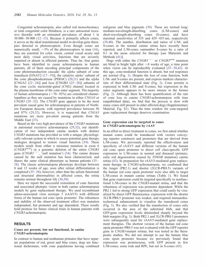
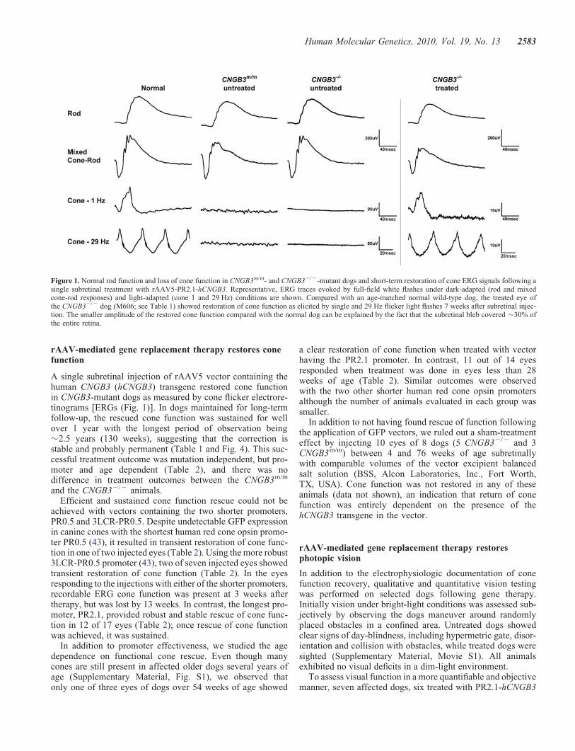
Dogs with either the CNGB32/2 or CNGB3m/m mutationare blind in bright light after �8 weeks of age, a time pointwhen vision can be reproducibly evaluated (35–38,42). Atthis age, cone-mediated function is absent, but rod responsesare normal (Fig. 1). Despite the loss of cone function, bothL/M- and S-cones are present, and express markers character-istic of their differentiated state (Fig. 2). Cone arrestin isexpressed in both L/M- and S-cones, but expression in theouter segments appears to be more intense in the former(Fig. 2). Although there has been quantification of the rateof cone loss in canine CNGB3-achromatopsia with time(unpublished data), we find that the process is slow withmany cones still present in older affected dogs (SupplementaryMaterial, Fig. S1). Thus the time window for cone-targetedgene replacement therapy deserves examination.
Gene expression can be targeted to conesin CNGB3-achromatopsia by rAAV
In an effort to direct treatment to cones, we first asked whethermutant cones could be transduced with vectors convey-ing reporter constructs and promoters that are derived fromL/M-cones. We previously reported the effectiveness andspecificity of rAAV5 and different versions of the humanred cone opsin promoter to direct cell class-specific GFPexpression to L/M-cones in normal and diseased (primary,early rod degeneration caused by PDE6B mutation) canineretina (43). In preparation for rAAV5-mediated gene replace-ment therapy in CNGB3-achromatopsia, we confirmed thatthe longer (PR2.1) and shorter (3LCR-PR0.5) variants ofthe human red cone opsin promoter were also able to targetL/M-cones in mutant canine retinas (Table 1). We foundthat gene expression could be targeted specifically to nonfunc-tional L/M-cones in the CNGB3-mutant retina, and that therobustness of expression was promoter dependent. While thePR2.1 led to strong GFP expression that could easily be visu-alized by direct GFP fluorescence, expression with the shorter3LCR-PR0.5 promoter was weaker, and required immunohis-tochemical enhancement to visualize the transduced cones(Fig. 3). We also verified that the transfection of cones onlyoccurred in the area of the subretinal bleb, and thatGFP-gene expression levels diminished sharply beyond thebleb margins (Fig. 3). Both PR2.1 and 3LCR-PR0.5 promoterswere subsequently used for rAAV5-mediated gene replace-ment therapies. The shortest version of the human red coneopsin promoter PR0.5 was not evaluated with the GFP reportergene in CNGB3-mutant retinas, but was tested in the thera-peutic studies. We did not attempt to use the human bluecone opsin promoter because we previously found thatexpression was promiscuous, with GFP present in fewL/M-cones, some rods and RPE, but not in S-cones (43).
2582 Human Molecular Genetics, 2010, Vol. 19, No. 13
rAAV-mediated gene replacement therapy restores conefunction
A single subretinal injection of rAAV5 vector containing thehuman CNGB3 (hCNGB3) transgene restored cone functionin CNGB3-mutant dogs as measured by cone flicker electrore-tinograms [ERGs (Fig. 1)]. In dogs maintained for long-termfollow-up, the rescued cone function was sustained for wellover 1 year with the longest period of observation being�2.5 years (130 weeks), suggesting that the correction isstable and probably permanent (Table 1 and Fig. 4). This suc-cessful treatment outcome was mutation independent, but pro-moter and age dependent (Table 2), and there was nodifference in treatment outcomes between the CNGB3m/m
and the CNGB32/2 animals.Efficient and sustained cone function rescue could not be
achieved with vectors containing the two shorter promoters,PR0.5 and 3LCR-PR0.5. Despite undetectable GFP expressionin canine cones with the shortest human red cone opsin promo-ter PR0.5 (43), it resulted in transient restoration of cone func-tion in one of two injected eyes (Table 2). Using the more robust3LCR-PR0.5 promoter (43), two of seven injected eyes showedtransient restoration of cone function (Table 2). In the eyesresponding to the injections with either of the shorter promoters,recordable ERG cone function was present at 3 weeks aftertherapy, but was lost by 13 weeks. In contrast, the longest pro-moter, PR2.1, provided robust and stable rescue of cone func-tion in 12 of 17 eyes (Table 2); once rescue of cone functionwas achieved, it was sustained.
In addition to promoter effectiveness, we studied the agedependence on functional cone rescue. Even though manycones are still present in affected older dogs several years ofage (Supplementary Material, Fig. S1), we observed thatonly one of three eyes of dogs over 54 weeks of age showed
a clear restoration of cone function when treated with vectorhaving the PR2.1 promoter. In contrast, 11 out of 14 eyesresponded when treatment was done in eyes less than 28weeks of age (Table 2). Similar outcomes were observedwith the two other shorter human red cone opsin promotersalthough the number of animals evaluated in each group wassmaller.
In addition to not having found rescue of function followingthe application of GFP vectors, we ruled out a sham-treatmenteffect by injecting 10 eyes of 8 dogs (5 CNGB32/2 and 3CNGB3m/m) between 4 and 76 weeks of age subretinallywith comparable volumes of the vector excipient balancedsalt solution (BSS, Alcon Laboratories, Inc., Fort Worth,TX, USA). Cone function was not restored in any of theseanimals (data not shown), an indication that return of conefunction was entirely dependent on the presence of thehCNGB3 transgene in the vector.
rAAV-mediated gene replacement therapy restoresphotopic vision
In addition to the electrophysiologic documentation of conefunction recovery, qualitative and quantitative vision testingwas performed on selected dogs following gene therapy.Initially vision under bright-light conditions was assessed sub-jectively by observing the dogs maneuver around randomlyplaced obstacles in a confined area. Untreated dogs showedclear signs of day-blindness, including hypermetric gate, disor-ientation and collision with obstacles, while treated dogs weresighted (Supplementary Material, Movie S1). All animalsexhibited no visual deficits in a dim-light environment.
To assess visual function in a more quantifiable and objectivemanner, seven affected dogs, six treated with PR2.1-hCNGB3
Figure 1. Normal rod function and loss of cone function in CNGB3m/m- and CNGB32/2-mutant dogs and short-term restoration of cone ERG signals following asingle subretinal treatment with rAAV5-PR2.1-hCNGB3. Representative, ERG traces evoked by full-field white flashes under dark-adapted (rod and mixedcone-rod responses) and light-adapted (cone 1 and 29 Hz) conditions are shown. Compared with an age-matched normal wild-type dog, the treated eye ofthe CNGB32/2 dog (M606; see Table 1) showed restoration of cone function as elicited by single and 29 Hz flicker light flashes 7 weeks after subretinal injec-tion. The smaller amplitude of the restored cone function compared with the normal dog can be explained by the fact that the subretinal bleb covered �30% ofthe entire retina.
Human Molecular Genetics, 2010, Vol. 19, No. 13 2583
and one with PR0.5-hCNGB3, had their visual function testedobjectively under scotopic and photopic conditions in anobstacle avoidance course. Objective identification of achroma-topsia was based on transit time, a quantitative response vari-able (42). There was a significant increase in mean transittimes of achromatopsia-affected dogs in ambient light intensi-ties of ≥25 lux, while the mean transit times of normalcontrol dogs were not affected by any ambient light conditions(Fig. 5). In contrast, following gene therapy in affected dogs,transit times under photopic conditions approached normalvalues, but were still slower. This remaining differencebetween normal and treated affected dogs is expected, giventhat normal dogs function binocularly in the obstacle course,whereas the treated dogs received the vector injection in onlyone eye. Additionally, only �30% of the retina was treated.Given these differences, the behavioral similarities betweennormal and treated dogs are remarkable. Performance in theobstacle course is illustrated in Supplementary Material,Movie S2.
Normalization of protein localization accompaniesrAAV-mediated rescue of cone function
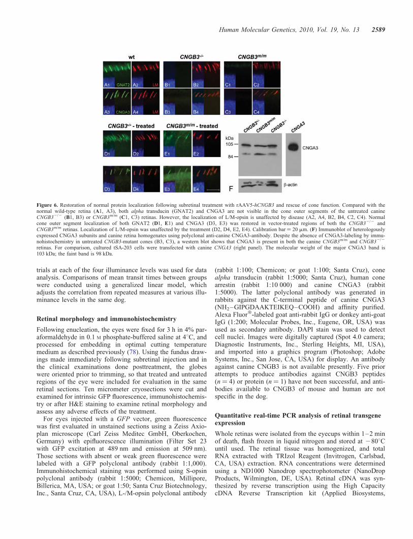
To more fully assess rescue of the cone phenotype, theexpression and localization of cone alpha transducinGNAT2 and CNGA3 proteins was evaluated by immunohisto-chemistry. In the normal retina, both GNAT2 and CNGA3were found in the cone outer segments. In contrast, the non-functional cones in the CNGB32/2 and CNGB3m/m retinasdid not have either GNAT2 or CNGA3 in the outer segments(Fig. 6), but western blot analysis demonstrated that the
CNGA3 protein was present in the retina (Fig. 6). This waspreviously reported for other transducin subunits in thecanine CNGB32/2 retina (44,45). At all time points evaluated,the examination of successfully treated retinal regions showedrestoration of protein localization to cone outer segments forboth GNAT2 and CNGA3 associated with rescued function(Fig. 6). This was limited to the vector-treated area and notvisible in untreated regions (data not shown). We interpretthese findings to indicate that successful restoration ofcone function was accompanied by relocation of CNGchannel components and transducin, both elements of thephototransduction cascade, to their proper cone outersegment compartment.
A minimum level of transgene expression is necessary, butnot sufficient, to restore the cone function
All the retinas analyzed at various times following subretinalrAAV5-hCNGB3 injection showed expression of thehCNGB3 transgene as measured by quantitative real-timePCR [qRT-PCR (Fig. 7)]. The robustness of transgeneexpression with the PR2.1 promoter confirmed our previousfindings using reporter GFP expression with this promoter(43). For comparison we also measured mRNA levels ofcanine CNGB3 (cCNGB3), S-opsin, L/M-opsin and rod opsinrelative to the 18S rRNA endogenous control. In treatedeyes that did not show recovery of cone function, thehCNGB3-transgene expression was lower, by �3.6–150-foldwhen compared with successfully treated eyes, but clearlystill detectable (Fig. 7). This means that hCNGB3 transgeneexpression must exceed a threshold level to be sufficient torestore the cone function. As expected, expression ofcCNGB3 mRNA was not present in null mutant dogs, butwas comparable with wild-type animals in the missensemutant dogs (Fig. 7). When we compared the detectableretinal hCNGB3 transgene expression levels with the coneflicker ERG amplitudes measured before tissue collection,we found a highly significant nonparametric Spearman rankcorrelation of 0.85 (P , 0.0001); that is, the cone ERGflicker amplitude increased with higher hCNGB3 transgeneexpression (Fig. 7).
DISCUSSION
We report that gene replacement therapy restores retinal conefunction in two canine models of CNGB3 achromatopsia, aneuronal channelopathy. Restored cone function was docu-mented by cone-specific ERG and recovery of day vision.As mutations in CNGB3 are the most common cause of achro-matopsia in humans, our results hold promise for future clini-cal trials. We also showed that the restoration of cone functionis mutation independent, but promoter and age dependent.Using vectors with the PR2.1 promoter, phenotypic correctionis stable and probably permanent.
Results with vectored GFP reporter gene in mutant retinasconfirmed that cones are present despite total lack of functionafter 8 weeks of age, and that gene expression can selectivelybe targeted to them. We previously showed the specificity androbustness of some human red cone opsin promoters, the
Figure 2. Presence of L/M- and S-cones in CNGB3-mutant canine retinas,expressing markers that characterize the differentiated state. Comparison ofcanine retinal structure and cone-specific protein expression in normal(A1—A4; 12 weeks), CNGB32/2 (B1—B4; 17 weeks), and CNGB3m/m
(C1—C4; 12 weeks) dogs. The H&E-stained sections show normal outerretinal structure independent of disease status (A1, B1, C1). The expressionand distribution of L/M- and S-opsin is not affected by the CNGB3-mutation.L/M-opsin labeling (green) co-localizes with cone arrestin expression (hCARlabeling in red) in the outer segments of the L/M-cones (A2, B2, C2). In con-trast, cone arrestin labeling is weak in S-cone outer segments; hence, theco-localized signal is dominated by the green S-opsin labeling (arrows inA3, B3, and C3). Closer analysis of cone arrestin distribution shows that thehCAR labeling is much weaker in the S-cone outer segments compared withthe L/M-cones of the canine WT retina (arrows in A4). hCAR labeling isessentially not visible in the S-cone outer segments of the CNGB3-mutantdogs (arrows in B4 and C4). Calibration bars ¼ 20 mm. RPE, retinalpigment epithelium; OS, outer segment; IS, inner segment.
2584 Human Molecular Genetics, 2010, Vol. 19, No. 13

shorter 3LCR-PR0.5 and the more robust longer PR2.1, totarget GFP reporter gene expression to normal canine cones(43). The same two promoters allowed us to also target thenonfunctional cones in the two strains of CNGB3-mutantdogs with the same robustness as in normals. We also con-firmed that transgene expression is limited to the area of thesubretinal bleb, and then gradually fades beyond the treatmentborders (43,46,47). In contrast to the high efficiency and speci-ficity obtained with the human red cone opsin promoters, wehave not been able to target canine S-cones with the currentlyavailable human S-cone promoter (43). Work in severalspecies has showed that S-cones can be targeted, but withlow specificity (48–51). Even though optimizing targeting toall classes of cones would be desirable from a therapeutic per-spective, S-cones represent the minority in both dogs [9–12%of the total cone population (41)], and most other mammalianspecies including human and nonhuman primates (52,53).Thus significant clinical improvement of quality of life andvision can be expected even with restoration of only M- andL-cone functions.
The robustness of GFP expression observed with each pro-moter matched the outcome of rescued cone function, i.e. thehighest success rate with the longest lasting treatment effectwas achieved with the longest promoter, PR2.1. The shorter3LCR-PR0.5 promoter also resulted in recordable restoredcone function, but with a much smaller success rate and
only transiently. The use of the shortest promoter, PR0.5,resulted in transient restoration of cone function even thoughwe could not detect any expression of GFP reporter gene innormal dogs (43). We do not have a clear explanation forthe transient treatment effect observed with the shorter promo-ters other than postulating an initial peak in hCNGB3 trans-gene expression that then decreased below a therapeuticthreshold. This would be reflected in longitudinal changes inexpression during the first few weeks after subretinal injection,but such a study has not been done yet. A trophic factorresponse to the insult of the subretinal injection is not likelyas we have not found such an effect in the GFP-vector andBSS injected eyes.
The level of hCNGB3 transgene expression significantlydiffered between retinas that were successfully treated withthe PR2.1 promoter and retinas that only transiently respondedwith the 3LCR-PR0.5 promoter. The importance of transgeneexpression levels is further supported by its significant corre-lation with cone flicker ERG amplitudes. What remainsunclear is the minimum level needed to restore function.Several animals that failed to respond to the treatment withthe longer PR2.1 promoter still showed levels of expressionclose to those with rescued cone function. In order to gainclarity about some of these questions, transgene expressionshould be monitored over time to determine its kinetics andto compare it to the timeline of rescued cone function and
Table 1. Eyes injected subretinally with rAAV5-GFP or rAAV5-hCNGB3
Promoter/vector construct Dog ID/gender Eye Age at Tx(weeks)
Vol. (ml) Conc. (vg ml21) Studies and gene expression longest follow-up period(weeks)ERG Obstacle Morphology/IHC qRT-PCR
PR0.5-hCNGB3 M577/m L 4 60 1.53 × 10¹⁴ 11 33GS80/m L 8 70 1.53 × 10¹⁴ 39 56 74
3LCR-PR0.5-GFP M582/m R 7 110 2.55 × 10¹² 10 25M583/m R 7 110 2.55 × 10¹² 31 38
3LCR-PR0.5- hCNGB3 M583/m L 7 110 7.48 × 10¹² 4 39GS89/f L 8 110 7.48 × 10¹² 115 115M608/m L 9 120 1.73 × 10¹² 3 5M596/f R 23 140 5.28 × 10¹² 37 37M574/f L 28 110 2.05 × 10¹² 31 88M589/m R 60 140 5.28 × 10¹² 34 36M575/f R 81 120 1.73 × 10¹² 30 38
PR2.1-GFP M570/m L 6 80 8 × 10¹⁴ 4M573/f L 3 90 1.36 × 10¹³ 4
PR2.1-hCNGB3 M606/m R 3 120 9.43 × 10¹² 27 41GS118/f L 7 130 8.47 × 10¹⁰ 27 28GS94/m L 10 80 7.23 × 10¹² 130 42M598/m L 11 110 4.33 × 10¹² 48 48
R 11 130 4.33 × 10¹² 48 48GS99/f L 12 120 1.29 × 10¹³ 45 45M625/m R 12 110 9.39 × 10¹² 9 12M617/m R 13 110 1.65 × 10¹³ 45M584/m L 14 180 7.23 × 10¹² 130 42M625/m L 17 150 8.67 × 10¹² 4 7M626/m L 17 190 8.67 × 10¹² 9 8 26
R 17 190 8.67 × 10¹² 9 8 26M627/m L 17 140 6.50 × 10¹² 4 5 26
R 17 140 8.67 × 10¹² 4 5 26GS87/m R 54 150 7.73 × 10¹² 44 44GS89/f R 54 150 7.73 × 10¹² 68 68GS80/m L 66 160 7.28 × 10¹² 4 16
All dogs with M prefix in ID are Alaskan Malamute-derived and are homozygous for the genomic deletion of CNGB3 (CNGB32/2). All dogs with the GS prefixare derived from German Shorthaired Pointer and are homozygous for the D262N missense mutation of CNGB3 (CNGB3m/m).
Human Molecular Genetics, 2010, Vol. 19, No. 13 2585

recovery of day vision. Because none of the therapeutic fail-ures could be explained solely by lack of transgene expression,other factors may also determine the restoration of conefunction, e.g. the synthesis of CNGB3 protein in sufficientquantities and the proper assembly of the CNGA3 andCNGB3 protein subunits into a functional tetrameric outersegment channel (54). As well, there was no observableretinal damage that could have explained a failed therapeuticeffect despite detectable hCNGB3 transgene expression.
In addition to promoter effects, we also observed a reprodu-cible reduction in the cone therapy success rate in dogs treatedat 54 weeks of age or older. The underlying molecular mech-anism for this phenomenon remains to be determined, particu-larly in view of the fact that many cones that appearstructurally normal are still present in older animals. Wehave not observed such an age-effect in our previous workon rAAV-mediated gene therapy in dogs with Leber Congeni-tal Amaurosis caused by RPE65-mutation (RPE65-LCA) (55).One possibility is that it may result from the greater complex-ity of interacting phototransduction elements in photo-receptors, either rods or cones, in comparison to those of theretinoid cycle in the RPE. This might suggest that a returnto ‘normal’ following gene replacement therapy in photo-receptors requires more protein reorganization than it doesfor comparable therapy in the RPE. This issue may have apotential impact on the age of patients initially selected foran achromatopsia gene therapy clinical trial.
We have not observed a detrimental effect of potentialhCNGB3 transgene overexpression, which probably occurredwith the PR2.1 promoter, on the cone photoreceptors, particu-larly in missense mutant dogs. One can imagine that too many
CNG channels on the cone outer segment membrane maynegatively affect the cones. However, CNG channels are het-erotetramers that are composed of homologous alpha and betasubunits (CNGA3 and CNGB3 in cones) (56,57), and it ispossible that the number of CNG channels in the cone outersegment membrane is determined primarily by the expressionlevels of CNGA3 rather than CNGB3.
Our results represent the second successful cone-directedgene replacement therapy in achromatopsia animal models;the first being rAAV-mediated gene therapy of the GNAT2cpfl3
mutant mouse (58). The value of our results lies in the fact thatthe achromatopsia dogs represent the only natural large animalmodel of CNGB3-achromatopsia, the most common form ofthe disease in man (29,32). Furthermore, the canine diseaseis unlike some of the naturally occurring or experimentalmouse achromatopsia models which are characterized byearly and rapid cone cell loss (59,60). The CNGB3 mutantdog retina shows normal development and preservation ofcones, and degeneration and loss occur slowly, thus increasingthe therapeutic time window.
We documented the successful gene therapy in theCNGB3-mutant dogs by the restoration of both the coneERG and by objective measure of day vision behavior. The be-havioral results suggest that inner retinal cells and centralvisual pathways were able to usefully process the input fromthe recovered cones, at least within the ages treated. The cor-tical effects of the functional deficit, however, are not clear atpresent, and are important areas for further studies of transla-tional significance. That visual cortical plasticity occurs inresponse to changes in peripheral inputs has been describedin recent work in the mouse and squirrel monkey whereinner retinal cells and central visual pathways are able toadjust an additional cone cell class (61,62). Achromatopsiastudies in man have shown a large-scale reorganization ofthe visual cortex in which the cortical region that normallyresponds to signals from the all-cone foveola, and is inactivein normal subjects under rod viewing conditions, becomeshighly responsive to rod-initiated signals (63). One possibilityis that restoration of central cone function in such patientswould result in remapping of the visual cortex, althoughsuch studies would need to be carried out only after confirmingcone ERG restoration upon treatment. Even though dogslack a foveo-macular region, they are an excellent modelto examine cortical plasticity upon vector treatment asshown by the functional cortical imaging methods being cur-rently developed (55) using ‘silent substitution methods’ thatare able to separate rod versus cone, and S-cone versus L/M-cone cortical responses (64).
In conclusion, robust, long-term rescue of cone function wasachieved in 2 canine models of CNGB3-achromatopsia. Asmutations in CNGB3 are the most common cause of humanachromatopsia, studies on a corresponding natural diseasein dogs offer unique opportunities for proof-of-principleexperiments examining cone-directed gene therapy for eventualtranslation to patients. Because cones are also compromised inmany other retinal diseases, e.g. cone dystrophies (21,22), orcone-rod dystrophies (65,66), and secondarily in many formsof age-related macular degeneration (AMD) (67,68) and retinitispigmentosa (69–71), results reported here offer a promise forfuture cone-directed gene therapy in humans.
Figure 3. Promoter-dependent robustness of GFP-transgene expression inCNGB32/2-mutant cone photoreceptors. (A) 25 weeks after subretinal injec-tion, GFP expression is cone-specific, but weak when using the 3LCR-PR0.5promoter; only one GFP-positive cone is seen without immunohistochemicallabeling. (B) Thirty-eight weeks following injection with 3LCR-PR0.5 promo-ter, immunohistochemistry using a GFP antibody shows that most cones areGFP positive. (C) Four weeks after subretinal injection, GFP-transgeneexpression was most robust with the PR2.1 promoter, and GFP protein specifi-cally located in L/M-cones (red) and visible as native fluorescence. (D) Wide-field fundus photograph shows the posterior segment of a canine eye with thetriangular shaped yellow-green tapetal fundus. It contains the round subretinalbleb superior to the optic nerve head visible immediately following successfulinjection. (E) Schematic representation of treated area in right eye 4 weeksafter subretinal injection with PR2.1-GFP vector. The vector bleb (blue) islocated within the canine tapetal fundus (green triangle) and covers part ofthe horizontally elongated area centralis (black), the site of maximal conedensity. Robust GFP expression is visible without immunohistochemicalenhancement in the center of the original subretinal vector bleb (E1). Trans-gene expression tapers off beyond the periphery of the bleb area (E2, E3).Calibration bars ¼ 40 mm.
2586 Human Molecular Genetics, 2010, Vol. 19, No. 13

MATERIALS AND METHODS
Animals
A total of 33 achromatopsia-affected dogs were studied, bothmales and females, between 3 and 81 weeks of age. These hadeither a genomic deletion (CNGB32/2; n ¼ 24) or a missensemutation (CNGB3m/m; n ¼ 9) of the CNGB3 gene (Table 1)(34). The animals were part of a research colony maintainedat the Retinal Disease Studies Facility (Kennett Square, PA,USA) and supported by the National Eye Institute, NIH(EY-06855), and a Foundation Fighting Blindness Centergrant. For terminal procedures, the dogs were euthanatizedwith an overdose of sodium pentobarbital, and the eyes enu-cleated for molecular and protein studies and immunohisto-chemistry. All procedures in this study were approved bythe University of Pennsylvania IACUC, and were done inaccordance with the ARVO Statement for the Use ofAnimals in Ophthalmic and Vision Research.
Vectors
Recombinant adeno-associated virus vectors of serotype 5(rAAV5) were used. The vector constructs with the green flu-orescent protein (GFP) reporter gene have recently beendescribed (43). The CNGB3 therapeutic vectors were con-structed similarly with the hCNGB3 cDNA replacing theGFP coding sequence. Promoter constructs were based onthe same red-cone opsin promoter derived from thepR2.1-LacZ plasmid containing bases spanning -4564 to-3009 and -496 to 0 of the human red cone pigment genedriving LacZ (49). Briefly, the vector plasmidpTR-PR2.1-Gnat2 was digested with NotI to release themurine Gnat2 gene, and the NotI linearized pTR-PR2.1 back-bone was isolated. A plasmid containing the human CNGB3gene (2430 bp) was use as a template for a PCR reactionwith primers (forward 5′-TTTGCGGCCGCATGTTTAAATCGCTGACAAAAGTCA-3′ and reverse 5′-TTTGCGGCCGCTTATTGCTTAGCCTTTTCTTTGACT-3′) to introduce NotIsites 5′ and 3′ of the ATG start and TAA stop codons, respect-ively, of CNGB3. The CNGB3 PCR product was sub-clonedinto a pCR-Blunt vector, digested with NotI, isolated, andcloned into the pTR-PR2.1 backbone to create the vectorplasmid pTR-PR2.1-hCNGB3. The vector plasmid also con-tains an SV40 Poly-A tail located 3′ of the stop codon. TherAAV constructs for PR0.5 and 3LCR-PR0.5 drivinghCNGB3 were created in a similar manner. Briefly, only the‘core’ 2496 to 0 promoter sequence was used for PR0.5,and three copies of the 37 bp locus control region fused tothe upstream sequence of the core promoter was used for3LCR-PR0.5. SURE cells (Stratagene, La Jolla, CA) wereused to propagate all rAAV constructs. The final sequencefor the hCNGB3 gene used in our study differed from the pub-lished sequence in two places: (i) at +540 were an A to G atthe third position in the codon resulted in a silent mutation(Pro to Pro) and (ii) at +892 where an A to C at the first pos-ition in the codon resulted in a missense mutation (Thr to Pro),a known polymorphism in the gene (28). Production and puri-fication of rAAV5 was carried out by procedures similar tothose previously described (72,73). Quantitative real-timePCR (qRT-PCR) was used to determine titer, and the finalrAAV5 aliquots in BSS (Alcon Laboratories, Inc.) with0.014% Tween 20 were stored at 2808C.
Vector administration
Subretinal administration of vector containing either a reporter ortherapeutic gene was performed in 25 dogs (CNGB32/2, n ¼ 19;
Figure 4. Long-term restoration of cone retinal function after a single subret-inal treatment with rAAV5-PR2.1-hCNGB3. (A) Representative 29 Hz coneflicker responses recorded from a CNGB3m/m- and CNGB32/2-mutant dogover 33 months after subretinal injection. The successful restoration of conefunction was sustained in both animals without any long-term deteriorationof the rescue effect. (B) Long-term restoration of the 29 Hz cone flickerresponses are shown for four treated dogs (see also Table 1). There was nodeterioration of the ERG amplitudes over time.
Table 2. Summary of treatment outcome as a function of vector promoter andage of dogs
Human red coneopsin promoter
Treatment outcome(rescue of cone function)
Number of eyesPR0.5 3LCR-PR0.5 PR2.1
Younger dogs(≤28 weeks)
No response 1 3 3Transient response 1 2 0Sustained response 0 0 11
Older dogs(≥54 weeks)
No response — 2 2Transient response — 0 0Sustained response — 0 1
Human Molecular Genetics, 2010, Vol. 19, No. 13 2587

CNGB3m/m, n ¼ 6) ranging in age from 3 to 81 weeks of age witha RetinaJectTM subretinal injector (SurModics, Inc., EdenPrairie, MN, USA) through a transvitreal approach undergeneral anesthesia using methods previously described (43,74).The majority of dogs had unilateral injections, and the felloweye served as control. In all cases, the area treated was in thetapetal zone superior to the optic disc, a region with very highcone density (41). Because eye size changes dramatically withage, and there are limits to the volume that can be delivered tothe subretinal space, vector volumes were chosen so that mostof this region was covered; these ranged between 60 and190 ml. Vector concentrations varied somewhat in different pro-duction lots, but most were in the range of 1012 vector genomes(vg)/ml. In order to rule out a sham-treatment effect by the sub-retinal injection alone, 10 eyes of 8 dogs (5 CNGB32/2 and 3CNGB3m/m) aged between 4 and 76 weeks were injected withcomparable volumes of BSS. Immediately following surgery,the retinal location and the extent of the subretinal blebs weredocumented by fundus drawings for reference in the morpho-logic studies. Details of the injections and experimentalprocedures performed in animals receiving either the therapeuticor reporter gene constructs are summarized in Table 1.
Postoperative management included a combination of subcon-junctival steroids, topical antibiotic-steroids and mydriatics, andshort-term systemic steroids and antibiotics as describedpreviously (43,74). Using this protocol, the uveitis induced bythe surgical trauma remained mild, and no signs of ocular painor periocular swelling were noticed. Following surgery, thedogs were monitored by routine ophthalmic examination withbinocular indirect ophthalmoscopy and slitlamp biomicroscopy.Flattening of the subretinal bleb occurred within 24–36 h. Ingeneral, the surgical procedure and vectors were well tolerated.
Expected side effects were limited to mild anterior and posterioruveitis immediately after subretinal injection that was easilycontrolled with the routine medical management (43,74). In3 of the 17 eyes injected with the rAAV5-PR2.1-hCNGB3vector, a multifocal chorioretinitis developed between 3 and 5weeks postinjection. This abnormality developed only at highervector concentrations was easily controlled with systemicsteroid treatment and no longer occurred when vector doseswere lowered. Long-term we found variable degrees of retinalscarring in the region of the retinotomy site; these changeshave been documented before (75). However, four animalsinjected with a particular rAAV5 vector lot developed severe,sterile, endophthalmitis within 12–24 h subretinal injection dueto endotoxin contamination of the vector preparation. Theseanimals were not included in this report, and the problem hasbeen resolved by additional purification steps.
Electroretinography
Standard Ganzfeld scotopic (dark-adapted) and photopic(light-adapted) ERGs were obtained from anesthetized dogs,using a modified Ganzfeld dome fitted with the LED lightstimuli transferred from a ColorDome stimulator (DiagnosysLLC, Lowell, MA, USA). The ERGs were recorded withcustom-built Burian-Allen bipolar contact lens electrodes(Hansen Labs, Coralville, IA, USA), commercially availableplatinum subdermal needle electrodes (Grass SafeleadNeedle electrodes; Grass Technologies, Astro-Med, Inc.,West Warwick, RI, USA) and the Espion E2 computer-basedsystem (Diagnosys LLC). Following premedication (acepro-mazine maleate, 0.5 mg/kg SQ; atropine sulfate, 0.03 mg/kgSQ) and intravenous thiobarbiturate induction (thiopentalsodium, 25 mg/kg), the dogs were maintained under isofluraneinhalation anesthesia for the ERG recordings. The proceduresfor electroretinography setup were standard and have beenreported previously (76,77). Rod and mixed cone-rodmediated responses were recorded after 20 min ofdark-adaptation with scotopic single white flash stimuli ofincreasing intensities (from 0.000577 to 10.26 cd s/m2). Fol-lowing 10 min of light-adaptation to a background illumina-tion of 34.26 cd/m2, 1 Hz single flash (from 0.00577 to10.26 cd s/m2) and 29.41 Hz flicker (from 0.00577 to5.77 cd s/m2) cone-mediated signals were recorded. Exceptfor the brighter scotopic light stimuli (≥0.577 cd s/m2), mul-tiple responses were averaged.
Objective vision testing
To complement the retinal functional studies, we developed anobstacle course that provides an objective assessment of visualperformance in dogs under variable scotopic and photopicconditions. Details of the apparatus and testing method havebeen described previously (42). The testing is comparablewith that used in human patients in one of the RPE65 clinicaltrials [PAMELA—Pedestrian Accessibility and MovementEnvironment Laboratory (7)]. With this testing modality, wecan reliably and reproducibly distinguish affected achromatop-sia dogs from normal dogs because of their significantly longertransit time through the course at light intensities ≥25 lux(42). For every dog, the averaged transit time from three
Figure 5. Restoration of day vision evaluated by objective behavioral testingwith an obstacle avoidance course. The graph shows the transit time in secondsfor dogs navigating a 3.6 m obstacle course as a function of ambient lightintensity. CNGB3m/m- and CNGB32/2-mutant animals were combined inthis figure. Compared with untreated CNGB3-mutant dogs, transit times aresignificantly shorter in the unilaterally treated dogs at ≥25 lux. At higherlight intensities of 65 and 646 lux, there were significant differencesbetween the untreated and treated dogs, with the transit times in treatedanimals being close to the normal control values. Even though the transittime significantly improved with gene replacement therapy, it did not comple-tely normalize, probably because only �30% of the retina was treated in onlyone eye. See Supplementary Material, Movie S2, for examples of dogs navi-gating the obstacle avoidance course. Data of normal controls and untreatedCNGB3-mutants taken from Ref. (42). P-values: ∗P , 0.01; ∗∗P , 0.001;∗∗∗P , 0.0001.
2588 Human Molecular Genetics, 2010, Vol. 19, No. 13
trials at each of the four illuminance levels was used for dataanalysis. Comparisons of mean transit times between groupswere conducted using a generalized linear model, whichadjusts the correlation from repeated measures at various illu-minance levels in the same dog.
Retinal morphology and immunohistochemistry
Following enucleation, the eyes were fixed for 3 h in 4% par-aformaldehyde in 0.1 M phosphate-buffered saline at 48C, andprocessed for embedding in optimal cutting temperaturemedium as described previously (78). Using the fundus draw-ings made immediately following subretinal injection and inthe clinical examinations done posttreatment, the globeswere oriented prior to trimming, so that treated and untreatedregions of the eye were included for evaluation in the sameretinal sections. Ten micrometer cryosections were cut andexamined for intrinsic GFP fluorescence, immunohistochemis-try or after H&E staining to examine retinal morphology andassess any adverse effects of the treatment.
For eyes injected with a GFP vector, green fluorescencewas first evaluated in unstained sections using a Zeiss Axio-plan microscope (Carl Zeiss Meditec GmbH, Oberkochen,Germany) with epifluorescence illumination (Filter Set 23with GFP excitation at 489 nm and emission at 509 nm).Those sections with absent or weak green fluorescence werelabeled with a GFP polyclonal antibody (rabbit 1:1,000).Immunohistochemical staining was performed using S-opsinpolyclonal antibody (rabbit 1:5000; Chemicon, Millipore,Billerica, MA, USA; or goat 1:50; Santa Cruz Biotechnology,Inc., Santa Cruz, CA, USA), L-/M-opsin polyclonal antibody
(rabbit 1:100; Chemicon; or goat 1:100; Santa Cruz), conealpha transducin (rabbit 1:5000; Santa Cruz), human conearrestin (rabbit 1:10 000) and canine CNGA3 (rabbit1:5000). The latter polyclonal antibody was generated inrabbits against the C-terminal peptide of canine CNGA3(NH2–GIPGDAAKTEIKEQ–COOH) and affinity purified.Alexa Fluorw-labeled goat anti-rabbit IgG or donkey anti-goatIgG (1:200; Molecular Probes, Inc., Eugene, OR, USA) wasused as secondary antibody. DAPI stain was used to detectcell nuclei. Images were digitally captured (Spot 4.0 camera;Diagnostic Instruments, Inc., Sterling Heights, MI, USA),and imported into a graphics program (Photoshop; AdobeSystems, Inc., San Jose, CA, USA) for display. An antibodyagainst canine CNGB3 is not available presently. Five priorattempts to produce antibodies against CNGB3 peptides(n ¼ 4) or protein (n ¼ 1) have not been successful, and anti-bodies available to CNGB3 of mouse and human are notspecific in the dog.
Quantitative real-time PCR analysis of retinal transgeneexpression
Whole retinas were isolated from the eyecups within 1–2 minof death, flash frozen in liquid nitrogen and stored at 2808Cuntil used. The retinal tissue was homogenized, and totalRNA extracted with TRIzol Reagent (Invitrogen, Carlsbad,CA, USA) extraction. RNA concentrations were determinedusing a ND1000 Nanodrop spectrophotometer (NanoDropProducts, Wilmington, DE, USA). Retinal cDNA was syn-thesized by reverse transcription using the High CapacitycDNA Reverse Transcription kit (Applied Biosystems,
Figure 6. Restoration of normal protein localization following subretinal treatment with rAAV5-hCNGB3 and rescue of cone function. Compared with thenormal wild-type retina (A1, A3), both alpha transducin (GNAT2) and CNGA3 are not visible in the cone outer segments of the untreated canineCNGB32/2 (B1, B3) or CNGB3m/m (C1, C3) retinas. However, the localization of L/M-opsin is unaffected by disease (A2, A4, B2, B4, C2, C4). Normalcone outer segment localization of both GNAT2 (D1, E1) and CNGA3 (D3, E3) was restored in vector-treated regions of both the CNGB32/2 andCNGB3m/m retinas. Localization of L/M-opsin was unaffected by the treatment (D2, D4, E2, E4). Calibration bar ¼ 20 mm. (F) Immunoblot of heterologouslyexpressed CNGA3 subunits and canine retina homogenates using polyclonal anti-canine CNGA3-antibody. Despite the absence of CNGA3-labeling by immu-nohistochemistry in untreated CNGB3-mutant cones (B3, C3), a western blot shows that CNGA3 is present in both the canine CNGB3m/m and CNGB32/2
retinas. For comparison, cultured tSA-203 cells were transfected with canine CNGA3 (right panel). The molecular weight of the major CNGA3 band is103 kDa; the faint band is 98 kDa.
Human Molecular Genetics, 2010, Vol. 19, No. 13 2589

Carlsbad, CA, USA) and following the manufacturer’s instruc-tions. Primers and TaqManw MGBTM probes were designedfor the hCNGB3 transgene, canine CNGA3 (cCNGA3),cCNGB3, L/M-opsin, S-opsin and rod opsin using PrimerExpress Software Version 2.0 [Applied Biosystems (Sup-plementary Material, Table S1)]. To ensure that only cDNA,and not genomic DNA, was amplified, primer pairs weredesigned to neighboring exon sequences. Eukaryotic 18SrRNA was used as an endogenous control (Hs99999901_s1;Applied Biosystems). Quantitative RT-PCR was conductedwith TaqManw Gene Expression Mastermix (Applied Biosys-tems) using an ABI 7500 Real-Time PCR System (AppliedBiosystems). Each reaction was run in quadruplicate, and con-tained 50 ng of cDNA template along with 900 nM forwardand reverse primers for each gene in a final reaction volumeof 30 ml. PCR cycling parameters were 508C for 2 min, then958C for 10 min, followed by 40 cycles of 958C for 15 s and608C for 1 min. Relative gene expression for each gene
compared with the 18S rRNA product was calculated as1/[2CtGene−Ct18S ] and compared for each gene among doggroups by one-way analysis of variance. Expression of thehCNGB3 transgene was compared with the 29 Hz flickeramplitudes by nonparametric Spearman rank correlation.
Western blotting
Retinas were harvested as described for qRT-PCR analysisand frozen at 2808C. Each retina was homogenized withfive strokes on ice in 0.5 ml of RIPA buffer (1% TritonX-100, 1% sodium deoxycholate, 0.1% sodium dodecylsul-phate, 158 mM NaCl, 10 mM Tris), containing protease inhibi-tor cocktail (Sigma-Aldrich Corp.). The homogenate wascentrifuged for 10 min at 48C at 14 000 rpm. Forty microgramprotein was loaded onto each lane. For comparison, culturedtSA-203 cells were transfected with 1.5 mg of canineCNGA3 cDNA and cell extracts prepared as described
Figure 7. Representative histogram showing relative mRNA expression levels of the hCNGB3 transgene as determined by qRT-PCR following subretinal injec-tion with rAAV5-hCNGB3. (A) While no hCNGB3 transgene expression was found in the wild-type (wt) and untreated achromatopsia (ACHM) retinas, allretinas injected with the therapeutic vector had detectable transgene expressions. The expression was highest with the PR2.1 promoter. The expressionlevels were significantly higher in the successfully PR2.1-injected retinas (with rescued cone function) compared with non-successfully 3LCR-PR0.5-injectedretinas (with no/transient rescue of cone function). For comparison, expression levels of L/M-, S- and rod opsin are shown. The expression levels of both rod andcone opsins are similar between wt and ACHM-affected treated and untreated retinas. Only the S-opsin mRNA expression was significantly higher in the wtcompared with the unsuccessfully treated ACHM retinas. P-values: ∗P , 0.05. (B) For comparison, a scatter plot shows the expression levels of the cone-specificgenes cCNGA3, cCNGB3, L/M- and S-opsin in untreated normal wild-type (wt, n ¼ 7), carrier (CNGB3+/m, n ¼ 3; CNGB3+/2, n ¼ 4) and ACHM-affected dogs(CNGB3m/m, n ¼ 3; CNGB32/2, n ¼ 3). Except for the non-detectable cCNGB3 in the CNGB32/2 dogs, the expression levels did not differ between animalgroups. (C) Cone ERG flicker amplitude increased with higher hCNGB3 transgene expression: comparison of detectable retinal hCNGB3 transgene expressionlevels with the cone flicker ERG amplitudes measured before tissue collection in 12 eyes revealed a highly significant nonparametric Spearman rank correlationof 0.85 (P , 0.0001). Relative gene expression for each gene compared to 18S was calculated as 1/[2CtGene−Ct18S ].
2590 Human Molecular Genetics, 2010, Vol. 19, No. 13

previously (79). The primary cCNGA3 antibody was diluted at1:500 and detected with ECLTM horseradish peroxidase-conjugated secondary anti-rabbit antibody (GE HealthcareBiosciences, Pittsburgh, PA, USA) diluted to 1:1000. For thehousekeeping protein primary polyclonal beta actin antibody(rabbit; Abcam, Inc., Cambridge, MA, USA) was diluted1:1000 and the secondary anti-rabbit antibody 1:5000. Immu-noreactivity was visualized using Amersham ECLTM WesternBlotting Detection Reagents (GE Healthcare).
SUPPLEMENTARY MATERIAL
Supplementary Material is available at HMG online.
ACKNOWLEDGEMENTS
We thank Jeremy Nathans (Johns Hopkins University—Howard Hughes Medical Institute) for the pR2.1-LacZplasmid; Bernd Wissinger (University Clinics Tubingen) forthe hCNGB3 cDNA; W. Clay Smith (University of Florida)for the GFP antibody; Cheryl M. Craft (University of SouthernCalifornia) for the hCAR antibody; and Tom Doyle and MinDing (University of Florida) for assistance in vector pro-duction. We also thank Ann Cooper, Amanda Nickle, GerriAntonini, Alice Eidsen, Tracy Greiner, Sanford Boye,Gui-shuang Ying, Jason Hinmon and the staff of the RetinalDisease Studies Facility at the University of Pennsylvaniafor technical support, and Mary Leonard for excellent illus-trations. We thank Paul A. Sieving (National Eye Institute)for encouraging one of the investigators (A.M.K.) to enterthe retinal research field.
Conflict of Interest statement. W.W.H. and the University ofFlorida have a financial interest in the use of rAAV therapies,and own equity in a company (AGTC Inc.) that may commer-cialize some aspects of this work. University of Pennsylvania,University of Florida and Cornell University hold a patent onthe described gene therapy technology (United States Patent20070077228, ‘Method for Treating or Retarding the Develop-ment of Blindness’). The remaining authors have declared thatno conflict of interest exists.
FUNDING
This work was supported by the National Institutes of Health[K12-EY015398 to A.M.K. (PI: Maureen G. Maguire), R01-EY019304 to A.M.K., R01-EY013132 to G.D.A.,R01-EY006855 to G.M.A., T32-EY007132 to W.W.H., P30-EY008571 to W.W.H., R01-EY011123 to W.W.H., R01-EY017549 to G.D.A., and P30-EY001583 (NEI Core Grantto the University of Pennsylvania; PI: David H. Brainard)];the Foundation Fighting Blindness (Center grant to G.D.A.);the Macula Vision Research Foundation (to W.W.H.); theMcCabe Fund (to A.M.K.); the ONCE International Prize(to G.D.A.); the Van Sloun Fund for Canine Genetic Research(to G.D.A.); Hope for Vision (to G.D.A.) and an unrestrictedgift from Brittany Rockefeller and family (to G.D.A. andA.M.K.).
REFERENCES
1. Acland, G.M., Aguirre, G.D., Ray, J., Zhang, Q., Aleman, T.S., Cideciyan,A.V., Pearce-Kelling, S.E., Anand, V., Zeng, Y., Maguire, A.M. et al.
(2001) Gene therapy restores vision in a canine model of childhoodblindness. Nat. Genet., 28, 92–95.
2. Acland, G.M., Aguirre, G.D., Bennett, J., Aleman, T.S., Cideciyan, A.V.,Bennicelli, J., Dejneka, N.S., Pearce-Kelling, S.E., Maguire, A.M.,Palczewski, K. et al. (2005) Long-term restoration of rod and cone visionby single dose rAAV-mediated gene transfer to the retina in a caninemodel of childhood blindness. Mol. Ther., 12, 1072–1082.
3. Pang, J.J., Chang, B., Kumar, A., Nusinowitz, S., Noorwez, S.M., Li, J.,Rani, A., Foster, T.C., Chiodo, V.A., Doyle, T. et al. (2006) Gene therapyrestores vision-dependent behavior as well as retinal structure andfunction in a mouse model of RPE65 Leber congenital amaurosis. Mol.
Ther., 13, 565–572.
4. Hauswirth, W.W., Aleman, T.S., Kaushal, S., Cideciyan, A.V., Schwartz,S.B., Wang, L., Conlon, T.J., Boye, S.L., Flotte, T.R., Byrne, B.J. et al.
(2008) Treatment of Leber congenital amaurosis due to RPE65 mutationsby ocular subretinal injection of adeno-associated virus gene vector:short-term results of a phase I trial. Hum. Gene Ther., 19, 979–990.
5. Cideciyan, A.V., Aleman, T.S., Boye, S.L., Schwartz, S.B., Kaushal, S.,Roman, A.J., Pang, J.J., Sumaroka, A., Windsor, E.A., Wilson, J.M. et al.
(2008) Human gene therapy for RPE65 isomerase deficiency activates theretinoid cycle of vision but with slow rod kinetics. Proc. Natl Acad. Sci.
USA, 105, 15112–15117.
6. Maguire, A.M., Simonelli, F., Pierce, E.A., Pugh, E.N. Jr, Mingozzi, F.,Bennicelli, J., Banfi, S., Marshall, K.A., Testa, F., Surace, E.M. et al.
(2008) Safety and efficacy of gene transfer for Leber’s congenitalamaurosis. N. Engl. J. Med., 358, 2240–2248.
7. Bainbridge, J.W., Smith, A.J., Barker, S.S., Robbie, S., Henderson, R.,Balaggan, K., Viswanathan, A., Holder, G.E., Stockman, A., Tyler, N.et al. (2008) Effect of gene therapy on visual function in Leber’scongenital amaurosis. N. Engl. J. Med., 358, 2231–2239.
8. Cideciyan, A.V., Hauswirth, W.W., Aleman, T.S., Kaushal, S., Schwartz,S.B., Boye, S.L., Windsor, E.A., Conlon, T.J., Sumaroka, A., Roman, A.J.et al. (2009) Vision 1 year after gene therapy for Leber’s congenitalamaurosis. N. Engl. J. Med., 361, 725–727.
9. Cideciyan, A.V., Hauswirth, W.W., Aleman, T.S., Kaushal, S., Schwartz,S.B., Boye, S.L., Windsor, E.A., Conlon, T.J., Sumaroka, A., Pang, J.J.et al. (2009) Human RPE65 gene therapy for Leber congenital amaurosis:persistence of early visual improvements and safety at 1 year. Hum. Gene
Ther., 20, 999–1004.10. Maguire, A.M., High, K.A., Auricchio, A., Wright, J.F., Pierce, E.A.,
Testa, F., Mingozzi, F., Bennicelli, J.L., Ying, G.S., Rossi, S. et al. (2009)Age-dependent effects of RPE65 gene therapy for Leber’s congenitalamaurosis: a phase 1 dose-escalation trial. Lancet, 374, 1597–1605.
11. Simonelli, F., Maguire, A.M., Testa, F., Pierce, E.A., Mingozzi, F.,Bennicelli, J.L., Rossi, S., Marshall, K., Banfi, S., Surace, E.M. et al.
(2010) Gene therapy for Leber’s congenital amaurosis is safe andeffective through 1.5 years after vector administration. Mol. Ther., 18,643–650.
12. Sharpe, L.T. and Nordby, K. (1990) Total colour blindness: anintroduction. In Hess, R.F., Sharpe, L.T. and Nordby, K. (eds), Night
Vision: Basic, Clinical and Applied Aspects. Cambridge University Press,Cambridge, pp. 253–289.
13. Michaelides, M., Hunt, D.M. and Moore, A.T. (2004) The conedysfunction syndromes. Br. J. Ophthalmol., 88, 291–297.
14. Michaelides, M., Hardcastle, A.J., Hunt, D.M. and Moore, A.T. (2006)Progressive cone and cone-rod dystrophies: phenotypes and underlyingmolecular genetic basis. Surv. Ophthalmol., 51, 232–258.
15. Simunovic, M.P. and Moore, A.T. (1998) The cone dystrophies. Eye
(Lond), 12 (Pt 3b), 553–565.
16. Curcio, C.A., Sloan, K.R., Kalina, R.E. and Henrickson, A.E. (1990)Human photoreceptor topography. J. Comp. Neurol., 292, 497–523.
17. Kohl, S., Baumann, B., Rosenberg, T., Kellner, U., Lorenz, B., Vadala,M., Jacobson, S.G. and Wissinger, B. (2002) Mutations in the conephotoreceptor G-protein alpha-subunit gene GNAT2 in patients withachromatopsia. Am. J. Hum. Genet., 71, 422–425.
18. Aligianis, I.A., Forshew, T., Johnson, S., Michaelides, M., Johnson, C.A.,Trembath, R.C., Hunt, D.M., Moore, A.T. and Maher, E.R. (2002)Mapping of a novel locus for achromatopsia (ACHM4) to 1p and
Human Molecular Genetics, 2010, Vol. 19, No. 13 2591

identification of a germline mutation in the alpha subunit of conetransducin (GNAT2). J. Med. Genet., 39, 656–660.
19. Rosenberg, T., Baumann, B., Kohl, S., Zrenner, E., Jorgensen, A.L. andWissinger, B. (2004) Variant phenotypes of incomplete achromatopsia intwo cousins with GNAT2 gene mutations. Invest. Ophthalmol. Vis. Sci.,45, 4256–4262.
20. Chang, B., Grau, T., Dangel, S., Hurd, R., Jurklies, B., Sener, E.C.,Andreasson, S., Dollfus, H., Baumann, B., Bolz, S. et al. (2009) Ahomologous genetic basis of the murine cpfl1 mutant and humanachromatopsia linked to mutations in the PDE6C gene. Proc. Natl Acad.
Sci. USA, 106, 19581–19586.21. Thiadens, A.A., den Hollander, A.I., Roosing, S., Nabuurs, S.B.,
Zekveld-Vroon, R.C., Collin, R.W., De Baere, E., Koenekoop, R.K., vanSchooneveld, M.J., Strom, T.M. et al. (2009) Homozygosity mappingreveals PDE6C mutations in patients with early-onset cone photoreceptordisorders. Am. J. Hum. Genet., 85, 240–247.
22. Wissinger, B., Gamer, D., Jagle, H., Giorda, R., Marx, T., Mayer, S.,Tippmann, S., Broghammer, M., Jurklies, B., Rosenberg, T. et al. (2001)CNGA3 mutations in hereditary cone photoreceptor disorders.Am. J. Hum. Genet., 69, 722–737.
23. Kohl, S., Marx, T., Giddings, I., Jagle, H., Jacobson, S.G.,Apfelstedt-Sylla, E., Zrenner, E., Sharpe, L.T. and Wissinger, B. (1998)Total colourblindness is caused by mutations in the gene encoding thealpha-subunit of the cone photoreceptor cGMP-gated cation channel. Nat.Genet., 19, 257–259.
24. Wissinger, B., Jagle, H., Kohl, S., Broghammer, M., Baumann, B., Hanna,D.B., Hedels, C., Apfelstedt-Sylla, E., Randazzo, G., Jacobson, S.G. et al.(1998) Human rod monochromacy: linkage analysis and mapping of acone photoreceptor expressed candidate gene on chromosome 2q11.Genomics, 51, 325–331.
25. Sundin, O.H., Yang, J.M., Li, Y., Zhu, D., Hurd, J.N., Mitchell, T.N.,Silva, E.D. and Maumenee, I.H. (2000) Genetic basis of totalcolourblindness among the Pingelapese islanders. Nat. Genet., 25, 289–293.
26. Winick, J.D., Blundell, M.L., Galke, B.L., Salam, A.A., Leal, S.M. andKarayiorgou, M. (1999) Homozygosity mapping of the achromatopsialocus in the Pingelapese. Am. J. Hum. Genet., 64, 1679–1685.
27. Milunsky, A., Huang, X.L., Milunsky, J., DeStefano, A. and Baldwin,C.T. (1999) A locus for autosomal recessive achromatopsia on humanchromosome 8q. Clin. Genet., 56, 82–85.
28. Kohl, S., Baumann, B., Broghammer, M., Jagle, H., Sieving, P., Kellner,U., Spegal, R., Anastasi, M., Zrenner, E., Sharpe, L.T. et al. (2000)Mutations in the CNGB3 gene encoding the beta-subunit of the conephotoreceptor cGMP-gated channel are responsible for achromatopsia(ACHM3) linked to chromosome 8q21. Hum. Mol. Genet., 9, 2107–2116.
29. Kohl, S., Varsanyi, B., Antunes, G.A., Baumann, B., Hoyng, C.B., Jagle,H., Rosenberg, T., Kellner, U., Lorenz, B., Salati, R. et al. (2005) CNGB3mutations account for 50% of all cases with autosomal recessiveachromatopsia. Eur. J. Hum. Genet., 13, 302–308.
30. Varsanyi, B., Wissinger, B., Kohl, S., Koeppen, K. and Farkas, A. (2005)Clinical and genetic features of Hungarian achromatopsia patients. Mol.Vis., 11, 996–1001.
31. Wiszniewski, W., Lewis, R.A. and Lupski, J.R. (2007) Achromatopsia:the CNGB3 p.T383fsX mutation results from a founder effect and isresponsible for the visual phenotype in the original report of uniparentaldisomy 14. Hum. Genet., 121, 433–439.
32. Thiadens, A.A., Slingerland, N.W., Roosing, S., van Schooneveld, M.J.,van Lith-Verhoeven, J.J., van Moll-Ramirez, N., van den Born, L.I.,Hoyng, C.B., Cremers, F.P. and Klaver, C.C. (2009) Genetic etiology andclinical consequences of complete and incomplete achromatopsia.Ophthalmology, 116, 1984–1989.e1.
33. Ahuja, Y., Kohl, S. and Traboulsi, E.I. (2008) CNGA3 mutations in twoUnited Arab Emirates families with achromatopsia. Mol. Vis., 14, 1293–1297.
34. Sidjanin, D.J., Lowe, J.K., McElwee, J.L., Milne, B.S., Phippen, T.M.,Sargan, D.R., Aguirre, G.D., Acland, G.M. and Ostrander, E.A. (2002)Canine CNGB3 mutations establish cone degeneration as orthologous tothe human achromatopsia locus ACHM3. Hum. Mol. Genet., 11, 1823–1833.
35. Aguirre, G.D. and Rubbin, L.F. (1975) The electroretinogram in dogs withinherited cone degeneration. Invest. Ophthalmol., 14, 840–847.
36. Aguirre, G.D. and Rubin, L.F. (1974) Pathology of hemeralopia in theAlaskan malamute dog. Invest. Ophthalmol., 13, 231–235.
37. Rubin, L.F. (1971) Hemeralopia in Alaskan malamute pups. J. Am. Vet.Med. Assoc., 158, 1699–1701.
38. Rubin, L.F. (1971) Clinical features of hemeralopia in the adult Alaskanmalamute. J. Am. Vet. Med. Assoc., 158, 1696–1698.
39. Jacobs, G.H., Deegan, J.F. II, Crognale, M.A. and Fenwick, J.A. (1993)Photopigments of dogs and foxes and their implications for canid vision.Vis. Neurosci., 10, 173–180.
40. Neitz, J., Geist, T. and Jacobs, G.H. (1989) Color vision in the dog. Vis.
Neurosci., 3, 119–125.41. Mowat, F.M., Petersen-Jones, S.M., Williamson, H., Williams, D.L.,
Luthert, P.J., Ali, R.R. and Bainbridge, J.W. (2008) Topographicalcharacterization of cone photoreceptors and the area centralis of thecanine retina. Mol. Vis., 14, 2518–2527.
42. Garcia, M.M., Ying, G.S., Cocores, C.A., Tanaka, J.C. and Komaromy,A.M. (2010) Evaluation of a behavioral method for objective visiontesting and identification of achromatopsia in dogs. Am. J. Vet. Res., 71,97–102.
43. Komaromy, A.M., Alexander, J.J., Cooper, A.E., Chiodo, V.A.,Glushakova, L.G., Acland, G.M., Hauswirth, W.W. and Aguirre, G.D.(2008) Targeting gene expression to cones with human cone opsinpromoters in recombinant AAV. Gene Ther., 15, 1049–1055.
44. Gropp, K.E., Szel, A., Huang, J.C., Acland, G.M., Farber, D.B. andAguirre, G.D. (1996) Selective absence of cone outer segment beta
3-transducin immunoreactivity in hereditary cone degeneration (cd). Exp.Eye Res., 63, 285–296.
45. Akhmedov, N.B., Piriev, N.I., Pearce-Kelling, S., Acland, G.M., Aguirre,G.D. and Farber, D.B. (1998) Canine cone transducin-gamma gene andcone degeneration in the cd dog. Invest. Ophthalmol. Vis. Sci., 39, 1775–1781.
46. Le Meur, G., Weber, M., Pereon, Y., Mendes-Madeira, A., Nivard, D.,Deschamps, J.Y., Moullier, P. and Rolling, F. (2005) Postsurgicalassessment and long-term safety of recombinant adeno-associatedvirus-mediated gene transfer into the retinas of dogs and primates. Arch.Ophthalmol., 123, 500–506.
47. Weber, M., Rabinowitz, J., Provost, N., Conrath, H., Folliot, S., Briot, D.,Cherel, Y., Chenuaud, P., Samulski, J., Moullier, P. et al. (2003)Recombinant adeno-associated virus serotype 4 mediates unique andexclusive long-term transduction of retinal pigmented epithelium in rat,dog, and nonhuman primate after subretinal delivery. Mol. Ther., 7,774–781.
48. Glushakova, L.G., Timmers, A.M., Pang, J., Teusner, J.T. and Hauswirth,W.W. (2006) Human blue-opsin promoter preferentially targets reportergene expression to rat s-cone photoreceptors. Invest. Ophthalmol. Vis.Sci., 47, 3505–3513.
49. Wang, Y., Macke, J.P., Merbs, S.L., Zack, D.J., Klaunberg, B., Bennett, J.,Gearhart, J. and Nathans, J. (1992) A locus control region adjacent to thehuman red and green visual pigment genes. Neuron, 9, 429–440.
50. Khani, S.C., Pawlyk, B.S., Bulgakov, O.V., Kasperek, E., Young, J.E.,Adamian, M., Sun, X., Smith, A.J., Ali, R.R. and Li, T. (2007)AAV-mediated expression targeting of rod and cone photoreceptors with ahuman rhodopsin kinase promoter. Invest. Ophthalmol. Vis. Sci., 48,3954–3961.
51. Mauck, M.C., Mancuso, K., Kuchenbecker, J.A., Connor, T.B.,Hauswirth, W.W., Neitz, J. and Neitz, M. (2008) Longitudinal evaluationof expression of virally delivered transgenes in gerbil conephotoreceptors. Vis. Neurosci., 25, 273–282.
52. Curcio, C.A., Allen, K.A., Sloan, K.R., Lerea, C.L., Hurley, J.B., Klock,I.B. and Milam, A.H. (1991) Distribution and morphology of human conephotoreceptors stained with anti-blue opsin. J. Comp. Neurol., 312, 610–624.
53. Roorda, A., Metha, A.B., Lennie, P. and Williams, D.R. (2001) Packingarrangement of the three cone classes in primate retina. Vision Res., 41,1291–1306.
54. Peng, C., Rich, E.D. and Varnum, M.D. (2004) Subunit configuration ofheteromeric cone cyclic nucleotide-gated channels. Neuron, 42, 401–410.
55. Aguirre, G.K., Komaromy, A.M., Cideciyan, A.V., Brainard, D.H.,Aleman, T.S., Roman, A.J., Avants, B.B., Gee, J.C., Korczykowski, M.,Hauswirth, W.W. et al. (2007) Canine and human visual cortex intact andresponsive despite early retinal blindness from RPE65 mutation. PLoSMed., 4, e230.
56. Bonigk, W., Altenhofen, W., Muller, F., Dose, A., Illing, M., Molday,R.S. and Kaupp, U.B. (1993) Rod and cone photoreceptor cells expressdistinct genes for cGMP-gated channels. Neuron, 10, 865–877.
2592 Human Molecular Genetics, 2010, Vol. 19, No. 13
57. Gerstner, A., Zong, X., Hofmann, F. and Biel, M. (2000) Molecularcloning and functional characterization of a new modulatory cyclicnucleotide-gated channel subunit from mouse retina. J. Neurosci., 20,1324–1332.
58. Alexander, J.J., Umino, Y., Everhart, D., Chang, B., Min, S.H., Li, Q.,Timmers, A.M., Hawes, N.L., Pang, J.J., Barlow, R.B. et al. (2007)Restoration of cone vision in a mouse model of achromatopsia. Nat. Med.,13, 685–687.
59. Michalakis, S., Geiger, H., Haverkamp, S., Hofmann, F., Gerstner, A. andBiel, M. (2005) Impaired opsin targeting and cone photoreceptormigration in the retina of mice lacking the cyclic nucleotide-gated channelCNGA3. Invest. Ophthalmol. Vis. Sci., 46, 1516–1524.
60. Ding, X.Q., Harry, C.S., Umino, Y., Matveev, A.V., Fliesler, S.J. andBarlow, R.B. (2009) Impaired cone function and cone degenerationresulting from CNGB3 deficiency: down-regulation of CNGA3biosynthesis as a potential mechanism. Hum. Mol. Genet., 18, 4770–4780.
61. Jacobs, G.H., Williams, G.A., Cahill, H. and Nathans, J. (2007)Emergence of novel color vision in mice engineered to express a humancone photopigment. Science, 315, 1723–1725.
62. Mancuso, K., Hauswirth, W.W., Li, Q., Connor, T.B., Kuchenbecker, J.A.,Mauck, M.C., Neitz, J. and Neitz, M. (2009) Gene therapy for red-greencolour blindness in adult primates. Nature, 461, 784–787.
63. Baseler, H.A., Brewer, A.A., Sharpe, L.T., Morland, A.B., Jagle, H. andWandell, B.A. (2002) Reorganization of human cortical maps caused byinherited photoreceptor abnormalities. Nat. Neurosci., 5, 364–370.
64. Estevez, O. and Spekreijse, H. (1982) The ‘silent substitution’ method invisual research. Vision Res., 22, 681–691.
65. Mears, A.J., Hiriyanna, S., Vervoort, R., Yashar, B., Gieser, L., Fahrner,S., Daiger, S.P., Heckenlively, J.R., Sieving, P.A., Wright, A.F. et al.(2000) Remapping of the RP15 locus for X-linked cone-rod degenerationto Xp11.4-p21.1, and identification of a de novo insertion in the RPGRexon ORF15. Am. J. Hum. Genet., 67, 1000–1003.
66. Fishman, G.A., Stone, E.M., Eliason, D.A., Taylor, C.M., Lindeman, M.and Derlacki, D.J. (2003) ABCA4 gene sequence variations in patientswith autosomal recessive cone-rod dystrophy. Arch. Ophthalmol., 121,851–855.
67. Curcio, C.A., Medeiros, N.E. and Millican, C.L. (1996) Photoreceptor lossin age-related macular degeneration. Invest. Ophthalmol. Vis. Sci., 37,1236–1249.
68. Phipps, J.A., Guymer, R.H. and Vingrys, A.J. (2003) Loss of conefunction in age-related maculopathy. Invest. Ophthalmol. Vis. Sci., 44,2277–2283.
69. Rosenfeld, P.J., Cowley, G.S., McGee, T.L., Sandberg, M.A., Berson,E.L. and Dryja, T.P. (1992) A null mutation in the rhodopsin gene causesrod photoreceptor dysfunction and autosomal recessive retinitispigmentosa. Nat. Genet., 1, 209–213.
70. McLaughlin, M.E., Sandberg, M.A., Berson, E.L. and Dryja, T.P. (1993)Recessive mutations in the gene encoding the beta-subunit of rodphosphodiesterase in patients with retinitis pigmentosa. Nat. Genet., 4,130–134.
71. Kajiwara, K., Berson, E.L. and Dryja, T.P. (1994) Digenic retinitispigmentosa due to mutations at the unlinked peripherin/RDS and ROM1
loci. Science, 264, 1604–1608.
72. Zolotukhin, S. (2005) Production of recombinant adeno-associated virusvectors. Hum. Gene Ther., 16, 551–557.
73. Zolotukhin, S., Potter, M., Zolotukhin, I., Sakai, Y., Loiler, S., Fraites,T.J. Jr, Chiodo, V.A., Phillipsberg, T., Muzyczka, N., Hauswirth, W.W.et al. (2002) Production and purification of serotype 1, 2 and 5recombinant adeno-associated viral vectors. Methods, 28, 158–167.
74. Komaromy, A.M., Varner, S.E., de Juan, E., Acland, G.M. and Aguirre,G.D. (2006) Application of a new subretinal injection device in the dog.Cell Transplant., 15, 511–519.
75. Jacobson, S.G., Acland, G.M., Aguirre, G.D., Aleman, T.S., Schwartz, S.B.,Cideciyan, A.V., Zeiss, C.J., Komaromy, A.M., Kaushal, S., Roman, A.J.et al. (2006) Safety of recombinant adeno-associated virus type 2-RPE65
vector delivered by ocular subretinal injection. Mol. Ther., 13, 1074–1084.
76. Acland, G.M. and Aguirre, G.D. (1987) Retinal degenerations in the dog:IV. Early retinal degeneration (erd) in Norwegian elkhounds. Exp. Eye
Res., 44, 491–521.
77. Acland, G.M., Ray, K., Mellersh, C.S., Langston, A.A., Rine, J.,
Ostrander, E.A. and Aguirre, G.D. (1999) A novel retinal degenerationlocus identified by linkage and comparative mapping of canine earlyretinal degeneration. Genomics, 59, 134–142.
78. Beltran, W.A., Hammond, P., Acland, G.M. and Aguirre, G.D. (2006) Aframeshift mutation in RPGR exon ORF15 causes photoreceptordegeneration and inner retina remodeling in a model of X-linked retinitispigmentosa. Invest. Ophthalmol. Vis. Sci., 47, 1669–1681.
79. Patel, K.A., Bartoli, K.M., Fandino, R.A., Ngatchou, A.N., Woch, G.,Carey, J. and Tanaka, J.C. (2005) Transmembrane S1 mutations inCNGA3 from achromatopsia 2 patients cause loss of function andimpaired cellular trafficking of the cone CNG channel. Invest.
Ophthalmol. Vis. Sci., 46, 2282–2290.
Human Molecular Genetics, 2010, Vol. 19, No. 13 2593