original article transplacental sildenafil rescues lung
TRANSCRIPT

ORIGINAL ARTICLE
Transplacental sildenafil rescues lung abnormalitiesin the rabbit model of diaphragmatic herniaFrancesca M Russo,1,2 Jaan Toelen,1,3 M Patrice Eastwood,1 Julio Jimenez,1,4
Andre Hadyme Miyague,1 Greetje Vande Velde,5 Philip DeKoninck,1
Uwe Himmelreich,3 Patrizia Vergani,6 Karel Allegaert,1,7 Jan Deprest1,2,8
▸ Additional material ispublished online only. To viewplease visit the journal online(http://dx.doi.org/10.1136/thoraxjnl-2015-207949).1Cluster Organ Systems,Department of Developmentand Regeneration, BiomedicalSciences, KU Leuven, Leuven,Belgium2Department of Obstetrics andGynecology, UniversityHospitals Leuven, Leuven,Belgium3Department of Pediatrics,University Hospitals Leuven,Leuven, Belgium4Department of Obstetrics andGynecology, Clinica Alemana,Santiago, Chile5Department of Imaging andPathology, BiomedicalMRI/MoSAIC, KU Leuven,Leuven, Belgium6Department of Obstetrics andGynecology, University ofMilano-Bicocca, Monza, Italy7Department of Neonatalogy,University Hospitals Leuven,Leuven, Belgium8Department of Obstetrics andGynecology, Institute ofWomen’s Health, UniversityCollege London, London, UK
Correspondence toProfessor Jan Deprest, FetalMedicine Unit, Division ofWoman and Child, Departmentof Obstetrics and Gynaecology,University Hospitals Leuven,Herestraat 49, Leuven 3000,Belgium;[email protected]
Received 20 October 2015Revised 17 February 2016Accepted 20 February 2016Published Online First17 March 2016
To cite: Russo FM,Toelen J, Eastwood MP,et al. Thorax 2016;71:517–525.
ABSTRACTIntroduction The management of congenitaldiaphragmatic hernia (DH) would benefit from anantenatal medical therapy, which addresses both lunghypoplasia and persistent pulmonary hypertension. Weaimed at evaluating the pulmonary effects of sildenafil inthe fetal rabbit model for DH.Methods We performed a dose-finding study toachieve therapeutic fetal plasmatic concentrationswithout toxicity following maternal sildenafiladministration. Subsequently, DH fetuses were randomlyexposed to transplacental placebo or sildenafil 10 mg/kg/day from gestational day 24 until examination at term(day 30). Efficacy measures were ipsilateral pulmonaryvascular and airway morphometry, micro-CT-basedbranching analysis, Doppler flow in the main pulmonaryartery and postnatal lung mechanics.Results Fetal sildenafil plasmatic concentration wasabove the minimal therapeutic level for at least 22 h/daywithout maternal and fetal side effects. The placebo-exposed DH fetuses had increased wall thickness inperipheral pulmonary vessels and significantly lessfifth-order vessels compared with controls (CTR).Sildenafil-exposed DH fetuses, instead, had a medial andadventitial thickness in peripheral pulmonary vessels inthe normal range and normal vascular branching. Fetalpulmonary artery Doppler showed a reduction ofpulmonary vascular resistances both in DH and in CTRfetuses treated by sildenafil compared with the placebo-treated ones. Sildenafil also reversed the mean terminalbronchiolar density to normal and improved lungmechanics, yet without measurable impact onlung-to-bodyweight ratio.Conclusions In the rabbit model for DH, antenatalsildenafil rescues vascular branching and architecture,reduces pulmonary vascular resistances and alsoimproves airway morphometry and respiratory mechanics.
INTRODUCTIONThe prevalence of congenital diaphragmatic hernia(DH) ranges between 1 and 4/10 000 births. Thismeans that in Europe around 2000 children areborn with this condition every year (2008).1 Lungdevelopment is first hit already in the embryonicperiod, but is further disturbed by lack of spacewhen viscera herniate through the defect into thechest. As a consequence, DH lungs have fewer andless mature airway branches, a smaller cross-sectional area of pulmonary vessels with structuralremodelling and altered vasoreactivity.2 At birth,
these changes lead to respiratory insufficiency andpersistent pulmonary hypertension (PPHT).3 Thelatter is lethal in up to 30%, despite prenatal refer-ral to high volume centres that offer standardisedneonatal care.4 5 Survivors may suffer from add-itional morbidities such as chronic PPHT in up to50% of cases, chronic lung disease, gastro-oesophageal reflux and, later in life, thoracicdeformations.6
Prenatal medical imaging methods can be used toidentify future non-survivors7 and therefore offerthe potential for prenatal interventions that couldavoid that outcome. Fetoscopic endoluminal tra-cheal occlusion (FETO) is an investigational minim-ally invasive procedure that prevents egress of lungliquid, causing increased pulmonary stretch leadingto accelerated lung growth.8 Observational studieshave shown an apparent increase in survival and
Key messages
What is the key question?▸ Data from rodent studies suggest that antenatal
sildenafil could attenuate the lung vascularchanges, which lead to pulmonaryhypertension in infants affected by congenitaldiaphragmatic hernia, but the safety andefficacy of the drug still need to be validated ina higher animal model.
What is the bottom line?▸ In the rabbit model for diaphragmatic hernia,
maternally administered sildenafil reverses allthe pathological changes in lung peripheralvessels and also results in a morphological andfunctional improvement in lung parenchymawithout obvious fetal and maternal toxicity,except for fetuses with normally developedlungs in whom it seems to decrease vascularbranching.
Why read on?▸ This study provides proof of the efficacy and
safety of antenatal sildenafil in rabbit fetuseswith experimental diaphragmatic hernia, andpaves the way for translation towards clinicalevaluation, which eventually may reduce thehigh mortality and morbidity related to thiscondition.
Russo FM, et al. Thorax 2016;71:517–525. doi:10.1136/thoraxjnl-2015-207949 517
Paediatric lung disease on O
ctober 28, 2021 by guest. Protected by copyright.
http://thorax.bmj.com
/T
horax: first published as 10.1136/thoraxjnl-2015-207949 on 17 March 2016. D
ownloaded from

reduced early neonatal respiratory morbidity compared withhistorical controls (CTR).9 Still, the procedure leads to pretermdelivery in 25% of cases and the maximum post-FETO survivalis 50%–60%. The remainder mortality is caused by insufficientairway growth and/or limited vascular development. Therefore,complementary or preferentially alternative non-invasive thera-peutic treatments would be welcomed. These should ideallyaddress both the vascular and airway problems in the disease.
The phosphodiesterase type 5 (PDE5) inhibitor sildenafilinduces pulmonary vascular dilatation and pulmonary angiogen-esis, and it inhibits pulmonary artery remodelling and perivascu-lar smooth muscle proliferation.10 It has been approved by theFood and Drug Administration (FDA) for use in pulmonaryhypertension in adults11 and often used as second-line therapyin PPHT in the newborn.12 Furthermore, clinical trials havebeen recently approved to evaluate its effect in pregnancies withsevere intrauterine growth restriction.13 The drug has alsoalready been considered for prenatal treatment of DH. In therat model, maternally administered sildenafil improved allpathological features which are the basis for PPHT, withoutmeasurable side effects in other PDE5-expressing organs.14
However, before these findings can be translated into the clinicalsetting, a study in a larger animal model with a lung develop-ment more similar to that of man is needed. In this study, weassessed the safety and transplacental passage of sildenafil andevaluated its efficacy in the rabbit DH model.
METHODSThis study was approved by the Ethics Committee on AnimalExperimentation of the Faculty of Medicine, KU Leuven,Leuven, Belgium (Project number: P014/2013) and follows theAnimal Research: Reporting of In Vivo Experiments (ARRIVE)guidelines for reporting animal research.
Below is a summary of the Methods; additional details can befound in the online supplementary material.
Determination of the therapeutic intervalTotal target sildenafil concentration (calculated as sildenafilconcentration +50% of its active N-desmethyl metaboliteDM–sildenafil concentration15) was between 47 and 500 ng/mL, according to previously published literature. Peak concen-trations in excess of 500 ng/mL in humans have been previ-ously shown to cause a 40% incidence of visual disturbancesand 25% of vascular events.16 A target of 47 ng/mL wasselected so that the concentration of unbound sildenafil wouldbe expected to produce a 53% inhibition of PDE5 activity.17
This was also the target concentration used in the trial forFDA approval of treatment of pulmonary arterial hypertensionin children.18
Dose-finding and tolerance studyThe optimal sildenafil dose defined by a pilot study (describedin details in the online supplementary material) was tested in alarger sample size to study the kinetics and assess the safety ofthe drug. Does underwent a sham laparotomy on gestationalday (GD)23 (term=31 days) to take into account the effects ofsurgery on tolerance indices. After the operation, does wererandomly assigned to two groups, receiving either placebo orsildenafil 10 mg/kg/day for a maximum of 7 days (from GD24to GD30) (figure 1A). In the sildenafil group, does were ter-minally anaesthetised at 30 and 60 min, 6, 12 and 22 h afterthe first injection and an additional time point 22 h after thethird injection, and maternal blood samples were collected bycentral ear artery puncture. Immediately after maternal
euthanasia, three randomly chosen fetuses from each doe weredelivered via caesarean section and anaesthetised with 200 mLketamine given intraperitoneally. Fetal blood samples wereobtained by cardiac puncture. Blood samples were centrifugedat 3000 rpm for 15 min, frozen and analysed for sildenafil andDM-sildenafil by high-performance liquid chromatography withmass spectrometric detection (VU Medisch CentrumLaboratory for Clinical Pharmacology and Pharmacy,Amsterdam, The Netherlands).19
Remaining does were monitored daily for tolerance indicesuntil harvest at GD31. These were maternal weight change per-centage (WCP=(body weight before therapy−body weight atend of therapy)/body weight at end of therapy×100); changesfrom baseline in behaviour, assessed by a combination ofvalidated behavioural scales20 21 (see online supplementarytable S1); heart rate change percentage (HRCP=(HR beforethe sham laparotomy−HR before the harvest)/HR before theharvest×100) monitored by a pulse oximeter. Fetal toleranceindices were fetal body weight (FBW) and fetal loss rate.
Efficacy studyTime-mated does underwent feto-maternal surgery on GD23for induction of left-sided DH as previously described.22
In each doe, a maximum of three fetuses were operated, that is,on the ovarian ends and one additional fetus at least one gesta-tional sac away from the operated fetuses. After surgery, doeswere randomly assigned to receive a daily injection of eitherplacebo or the selected dose of sildenafil from GD24 untilGD30 by using randomisation software (GraphPad Software).Tolerance indices were recorded till GD31, when does wereeuthanised to harvest the fetuses. Non-operated littermates ofcomparable size to the operated ones served as CTR.
Gross anatomyGross anatomy was evaluated in fetuses further used for lungmorphometry analysis. Fetuses were delivered 20 min aftermaternal sacrifice to ensure fetal death and prevent fetal breath-ing. FBW and total fetal lung weight were measured, allowingcalculation of the lung-to-body weight ratio (LBWR).
Airway and vascular morphometryFetal lungs and the trachea were removed en bloc and processedas described in the online supplementary material23 for histo-logical and immunohistochemical analysis.
Vascular architecture assessed by micro-CT scansDetails on the method for in vivo barium perfusion and CTscan acquisition are provided in the online supplementarymaterial. The vessel volume/total volume ratio was calculatedbased on full stack histograms of a manually delimited volumeof interest.24 Finally, manual counting of vessel branching andorder on reconstructed three-dimensional (3D) images wasperformed.25
Fetal ECGFetal ECG was performed on GD30.5, 3 h after the last dose ofsildenafil. The VisualSonics VEVO 2100 (Toronto, Canada)high-resolution microultrasound platform and a VisualSonicsMS-250 transducer (13–24 MHz, VisualSonics) were used fordata acquisition, which was done in accordance with theAmerican Society of Echocardiography guidelines and standardsfor performance of the fetal ECG.26 27 M-mode ECG indicesand Doppler measurements are described in the online supple-mentary material.
518 Russo FM, et al. Thorax 2016;71:517–525. doi:10.1136/thoraxjnl-2015-207949
Paediatric lung disease on O
ctober 28, 2021 by guest. Protected by copyright.
http://thorax.bmj.com
/T
horax: first published as 10.1136/thoraxjnl-2015-207949 on 17 March 2016. D
ownloaded from

Measurements of ventilatory mechanicsLung function was assessed in fetuses used previously for ECGmeasurements. After ultrasound examination of selected fetuses,involved fetuses were delivered via caesarean section for lungfunction testing as earlier described.28 The hysterotomy wassutured to prevent excessive bleeding and the does were main-tained under general anaesthesia until the extraction of the lastselected fetus, after which they were euthanised.
Invasive lung function testing was performed after 3–5 min ofspontaneous breathing using a forced oscillation technique withthe FlexiVent system (FlexiVent; SCIREQ, Montreal, Canada).
StatisticsAccording to the previously published literature,29 we deter-mined that a sample size of five and six fetuses/group wouldprovide a power of ≥90% with a two-sided type I error of 5%to detect a 10% reduction in proportionate medial thickness(%MT) and a 20% reduction in mean terminal bronchiolardensity (MTBD) respectively.
The Kolmogorov–Smirnov test was used to assess the distribu-tion of variables. Those with a normal distribution are presentedas mean±SD and t test or analysis of variance combined withTukey’s multiple comparisons test were used to evaluate differ-ences between treatment groups. Variables not normally distrib-uted were expressed as median and IQR and were analysedusing the Wilcoxon rank-sum test. A detailed overview of
individual outcome measures and statistical tests is provided inthe online supplementary table S2. A 2-tailed p<0.05 was con-sidered significant. All comparisons were performed using Prismfor Windows V.5.0 (GraphPad software, San Diego, California).
RESULTSSildenafil crosses the placenta and reaches stable fetallevels without inducing fetal toxicityFigure 1B shows maternal and fetal total plasma concentrationprofiles after subcutaneous administration of 10 mg/kg sildenafilstarting on GD24. This dose led to therapeutic fetal plasmaticconcentrations for at least 22 h/day. Sildenafil kinetics was
Figure 1 Kinetics and tolerance ofsildenafil 10 mg/kg/day. (A) Timeline ofthe dose-finding and tolerance study.(B) Maternal and fetal plasma totalsildenafil levels at different timepoints. Error bars indicate SDs. Thedotted lines depict the therapeuticrange. (C) Mean course of behaviouralscore by gestational age. There weresignificant differences to baseline atgestational day (GD)24–26, but bothgroups were equally affected. Squaresindicate mean behavioural scores; errorbars indicate SD. Letters indicate posthoc corrected p values of comparisonsto baseline values; sildenafil:a: p=0.035, b: p=0.019, c: p=0.008d: p=0.019, e: p=0.003.
Table 1 Tolerance parameters in placebo-treated andsildenafil-treated animals
Placebo Sildenafil 10 mg/kg/day p Value
WCP 3.8±1.9 0.4±6.9 0.3HRCP −6.8±18.25 −6.0±21.2 0.9Mean fetal loss rate (%) 9.1±5.1 8.2±12.7 0.7FBW (g) 43.2±7.5 46.2±7.2 0.1
Values represent mean±SD.N=5 does and 30 fetuses: placebo group; N=5 does and 34 fetuses sildenafil group.FBW, fetal body weight; HRCP, heart rate change percentage; WCP, weight changepercentage.
Russo FM, et al. Thorax 2016;71:517–525. doi:10.1136/thoraxjnl-2015-207949 519
Paediatric lung disease on O
ctober 28, 2021 by guest. Protected by copyright.
http://thorax.bmj.com
/T
horax: first published as 10.1136/thoraxjnl-2015-207949 on 17 March 2016. D
ownloaded from

similar in the maternal and fetal circulation, with a peak at30 min and concentrations above the maximal target for thefirst hour. Of note, plasmatic sildenafil concentrations in fetaland maternal plasma 22 h after the third day of therapy did notdiffer from those 22 h after the first injection, indicative that thedrug is not accumulating.
Tolerance indices were compared in animals treated with sil-denafil or placebo for 7 days. There were no significant differ-ences in WCP, HRCP, fetal loss rate and FBW (table 1).Similarly, the course of the behavioural score was comparablebetween the sildenafil and placebo-treated does (figure 1C).
Subsequently, 18 does underwent laparotomy on GD23 withsurgical creation of DH in 45 fetuses. Of these, 36 fetuses
(80%, 19/22 placebo-exposed and 17/23 sildenafil-exposed,p=0.4) survived until harvesting. In all pups, the presence of adiaphragmatic defect and liver herniation was confirmed atobduction. Out of a total of 106 unoperated littermates alive atharvesting, 37 (19 placebo exposed and 18 sildenafil exposed)were used as CTR for further histological and functional ana-lyses (table 2). FBW did not differ between the four studygroups, nor the other fetal tolerance indices (see onlinesupplementary figure S1A). DH fetuses had a significantly lowerLBWR compared with CTR (12.4±0.5% vs 25.2±4.9%,p<0.001). Sildenafil-exposed DH fetuses had a LBWR withinthe range of DH fetuses exposed to placebo (see onlinesupplementary figure S1B).
Table 2 Number of fetuses and pregnancy loss rate in the different study groups
Medication Induction of DH Survival at CS
Read outs
Histology mCT-arteriograms Functional measures
PlaceboDoes 9 9DH fetuses 22 19/22 6 6 7CTR fetuses 57 52/57 6 6 7–5*
Sildenafil
Does 9 9DH fetuses 23 17/23 6 5† 5CTR fetuses 60 54/60 6 6 6
*Two fetuses underwent ECG examination, but were excluded for the lung mechanics analysis due to occurrence of pneumothorax during ventilation.†One fetus was excluded for suboptimal barium perfusion.mCT, micro-CT; CS, caesarean section; CTR, control; DH, diaphragmatic hernia; GD, gestational day.
Figure 2 Airway and vascular morphometry. (A and B) Representative H&E (A) and Miller’s (B) stained sections showing differences in lungparenchyma and in medial and adventitial thickness in the four study groups. (C) Mean terminal bronchiolar density (MTBD). (D) Proportionatemedial thickness (%MT) and proportionate adventitial thickness (%AT). Scale bar: 20 μm. Large horizontal bars in the middle indicate mean, errorbars indicate SD. Significant post hoc corrected p values are reported above comparisons: a: p<0.001, b: p=0.008, c: p=0.002, d: p=0.034,e: p=0.049. CTR, control group; DH, diaphragmatic hernia; plac, placebo; sild, sildenafil.
520 Russo FM, et al. Thorax 2016;71:517–525. doi:10.1136/thoraxjnl-2015-207949
Paediatric lung disease on O
ctober 28, 2021 by guest. Protected by copyright.
http://thorax.bmj.com
/T
horax: first published as 10.1136/thoraxjnl-2015-207949 on 17 March 2016. D
ownloaded from

Sildenafil reverses the parenchymal and vascular changesof DHAirway morphometric measures were comparable in DH andCTR fetuses with the exception of MTBD, a measure of distalairway complexity, which was increased in DH fetuses treatedwith placebo. Sildenafil-exposed DH fetuses had an MTBDwithin the range of CTR fetuses (figure 2A, C). Vascular mor-phometric indices are displayed in figure 2B, D. The %MTand proportionate adventitial thickness (%AT) were signifi-cantly higher in DH pups compared with CTR, yet fell in thenormal range when DH fetuses were exposed to sildenafil.The differences were more pronounced in vessels with anexternal diameter (ED) ≤60 mm, which correspond to theintra-acinar or resistance vessels30 (see online supplementaryfigures S2A and S2B). The drug had no effect on vascularindices in CTR pups.
Sildenafil reduces peripheral vascular muscularisation andincreases parenchymal Vascular Endothelial Growth Factor(VEGF) expressionDH fetuses had proportionally more muscularised peripheralvessels than CTR (mean±SD: 36±7% vs 13±4%, p<0.001)(figure 3A, D), whereas sildenafil treatment was associated with
a degree of peripheral muscularisation in the normal range (20±2%). There was no measurable effect of sildenafil on CTRfetuses. Again, these differences were more evident in vesselswith an ED ≤60 mm (see online supplementary figure 2C).Sildenafil was also associated with an increase in the immunor-eactivity for VEGF and its receptor Flk-1 in the lung paren-chyma, both in DH fetuses and in CTR (figure 3B, C, E, F).The expression of VEGF and Flk-1 in vascular wall cells,instead, did not differ between the four study groups (see onlinesupplementary figure S3).
Sildenafil does not affect vessel volume but restore vesselbranching in DH lungsQuantification of vessel volume using micro-CT (mCT) showeda high variability in the vessel volume/total lung volume ratio,with no significant differences within the four groups(figure 4A). The analysis of vessel branching on reconstructed3D images demonstrated that the placebo-exposed DH fetuseshad significantly fewer vessels of the fifth order or higher(further called ‘peripheral’) compared with CTR (mean±SD: 18±10% vs 49±10%, p<0.01). The number of peripheral vesselsof sildenafil-exposed DH fetuses was in the normal range (43±7%). In CTR fetuses exposed to sildenafil, instead, the
Figure 3 Effect of sildenafil on muscularisation of peripheral vessels and on the VEGF pathway. (A) Representative sections stained for smoothmuscle actin (SMA) showing muscularised or partially/non-muscularised vessels. (B and C) Representative immunohistochemical staining for VEGF (B)and Flk-1 (C) in lung parenchyma. (D) Percentage of SMA-positive vessels in all vessels with external diameter <100 μm. (E and F) Quantification ofthe immunoreactivity for VEGF (E) and Flk-1 (F) in lung parenchyma. Scale bar: 20 μm. Large horizontal bars in the middle indicate mean, error barsindicate SD. Significant post hoc corrected p values are reported above comparisons: a: p<0.001, b: p=0.012, c: p<0.042, d: p= 0.02, e: p<0.035.CTR, control group; DH, diaphragmatic hernia; plac, placebo; sild, sildenafil.
Russo FM, et al. Thorax 2016;71:517–525. doi:10.1136/thoraxjnl-2015-207949 521
Paediatric lung disease on O
ctober 28, 2021 by guest. Protected by copyright.
http://thorax.bmj.com
/T
horax: first published as 10.1136/thoraxjnl-2015-207949 on 17 March 2016. D
ownloaded from
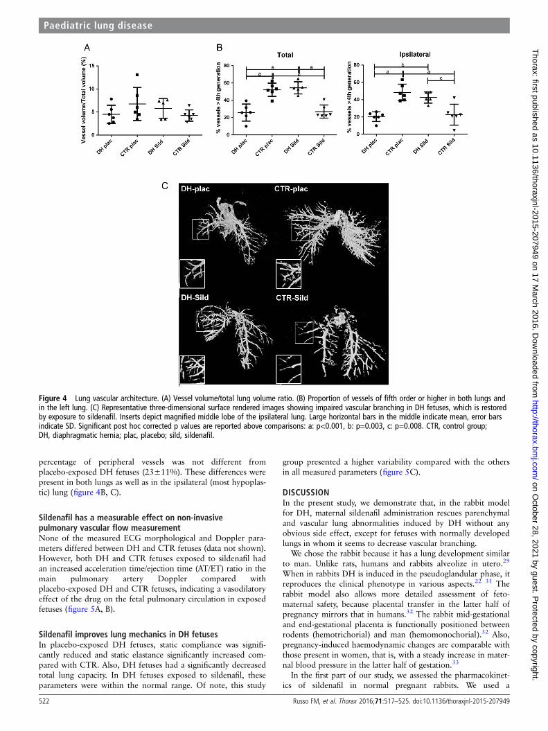
percentage of peripheral vessels was not different fromplacebo-exposed DH fetuses (23±11%). These differences werepresent in both lungs as well as in the ipsilateral (most hypoplas-tic) lung (figure 4B, C).
Sildenafil has a measurable effect on non-invasivepulmonary vascular flow measurementNone of the measured ECG morphological and Doppler para-meters differed between DH and CTR fetuses (data not shown).However, both DH and CTR fetuses exposed to sildenafil hadan increased acceleration time/ejection time (AT/ET) ratio in themain pulmonary artery Doppler compared withplacebo-exposed DH and CTR fetuses, indicating a vasodilatoryeffect of the drug on the fetal pulmonary circulation in exposedfetuses (figure 5A, B).
Sildenafil improves lung mechanics in DH fetusesIn placebo-exposed DH fetuses, static compliance was signifi-cantly reduced and static elastance significantly increased com-pared with CTR. Also, DH fetuses had a significantly decreasedtotal lung capacity. In DH fetuses exposed to sildenafil, theseparameters were within the normal range. Of note, this study
group presented a higher variability compared with the othersin all measured parameters (figure 5C).
DISCUSSIONIn the present study, we demonstrate that, in the rabbit modelfor DH, maternal sildenafil administration rescues parenchymaland vascular lung abnormalities induced by DH without anyobvious side effect, except for fetuses with normally developedlungs in whom it seems to decrease vascular branching.
We chose the rabbit because it has a lung development similarto man. Unlike rats, humans and rabbits alveolize in utero.29
When in rabbits DH is induced in the pseudoglandular phase, itreproduces the clinical phenotype in various aspects.22 31 Therabbit model also allows more detailed assessment of feto-maternal safety, because placental transfer in the latter half ofpregnancy mirrors that in humans.32 The rabbit mid-gestationaland end-gestational placenta is functionally positioned betweenrodents (hemotrichorial) and man (hemomonochorial).32 Also,pregnancy-induced haemodynamic changes are comparable withthose present in women, that is, with a steady increase in mater-nal blood pressure in the latter half of gestation.33
In the first part of our study, we assessed the pharmacokinet-ics of sildenafil in normal pregnant rabbits. We used a
Figure 4 Lung vascular architecture. (A) Vessel volume/total lung volume ratio. (B) Proportion of vessels of fifth order or higher in both lungs andin the left lung. (C) Representative three-dimensional surface rendered images showing impaired vascular branching in DH fetuses, which is restoredby exposure to sildenafil. Inserts depict magnified middle lobe of the ipsilateral lung. Large horizontal bars in the middle indicate mean, error barsindicate SD. Significant post hoc corrected p values are reported above comparisons: a: p<0.001, b: p=0.003, c: p=0.008. CTR, control group;DH, diaphragmatic hernia; plac, placebo; sild, sildenafil.
522 Russo FM, et al. Thorax 2016;71:517–525. doi:10.1136/thoraxjnl-2015-207949
Paediatric lung disease on O
ctober 28, 2021 by guest. Protected by copyright.
http://thorax.bmj.com
/T
horax: first published as 10.1136/thoraxjnl-2015-207949 on 17 March 2016. D
ownloaded from

comprehensive number of maternal as well as fetal safety mea-sures when testing dosages and administration regimens. We didnot observe obvious clinical side effects with 10 mg/kg/day eventhough this dose was associated with a short period of above-target sildenafil levels. We also excluded drug accumulation afterserial administration, a process that otherwise with chronic usecould result in toxicity. Thus, we corroborated the aforemen-tioned data from the rodent model, that is, that sildenafil is welltolerated by fetuses in the later stages of gestation.14
In addition to this, we investigated the effects of sildenafiltreatment on the developing lung. The drug had an effect onparenchymal development with significant improvement inMTBD. The interactions between the airways and blood vesselsare critical during lung development.34 Therefore, it is postu-lated that antenatal interventions designed to prevent vascularremodelling may rescue vascular and airway changes.14 35 Thefunctional analysis of the lungs with forced oscillation techniqueparallels the histological findings. As previously experimentallyand clinically demonstrated, lung mechanics are significantlycompromised in DH.31 We showed that such changes arereverted or at least attenuated by sildenafil, though with a highvariability among fetuses. Nevertheless, other relevant param-eter of lung function (like tissue damping or hysteresivity) couldnot be extensively investigated due to technical difficultiesrelated to DH lung fragility. Thus, it is possible that parenchy-mal lung growth is still not ideal as there was no major con-comitant increase in LBWR.
The vascular changes associated with DH are known toinduce PPHT. Although PPHT is best known as a postnatalevent in DH, the structural changes are already present inutero.22 36 The potential benefit of sildenafil on the
pathophysiology of PPHT has already been demonstrated in thenitrofen rat model.14 In the rabbit, sildenafil reversed the patho-logical changes in peripheral vessels and improved vascularbranching in DH. At the structural level, sildenafil restored vas-cular wall thickness and muscularisation of peripheral arteries tonormal levels. We also performed immunohistochemical analysisfor VEGF and for its receptor Flk-1, but in the vasculature wedid not detect a difference between the four cohorts. Yet in thealveolar parenchyma, sildenafil increased VEGF-positive andFlk-1-positive cell density both in DH and CTR lungs. VEGFhas been shown to interact with the nitric oxide (NO) pathway,by inducing release of NO from vascular endothelial cells andregulating endothelial NO synthase (eNOS) expression.37 Theactivation of eNOS mediated by sildenafil could, in turn, upre-gulate VEGF expression.38
To our knowledge, this is the first study in animal models forDH, where specific methods for quantitative assessment ofvessel density and branching have been used. We did so usingbarium–gelatin angiograms and 3D reconstruction on mCTimages. We observed decreased peripheral vascular branching inDH fetuses, which was reversed by sildenafil. The latter had alsoa measurable effect on CTR fetuses, where it reduced vascularbranching. This is in line with what has been demonstrated inthe rat model.14 The mechanism behind this finding remainsunclear, but may be related to a reduction in pulmonary bloodpressure.39 This could be beneficial in lungs with increased vas-cular resistances, but on the other hand it cannot be excludedthat it has adverse effect in more normal lungs, such as hypoper-fusion and impaired vessel growth in normal lungs. This findingshould prompt cautious use of sildenafil treatment in DH caseswith milder forms of lung hypoplasia.
Figure 5 Functional measurements. (A) Representative images of the Doppler waveforms of the main pulmonary artery. (B) Acceleration time/ejection time (AT/ET) ratio; N=5 per DH-sildenafil, 6 per CTR sildenafil, 7 per the placebo-exposed DH fetuses and placebo-exposed CTR groups.(C) Measures of lung mechanics: total lung capacity, static compliance and static elastance. Large horizontal bars in the middle indicate mean,error bars indicate SD. Significant post hoc corrected p values are reported above comparisons: a: p<0.001, b: p=0.01, c: p=0.023, d: p=0.002,e: p=0.004, f: p=0.003. CTR, control group; DH, diaphragmatic hernia; plac, placebo; sild, sildenafil.
Russo FM, et al. Thorax 2016;71:517–525. doi:10.1136/thoraxjnl-2015-207949 523
Paediatric lung disease on O
ctober 28, 2021 by guest. Protected by copyright.
http://thorax.bmj.com
/T
horax: first published as 10.1136/thoraxjnl-2015-207949 on 17 March 2016. D
ownloaded from

Finally, we demonstrated that sildenafil causes a measurablevasodilation of the pulmonary vasculature, resulting in anincreased AT/ET ratio of the pulmonary artery Doppler wave-form. However, we did not observe any difference between DHfetuses and CTR either in this Doppler parameter or in thecardiac structural dimensions. This is in contrast to what wasobserved in the nitrofen model.36 DH rats had lower pulmonaryartery Doppler ET/ET ratios and PI compared with CTR. Inthat study, Doppler measurements were performed on thecontralateral first branch of the pulmonary artery branch asopposed to the main pulmonary artery in our study. This differ-ence may be caused by the measurable impact of shunting viathe ductus arteriosus in our measurement.
Our study design, as well as the animal model, has some lim-itations. These include the lack of vascular assessment of extra-pulmonary vessels, in particular in the brain and liver. Weneither did perform quantitative PCR analysis of relevant genesinvolved in the pathway. PCR, however, only allows a quantifi-cation of expression levels in the lung as a whole and not in spe-cific structures such as conductive airways, parenchyma orvasculature, unless structures have been previously discriminatedby laser-capture microdissection. Nor do we have postnatal out-comes with regard to long-term toxicity of the drug. Also, ourassessment methods of pulmonary flow and resistance or struc-tural cardiac impact failed to demonstrate a difference betweentreatment groups. This is not different from clinical reality, as inhumans there is no robustly validated prenatal ultrasound pre-dictor for PPHT. It would also be interesting to evaluate moreselective PDE5 inhibitors, like tadalafil.40 However, use of tada-lafil in the neonatal population is contraindicated due to lack ofmaturation of the glucuronidation pathway vital for drug metab-olism.41 Finally, additional studies may be justified to furtherinvestigate the relevant pathways involved in the therapeuticeffect of the drug, measurement of PPHT (either invasively ornon-invasively) and extensive performance of lung functiontesting. This may be done both in the rabbit model as well assheep.
In conclusion, we proved that sildenafil is effective in revers-ing many of the parenchymal and vascular effects of DH in thefetal rabbit. Our results corroborate the clinical safety of thisdrug during pregnancy and might pave the way for clinicalapplication of a novel antenatal medical strategy for DH.
Acknowledgements We thank Rieta Van Bree, Catherina Luyten and Lieve Verbistfor their technical help in the histological studies.
Contributors FMR designed the study, conducted the experiments, analysed thedata and wrote the manuscript. MPE conducted the experiments and analysed thedata. AHM, JJ and PDK conducted the experiments. GVV, UH and PV participated inthe study design. JT and KA participated in the study design and revised themanuscript. JD designed the study, revised the manuscript and organised the studyas overall supervisor.
Funding Our translational research programme is being funded by the Fonds voorWetenschappelijk Onderzoek Vlaanderen (FWO; JD as clinical researcher; 1.801207;KA 1.800214; GVV as postdoctoral fellow; 12N7615N), the European Commissionvia its Erasmus Joint Doctoral programme (MPE and JJ; 2013-0040). JT is beneficiaryof a clinical research grant from the ‘Klinische Opleidings-en Onderzoeks-Raad’ ofthe University Hospitals Leuven.
Competing interests None declared.
Provenance and peer review Not commissioned; externally peer reviewed.
REFERENCES1 Kotecha S, Barbato A, Bush A, et al. Congenital diaphragmatic hernia. Eur Respir J
2012;39:820–9.2 Rottier R, Tibboel D. Fetal lung and diaphragm development in congenital
diaphragmatic hernia. Semin Perinatol 2005;29:86–93.
3 Neonatal Inhaled Nitric Oxide Study Group. Inhaled nitric oxide in full-term andnearly full-term infants with hypoxic respiratory failure. N Engl J Med1997;336:597–604.
4 Hayakawa M, Ito M, Hattori T, et al. Effect of hospital volume on the mortality ofcongenital diaphragmatic hernia in Japan. Pediatr Int 2013;55:190–6.
5 Reiss I, Schaible T, van den Hout L, et al. Standardized postnatal management ofinfants with congenital diaphragmatic hernia in Europe: the CDH EURO Consortiumconsensus. Neonatology 2010;98:354–64.
6 Tovar JA. Congenital diaphragmatic hernia. Orphanet J Rare Dis 2012;7:1.7 Claus F, Sandaite I, DeKoninck P, et al. Prenatal anatomical imaging in fetuses with
congenital diaphragmatic hernia. Fetal Diagn Ther 2011;29:88–100.8 DiFiore JW, Fauza DO, Slavin R, et al. Experimental fetal tracheal ligation reverses
the structural and physiological effects of pulmonary hypoplasia in congenitaldiaphragmatic hernia. J Pediatr Surg 1994;29:248–56; discussion 56–7.
9 Done’ E, Lewi P, Rayyan M, et al. 56: Neonatal morbidity in fetuses with severeisolated congenital diaphragmatic hernia (CDH) in the FETO era. Am J ObstetGynecol 2011;201:S33.
10 Schermuly RT, Kreisselmeier KP, Ghofrani HA, et al. Chronic sildenafil treatmentinhibits monocrotaline-induced pulmonary hypertension in rats. Am J Respir CritCare Med 2004;169:39–45.
11 Wharton J, Strange JW, Møller GM, et al. Antiproliferative effects ofphosphodiesterase type 5 inhibition in human pulmonary artery cells. Am J RespirCrit Care Med 2005;172:105–13.
12 Baquero H, Soliz A, Neira F, et al. Oral sildenafil in infants with persistentpulmonary hypertension of the newborn: a pilot randomized blinded study.Pediatrics 2006;117:1077–83.
13 Ganzevoort W, Alfirevic Z, von Dadelszen P, et al. STRIDER: Sildenafil Therapy InDismal prognosis Early-onset intrauterine growth Restriction—a protocol for asystematic review with individual participant data and aggregate data meta-analysisand trial sequential analysis. Syst Rev 2014;3:23.
14 Luong C, Rey-Perra J, Vadivel A, et al. Antenatal sildenafil treatment attenuatespulmonary hypertension in experimental congenital diaphragmatic hernia.Circulation 2011;123:2120–31.
15 Cheitlin MD, Hutter AM Jr, Brindis RG, et al. ACC/AHA expert consensus document.Use of sildenafil (Viagra) in patients with cardiovascular disease. American Collegeof Cardiology/American Heart Association. J Am Coll Cardiol 1999;33:273–82.
16 Goldenberg MM. Safety and efficacy of sildenafil citrate in the treatment of maleerectile dysfunction. Clin Ther 1998;20:1033–48.
17 Ballard SA, Gingell CJ, Tang K, et al. Effects of sildenafil on the relaxation ofhuman corpus cavernosum tissue in vitro and on the activities of cyclic nucleotidephosphodiesterase isozymes. J Urol 1998;159:2164–71.
18 Barst RJ, Ivy DD, Gaitan G, et al. A randomized, double-blind, placebo-controlled,dose-ranging study of oral sildenafil citrate in treatment-naive children withpulmonary arterial hypertension. Circulation 2012;125:324–34.
19 Vos RM, Chahbouni A, Sinjewel A, et al. Quantitative analysis of sildenafil anddesmethylsildenafil in human serum by liquid chromatography-mass spectrometrywith minimal sample pretreatment. J Chromatogr B Analyt Technol Biomed Life Sci2008;876:283–7.
20 Leach MC, Allweiler S, Richardson C, et al. Behavioural effects ofovariohysterectomy and oral administration of meloxicam in laboratory housedrabbits. Res Vet Sci 2009;87:336–47.
21 Keating SC, Thomas AA, Flecknell PA, et al. Evaluation of EMLA cream forpreventing pain during tattooing of rabbits: changes in physiological, behaviouraland facial expression responses. PLoS ONE 2012;7:e44437.
22 Wu J, Yamamoto H, Gratacos E, et al. Lung development following diaphragmatichernia in the fetal rabbit. Hum Reprod 2000;15:2483–8.
23 Roubliova XI, Lewi PJ, Vaast P, et al. Effects of betamethasone on peripheralarterial development in term fetal rabbit. Pediatr Pulmonol 2008;43:795–805.
24 De Langhe E, Vande Velde G, Hostens J, et al. Quantification of lung fibrosis andemphysema in mice using automated micro-computed tomography. PLoS ONE2012;7:e43123.
25 Counter WB, Wang IQ, Farncombe TH, et al. Airway and pulmonary vascularmeasurements using contrast-enhanced micro-CT in rodents. Am J Physiol Lung CellMol Physiol 2013;304:L831–43.
26 Rychik J, Ayres N, Cuneo B, et al. American Society of Echocardiography guidelinesand standards for performance of the fetal echocardiogram. J Am Soc Echocardiogr2004;17:803–10.
27 Hodges R, Endo M, La Gerche A, et al. Fetal echocardiography and pulsed-waveDoppler ultrasound in a rabbit model of intrauterine growth restriction. J Vis Exp2013;(76):50392.
28 Jani JC, Flemmer AW, Bergmann F, et al. The effect of fetal tracheal occlusion on lungtissue mechanics and tissue composition. Pediatr Pulmonol 2009;44:112–21.
29 Roubliova XI, Deprest JA, Biard JM, et al. Morphologic changes and methodologicalissues in the rabbit experimental model for diaphragmatic hernia. Histol Histopathol2010;25:1105–16.
30 Roubliova XI, Verbeken EK, Wu J, et al. Effect of tracheal occlusion on periphericpulmonary vessel muscularization in a fetal rabbit model for congenitaldiaphragmatic hernia. Am J Obstet Gynecol 2004;191:830–6.
524 Russo FM, et al. Thorax 2016;71:517–525. doi:10.1136/thoraxjnl-2015-207949
Paediatric lung disease on O
ctober 28, 2021 by guest. Protected by copyright.
http://thorax.bmj.com
/T
horax: first published as 10.1136/thoraxjnl-2015-207949 on 17 March 2016. D
ownloaded from

31 Flemmer AW, Jani JC, Bergmann F, et al. Lung tissue mechanics predict lunghypoplasia in a rabbit model for congenital diaphragmatic hernia. Pediatr Pulmonol2007;42:505–12.
32 Fischer B, Chavatte-Palmer P, Viebahn C, et al. Rabbit as a reproductive model forhuman health. Reproduction 2012;144:1–10.
33 McArdle AM, Denton KM, Maduwegedera D, et al. Ontogeny of placental structuraldevelopment and expression of the renin-angiotensin system and 11beta-HSD2genes in the rabbit. Placenta 2009;30:590–8.
34 Stenmark KR, Abman SH. Lung vascular development: implications for thepathogenesis of bronchopulmonary dysplasia. Annu Rev Physiol 2005;67:623–61.
35 Deprest J, Gucciardo L, Eastwood P, et al. Medical and regenerative solutions forcongenital diaphragmatic hernia: a perinatal perspective. Eur J Pediatr Surg2014;24:270–7.
36 Yamamoto Y, Thebaud B, Vadivel A, et al. Doppler parameters of fetal lunghypoplasia and impact of sildenafil. Am J Obstet Gynecol 2014;211:263.e1–8.
37 Frelin C, Ladoux A, D’Angelo G. Vascular endothelial growth factors andangiogenesis. Ann Endocrinol (Paris) 2000;61:70–4.
38 Zhao X, Lu X, Feng Q. Deficiency in endothelial nitric oxide synthase impairsmyocardial angiogenesis. Am J Physiol Heart Circ Physiol 2002;283:H2371–8.
39 Reffelmann T, Kloner RA. Therapeutic potential of phosphodiesterase 5 inhibition forcardiovascular disease. Circulation 2003;108:239–44.
40 Shue EH, Schecter SC, Gong W, et al. Antenatal maternally-administeredphosphodiesterase type 5 inhibitors normalize eNOS expression in the fetal lambmodel of congenital diaphragmatic hernia. J Pediatr Surg 2014;49:39–45;discussion 45.
41 Vorhies EE, Ivy DD. Drug treatment of pulmonary hypertension in children. PaediatrDrugs 2014;16:43–65.
Russo FM, et al. Thorax 2016;71:517–525. doi:10.1136/thoraxjnl-2015-207949 525
Paediatric lung disease on O
ctober 28, 2021 by guest. Protected by copyright.
http://thorax.bmj.com
/T
horax: first published as 10.1136/thoraxjnl-2015-207949 on 17 March 2016. D
ownloaded from