gastrointestinal stromal tumor: a rare cause of neonatal intestinal obstruction
TRANSCRIPT

BRIEF REPORTGastrointestinal Stromal Tumor: A Rare Cause of Neonatal
Intestinal Obstruction
Manoj U. Shenoy, MS, MCh, FRCS, FRCS-Paed Surg,1*Shailinder J. Singh, MS, MCh, FRCS, FRCS-Paed Surg,1 Keith Robson, MBBS, FRCPath,2
and Richard J. Stewart, MD, FRCS, FRCS-Paed Surg1
Key words: gastrointestinal stromal tumor; neonatal intestinal obstruction;childhood cancer
Neonatal intestinal obstruction is commonly due toatresia, meconium ileus, or Hirschsprung disease; a tu-mor is an uncommon cause. Our experience with such aneonate is of interest especially insofar as the neoplasmproved to be the extremely rare stromal tumor of thegastrointestinal tract. The tumor developed in associationwith ileal atresia. We have not found a previous record inthe English-language literature.
The baby boy was born at 41 weeks of gestation, witha birth weight of 3.75 kg, to a primigravida mother. Hewas 1 day old when seen by us because of abdominaldistention, bilious vomiting, and delayed passage of me-conium. Examination revealed a distended and tenderabdomen, with visible bowel loops. Abdominal X-rayfilms showed multiple air fluid levels. Contrast enemashowed a small-calibre left colon, normal calibre of theremaining large intestine, but no proximal flow past theterminal ileum. A laparotomy was arranged, and an atre-sia of the terminal ileum was found. There was also avascular mass in the terminal ileum, which measuredabout 2.5 cm in diameter. Twelve centimeters of smallbowel was resected, and an end-to-end anastomosis wasperformed about 20 cm proximal to the ileocaecal valve.The postoperative course was uneventful.
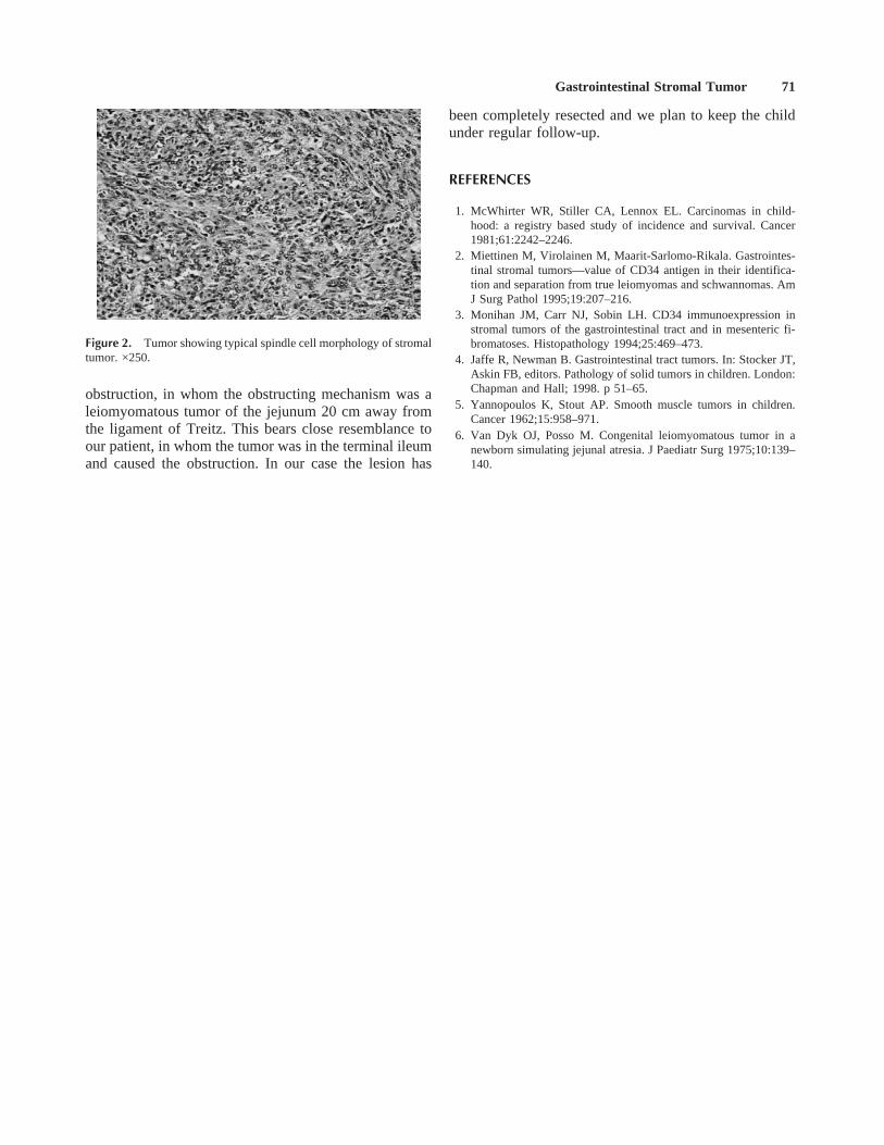
Histopathology (Figs. 1, 2) showed the presence ofsolid pale tissue 2.5 cm in diameter obstructing the lumenand causing proximal dilatation. Microscopically the tu-mor was a spindle cell lesion that encircled the bowel, thelumen of which could be seen as a tiny cyst-like area.Elements of residual bowel tissue (smooth muscle andnerve bundles) could be seen within the tumor on immu-nohistochemistry, showing that the tumor diffusely in-vaded throughout the bowel wall elements and replacedthem. There was mild pleomorphism but no necrosis andonly one mitosis per ten high-power fields. There werelarge numbers of lymphocytes and macrophages betweenthe tumor cells, which were positive for vimentin andmuscle differentiation markers (actin and, focally, myo-
sin). CD 34 was positive. Neuronal markers (S100 andCD56) were negative. The features were in keeping witha gastrointestinal stromal tumor. At 1 year of follow-upthe patient was thriving, with no evidence of tumor re-currence.
Neonatal tumors are rare, accounting for 0.5–2% of allpaediatric tumors [1]. The termgastrointestinal stromaltumor (GIST) has been applied to mesenchymal tumorsthat represent neither typical leiomyomas nor schwanno-mas [2–4]. Smooth muscle tumor is a relatively rarecause of intestinal obstruction in the newborn [5]. VanDyk and Posso [6] report a newborn girl with intestinal
1Department of Paediatric Surgery, University Hospital, Queen’sMedical Centre, Nottingham, United Kingdom2Department of Pathology, University Hospital, Queen’s Medical Cen-tre, Nottingham, United Kingdom
*Correspondence to: Mr. M.U. Shenoy, Registrar in Paediatric Sur-gery, c/o Mr. Rance’s Secretary, Department of Paediatric Surgery,Nottingham City Hospital, Hucknall Road, Nottingham NG5 1PB,United Kingdom. E-mail: [email protected]
Received 26 April 1999; Accepted 2 June 1999
Figure 1. Tumor underlying mucosa. ×40.
Medical and Pediatric Oncology 34:70–71 (2000)
© 2000 Wiley-Liss, Inc.
obstruction, in whom the obstructing mechanism was aleiomyomatous tumor of the jejunum 20 cm away fromthe ligament of Treitz. This bears close resemblance toour patient, in whom the tumor was in the terminal ileumand caused the obstruction. In our case the lesion has
been completely resected and we plan to keep the childunder regular follow-up.
REFERENCES
1. McWhirter WR, Stiller CA, Lennox EL. Carcinomas in child-hood: a registry based study of incidence and survival. Cancer1981;61:2242–2246.
2. Miettinen M, Virolainen M, Maarit-Sarlomo-Rikala. Gastrointes-tinal stromal tumors—value of CD34 antigen in their identifica-tion and separation from true leiomyomas and schwannomas. AmJ Surg Pathol 1995;19:207–216.
3. Monihan JM, Carr NJ, Sobin LH. CD34 immunoexpression instromal tumors of the gastrointestinal tract and in mesenteric fi-bromatoses. Histopathology 1994;25:469–473.
4. Jaffe R, Newman B. Gastrointestinal tract tumors. In: Stocker JT,Askin FB, editors. Pathology of solid tumors in children. London:Chapman and Hall; 1998. p 51–65.
5. Yannopoulos K, Stout AP. Smooth muscle tumors in children.Cancer 1962;15:958–971.
6. Van Dyk OJ, Posso M. Congenital leiomyomatous tumor in anewborn simulating jejunal atresia. J Paediatr Surg 1975;10:139–140.
Figure 2. Tumor showing typical spindle cell morphology of stromaltumor. ×250.
Gastrointestinal Stromal Tumor 71