fusion bacterialprotoplasts - pnas microbiology: schaefferetal. table 1. derivation ofthe...
TRANSCRIPT

Proc. Natl. Acad. Sci. USAVol. 73, No. 6, pp. 2151-2155, June 1976Microbiology
Fusion of bacterial protoplasts(Bacillus subtilis/diploid bacteria/polyethylene glycol/chromosome recombination)
PIERRE SCHAEFFER, BRIGITTE CAMI, AND ROLLIN D. HOTCHKISS*Institut de Microbiologie, Universit6 de Paris-Sud, 91405, Orsay, France
Contributed by Rollin D. Hotchkiss, April 7,1976
ABSTRACT Prototrophic Bacillus subtifis cells can beformed in the presence of DNase as a result of cell fusion oc-curring in mixed populations of protoplasts derived from twoparental strains which are both nutritionally-complementingand polyauxotrophic. No prototrophs ever appear from mixednonprotoplasted bacteria, or from the auxotrophic parentalprotoplasts plated separately. The frequency of prototrophformation, which is appreciable only when the mixed proto-plasts are exposed to polyethylene glycol treatment, may exceed
10-4 ofthe total protoplast population initially present,which is 1 to 4 X 10⁻³ of those protoplasts which reverted to thebacillary form. It is strongly dependent on the number andchromosomal location of the markers used in the selection ofthe prototrophs, and it is unaffected when either one of theparental strains bears the phage k105 in the inducible prophagestate. No auxotrophic bacteria, parental or otherwise, werefound as segregants from repeatedly isolated prototrophic clonesgrowing in a nonselective medium. Unselected markers segre-gate among the selected recombinants. It is concluded that theobserved formation of prototrophic bacteria is due to protoplastfusion, a process which does not induce prophage development,and that the only stable products of the resulting diploid stateare haploid recombinants.
Hybridization of mammalian somatic cells, which was intro-duced 15 years ago (1), is being widely used to study the ex-pression of differentiated functions (2-4) and the genetics ofhuman cells in culture (5, 6). Fusion of protoplasts from higherplant cells has also been achieved (7), and in some cases wholeflowering hybrid plants have been regenerated, starting fromfused protoplasts (8). A broad and most promising field has,thus, been opened for rationally combining desirable propertiesfrom two sexually incompatible plant lines (9, 10). In this con-text, it seemed surprising that, to our knowledge, no sustainedattempt at fusing bacterial protoplasts has been reported. Withsuch purposes in mind we decided to try fusion of protoplastsof Bacillus subtilis, a Gram-positive bacterium in which manynutritional markers are available. Although no conjugation,mediated by sex factors, has ever been found in this organism,a detailed chromosomal map is available (11), constructed fromtransduction and transformation data. The cell wall consistsessentially of a peptidoglycan layer, easily removed by lyso-zyme treatment, and regenerated under osmotic protection (12,13). An important element in the choice of this material was theabsence of an outer membrane, a potential obstacle to cyto-plasmic membrane contact and fusion.Assuming that fusion occurs between protoplasts from two
polyauxotrophic strains, the appearance of prototrophic cloneswould presumably require wall regeneration and selection ofthe prototrophs. Plating the mixed protoplasts on an hypertonicminimal medium efficiently supporting these two processes hasbeen tried repeatedly, with only occasional success. Regener-
Abbreviations: PEG, polyethylene glycol; SMM, sucrose-magne-sium-maleate buffer; RDR, rich regeneration agar medium; SDR,nonhypertonic minimal media; SMMD, sucrose-magnesium-maleatebuffer containing 5 ,g/ml DNase.* Present address: The Rockefeller University, New York, N.Y. 10021.
2151
ation itself, with our material, has been rare and unreliable insuch media. Attempting the required selection first, and thenproceeding to wall regeneration seemed excluded, becauseprotoplasts produced by lysozyme treatment do not divide inliquid minimal medium (13). We were, thus, led to carry outwall regeneration upon the mixed protoplasts first, on a richhypertonic agar medium (13), and in a second step the selectionby replica plating on various deficient agar media.
MATERIALS AND METHODSBacterial Strains and Media. The strains used in fusion
experiments were constructed as described in Table 1. Thechromosomal location of their markers is given in Fig. 1. Bac-teria were first grown in nutrient broth (17), and protoplastedin SMM, the sucrose-magnesium-maleate buffer of Wyrick andRogers (13), to which 5 ,ug/ml of DNase I (Worthington Bio-chem. Corp.) had been added (SMMD). Protoplasts were madeto revert to bacillary forms by plating on RDR, a rich regen-eration agar medium of high tonicity (13), to which 5 /g/mleach of DNase I and rifamycin (Lepetit Labs, Milano) wereadded. Prototrophic clones within the film of growth that ap-peared on RDR plates after incubation were selected out byreplica plating onto variously supplemented SDR medium. Thisis a nonhypertonic minimal medium (14), to which 20 ,MMnCl2, 5 gg/ml of DNase, 1 Ag/ml of rifamycin, and 15 g/literof (Difco) agar have been added. It was used as a selectionmedium, both unsupplemented (SDR) and supplemented (seeTable 2).
Procedure Adopted for Fusion Experiments. Overnightprecultures of both parental strains in nutrient broth at 300 wereinoculated, before growth ceased, into 20 ml of broth, to givean initial optical density (OD570) = 0.05. These cultures wereincubated with shaking at 370 until an OD of 0.4 was reached.From each culture, 15 ml were centrifuged, the pellets weretaken up in 3 ml ofSMMD (OD570 = 2, or about 4 X 10⁸ colonyforming units/ml), and lysozyme was added to a concentrationof 200,ug/ml. Complete protoplast formation was usually seenafter 10 min of gentle shaking at 420, but exposure to lysozymewas continued for 20 more minutes. Samples (0.1 ml) of eachsuspension were then plated on ordinary nutrient agar. Theplates usually remained sterile, and indicated that the frequencyof osmotic shock resistant forms was below 2.5 X 10⁻⁸.One milliliter samples from each of the two suspensions were
mixed in a third tube, the three tubes were centrifuged, andeach pellet was resuspended in 0.2 ml of SMMD. To one tube,1.8 ml of a 40% (wt/vol) solution of polyethylene glycol (PEG)tin SMM was added and the suspension immediately homoge-nized by shaking. After a 1 min exposure to PEG, either at 200or at 00, several 0.05 ml samples were spread on the surface ofduplicate RDR plates and used to make 10-1 and 10-2 dilutionsin SMMD, from which, in turn, further reversion plates and also
tThe molecular weight is not critical; PEG 6000 from Merck wasusually used.

2152 Microbiology: Schaeffer et al.
Table 1. Derivation of the parental strains used
Strains constructed by transformation Recipient DNA donor Phenotypic changes
S, (rfm-486 metB5 leu-8 thr-5) Mu8u5u5* MO21t RfmrS3 (rfm-486 purB34 ura-1 trpC7) GSY1104t MO21t RfmrS, (rfm-486 ura-1 trpC7 thr-5) S3 Si Ade+ Thr-S7 (rfm-486 purB34 metB5 leu-8) S1 S3 Thr+ AdeSs (rfm-486 purB34 leu-8 thr-5) S1 S3 Met+ Ade-S9 (rfm-486 ura-1 metB5 trpC7) S3 S1 Adel MetF
In the transformation experiments (14) excess DNA (5 ,ug/ml) was used whenever double transformants were wanted. Rfmr refers torifamycin resistance.* This strain is metB5 leu-8 thr-.5 (15).t This strain is rfm-486 trpC2 leu-2 (16).T Supplied by C. Anagnostopoulos, this strain is purB34 ura- I trpC7.
further dilutions were prepared. The three pellets were pro-cessed and plated in succession, so that prolonged exposure toPEG, which can diminish the number of prototrophs, wasavoided. After 48 hr of incubation at 370, the most heavily in-oculated RDR plates were replicated with a velvet surface onSDR plates, or SDR plates carrying various limited combina-tions of the six growth factors. The prototrophic or partiallyprototrophic colonies were counted after at least 72 hr of in-cubation at 37'. An unexplained crowding effect was noted,i.e., 3 to 10 times higher counts of prototrophs per ml wereobtained from RDR plates inoculated with 2 X 10⁶ protoplastsor less than from plates that received the highest inoculum (2X 10⁷ protoplasts). Regeneration frequency for the parentalstrains themselves varied from 0.3 to 20%, such extreme valuesbeing rarely observed. Within this range, this variation did notgreatly influence the numbers of prototrophs obtained on agiven selection medium.
RESULTSAttempts to produce prototrophic clones by fusion occurringspontaneously in mixtures of protoplasts from two triply aux-otrophic strains (Mu8u5u5 and GSY1104, see Table 1) wereunsuccessful. In order to avoid possible contaminants, a rif-amycin-resistant marker was introduced into each of thesestrains by transformation, so that subsequent work would bedone under the protection of the antibiotic. When triply aux-otrophic strains (Si and S3, see Table 1), are used in fusion ex-periments, selection can be instituted not only for true proto-trophs, on rifamycin-containing minimal medium, but also forwhat we will call here "partial phototrophs", on the same me-dium supplemented with two growth factors, of which one isrequired by each parent. When selection was against all sixmarkers, no prototrophic growth was noted, but when two of
t rpCFIG. 1. Chromosomal map of B. subtilis. The map is drawn to
scale according to ref. 11. The loci mentioned are those in which themarkers used are located. 0 and T stand for origin and terminus ofreplication. Starting from 0 the two arrows in opposite directionsindicate that replication is bidirectional.
these markers were not selected against, a very few partialprototrophs did grow out, even in the presence of DNase. Notall combinations of two growth factors seemed to be effective(see the first column of Table 2), but no conclusions could bedrawn from such low numbers of colonies.
Recognition of fusion products became possible only whena PEG treatment was appliedt, in awareness of its usefulnessin the fusion of plant protoplasts (18). The treatment turned outto be effective (Table 2), but only when PEG concentrationsapproaching 40% were used. Since control plates bearingPEG-treated protoplasts from only one parent in every caseremained sterile, it appeared that prototroph production didoccur as a result of protoplast fusion, but was very rare in theabsence of an appropriate fusion-enhancing treatment, or whena large number of markers were involved. An important notionalready suggested by these observations was that the prototro-phic growth observed is more likely to result from postfusionalgenetic recombination, than from divisions of the initiallycreated heterodiploid cells; appearance of stable diploid pro-totrophs should be independent of the growth factors suppliedduring selection. Support for this interpretation will now besought in various ways in the sections to follow.
Fusion experiments subjected to alternative markerselectionsThe progeny obtained on 10 different selection media from SIX Sa fusion in three independent experiments appear in Table3. After PEG treatment and wall regeneration, they may con-tain a few true prototrophs appearing on minimal medium(SDR), but they generally contain much larger numbers ofpartial prototrophs which are revealed when the medium issupplemented with two growth factors. The yield of fusionproducts detected is dependent upon not only the number butalso the particular pair of markers involved in the selection.Each selected colony implies the acquisition of some combi-nation of the wild-type genes derived from both parents. Pre-sumably these could arise only from fusion of the parents, be-cause a known fusion-enhancing agent significantly increasestheir number, they are only obtained when protoplasts-notcells-are mixed, and they are readily produced and developedin the constant presence of DNase. The widely varying yieldswith different selection media indicate that we are not, in anycase, dealing with the total number of diploids produced. In allor most cases, the diploid phase must have lasted a rather shorttime and must have soon been succeeded by appearance of aparticular one or a few of the many possible haploid recombi-nants. The pattern of particular recombinants obtained is thent It is pleasant to acknowledge that Dr. M. Fox first suggested to us theuse of PEG as a possible fusion-enhancing agent, as early as June,1974.
Proc. Natl. Acad. Sci. USA 73 (1976)
Proc. Natl. Acad. Sci. USA 73 (1976) 2153
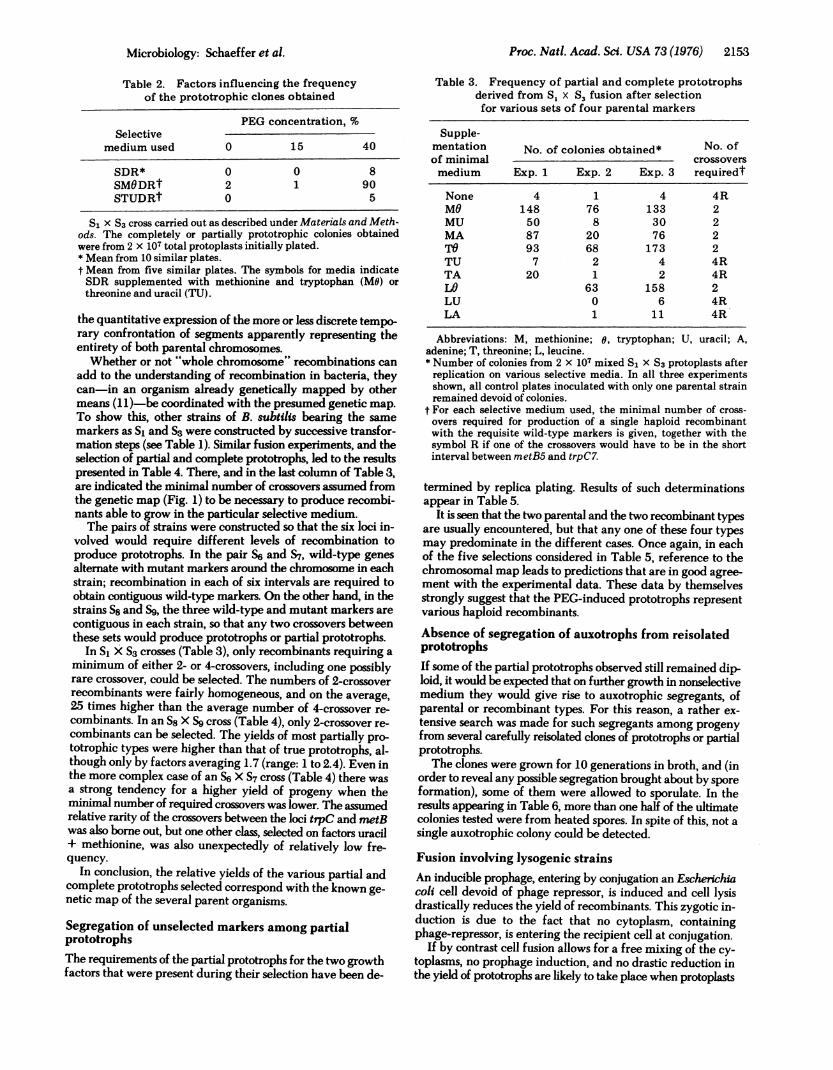
Table 2. Factors influencing the frequencyof the prototrophic clones obtained
PEG concentration, %Selective
medium used 0 15 40
SDR* 0 0 8SMODRt 2 1 90STUDRt 0 5
SI X S3 cross carried out as described under Materials and Meth-ods. The completely or partially prototrophic colonies obtainedwere from 2 X 10⁷ total protoplasts initially plated.* Mean from 10 similar plates.t Mean from five similar plates. The symbols for media indicateSDR supplemented with methionine and tryptophan (MO) orthreonine and uracil (TU).
the quantitative expression of the more or less discrete tempo-rary confrontation of segments apparently representing theentirety of both parental chromosomes.Whether or not "whole chromosome" recombinations can
add to the understanding of recombination in bacteria, theycan-in an organism already genetically mapped by othermeans (11)-be coordinated with the presumed genetic map.To show this, other strains of B. subtilis bearing the samemarkers as Si and S3 were constructed by successive transfor-mation steps (see Table 1). Similar fusion experiments, and theselection of partial and complete prototrophs, led to the resultspresented in Table 4. There, and in the last column of Table 3,are indicated the minimal number of crossovers assumed fromthe genetic map (Fig. 1) to be necessary to produce recombi-nants able to grow in the particular selective medium.The pairs of strains were constructed so that the six loci in-
volved would require different levels of recombination toproduce prototrophs. In the pair S6 and S7, wild-type genesalternate with mutant markers around the chromosome in eachstrain; recombination in each of six intervals are required toobtain contiguous wild-type markers. On the other hand, in thestrains S8 and S9, the three wild-type and mutant markers arecontiguous in each strain, so that any two crossovers betweenthese sets would produce prototrophs or partial prototrophs.
In SI X S3 crosses (Table 3), only recombinants requiring aminimum of either 2- or 4-crossovers, including one possiblyrare crossover, could be selected. The numbers of 2-crossoverrecombinants were fairly homogeneous, and on the average,25 times higher than the average number of 4-crossover re-combinants. In an S8 X S9 cross (Table 4), only 2-crossover re-combinants can be selected. The yields of most partially pro-totrophic types were higher than that of true prototrophs, al-though only by factors averaging 1.7 (range: 1 to 2.4). Even inthe more complex case of an S6 X S7 cross (Table 4) there wasa strong tendency for a higher yield of progeny when theminimal number of required crossovers was lower. The assumedrelative rarity of the crossovers between the loci trpC and metBwas also borne out, but one other class, selected on factors uracil+ methionine, was also unexpectedly of relatively low fre-quency.
In conclusion, the relative yields of the various partial andcomplete prototrophs selected correspond with the known ge-netic map of the several parent organisms.
Segregation of unselected markers among partialprototrophsThe requirements of the partial prototrophs for the two growthfactors that were present during their selection have been de-
Table 3. Frequency of partial and complete prototrophsderived from 5, x S3 fusion after selectionfor various sets of four parental markers
Supple-mentation No. of colonies obtained* No. ofof minimal crossoversmedium Exp. 1 Exp. 2 Exp. 3 requiredt
None 4 1 4 4RMO 148 76 133 2MU 50 8 30 2MA 87 20 76 2TO 93 68 173 2TU 7 2 4 4RTA 20 1 2 4RLO 63 158 2LU 0 6 4RLA 1 11 4R
Abbreviations: M, methionine; o, tryptophan; U, uracil; A,adenine; T, threonine; L, leucine.* Number of colonies from 2 X 10⁷ mixed Si x S3 protoplasts afterreplication on various selective media. In all three experimentsshown, all control plates inoculated with only one parental strainremained devoid of colonies.
t For each selective medium used, the minimal number of cross-overs required for production of a single haploid recombinantwith the requisite wild-type markers is given, together with thesymbol R if one of the crossovers would have to be in the shortinterval between metB5 and trpC7.
termined by replica plating. Results of such determinationsappear in Table 5.
It is seen that the two parental and the two recombinant typesare usually encountered, but that any one of these four typesmay predominate in the different cases. Once again, in eachof the five selections considered in Table 5, reference to thechromosomal map leads to predictions that are in good agree-ment with the experimental data. These data by themselvesstrongly suggest that the PEG-induced prototrophs representvarious haploid recombinants.Absence of segregation of auxotrophs from reisolatedprototrophsIf some of the partial prototrophs observed still remained dip-loid, it would be expected that on further growth in nonselectivemedium they would give rise to auxotrophic segregants, ofparental or recombinant types. For this reason, a rather ex-tensive search was made for such segregants among progenyfrom several carefully reisolated clones of prototrophs or partialprototrophs.The clones were grown for 10 generations in broth, and (in
order to reveal any possible segregation brought about by sporeformation), some of them were allowed to sporulate. In theresults appearing in Table 6, more than one half of the ultimatecolonies tested were from heated spores. In spite of this, not asingle auxotrophic colony could be detected.
Fusion involving lysogenic strainsAn inducible prophage, entering by conjugation an Escherichiacoli cell devoid of phage repressor, is induced and cell lysisdrastically reduces the yield of recombinants. This zygotic in-duction is due to the fact that no cytoplasm, containingphage-repressor, is entering the recipient cell at conjugation.
If by contrast cell fusion allows for a free mixing of the cy-toplasms, no prophage induction, and no drastic reduction inthe yield of prototrophs are likely to take place when protoplasts
Microbiology: Schaeffer et al.

2154 Microbiology: Schaeffer et al.
Table 4. Pattern of complete and partial prototrophs obtained fromparental protoplasts bearing mutant markers in alternating (A) or in clustered (B) arrangements
Fusion A, S6 x S? Fusion B, Ss X S9
Selective No. of No. of Selective No. of No. ofmedium colonies crossovers medium colonies crossovers
Minimal 31 6R Minimal 75 2UL 192 2R TO 114 2UA 52 4R TM 125 2UM 149 4 TU 181 2OL 357 4 LO 78 2OA 362 2 LM 91 2OM 248 4 LU 165 2TL 189 4R AO 143 2TA 154 4R AM 120 2TM 280 2 AU 133 2
Standard fusion experiments with the strains indicated. Single parental protoplasts were, in all cases, carried through the same procedure,and in each of the many selective media, each parental type gave no colonies whatever. Yield is from 2 x 10⁷ mixed protoplasts.
of lysogenic and phage-sensitive bacteria are fused. In orderto verify this point, use has been made of 4105, a temperatephage of B. subtilis 168. Si and S3 were lysogenized, and in-ducibility of their derivatives was tested with mitomycin as
described (19). No clearcut reduction in the yield of partialprototrophs was observed, when one of the parental strains waslysogenic (Table 7).
DISCUSSION
When mixed together, lysozyme-induced protoplasts of twotriply auxotrophic strains of B. subtilis were shown to produce,after regeneration of cell wall, clones of prototrophic bacteriawhich do not form from similarly treated protoplasts of thesingle parental strains. Since this appearance of prototrophs wasnot observed from mixtures of nonprotoplasted bacteria, andwas unaffected in the continued presence of DNase, it is be-lieved to result from protoplast fusion. This conclusion is furthersupported by the observation that prototroph formation is un-affected when one of the parents is supplied as an induciblelysogenic strain, and also by the fact that it is increased sub-stantially when the mixed protoplasts are treated with poly-ethylene glycol, an effective, apparently universal, fusion-enhancing agent (18, 20-22). Similar conclusions were reachedsimultaneously by Fodor and Alfoldi, working on Bacillusmegaterium (23). We thank these workers for the benefit offree exchange of information as the two investigations, com-menced independently, have proceeded to the stage of thesecollateral publications.
When one of the three growth factors required by each ofthe parental lines was supplied during the selection of the fusionproducts-i.e., when two of the six markers were not selectedagainst-the following observations were made: (a) higheryields were generally obtained than when complete prototrophswere selected for, and (b) not all combinations of the two growthfactors added were equally effective in increasing the yield.(This was true with the three pairs of parental strains used,which all bore the same six markers, but in different combi-nations.) Confronted with the known chromosomal map of theorganism (Fig. 1), these data clearly establish that in a givenfusion experiment the yields observed on 10 different selectionmedia are an inverse function of the minimal number of thecrossovers required to allow growth on the various selectionmedia (Tables 3 and 4). Therefore, for given rates of fusion andregeneration, the yields observed are largely determined by thefrequency of the recombination events required to allow growthon the medium being used. Such events as chance associationof cells of the two parental lines at the time colonies are ini-tiated, appear to be relatively uniform. On the other hand, theconcurrence of a second pair of required crossovers with a re-
quired first pair is commonly greater than 10-1, so it is clear thatparental chromosomes are, at some stage, liable to multipleexchanges, once being liable to any exchange.The precise cellular mechanism of genome-to-genome in-
teraction is not of course defined, but it appears to be unre-
stricted in completeness. In this connection, in a study of ex-
perimentally attempted fusion of bacterial protoplasts,Lederberg and St. Clair (24) indicate that penicillin-protoplasts
Table 5. Segregation of unselected markers among partial prototrophs from one S, x S3 fusion experiment
Number of colonies having indicated constitution
Unselected S, parental type (-/+) S3 parental type (+/-) +/+ Recombinant -/-Recombinantmarkers S % % S
met/trp 20 78 0 2met!ura 74 3 2 21met/pur 60 1 2 38thr/thr 0 84 2 14thr/pur 27 16 55 2
Colonies obtained on the variously supplemented selection media were transferred and set in rows on plates of their original selectionmedium, containing two growth factors. After overnight incubation, these plates were replicated on minimal medium and on mediumcontaining either growth factor or both.
Proc. Natl. Acad. Sci. USA 73 (1976)

Proc. Natl. Acad. Sci. USA 73 (1976) 2155
Table 6. Stability of prototrophic clones during growth in nonselective medium
Medium in which originally Number of reisolated Total no. of ultimate Maximal frequency ofisolated and finally retested clones tested colonies tested* auxotrophic subcolonies
Minimal 4 1122 <9 x 10⁻⁴Minimal + TO 4 1781 <6 X 10⁻⁴Minimal + MO 3 3072 <3 x 10⁻⁴
Several original potentially-prototrophic clones derived from Si X S3 fusion upon selective media were reisolated twice as clones from thesame media, then grown for 10 generations in nonselective broth, and retested for possible regain of auxotrophic markers derived from theoriginal parents. See Table 3 for abbreviations.* The tested colonies were derived in approximately equal numbers from the reisolated clones within each group. Not one auxotrophiccolony was recovered in any case. The upper limits of possible auxotrophs per clone would therefore be somewhat higher than the limitsshown.
of Escherichia coli show only such rare unidirectional chro-mosome transfer as is already possible from F+ to F- cellsthemselves, which we would therefore suppose is unidirectionaland incomplete. The evidence in our case is that neithercrossfeeding of some kind, nor limited chromosome transferwould explain the observations; they are, rather, explained bythe formation by fusion of protoplasts that must be at leastdiploids. The very large size of the aggregates that are seen toform as soon as PEG is added, may suggest that mostly poly-ploids are formed. Aggregation however is not to be confusedwith fusion, and may be at most a prerequisite. In fact, it largelydisappears when the PEG is eliminated by centrifugation,without drastically reducing the final yield of prototrophs. Themean ploidy level of the fused protoplasts, and the factors onwhich it may depend, remain to be determined.Whatever this level may be, it is clear that after two reiso-
lations all of the prototrophic bacteria produced by fusion arehaploid recombinants. Already inferred from the data in Tables3 and 4, this was confirmed directly when segregation of theunselected markers in partial prototrophs and of auxotrophsfrom the reisolated prototrophs were studied (see Tables 5 and6, respectively). If we think we know the initial (polyploid), andthe final (haploid) states of the fusion-induced formation ofprototrophs, the minimal number of generations required toproduce the change remains to be estimated.From the data presented, the highest yield of prototrophs
observed seems to be 1.8 X 10⁻⁵ (Table 4, Fusion A). This is anunderestimation, however. The experiments in Tables 3 and4 were aimed at comparing, in different crosses, the yields onvarious selection media. Because some of these yields were verylow, large inocula (2 X 10⁷ protoplasts per plate) were used.While such inocula will usually produce higher numbers ofprototrophic colonies per plate than smaller ones, they producea crowding effect, not all prototrophs showing up as colonies.When in other experiments prototroph frequency was morecorrectly measured by plating only 2 X 10⁶ protoplasts per
Table 7. Frequency of partial prototrophs fromlysogenic inducible parental strains
Cell Frequency ofregener- partial prototrophsation (on minimal +
Strains crossed % MO agar medium)
Si X S3 5.2 0.6 x 10⁻⁴SI (0105) x S3 7.5 1.0 x 10⁻⁴S1 X S3 (0105) 3.5 0.4 x 10⁻⁴Si (0105)X S3 q105) 7.0 0.75 x 10⁻⁴
See Table 3 for abbreviations.
plate, frequencies as high as 2 X 10⁻⁴ (0.4% of those protoplaststhat did regenerate a wall) have been recorded. For obviousreasons, the frequency of the fusion event itself cannot be de-duced from the experiments presented here.
Our thanks are due to C. Anagnostopoulos and L. Rutberg, whokindly supplied bacterial strains. This work was supported by theCentre National de la Recherche Scientifique (Contract L.A. 136) andthe Fondation pour la Recherche Medicale Francaise. One of us(R.D.H.) was a visiting Professor of the University of Paris-Sud in 1975.
1. Barski, G., Sorieul, S. & Cornefert F. (1960) C. R. Acad. Sci. Paris251, 1825-1827.
2. Harris, H. (1970) Cell Fusion (Clarendon Press, Oxford).3. Ephrussi, B. (1972) Hybridization of Somatic Cells (Princeton
University Press, Princeton, N.J.).4. Davidson, R. L. (1973) in Genetic Mechanisms of Development,
ed. Ruddle, F. H. (Academic Press, New York), pp. 295-328.5. Ephrussi, B. & Weiss, M. C. (1969) Sci. Am. 220-4,26-35.6. Ruddle, F. H. & Kucherlapati, R. S. (1974) Sci. Am. 231-1,36-44.7. Power, J. B., Cummins, S. E. & Cocking, E. C. (1970) Nature 225,
1016-1018.8. Carlson, P. S., Smith, H. H. & Dearing, R. D. (1972) Proc. Natl.
Acad. Sci. USA 69,2292-2294.9. Villanueva, J. R., Garcia-Acha, I., Gascon, S. & Uruburu, F. eds.
(1973) in Yeast, Mould and Plant Protoplasts (Academic Press,New York), Part V, pp. 307-344.
10. Centre National De La Recherche Scientifique (1973) Proto-plastes et fusion de cellules somatiques vegetales," Colloq. Intn.CNRS, no. 212 (INRA Publ. 73-1, Paris).
11. Young, F. E. & Wilson, G. A. (1975) in Spores VI, eds. Gerhardt,P., Costilow, R. N. & Sadoff, H. L. (American Society of Micro-biology), pp. 596-614.
12. Landman, 0. E. & Forman, A. (1969) J. Bacteriol. 99,576-589.13. Wyrick, P. B. & Rogers, H. J. (1973) J. Bacteriol. 116, 456-
465.14. Anagnostopoulos, C. & Spizizen, J. (1961) J. Bacteriol. 81,
741-746.15. Yoshikawa, H. & Sueoka, N. (1963), Proc. Natl. Acad. Sci. USA
49,559-566.16. Sonenshein, A. L., Cami, B., Brevet, J. & Cote, R. (1974) J. Bac-
teriol. 120, 253-265.17. Schaeffer, P., Millet, J. & Aubert, J. P. (1965) Proc. Natl. Acad.
Sci. USA 54,704-711.18. Kao, K. N. & Michayluk, M. R. (1974) Planta 115,355-367.19. Rutberg, L., Hoch, J. A. & Spizizen, J. (1969) J. Virol. 4, 50-
57.20. Ahkong, Q. F., Fischer, D., Tampion, W. & Lucy, J. A. (1975)
Nature 253, 194-195.21. Ahkong, Q. F., Howell, J. I., Lucy, J. A., Safwat, F., Davey, M.
R. & Cocking, E. C. (1975) Nature 255, 66-67.22. Vasil, I. K. & Giles, K. L. (1975) Science 190, 680.23. Fodor, K. & Alfoldi, L. (1976) Proc. Natl. Acad. Sci. USA 73,
2147-2150.24. Lederberg, J. & St. Clair, J. (1958) J. Bacteriol. 75, 143-160.
Microbiology: Schaeffer et al.