functional relevance of specific interactions between herpes
TRANSCRIPT

JOURNAL OF VIROLOGY, June 1990, p. 2620-2631 Vol. 64, No. 60022-538X/90/062620-12$02.00/0Copyright © 1990, American Society for Microbiology
Functional Relevance of Specific Interactions between HerpesSimplex Virus Type 1 ICP4 and Sequences from the Promoter-
Regulatory Domain of the Viral Thymidine Kinase GeneANTHONY N. IMBALZANO,"2 ALYSSA A. SHEPARD,"3 AND NEAL A. DELUCAl.2.3*
Laboratory of Tumor Virus Genetics, Dana-Farber Cancer Institute,' Department of Microbiology and MolecularGenetics,2 and Committee on Virology,3 Harvard Medical School, Boston, Massachusetts 02115
Received 14 December 1989/Accepted 20 February 1990
The herpes simplex virus (HSV) type 1 immediate-early regulatory protein ICP4 is required for inducedexpression of HSV early and late genes, yet the mechanism by which this occurs is not known. We examinedthe promoter and flanking sequences of the HSV early gene that encodes thymidine kinase for the ability tointeract specifically with ICP4 in gel retardation assays. Protein-DNA complexes containing ICP4 wereobserved with several distinct regions flanking the tk promoter. cis-Acting elements that interact with cellulartranscription factors were apparently not required for these interactions to form. Purified ICP4 formedprotein-DNA complexes with fragments from these regions, and Southwestern (DNA-protein blot) analysisindicated that the interaction between ICP4 and these sequences can be direct. None of the tk sequences thatinteract with ICP4 contains a consensus binding site for ICP4 (S. W. Faber and K. W. Wilcox, Nucleic AcidsRes. 14:6067-6083, 1986), reflecting the ability of ICP4 to interact with more than one DNA sequence. Amutated ICP4 protein expressed from the viral genome that retains the ability to bind to a consensus bindingsite but does not bind specifically to the identified sites flanking the tk promoter results in induced transcriptionof the tk gene. These data support hypotheses for ICP4-mediated transactivation of the tk promoter in Vero cellsthat do not require the intrinsic ability of ICP4 to bind specifically in or near the promoter of the tk gene.
Expression of herpes simplex type 1 (HSV-1) genes iscoordinately and sequentially controlled such that threemajor phases of protein synthesis occur (24, 25). Theseproduce the immediate-early (IE), early, and late geneproducts. IE gene products are expressed in the absence ofprior viral protein synthesis (2, 5, 46), and functional IEproteins are required to induce expression of both early andlate genes (24, 25).One IE protein, ICP4, has been shown to be required for
induction of HSV-1 early and late genes and for repression ofits own and possibly other IE genes (8, 13, 47, 54). Transientexpression assays using cloned ICP4 and an appropriatereporter gene have shown that ICP4 transactivates early andlate gene promoters (10, 18, 22, 40, 48), while genes underthe control of the ICP4 promoter are repressed (10, 41, 42,47). These studies are consistent with conclusions based onobservations that temperature-sensitive (ts) and deletionmutants of ICP4 are impaired for early and late gene expres-sion and overexpress the ICP4 gene and other IE genes (9,13).
It previously has been shown that ICP4 has the ability toform protein-DNA complexes with DNA sequences includ-ing the hexanucleotide ATCGTC (19). One such sequence ispresent at the transcription initiation site of the gene forICP4 (20, 28, 39). Several findings show that ICP4 binding tothis site contributes to autoregulation by ICP4 (11, 12, 41, 42,45, 49), including the observation that a mutation in this siteintroduced into the viral genome causes elevated levels ofICP4 (N. DeLuca, unpublished data).The transactivating function of ICP4 is not as well under-
stood. ICP4 is required for induced expression of early andlate genes during viral infection; however, attempts to dem-onstrate cis-acting sequences responsible for induction have
* Corresponding author.
not been definitive. The promoter region for the HSV-1thymidine kinase gene (tk) is perhaps the best characterizedamong ICP4-inducible gene products. Classified as an earlygene, tk can be expressed at a basal level in heterologoussystems, such as frog oocytes and mouse L cells (33, 34).Four cis-acting elements that correspond to binding sites forcellular transcription factors (26, 32, 35, 36) mediate thisbasal level of expression. However, in the context of thevirus, the tk gene is efficiently expressed only in the presenceof IE gene products. To address the possibility that expres-sion under the control of HSV-1 trans-acting factors isregulated differently from the basal level of expression,several studies have attempted to define tk promoter do-mains required for induced expression. While earlier reportssuggested that induction-specific sequences exist (15, 57),other studies (6, 14) have demonstrated that the promoterdomains required for basal expression are also requiredduring viral infection.
In contrast, studies of another HSV gene promoter, gly-coprotein D (gD), show the existence of consensus andnonconsensus binding sites for ICP4 and suggest that thesesites contribute to transactivation (3, 52, 53). Similarly, theIE protein of pseudorabies virus, a protein analogous to theHSV-1 ICP4 protein (31), binds to sites of limited homologyon gene promoters which can be stimulated in vitro by the IEprotein (7). Furthermore, multiple ICP4-binding sites, in-cluding consensus and nonconsensus sequences, have beenidentified in an HSV late gene (38), although no function hasbeen attributed to them. In addition, mutational analysis ofICP4 has identified regions of the protein responsible for itsregulatory activities based on the ability of mutant ICP4polypeptides to retain transactivating and transrepressingfunctions in transient expression assays (11, 44, 45, 50) andin the background of the virus (12, 50). On the basis of thesestudies, it was determined that the region of the ICP4
2620

INTERACTION OF ICP4 WITH THE tk PROMOTER 2621
polypeptide which conferred DNA binding to consensusbinding sites was required for regulatory activity. Theseobservations raise the possibility that DNA binding plays arole in transactivation by ICP4.
In this report, we describe sequences near the HSV-1 tkpromoter which interact with ICP4, confirming the ability ofICP4 to interact with multiple DNA sequences of, at best,limited homology to the consensus sequence. The behaviorof a mutant ICP4 polypeptide with greatly reduced affinityfor the binding sites in this region suggests that the specificsequence recognition of the -254 to +54 region of the tkgene by ICP4 is not required for transactivation.
MATERIALS AND METHODS
Virus and cells. Procedures for growth and maintenance ofCV-1 and Vero cells have been described previously (8, 13).The wild-type strain of HSV-1 used was KOS. The ICP4nonsense mutants n12 and n208 were previously described(12). The ICP4 insertion mutants ill and nill are describedbelow.
Whole-cell extracts. Approximately 106 Vero cells weremock infected or infected with wild-type KOS or an ICP4mutant virus at a multiplicity of infection of 20 PFU per cell.At 6 h postinfection, cells were washed and harvested inTBS (140 mM NaCl, 5 mM KCl, 25 mM tricine [pH 7.4], 0.7mM CaCl2, 0.5 mM MgCl2) and pelleted at 4°C. Pellets werestored at -80°C until use. Extracts were prepared by sus-pending the cells in 50 ,ul of cold lysis buffer (50 mM Trishydrochloride [pH 8.0], 0.5 M NaCl, 2% Nonidet P-40[NP-40], and 0.1 mM 1-chloro-3-tosylamido-7-amino-L-2-heptanone [TLCK] added immediately before use) and incu-bating them on ice for 30 min. The suspensions were thencentrifuged for 15 min at 13,000 x g, and the resultingsupernatant was carefully drawn off.
Radiolabeled probes. Probes were prepared from plasmidDNAs containing all or part of the tk promoter. Plasmidswere cleaved at a relevant restriction site, treated with calfintestinal phosphatase (Boehringer Mannheim Biochemi-cals), end labeled with [_y-32PIATP (Dupont, NEN ResearchProducts) by using T4 polynucleotide kinase (New EnglandBioLabs), and cleaved again with a restriction endonucleaseto release the desired end-labeled fragment. DNA fragmentswere separated on 8% or 10% polyacrylamide gels, stainedwith ethidium bromide, and cut out of the gel. DNA wasrecovered and purified by electroelution, followed by anElutip column (Schleicher & Schuell, Inc.) and a SephadexG50 spin column. Plasmids containing linker scanner (LS)mutations were a kind gift of Steven McKnight and DonaldCoen and have been described previously (6, 35).
Gel retardation assays. DNA-binding reactions were per-formed as follows. One nanogram of an end-labeled probe (3x 10⁴ to 6 x 10⁴ cpm/ng) and 10 to 15 ,ug (-3 ,ul) of awhole-cell extract from uninfected or infected cells (deter-mined by Bio-Rad protein assay) were combined in a bufferconsisting of 10 mM Tris hydrochloride (pH 7.5), 1 mMEDTA, 0.1% NP-40, 5% glycerol, and 50 ,ug of bovine serumalbumin per ml for 30 min at room temperature. A 2.0 to5.0-,ug sample of poly(dA-dT) (dA-dT) or poly(dI-dC)(Pharmacia) was included as a nonspecific competitor. Thetotal volume of the binding reaction was 30 ,ul. To identifyprotein-DNA complexes containing ICP4, 1 ,ul of a 1:10dilution of 58S, a monoclonal antibody to ICP4 (51), wasadded to the binding mixture after the initial 30-min reactionand was allowed to incubate for an additional 15 min at roomtemperature. Binding reactions were electrophoretically sep-
arated on a 4% native polyacrylamide gel at 200 V aspreviously described (12). The gels were dried and exposedovernight to Kodak XAR-5 film with intensifying screens.
Purification of ICP4. Approximately 2 x 10⁸ Vero cellswere infected with KOS at a multiplicity of infection of 10PFU per cell. At 12 h postinfection, the infected cells werescraped from culture bottles, pelleted at 4°C, suspended incold TBS containing 0.1 mM TLCK, and pelleted. The cellswere then suspended in RSB (10 mM NaCl, 10 mM Trishydrochloride [pH 7.4], 3 mM MgCl2, 0.1% NP-40) contain-ing 0.1 mM TLCK, pelleted, suspended in 25 ml ofRSB plus0.1% NP-40, and subjected to six strokes in a Douncehomogenizer. Nuclei were isolated by centrifugation in aBeckman GPR centrifuge at 2,000 rpm for 5 min. For lysis,nuclei were suspended in 1.0 ml of RSB, followed byaddition of an equal volume of cold 2x lysis buffer (100 mMTris hydrochloride [pH 8.0], 1 M KCl, 4% NP-40, 0.2 mMTLCK). After 40 min on ice with stirring every 10 min, thesample was centrifuged for 1 h at 45,000 rpm in an SW50.1rotor at 4°C. The supernatant was saved as the extract andsubsequently fractionated on the basis of size in a mannersimilar to that reported previously (37). The extract wasapplied to a Sephacryl S-300 column (1.6 by 90 cm) equili-brated with CBO.5 {20 mM Tris hydrochloride [pH 8.0], 0.5M KCl, 1 mM EDTA, 10 mM P-mercaptoethanol, 0.01%3-[(3-cholamidopropyl)-dimethylammonio]-1-propanesulfo-nate [CHAPS], 0.1 mM TLCK} at a rate of 12 ml/h. Frac-tions were assayed by gel retardation and Western dot blot(immunoblot). Briefly, 1 pul of each fraction was spotted ontonitrocellulose and analyzed for the presence of ICP4 byincubation with a 1:100 dilution of antibody 58S (51) by usingthe Protoblot Western blot alkaline phosphatase system(Promega Biotec). Fractions containing ICP4 were pooledand mixed with 10% glycerol and 100 ,ug of bovine serumalbumin per ml for storage.ICP4 was further purified by specific DNA affinity. Two
complementary single-stranded oligonucleotides specifyingBamHI sites at the ends and corresponding to the consensusbinding site at the start site of ICP4 transcription were usedto construct a 2-ml DNA affinity column by the method ofKadonaga and Tjian (27). The sequences of the oligonucle-otides were GATCCGCCCCGATCGTCCACACGGAGCGCG and GATCCGCGCTCCGTGTGGACGATCGGGGCG.The column was equilibrated with CBO.05 buffer (same as
CBO.5, except that CBO.05 contains 0.05 M KCI). Thesample was dialyzed against CBO.05 and subsequently ap-plied in 10 ml to the DNA affinity column at the rate of 12.5ml/h. The column was washed with 10 to 20 ml of CBO.05and then eluted with stepwise applications of 10 ml each ofCBO.3, CBO.6, and CBO.9. These solutions contained 0.3,0.6, and 0.9 M KCl, respectively. The flowthrough, wash,and eluted fractions were assayed by Western dot blot.ICP4-containing fractions corresponding to the 0.3 M elutionwere diluted to 0.05 M KCI and passed over the affinitycolumn an additional time as already described. The finalpreparation of ICP4 was frozen at -80°C.
Methylation interference. End-labeled DNA probes were
partially methylated as already described (30), ethanol pre-cipitated, and treated with RNase to degrade the tRNApresent in the stop buffer (30). The samples were phenolextracted and subsequently passed over a G50 spin column.The probe was then used in a gel retardation assay (de-scribed above), using affinity-purified ICP4. DNA was trans-ferred to NA45 paper (Schleicher & Schuell) for 2 h at 225mA in an electroblot apparatus, and the NA45 was exposedto film. Regions of the membrane corresponding to bound
VOL. 64, 1990

2622 IMBALZANO ET AL.
and unbound DNAs were cut out and eluted in 50 mMarginine-1 M NaCl for 30 min at 70°C. Following ethanolprecipitation, the samples were subject to piperidine treat-ment as already described (30). Bound and unbound DNAswere then electrophoresed on a 12% sequencing gel.
Southwestern and Western blots. Binding of the promotersequences to ICP4 was analyzed by a Southwestern assaybased on a modification of the procedure described byMichael et al. (38). Briefly, approximately 0.5 ,g of purifiedICP4 was electrophoresed on a sodium dodecyl sulfate(SDS)-9% polyacrylamide gel and subsequently electroblot-ted to nitrocellulose. The nitrocellulose was then cut intostrips corresponding to individual gel lanes. The protein onthe strips was renatured by washing three times for 1 h eachtime in buffer A (10 mM Tris hydrochloride [pH 7.2], 5%skim milk, 10% glycerol, 2.5% NP-40, 0.1% dithiothreitol,150 mM NaCl) at room temperature. Following a brief washin buffer B (10 mM Tris hydrochloride [pH 7.2], 50 mMNaCl, 0.25% skim milk), the strips were placed in hybrid-ization bags containing 4 ml of buffer B plus 1 ,ug of salmonsperm DNA per ml and 5 x 10⁵ cpm (approximately 5 to 10ng) of the indicated 32P-labeled probe. Inclusion of salmonsperm DNA was essential to eliminate nonspecific binding ofthe probe to ICP4. Hybridization proceeded with constantshaking overnight at 4°C. The strips were briefly rinsed intwo changes of cold buffer B without skim milk, dried, andexposed to film. For Western analysis, one strip was re-moved from the nitrocellulose sheet immediately after thetransfer procedure and subsequently analyzed for the pres-ence of ICP4 by incubation with a 1:100 dilution of antibody58S and the Protoblot Western blot alkaline phosphatasesystem (Promega).
Expression studies. Nuclear run-on transcription experi-ments were performed exactly as previously described (12),by using the modified procedure of Weinheimer et al. (55).Northern (RNA) blot analysis and analysis of [35S]me-thionine-labeled SDS-peptides on 9% polyacrylamide-N,N'-diallytartardiamide-cross-linked gels, were also performedas previously described (12). To probe the Northern blot fortk, a gel purified SacI-SmaI fragment internal to the tkmRNA sequence was used. To probe for UL42, a gel-purified PstI fragment internal to the UL42 mRNA sequencewas used. Chloramphenicol acetyltransferase (CAT) assayswere performed as previously described (10), except that thetransfection used lipofectin in accordance with manufacturer(Bethesda Research Laboratories, Inc.)-supplied instruc-tions.
RESULTS
This study was designed to determine whether ICP4, theHSV-1 IE protein essential for induction of expression ofearly and late genes, can form protein-DNA complexes withspecific promoter sequences of the tk gene and, if so, tolocalize the site(s) responsible for the interaction(s). Previ-ous reports have indicated that in the presence of nonspecificcompetitor DNAs, protein-DNA complexes containing ICP4can be visualized in gel retardation assays using probes thatinclude the start site of ICP4 transcription (20, 28, 39) orportions of the promoters from the genes for ICPO (28, 29)and gD (52, 53). Similar complexes have been shown withpromoter fragments from a late gene (38). To determinewhether ICP4 could interact with sequences in the vicinity ofthe tk transcription initiation site, we performed gel retarda-tion assays, using probes spanning the tk promoter from-480 to +54 (Fig. 1). ICP4-containing protein-DNA com-
plexes were identified by addition of 58S, a monoclonalantibody to ICP4 (51), to the binding reaction. The com-plexes which were reduced in mobility by addition of theantibody were inferred to contain ICP4 (for example, KOS-infected lanes marked +Ab in Fig. 1A). The results (Fig. 1)indicate that ICP4 can form a protein-DNA complex withseveral regions of the tk promoter. The last two lanes of Fig.1E showed two ICP4-containing complexes formed when aprobe spanning -13 to +54 was used. The first portion ofFig. 1E and the second half of Fig. 1D show gel retardationassays using probes spanning -75 to -13 and -115 to +17,respectively. The -75 to -13 probe contains the TATA boxand the proximal SPl-binding site, while the -115 to +17probe contains all four of the recognized cis-acting sites forcellular transcription factors, as well as the start site oftranscription. ICP4-containing protein-DNA complexeswere not observed with either of these probes. The firstportion of Fig. 1D shows a gel retardation assay with a probespanning -221 to -115, which includes sequences up to thePstI site, which is generally considered beyond the limit ofthe promoter. This fragment also contains an octamer motif,which has been implicated as a functional component of thetk promoter in some transient assays (43). No ICP4-con-taining protein-DNA complexes formed with this probe.We then examined sequences further upstream for the
ability to interact with ICP4. A fragment from -254 to -197was able to form a complex with ICP4 (Fig. 1C). Since thefragment from -221 to -115 did not interact with ICP4, thebinding site most probably is localized to the sequencebetween -254 and -221. The other two panels in Fig. 1show two ICP4-containing complexes that formed withfragments spanning -385 to -254 (Fig. 1B) and -480 to-385 (Fig. 1A). A summary of the ability of tk promoterfragments to form protein-DNA complexes with ICP4 isshown schematically in Fig. 2.To confirm that the formation of the observed ICP4-DNA
complexes was not influenced by the presence of functionalcis-acting elements that affect transcriptional efficiency, weperformed gel retardation assays by using probes containingLS mutations in these sequences. Probes containing LSmutations in the proximal SPl-binding site (LS-59/-49 andLS-56/-46) were as capable as the wild-type probe offorming a complex with ICP4. Similar results were obtainedwith a probe containing LS-29/-18, which mutates theTATA box. In addition, ICP4 was able to form a complexwith a probe containing LS+5/+15, a mutation downstreamof the mRNA cap site that, when inserted into the back-ground of the virus, has been shown to reduce tk transcrip-tion (6). ICP4 can also form a complex with a probecontaining the LS+16+36/+10 mutation. LS+16+36/+10 isa mutation that deletes + 16 to +36 and inserts a 10-base-pairBamHI linker (35). The ability of LS+16+36/+10 to interactwith ICP4, combined with the facts that a fragment from-115 to +17 does not interact with ICP4 and a fragmentfrom +17 to +54 does (see Fig. 2), effectively localizes theregion necessary for interaction with ICP4 to sequencesbetween +36 and +54. The data for the LS mutants aresummarized in Fig. 2.To further test the specificity of these interactions, com-
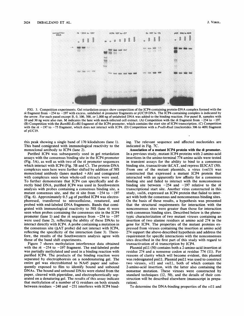
petition experiments were performed. ICP4-containing pro-tein-DNA complexes that formed with each of the fourdescribed regions of the tk promoter were specifically com-peted away when increasing amounts of the same unlabeledfragment were added to the binding reaction. Figure 3 showsa series of competition experiments in which tk -254 to-197 was the labeled probe. Increasing amounts of the
J. VIROL.

INTERACTION OF ICP4 WITH THE tk PROMOTER 2623
- 300 -200 -1 00 0Mr" mm.- mt I YZu O-ua
SP1 CBP SP1 TBP
v KOS.
-Ab
t. Om'se:ptmef t06S
M sot
*Ab *Ab -AOb
_ _
a o**
0_40
..
4 iM
M Eo$ M KOS
D -Et *Ab
~ ~
wm I"
I..: ^ }S -A 4'
FIG. 1. Protein-DNA interactions of ICP4 with tk promoter fragments. A schematic diagram of the tk promoter is shown, includingcis-acting sequences Which interact with cellular transcription factor SP1, proteins that bind to CCAAT boxes (CBP), and TATA box-bindingproteins (TBP). Nucleotides are indicated with reference to the transcription start site. (A) Gel retardation assay using the tk fragment from-480 to -385. Lanes 1 and 2 contained extract from mock-infected cells (M). Lanes 3 and 4 contained extract from KOS-infected cells. Amonoclonal antibody to ICP4 was added to lanes 2 and 4 (+Ab). (B and C) Gel retardation assays using tk fragments from -385 to -254 andfrom -254 to -197, respectively. Lane 1 in each panel contained mock-infected cell extract. Lanes 2 to 6 contained extract fromKOS-infected cells and increasing amounts of nonspecific competitor DNA. Lanes B4 and C6 contained a monoclonal antibody to ICP4. (D)Gel retardation assay using tk fragments from -221 to -115 (lanes 1 to 4) and from -115 to +17 (lanes 5 to 8). Addition of mock- andKOS-infected extracts and the monoclonal antibody was as for panel A. (E) Gel retardation assays using tk fragments from -75 to -13 (lanesI to 4) and from -13 to +54 (lanes 5 to 8). Addition of mock- and KOS-infected cell extracts and the monoclonal antibody was as for panel A.
unlabeled fragment (-254 to -197) were able to compete for complex and the free probe in each panel of Fig. 3).most of the ICP4-containing complex (Fig. 3A) while not at Competition was also observed when excess DNA contain-all affecting the binding of a nonspecific DNA-binding pro- ing the start site of ICP4 transcription (a consensus bindingtein (see the band located between the ICP4-containing site) was used as an unlabeled competitor (Fig. 3B). Very
little competition was observed when excess amounts of the-400 -300 -200 -100 +1 +60 tk -197 to -75 fragment or a piece of pUC19 DNA was used
Iablity to form(Fig. 3C and D). ICP4 did not form a complex with either of
SP1 CBP SP1 TBP with ICP4 these fragments in mobility shift experiments (Fig. 1D; datanot shown).
Binding of purified ICP4 to the tk-binding site. To confirmLS -56-46 + these results and better address the nature of the ICP4-DNA
+ interaction, we purified ICP4 from infected cell nuclei.Nuclear extracts were applied to a Sephacryl S-300 gel
LS -29-18 | + filtration column, and fractions were assayed for immuno-LS +5+15 I logical reactivity to 58S antibody by Western dot blot and for
LS +16+36/+10 + + DNA-binding activity by gel retardation assay. Figure 4A
shows results of a gel retardation assay of the relevantfractions from the Sephacryl S-300 column. The peak of
- DNA-binding activity also corresponded to the peak of 58Santibody reactivity (data not shown). The appropriate frac-
- tions were pooled and applied to a DNA affinity column as
-___________ + described in Materials and Methods. The affinity column was+ eluted stepwise with 10-ml washes of column buffer contain-+ ing 0.3, 0.6, and 0.9 M KCI. The fractions were analyzed by
* ~~~~~~~~~~~Westerndot blot. The elution profile is shown in Fig. 4B. AFIG. 2. Schematic diagram of the tk promoter and all tk promoter ster elution 4Befragments tested for the ability to form ICP4-containing protein- single, sharp peak was obtained with application of the
DNA complexes. Fragments containing LS mutations (35) and the column buffer containing 0.3 M KCl. The material from this
positions of the mutations are indicated. For abbreviations, see the peak was used for subsequent analyses. Figure 4C is a
legend to Fig. 1. Coomassie brilliant blue-stained SDS-polyacrylamide gel of
-400A
+60
4.64
,Z, so,
so.o _
VOL. 64, 1990
I I -T
0000
2624 IMBALZANO ET AL.
ICP4 -107 to +27
OO"Cb M o ° ^ < > °8
0°
_~~~~~~~~~~_
w f.
o *6 :aw _w _ - so *a_
- C
FIG. 3. Competition experiments. Gel retardation assays show competition of the ICP4-containing protein-DNA complex formed with thetk fragment from -254 to -197 with excess, unlabeled tk promoter fragments or pUC19 DNA. The ICP4-containing complex is indicated bythe arrow. For each panel except B, 0, 100, 300, or 1,000 ng of unlabeled DNA was added to the binding reaction. For panel B, samples with10 and 30 ng were also run. M indicates the lane with mock-infected cell extract. (A) Competition with the tk fragment from -254 to -197.(B) Competition with the BamHI-EcoRI fragment of the ICP4 promoter, which contains the start site of ICP4 transcription. (C) Competitionwith the tk -197 to -75 fragment, which does not interact with ICP4. (D) Competition with a PvuII-RsaI (nucleotides 306 to 409) fragmentof pUC19.
this peak showing a single band of 170 kilodaltons (lane 1).This band comigrated with immunological reactivity to themonoclonal antibody to ICP4 (lane 2).
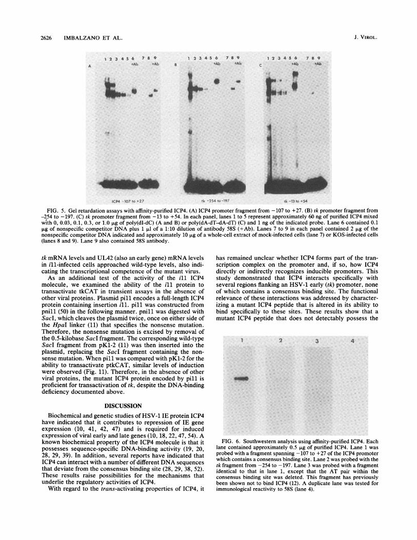
Purified ICP4 was subsequently used in gel retardationassays with the consensus binding site in the ICP4 promoter(Fig. 5A), as well as with two of the tk promoter sequenceswhich interact with ICP4 (Fig. 5B and C). The protein-DNAcomplexes seen here were further shifted by addition of 58Smonoclonal antibody (lanes marked +Ab) and comigratedwith complexes seen when whole-cell extracts were used.To further demonstrate that ICP4 can specifically and di-rectly bind DNA, purified ICP4 was used in Southwesternanalysis with probes containing a consensus binding site, amutant consensus site, and the tk site from -254 to -197(Fig. 6). Approximately 0.5 ,ug of purified ICP4 was electro-phoresed, transferred to nitrocellulose, renatured, andprobed with end-labeled DNA fragments. Bands that comi-grated with immunological reactivity to 58S (lane 4) wereseen when probes containing the consensus site in the ICP4promoter (lane 1) and the tk sequence from -254 to -197were used (lane 2), reflecting the ability of these DNAs tointeract directly with ICP4. A probe containing a mutation inthe consensus site (AAT probe) did not interact with ICP4,reflecting the specificity of the interaction (lane 3). There-fore, the results of the Southwestern analysis agree withthose of the band shift experiments.
Figure 7 shows methylation interference data obtainedwith the tk -254 to -197 fragment. The end-labeled probewas partially methylated and used in a binding reaction withpurified ICP4. The products of the binding reaction wereseparated by electrophoresis on a nondenaturing gel. Theentire gel was electroblotted to NA45 paper and subse-quently exposed to film to identify bound and unboundDNAs. The bound and unbound DNAs were eluted from thepaper, cleaved with piperidine, and electrophoretically sep-arated on a denaturing gel. The results of this assay indicatethat methylation of a number of G residues on both strandsbetween residues -240 and -231 interferes with ICP4 bind-
ing. The relevant sequence and affected nucleotides areindicated in Fig. 7C.
Association of a mutant ICP4 protein with the tk promoter.In a previous study, mutant ICP4 proteins with 2-amino-acidinsertions in the amino-terminal 774 amino acids were testedin transient assays for the ability to bind to a consensusbinding site, transactivate tkCAT, and repress IE3CAT (50).From one of the mutant plasmids, a virus (vnil3) wasconstructed that expressed a mutant ICP4 protein thatinteracted with an apparently low affinity for a consensusbinding site and failed to interact with the nonconsensusbinding site between -254 and -197 relative to the tktranscriptional start site. Another virus constructed in thisstudy, vnil6, expressed an ICP4 protein that failed to inter-act with both the consensus and nonconsensus binding sites.On the basis of these results, a hypothesis was presentedthat the structural requirements for interaction with thenonconsensus sites were greater than those for interactionwith consensus binding sites. Described below is the pheno-typic characterization of two mutant viruses containing aninsertion of two alanine residues at amino acid 274 in thegene for ICP4. The properties of the mutant proteins ex-pressed from viruses containing the insertion at amino acid274 support the above-described hypothesis and address therequirement for specific interactions with the nonconsensussites described in the first part of this study with regard totransactivation of tk transcription by ICP4.
Plasmid pill (50) contains both a 2-amino-acid insertion atresidue 274 and a nonsense codon at residue 774 (11). Forreasons of clarity which will become evident, this plasmidwas redesignated pnill. Plasmid pnill was used to constructtwo viruses, vill and vnill, both of which contain the2-amino-acid insertion, with the latter also containing thenonsense mutation. These viruses were constructed bystandard techniques (12, 50), and the details of their con-struction will be described elsewhere (manuscript in prepa-ration).To determine the DNA-binding properties of the vill and
competitorDNA
tk - 254 to -197 tk -197 to -75 pUC 306 to 4090
0 0 00 0(0
Ogmo(a
. m _ _
J. VIROL.

INTERACTION OF ICP4 WITH THE tk PROMOTER 2625
45 50
2 3
205 kO
116
97
66
45
29
FIG. 4. Purification of ICP4. (A) Gel retardation assay of rele-vant column fractions from a Sephacryl S-300 column. Approxi-mately 0.1 ng of the BamHI-EcoRI (-107 to +27) fragment from theICP4 promoter containing a consensus binding site was used in a
binding reaction with 0.03 ,ug of poly(dI-dC) and 2 ,ul of each columnfraction. (B) Elution profile from the DNA affinity column. Arrowsindicate the points at which the respective elution buffers were
applied. The y axis represents arbitrary units of ICP4 based on
densitometric scans of the Westem dot blot (see Materials andMethods). (C) SDS-gel of 1 ,ug of affinity-purified ICP4 (lane 1). Aduplicate lane was assayed for immunological reactivity to a ICP4-specific antibody 58S (lane 2). Molecular mass markers and theircorresponding sizes are shown in lane 3. kD, Kilodaltons.
vinll proteins relative to those of the full-length KOSprotein and the n208 (parent of vnill) protein, whole-cellextracts were prepared and used in gel retardation assays
with probes containing a consensus binding site and the tksites described in Fig. 1. Both the vill and vnill proteinsformed protein-DNA complexes with the consensus bindingsite but not with the sites in the tk promoter region (Fig. 8).To more closely examine the DNA-binding properties of an
ICP4 molecule containing the ill insertion, the vill proteinwas purified from vill-infected Vero cells by using theprocedure described above for the wild-type protein. Bind-ing of purified KOS ICP4 to the indicated probes was
compared with that of vill in the presence of differentconcentrations of a nonspecific competitor. Protein-DNAcomplexes were evident with the KOS protein at the highestconcentration (1 ,ug) of the nonspecific competitor used,while vill-DNA complexes were not detected at the lowest
concentration (0.03 ,ug) of the competitor (Fig. 8B). There-fore, the specific affinity of the vill protein for the tk siteswas drastically reduced or eliminated. In a similar experi-ment, it was determined that the purified vill protein re-tained the ability to bind to a consensus binding site (data notshown). This is consistent with the results of Fig. 8A andalso with the fact that DNA affinity was successfully used asa method for purification. In the absence of competitorDNA, a relatively intense heterogeneous shift was seen forboth the KOS and vill proteins. This most probably reflectsnonspecific binding of the vill and KOS proteins, becausean antibody further shifted this material (data not shown).However, despite the relatively homogeneous nature of theICP4 preparations used in the above-described experiments(Fig. 4C), we cannot rule out the possibility that the heter-ogeneous material was in part due to a small degree ofcontamination with other nonspecific DNA-binding proteinsor degradation products of ICP4.
Activities of ill insertion mutants. The observation thatboth the vnill and vill proteins had dramatically reducedabilities to associate specifically with the wild-type bindingsites within the -254 to +54 region of the tk gene provides atest for the involvement of these interactions in ICP4-mediated transinduction of the tk gene and ICP4 function ingeneral. Initial analysis of the phenotypes of vill and vnillwas performed by comparing the viral peptides synthesizedin nl2-, n208-, KOS-, vill-, and vnill-infected cells at 4 and9 h postinfection. ni2 contains a nonsense mutation at aminoacid 251 and does not specify ICP4 activities; therefore, itefficiently synthesizes only IE (a) proteins and ICP6 (12).n208, like vnill, contains a nonsense mutation at amino acid774 and is semipermissive for early and "leaky" late (yl)gene expression (12). As can be seen from the SDS-gelprofile of Fig. 9, the ill insertion had no detectable effect inthe KOS or n208 background; therefore, gene expression asmeasured by protein levels was not greatly affected by theill insertions. Consistent with the polypeptide profile shownin Fig. 9 is the observation that the virus vill grew as well asKOS on cells that do not provide ICP4 in trans (data notshown). n12, n208, and vnill require the wild-type ICP4provided by E5 cells for growth because of the presence ofthe nonsense mutations in the genes for ICP4.A direct examination of the effect of the ill mutation on
the transcription of the tk gene was performed by nuclearrun-on transcription. Nuclei were prepared at 6 h postinfec-tion from uninfected cells and from cells infected with ni2,vill, or KOS. Phosphonoacetic acid (300 ,ug/ml) was in-cluded to inhibit DNA synthesis and therefore restrict themeasurement of transcription to that from input templates.Nascent RNA molecules were elongated by in vitro incuba-tion of nuclei with nucleoside triphosphates, including[e&2P]GTP. RNA was isolated and used to probe filterscontaining serial dilutions of single-stranded DNA homolo-gous and complementary to the indicated mRNA. Thedifference between uninduced transcription and ICP4-in-duced transcription of the genes for tk and ICP8 is demon-strated in Fig. 10A by comparison of the ni2 and KOSsignals, respectively. The ill mutation did not affect inducedexpression, since the rates of transcription of the genes for tkand ICP8 in the vill background were similar to those in theKOS background. Also shown in Fig. 10A is the ability torepress representative IE gene (ICP4 and ICP27) transcrip-tion as a consequence of either the vill or KOS ICP4protein.
Figure 10B shows a Northern blot reflecting stable tran-script levels from cells infected with KOS, vill, or n12. Both
A.
35 40fraction
S.
0tIt
0 20
fraction
VOL. 64, 1990
2626 IMBALZANO ET AL.
1 2 3 4 5 6 7 8 9*Ab *Ab B
1 2 3 45 6 7 8 9+Ab $Ab c
1 2 3 4 5 6 7 8 9tAb tAb
a F
.-AX I
*. 1 .:.~...1
a..sP4" 0
ICP4 -10)7 to +27 tk -254 to -197 ik -13 to t54
FIG. 5. Gel retardation assays with affinity-purified ICP4. (A) ICP4 promoter fragment from -107 to +27. (B) tk promoter fragment from-254 to -197. (C) tk promoter fragment from -13 to +54. In each panel, lanes 1 to 5 represent approximately 60 ng of purified ICP4 mixedwith 0, 0.03, 0.1, 0.3, or 1.0 jig of poly(dI-dC) (A and B) or poly(dA-dT-dA-dT) (C) and 1 ng of the indicated probe. Lane 6 contained 0.1pug of nonspecific competitor DNA plus 1 ,ul of a 1:10 dilution of antibody 58S (+Ab). Lanes 7 to 9 in each panel contained 2 jig of thenonspecific competitor DNA indicated and approximately 10 p.g of a whole-cell extract of mock-infected cells (lane 7) or KOS-infected cells(lanes 8 and 9). Lane 9 also contained 58S antibody.
tk mRNA levels and UL42 (also an early gene) mRNA levelsin ill-infected cells approached wild-type levels, also indi-cating the transcriptional competence of the mutant virus.As an additional test of the activity of the ill ICP4
molecule, we examined the ability of the ill protein totransactivate tkCAT in transient assays in the absence ofother viral proteins. Plasmid pill encodes a full-length ICP4protein containing insertion ill. pill was constructed frompnill (50) in the following manner. pnill was digested withSac, which cleaves the plasmid twice, once on either side ofthe HpaI linker (11) that specifies the nonsense mutation.Therefore, the nonsense mutation is excised by removal ofthe 0.5-kilobase Sacl fragment. The corresponding wild-typeSacl fragment from pKl-2 (11) was then inserted into theplasmid, replacing the SacI fragment containing the non-sense mutation. When pill was compared with pKl-2 for theability to transactivate ptkCAT, similar levels of inductionwere observed (Fig. 11). Therefore, in the absence of otherviral proteins, the mutant ICP4 protein encoded by pill isproficient for transactivation of tk, despite the DNA-bindingdeficiency documented above.
DISCUSSION
Biochemical and genetic studies of HSV-1 IE protein ICP4have indicated that it contributes to repression of IE gene
expression (10, 41, 42, 47) and is required for inducedexpression of viral early and late genes (10, 18, 22, 47, 54). Aknown biochemical property of the ICP4 molecule is that itpossesses sequence-specific DNA-binding activity (19, 20,28, 29, 39). In addition, several reports have indicated thatICP4 can interact with a number of different DNA sequencesthat deviate from the consensus binding site (28, 29, 38, 52).These results raise possibilities for the mechanisms thatunderlie the regulatory activities of ICP4.With regard to the trans-activating properties of ICP4, it
has remained unclear whether ICP4 forms part of the tran-scription complex on the promoter and, if so, how ICP4directly or indirectly recognizes inducible promoters. Thisstudy demonstrated that ICP4 interacts specifically withseveral regions flanking an HSV-1 early (tk) promoter, noneof which contains a consensus binding site. The functionalrelevance of these interactions was addressed by character-izing a mutant ICP4 peptide that is altered in its ability tobind specifically to these sites. These results show that amutant ICP4 peptide that does not detectably possess the
I 2 3 4
FIG. 6. Southwestern analysis using affinity-purified ICP4. Eachlane contained approximately 0.5 jig of purified ICP4. Lane 1 wasprobed with a fragment spanning -107 to +27 of the ICP4 promoterwhich contains a consensus binding site. Lane 2 was probed with thetk fragment from -254 to -197. Lane 3 was probed with a fragmentidentical to that in lane 1, except that the AT pair within theconsensus binding site was deleted. This fragment has previouslybeen shown not to bind ICP4 (12). A duplicate lane was tested forimmunological reactivity to 58S (lane 4).
A
J. VIROL.

INTERACTION OF ICP4 WITH THE tk PROMOTER 2627
AU B B U
-240 _W239 *
0O-231
-2324_ -234
C _ -238
FIG. 7. Methylation interference. End-labeled fragments con-taining the tk -254 to -197 fragment were treated as indicated inMaterials and Methods. (A) G cleavage pattern of the coding strand(U, unbound DNA; B, bound DNA). Underrepresented G nucleo-tides are marked by dots and position numbers relative to themRNA start site. (B) G cleavage pattern of the noncoding strand.(C) Relevant nucleotide sequence. Underrepresented nucleotidesare marked with dots.
A.-,X"A.8 8~0
1ta a
intrinsic ability to interact specifically with these nonconsen-sus binding sites flanking the tk promoter induces transcrip-tion of the tk gene to the same level and rate as wild-typeICP4. Our interpretation of these results is that such inter-actions are not absolutely required for activation of tran-scription by ICP4.ICP4 specifically interacts with DNA sequences adjacent
to the tk promoter. One is in the 5'-untranslated regiondownstream of the promoter; the other three are located inupstream regions beyond the conventional limits of the tkpromoter. None of these sequences contains a consensus(ATCGTCnnnnYCGRC) binding site for ICP4. Similarly,protein-DNA complexes have been observed in regions ofother HSV-1 gene promoters that also do not contain con-sensus binding sites (28, 29, 38, 52). This confirms that ICP4can interact with a variety of DNA sequences. The mecha-nism by which this occurs is not understood. A possibleexplanation involves the existence of a single domain on theprotein capable of recognizing divergent DNA sequences.This implies that the nonconsensus ICP4-binding sites maybe degenerate homologies of the consensus and that protein-DNA contacts in flanking regions may contribute to a stableinteraction. In some of the nonconsensus sites identified forICP4, loose homologies of the consensus ATCGTCnnnnYCGRC can be identified. For example, the site locatedbetween -254 and -197 contains the sequence ATCtTgnnnnCCGGa (capital letters match the consensus) from-250 to -236. The nucleotides which interfere with bindingwhen methylated span residues -240 to -231 (Fig. 7). Theexperimentally determined contacts at positions -238,-239, and -240 are contained in the sequence given above,while four additional contacts are adjacent to it. Alterna-tively, certain regions of the protein may act to modify thespecificity of binding, as certain insertion mutations differ-entially affect binding to different sites (this report; 50).Diversity of sequence recognition may also be generated byprotein-protein contacts between ICP4 and other viral orcellular proteins. While this remains a possibility, our resultsindicate that diverse DNA-binding properties are inherent in
0
X X C > :
I-S
a
as
~~~~~~~~~4a i dos
ICP4 -107 to -27 tk -254 to -197 t1 -13 to -34
FIG. 8. Gel retardation analysis of ill and nill ICP4 molecules. (A) The indicated viruses were used to prepare infected cell extracts foruse in gel retardation assays with probes containing the consensus binding site in the ICP4 promoter, spanning tk -254 to -197 or spanningtk -13 to +54. (B) Gel retardation assay with purified wild-type (KOS) or ill ICP4 with tk promoter fragments -254 to -197 (left half) andtk -13 to +54 (right half). The first five lanes in each half represent 1.0 ng of the tk promoter fragment from -254 to -197 with approximately60 ng of purified KOS ICP4 in the presence of 0, 0.03, 0.1, 0.3, and 1.0 ,ug of poly(dI-dC). The second five lanes in each half were identical,except that 60 ng of purified ill ICP4 was used. The right half of the figure was identical, except that the probe used was the tk -13 to +54fragment.
VOL. 64, 1990
tk 254 to -Is97 t6 -13 to -i 54

2628 IMBALZANO ET AL.
ill KOS n12
ICP4
1,2 L
- x
tk
ICP27:::: :. .:_ iiK ..B; g s 4 - ............. ,iN.iX Z ...... .....tl!|!Ew :> i i- y :.f:.: :.:. j Z " . . ;.: *s, . v . . ,:. :z Fi ................. ,.i...* : _
.e
4 IE
5 L6
08 lEE
11 L4' IE15 L
OMdft* ~ 25 L
___ 27 IE
4S Sw
FIG. 9. Polypeptide profiles of the vill and vnill viruses. Cellswere infected at a multiplicity of infection of 20 in Ham F12 mediumand labeled for 30 min with [35S]methionine at 4 or 9 h postinfection.Samples were electrophoresed on a 9% polyacrylamide gel. Anautoradiogram of labeled polypeptides is shown. ICP designationsfor selected proteins are listed on the right. Each protein is alsoclassified as IE, early (E), or late (L). Lane M contained mock-infected cell extract.
the ICP4 protein itself. The mobilities of ICP4-DNA com-plexes seen in crude extracts were the same as those seen forpurified protein (Fig. 5). In addition, ICP4 bound to twodivergent sites when purified protein was used in a South-western protocol (Fig. 6). This result is consistent with theprevious results of Michael et al. (38), who demonstrated thediverse DNA-binding properties of ICP4 in Southwesternexperiments with a promoter-regulatory region of an HSVlate gene.
Examination of the properties of the ICP4 moleculesproduced by the mutant viruses vill and vnill has provideda means to examine the in vivo relevance of the ICP4-binding sites near the tk promoter. Neither the ICP4 inwhole-cell extracts from cells infected with vill or vnill nor
purified ICP4 from vill-infected cells specifically bound to tksequences (Fig. 8). Therefore, analyses of their regulatoryactivities were performed to assess function in the apparentabsence of binding by ICP4. The expression assays examin-ing vill and vnill are shown in Fig. 9 to 11. At the level of
tk
UL42
ICP8
FIG. 10. (A) Run-on transcription assay performed on infectedcell nuclei isolated at 6 h postinfection from cells infected with theindicated viruses in the presence of 300 ,ug of phosphonoacetic acidper ml. Autoradiographic images of 32P-labeled mRNA hybridizedto threefold dilutions (1.0, 0.3, and 0.1 ,ug) of a complementary DNAstrand immobilized on a nitrocellulose filter are shown. Hybridiza-tion to the homologous DNA strand was also performed, and theresulting signal was always greater than 10-fold less than thecomplementary strand signal (data not shown). (B) Northern blotindicating accumulation of stable tk or UL42 mRNA. Total cellularRNAs were prepared from cells infected with the indicated virusesin the presence of 300 ,ug of phophonoacetic acid per ml and thenelectrophoretically separated and transferred to nitrocellulose. Fil-ters were probed with a gel-isolated, 32P-labeled SacI-SmaI frag-ment of the tk gene or a PstI fragment of the UL42 gene.
protein expression, vill and vnill are almost indistinguish-able in the production of IE, early, and late proteins relativeto KOS and n208, respectively. In addition, the transcriptionof the early genes, tk, ICP8, and UL42 in the KOS and villbackgrounds was similar (Fig. 10). The level of IE gene(ICP4 and ICP27) transcription appeared to be slightlyelevated in the vill sample. This may reflect a lower affinityfor the consensus binding site, which has been shown tocorrelate with IE gene repression (49; unpublished data).
0.*
1.0 9.6 62
FIG. 11. Transient expression assay comparing expression ofptkCAT (10) following cotransfection with pUC19, pill, or pKl-2(which encodes wild-type ICP4). The numbers indicate the foldincrease in percent acetylation of chloramphenicol as a function ofpill and pKl-2 relative to that in the cotransfection with pUC19.
4 h"I............
£%A
9h
0 0C04 C4N 0 Z C14 c
c E Y. 1) c >
A. B.
J. VIROL.

INTERACTION OF ICP4 WITH THE tk PROMOTER 2629
The ability of the ill protein to transactivate the tk promoterin the absence of other viral gene products was demon-strated by the ability of pill to transactivate tkCAT intransient assays nearly as efficiently as the wild-type proteincoded for by pKl-2 (Fig. 11). On the basis of these data, wepropose that the interactions between ICP4 and the DNAsequences adjacent to the tk promoter, at best, marginallycontribute to transactivation of tk gene expression and arenot essential for the transinducing properties of ICP4.
It remains possible that the in vitro mobility shift experi-ments are not sensitive enough to measure functional ICP4-DNA interactions within a cell. If so, it is possible that theill protein retains some sequence-specific binding activity invivo, which may confer activity. Arguing against this possi-bility are the results of previous studies which showed thatdeletion of the sequence that encodes the untranslatedregion (23) or the region upstream of -119 relative to the tkmRNA start site (14) had little or no effect on transactiva-tion. Alternatively, functional ICP4-DNA interactions maydiffer from those detectable by in vitro band shift experi-ments.
Studies of the tk promoter have shown that the cissequences which interact with cellular transcription factorsare required for both tk basal-level expression in transientassays and tk expression in the context of the virus, leadingto the proposal that viral trans induction is mediated throughthe interaction of viral trans-activators with cellular tran-scription factors (6). Similar mechanisms have been pro-posed for the simian virus 40 large-T antigen (21), the ElAprotein of adenovirus (56; reviewed in reference 4), and theIE protein of pseudorabies virus (1). We favor a hypothesisinvolving the interaction of ICP4 with one or probablyseveral cellular transcription factors which interact with thetk promoter. The argument for multiple interactions followsfrom the observation that mutations in any one of the siteswhich specify an interaction with a cellular factor(s) do notlower tk expression below the uninduced level (6). Nonspe-cific contacts by ICP4 with the DNA may help to stabilizethese interactions. Note that purified ill protein retained theability to bind to the nonconsensus sites in the absence of anonspecific competitor (Fig. 8B) and that most mutations inthe DNA-binding domain of ICP4 that affect specific DNAbinding also reduce or eliminate transactivation (45, 50). Theeffects of these mutations on the nonspecific DNA-bindingproperties of ICP4 have not been extensively studied. Inview of the experimentally determined size of the ICP4molecule relative to that of the promoter, contact amongICP4, promoter sequences, and one or more cellular trans-activating factors is a reasonable possibility. Tests of thesehypotheses are under way in our laboratory.While our data tend to minimize the importance of specific
DNA binding for transactivation of the tk promoter, itremains possible that this activity is important for transacti-vation of other HSV genes. Other investigators who haveexamined the expression of the HSV-1 gene for gD haveproposed, on the basis of transient assays and in vitrotranscription reactions, that the ICP4-binding sites presentnear the promoter of the gene for gD contribute to transac-tivation (52, 53). In view of the size and complexity of ICP4,it is possible that it transactivates genes by a variety ofmechanisms. Some genes may require the sequence-specificbinding of ICP4 to give the level of expression seen inproductive infection, while other genes, like tk, may not.This may hold in particular for genes such as that for gD,which contains a consensus binding site located at -105 withrespect to the gD mRNA start site (19, 52, 53). The presence
of this site could contribute to transactivation by a differentmechanism used by ICP4, or, more specifically, ill ICP4, totransactivate the tk gene and numerous other genes whichare transactivated but do not contain consensus bindingsites. However, it was previously shown that this sequenceis not necessary for induced expression of the gene for gD intransient assays (16, 17).The possible contribution of the binding sites described
herein for transactivation may become more significant incell types other than those used to propagate productiveinfections in the laboratory. The relative abundance andactivities of cellular transcription factors which mediate tktranscription may vary in different cell types, such as epi-thelial cells and neurons. Therefore, direct sequence-specificbinding of ICP4 could possibly be used as an alternativemechanism to transactivate a gene in one cellular environ-ment and not be required in another.
ACKNOWLEDGMENTS
We thank Donald Coen for helpful discussions and reviewing themanuscript and Steve McKnight and Donald Coen for providingplasmids containing LS mutations. We also thank Sara Sherman formanuscript preparation.
This work was supported by Public Health Service grant A127431from the National Institute of Health and American Cancer Societygrant JFRA-195 to N.A.D.
LITERATURE CITED1. Abmayer, S. M., J. L. Workman, and R. G. Roeder. 1988. The
pseudorabies immediate early protein stimulates in vitro tran-scription by facilitating TFIID:promoter interactions. GenesDev. 2:542-553.
2. Batterson, W., and B. Roizman. 1983. Characterization of theHSV-associated factor responsible for the induction of ot genes.J. Virol. 46:371-377.
3. Beard, P., S. Faber, K. W. Wilcox, and L. I. Pizer. 1986. Herpessimplex virus immediate early infected-cell polypeptide 4 bindsto DNA and promotes transcription. Proc. Natl. Acad. Sci.USA 83:4016-4020.
4. Berk, A. J. 1986. Adenovirus promoters and ElA transactiva-tion. Annu. Rev. Genet. 20:45-79.
5. Campbell, M. E. M., J. W. Palfreyman, and C. M. Preston.1984. Identification of herpes simplex virus DNA sequenceswhich encode a trans-acting polypeptide responsible for stimu-lation of immediate early transcription. J. Mol. Biol. 180:1-19.
6. Coen, D. M., S. P. Weinheimer, and S. L. McKnight. 1986. Agenetic approach to promoter recognition during trans inductionof viral gene expression. Science 234:53-59.
7. Cromlish, W. A., S. M. Abmayer, J. L. Workman, M. Horiko-shi, and R. G. Roeder. 1989. Transcriptionally active immediate-early protein of pseudorabies virus binds to specific sites on
class II gene promoters. J. Virol. 63:1869-1876.8. DeLuca, N. A., M. A. Courtney, and P. A. Schaffer. 1984.
Temperature-sensitive mutants in HSV-1 ICP4 permissive forearly gene expression. J. Virol. 52:767-776.
9. DeLuca, N. A., A. McCarthy, and P. A. Schaffer. 1985. Isolationand characterization of deletion mutants of herpes simplex virustype 1 in the gene encoding immediate-early regulatory proteinICP4. J. Virol. 56:558-570.
10. DeLuca, N. A., and P. A. Schaffer. 1985. Activation of immedi-ate-early, early, and late promoters by temperature-sensitiveand wild-type forms of herpes simplex virus type 1 proteinICP4. Mol. Cell. Biol. 5:1997-2008.
11. DeLuca, N. A., and P. A. Schaffer. 1987. Activities of herpessimplex virus type 1 (HSV-1) ICP4 genes specifying nonsense
peptides. Nucleic Acids Res. 15:4491-4511.12. DeLuca, N. A., and P. A. Schaffer. 1988. Physical and functional
domains of the herpes simplex virus transcriptional regulatoryprotein ICP4. J. Virol. 62:732-743.
13. Dixon, R. A. F., and P. A. Schaffer. 1980. Fine-structure
VOL. 64, 1990

2630 IMBALZANO ET AL.
mapping and functional analysis of temperature-sensitive mu-tants in the gene encoding the herpes simplex virus type 1immediate early protein VP175. J. Virol. 36:189-203.
14. Eisenberg, S. P., D. M. Coen, and S. L. McKnight. 1985.Promoter domains required for expression of plasmid-bornecopies of the herpes simplex virus thymidine kinase gene invirus-infected mouse fibroblasts and microinjected frog oocytes.Mol. Cell. Biol. 5:1940-1947.
15. El Kareh, A., A. J. M. Murphy, T. Fichter, A. Efstratiadis, andS. Silverstein. 1985. "Transactivation" control signals in thepromoter of the herpes virus thymidine kinase gene. Proc. Natl.Acad. Sci. USA 82:1002-1006.
16. Everett, R. D. 1983. DNA sequence elements required forregulated expression of the HSV-1 glycoprotein D gene liewithin 83 bp of the RNA capsites. Nucleic Acids Res. 11:6647-6666.
17. Everett, R. D. 1984. A detailed analysis of an HSV-1 earlypromoter: sequences involved in trans-activation by viral imme-diate-early gene products are not early gene specific. NucleicAcids Res. 12:3037-3056.
18. Everett, R. D. 1984. Trans activation of transcription by herpesvirus products: requirement for two HSV-1 immediate-earlypolypeptides for maximum activity. EMBO J. 3:3135-3141.
19. Faber, S. W., and K. W. Wilcox. 1986. Association of the herpessimplex virus regulatory protein ICP4 with specific nucleotidesequences. Nucleic Acids Res. 14:6067-6083.
20. Faber, S. W., and K. W. Wilcox. 1988. Association of herpessimplex virus regulatory protein ICP4 with sequences spanningthe ICP4 gene transcription initiation site. Nucleic Acids Res.16:555-570.
21. Gallo, G. J., G. Gilinger, and J. C. Alwine. 1988. Simian virus 40T antigen alters the binding characteristics of specific simianDNA-binding factors. Mol. Cell. Biol. 8:1648-1656.
22. Gelman, I. H., and S. Silverstein. 1985. Identification of imme-diate-early genes from herpes simplex virus that transactivatethe virus thymidine kinase gene. Proc. Natl. Acad. Sci. USA82:5265-5269.
23. Halpern, M. E., and J. R. Smiley. 1984. Effects of deletions onexpression of the herpes simplex virus thymidine kinase genefrom the intact viral genome: the amino terminus of the enzymeis dispensable for catalytic activity. J. Virol. 50:733-738.
24. Honess, R. W., and B. Roizman. 1974. Regulation of herpesvirus macromolecular synthesis. I. Cascade regulation of thesynthesis of three groups of viral proteins. J. Virol. 14:8-19.
25. Honess, R. W., and B. Roizman. 1975. Regulation of herpesvirus macromolecular synthesis: sequential transition of poly-peptide synthesis requires functional viral polypeptides. Proc.Natl. Acad. Sci. USA 72:1276-1280.
26. Jones, K. A., K. R. Yamamoto, and R. Tjian. 1985. Two distincttranscription factors bind to the HSV thymidine kinase pro-moter in vitro. Cell. 42:559-572.
27. Kadonaga, J. T., and R. Tjian. 1986. Affinity purification ofsequence-specific DNA binding proteins. Proc. Natl. Acad. Sci.USA 83:5889-5893.
28. Kristie, T. M., and B. Roizman. 1986. a4, the major regulatoryprotein of herpes simplex virus type 1, is stably and specificallyassociated with promoter-regulatory domains of a genes and ofselected other viral genes. Proc. Natl. Acad. Sci. USA 83:3218-3222.
29. Kristie, T. M., and B. Roizman. 1986. DNA-binding site ofmajor regulatory protein a4 specifically associated with promot-er-regulatory domains of ot genes of herpes simplex virus type 1.Proc. Natl. Acad. Sci. USA 83:4700-4704.
30. Maniatis, T., E. F. Fritsch, and J. Sambrook. 1982. Molecularcloning: a laboratory manual. Cold Spring Harbor Laboratory,Cold Spring Harbor, N.Y.
31. McGeoch, D. J., A. Dolan, S. Donald, and D. H. K. Brauer.1986. Complete DNA sequence of the short repeat region in thegenome of herpes simplex virus type 1. Nucleic Acids Res.14:1727-1744.
32. McKnight, S. L. 1982. Functional relationships between tran-scriptional control signals of the thymidine kinase gene ofherpes simplex virus. Cell 31:355-365.
33. McKnight, S. L., and E. R. Gavis. 1980. Expression of theherpes thymidine kinase gene in Xenopus laevis oocytes: anassay for the study of deletion mutants constructed in vitro.Nucleic Acids Res. 8:5931-5948.
34. McKnight, S. L., E. R. Gavis, R. Kingsbury, and R. Axel. 1981.Analysis of transcriptional regulatory signals of the HSV thy-midine kinase gene: identification of an upstream control region.Cell 25:385-398.
35. McKnight, S. L., and R. Kingsbury. 1982. Transcriptionalcontrol signals of a eukaryotic protein-coding gene. Science217:316-324.
36. McKnight, S. L., R. C. Kingsbury, A. Spence, and M. Smith.1984. The distal transcription signals of the herpes virus tk geneshare a common hexanucleotide control sequence. Cell 37:253-262.
37. Metzler, D. W., and K. W. Wilcox. 1985. Isolation of herpessimplex regulatory protein ICP4 as a homodimeric complex. J.Virol. 55:329-337.
38. Michael, N., D. Spector, P. Mavromara-Nazos, T. M. Kristie,and B. Roizman. 1988. The DNA-binding properties of the majorregulatory protein a4 of herpes simplex virus. Science 238:1531-1534.
39. Muller, M. T. 1987. Binding of the herpes simplex virus type 1gene product ICP4 to its own transcription start site. J. Virol.61:858-865.
40. O'Hare, P., and G. S. Hayward. 1985. Evidence for a direct rolefor both the 175,000- and 110,000-molecular-weight immediate-early proteins of herpes simplex virus in the transactivation ofdelayed-early promoters. J. Virol. 53:751-760.
41. O'Hare, P., and G. S. Hayward. 1985. Three trans-actingregulatory proteins of herpes simplex virus modulate immedi-ate-early gene expression in a pathway involving positive andnegative feedback regulation. J. Virol. 56:723-733.
42. O'Hare, P., and G. S. Hayward. 1987. Comparison of upstreamsequence requirements for positive and negative regulation of aherpes simplex virus immediate-early gene by three virus-encoded trans-acting factors. J. Virol. 61:190-199.
43. Parslow, T. G., S. D. Jones, B. Bond, and K. R. Yamamoto.1987. The immunoglobulin octanucleotide: independent activityand selective interaction with enhancers. Science 235:1498-1501.
44. Paterson, T., and R. D. Everett. 1988. Mutational dissection ofthe HSV-1 immediate-early protein Vmwl75 involved in tran-scriptional transactivation and repression. Virology 166:186-196.
45. Paterson, T., and R. D. Everett. 1988. The regions of the herpessimplex virus type 1 immediate-early protein Vmwl75 requiredfor site specific DNA binding closely corresponds to thoseinvolved in transcriptional regulation. Nucleic Acids Res. 16:11005-11025.
46. Post, L. E., S. Mackem, and B. Roizman. 1981. Regulation of agenes of herpes simplex virus: expression of chimeric genesproduced by fusion of thymidine kinase with a gene promoters.Cell 24:555-565.
47. Preston, C. M. 1979. Abnormal properties of an immediate earlypolypeptide in cells infected with the herpes simplex type 1mutant tsK. J. Virol. 32:357-369.
48. Quinlan, M. P., and D. M. Knipe. 1985. Stimulation of expres-sion of a herpes simplex virus DNA-binding protein by two viralfunctions. Mol. Cell. Biol. 5:957-963.
49. Roberts, M. S., A. Boundy, P. O'Hare, M. C. Pizzorno, D. M.Ciufo, and G. S. Hayward. 1988. Direct correlation between anegative autoregulatory response element at the cap site ofherpes simplex virus type 1 IE175 (a4) promoter and a specificbinding site for the IE175 (ICP4) protein. J. Virol. 62:4307-4320.
50. Shepard, A. A., A. N. Imbalzano, and N. A. DeLuca. 1989.Separation of primary structural components conferring auto-regulation, transactivation, and DNA-binding properties to theherpes simplex virus transcriptional regulatory protein ICP4. J.Virol. 63:3714-3728.
51. Showalter, S. D., M. Zweig, and B. Hampar. 1981. Monoclonalantibodies to herpes simplex virus type 1 proteins, including theimmediate-early protein ICP4. Infect. Immun. 34:684-692.
J. VIROL.

INTERACTION OF ICP4 WITH THE tk PROMOTER 2631
52. Tedder, D. G., R. D. Everett, K. W. Wilcox, P. Beard, and L. I.
Pizer. 1989. ICP4-binding sites in the promoter and codingregions of the herpes simplex virus gD gene contribute toactivation of in vitro transcription by ICP4. J. Virol. 63:2510-2520.
53. Tedder, D. G., and L. I. Pizer. 1988. Role for DNA-proteininteraction in activation of the herpes simplex virus glycopro-tein D gene. J. Virol. 62:4661-4672.
54. Watson, R., and J. B. Clements. 1980. A herpes simplex virustype 1 function continuously required for early and late virus
RNA synthesis. Nature (London) 285:329-330.55. Weinheimer, S. P., and S. L. McKnight. 1987. Transcriptional
and post-transcriptional controls establish the cascade of herpessimplex virus protein synthesis. J. Mol. Biol. 195:819-833.
56. Wu, L., D. S. E. Rosser, M. C. Schmidt, and A. Berk. 1987. ATATA box is implicated in ElA transcriptional activation of a
simple adenovirus 2 promoter. Nature (London) 326:512-515.57. Zipser, D., L. Lipsich, and J. Kwoh. 1981. Mapping functional
domains in the promoter region of the herpes thymidine kinasegene. Proc. Natl. Acad. Sci. USA 78:6276-6280.
VOL. 64, 1990