functional recovery and rehabilitation after acquired ... · implementation of this unified model...
TRANSCRIPT
1
Functional Recovery and Rehabilitation After Acquired Brain Damage:
Mechanisms and Possibilities for Strengthening Treatment, with Special Focus on the Potentials of the Application of Environmental Enrichment.
Master’s Thesis
Program: Neuropsychology. 10th semester, psychology
Total number of characters in the
assignment
(incl. spaces and footnotes): 146.356
Equal to number of normal pages:
60,98
Name and Student number:
Louise Gade, 2009-2423
10th semester, psychology.
Master’s Thesis.
Neuropsychology.
Aalborg University,
October, 2014.

2
Abstract
Worldwide, traumatic brain injury is one of the leading causes of severe disability.
Therefore, it seems pivotal that we investigate the possibility for strengthening the
treatment of brain injury or even present some alternatives to existing treatment.
However, in order for us to fully grasp functional recovery, we need to learn more
about the neural mechanisms involved in this process. Studies using fMRI have
indicated that there are some correlation between activity in specific areas of the brain
and the conduction of particular tasks, indicating that the brain is modular to some
extent. However, in an extreme modular theory, recovery of a function that was lost to
injury is not possible, which indicates that the brain cannot be entirely modular, since
functional recovery does occur. An alternative to the theory of functional localization
is connectionism. Within this view, functional recovery occurs because the
informational input provided to the brain is actually distributed across the brain and
one structure is not the sole mediator of a specific task. However, in an extreme
connectionist view, the aforementioned correlation between brain activity and task
conduction cannot be explained. Therefore, neither of the two perspectives adequately
covers how, evidently, functional localization and functional recovery can coexist.
Thus, there is a need for a more comprehensive model of the brain, one that accounts
for both localization of brain function, as well as functional recovery. The model of
Reorganization of Elementary Functions, REF-model, does exactly that.
Implementation of this unified model has consequences for the construction of
rehabilitative training of brain injury, since, evidently, recovery of function is often
very task-specific and not necessarily transferable to other situations, for instance a
real life situation, something which seems essential for the recovering brain injury
patient. Therefore, the REF-model dictates that rehabilitative training must constructed
in a fashion that resembles everyday life, which further emphasizes the significant
impact that the situation in which the training is conducted has on the functional
outcome. The results of the experiment presented in this paper supports this theory.
Briefly, the main focus of the experiment was to study the effects of environmental
enrichment as a therapeutic tool. We studied the acquisition of the delayed alternation
test in a T-maze in six groups of rats, each group representing a different combination
of operation, sham or transection of the fimbria-fornix, and housing condition,
cognitively enriched, socially enriched or non-enriched housing. Results indicate that
3
environmental enrichment has a positive effect on the functional outcome. Animals
housed in both of the enriched environments made significantly less errors on the
delayed alternation test compared to the non-enriched animals. Further,
pharmacological testing indicated that the neural substrate employed by the animals,
post-traumatically, is not necessarily the most efficient. When we inhibited their
dopaminergic system, all lesioned animals performed significantly better compared to
control days. This might indicate that the functional reorganization exhibited by these
animals has the potential to be even more effective. This, I feel, stresses the importance
of environmental input in the formation of alternative neural substrates for mediation
of a given task. Therefore, I find it pivotal that we try to further investigate the neural
mechanisms involved in functional recovery, the situation in which rehabilitative
training is conducted and how training is constructed. This will allow us to more
efficiently treat our brain injury patients and provide them with the optimal mediation
of function.

4
Abbreviations
CCI – Controlled Cortical Impact
EE – Environmental Enrichment
FF – Fimbria-Fornix
FPI – Fluid Percussion Injury
LFPI – Lateral Fluid Percussion Injury
PFC – Prefrontal Cortex
REF – Reorganization of Elementary Functions
SE – Social Enrichment
SH – Standard Housing
TBI – Traumatic Brain Injury
UCN – Unit for Cognitive Neuroscience
5
Table of Contents 1. Introduction ............................................................................................................ 7
1.1. Thesis statement ................................................................................................ 8
1.1.2. Problem definition ...................................................................................... 8
1.2. Outline ............................................................................................................... 9
2. Models of the Brain .............................................................................................. 10
2.1. Theory of functional localization .................................................................... 11
2.2. Connectionism ................................................................................................ 14
2.3. Preliminary reflections .................................................................................... 16
2.4. Model for Reorganization of Elementary Functions (REF-model) ................ 16
2.4.1. Three principles of post-traumatic functional recovery .......................... 17
2.4.2. The REF-model ........................................................................................ 19
2.4.3. Implementation of the REF-model ........................................................... 22
3. Animal models ...................................................................................................... 23
3.1. Usefulness of animal models .......................................................................... 23
3.2. Ethical Considerations .................................................................................... 24
3.2.1. Contractarianism ..................................................................................... 24
3.2.2. Utilitarianism ........................................................................................... 26
3.2.3. Animal Rights View .................................................................................. 28
3.2.4. Conclusion ............................................................................................... 28
3.3. Brain Injury Models ........................................................................................ 29
3.3.1. Fluid Percussion Injury............................................................................ 30
3.3.2. Controlled Cortical Impact ...................................................................... 31
3.3.3. Transection of the Fimbria Fornix Fiber Bundle .................................... 32
4. Experimental section ............................................................................................ 37
4.1. Introduction ..................................................................................................... 38
4.2. Methods ........................................................................................................... 39
6
4.2.1. Subjects .................................................................................................... 39
4.2.2. Group randomization ............................................................................... 40
4.2.3. Housing conditions .................................................................................. 40
4.2.4. Surgical procedure ................................................................................... 40
4.2.5. Ethovision ................................................................................................. 41
4.2.6. Cognitive task: Delayed alternation ........................................................ 42
4.2.7. Pharmacological challenges .................................................................... 43
4.2.8. Histology .................................................................................................. 44
4.2.9. Statistical analysis .................................................................................... 44
4.3. Results ............................................................................................................. 44
4.3.1. Motor activity ........................................................................................... 45
4.3.2. Histology .................................................................................................. 46
4.3.3. Behavioral data ........................................................................................ 46
4.3.4. Pharmacological challenges .................................................................... 49
4.3.5. Summarizing main differences between the present data and the complete
data set ............................................................................................................... 55
4.4. Discussion ....................................................................................................... 56
4.4.1. Discussion of results ................................................................................ 56
4.4.2. Returning to the REF-model .................................................................... 64
4.5. Conclusion ...................................................................................................... 66
5. Concluding remarks ............................................................................................ 67
6. References ............................................................................................................. 72
7
1. Introduction
Traumatic brain injury (TBI) is one of the leading causes of death and disability in
people under the age of forty-five. Approximately ten million people worldwide are
afflicted with TBI every year and an estimated 57 million TBI-survivors are currently
living with the ramifications of brain damage (Langlois et al., 2006). Indeed, the
disabilities caused by trauma to the brain, can be devastating. Temporary or permanent
impairment of physical, psychosocial and cognitive functions is not unusual following
TBI (Mass et al., 2008), and the clinical symptoms include anxiety, sensory and
attentional impairments, as well as profound memory deficits (Johnson et al., 2013).
With this in mind, it seems evident that it is necessary to investigate ways to alleviate
these symptoms as quickly and as efficiently as possible. Studies on non-invasive
intervention strategies indicate that such strategies have beneficial effects in the
treatment of patients with cognitive disabilities caused by TBI (Pang & Hannan, 2013;
Cheng, 2012; Leggio et al., 2005; Johnson et al., 2013; De Bartholo et al., 2008). We
know that voluntary exercise may, on its own, facilitate neural rehabilitation after
acquired brain damage, because it leads to an up regulation of brain-derived
neurotrophic factor (BDNF), which in turn enhances neuroplasticity and the survival
of neurons (Griesbach et al., 2004). Since physical disabilities often follow TBI (Mass
et al., 2008), some patients might not be able to exercise enough, if at all, for it to have
an effect. In cases where physical exercise is not a possibility, the need to investigate
other non-invasive alternatives, becomes evident.
Animal models have showed us that environmental enrichment (EE) induces neuronal
changes in both the healthy and the injured brain (van Praag et al., 2000; Johnson et
al., 2013; Garcia et al., 2011). These changes include increased neuronal density, more
dendritic branching, as well as an increase in the number of neuronal synapses
(Kempermann et al., 1997; Leggio et al., 2005; Olson et al., 2006; van Praag et al.,
2000). Further, EE promotes neurogenisis, the birth of neurons, and angiogenesis, the
physiological process through which new blood vessels are formed from pre-existing
vessels. In addition, EE facilitates survival of hippocampal neurons (Garcia et al.,
2011; van Praag et al., 2000), which reduces TBI-induced deficits, especially learning
and memory deficits (Will et al., 2004; Garcia et al., 2011), and improves recovery
time (Johnson et al., 2013). As such, the concept of EE poses an eligible intervention

8
strategy (Garcia et al., 2011; Johnson et al, 2013; Cheng, 2012). However, in order for
us to fully grasp in what way alternative rehabilitative strategies, like enriched
environments, can be utilized, we need to learn more about the mechanisms involved
in functional recovery after brain damage in general, i.e. how can reorganization be
conceptualized?
The focus of this paper, therefore, is as follows:
1.1. Thesis statement
“Functional recovery and rehabilitation after acquired brain damage – mechanisms
and possibilities for strengthening treatment, with special focus on the potentials of
the application of environmental enrichment.”
1.1.2. Problem definition
With the thesis statement above, I outline the focal point of my paper: Functional
recovery and rehabilitation. Considering the pronounced disabilities of brain injury,
mentioned above, I feel no need to further elaborate why this is the focus of my thesis.
I will try to account for some of the mechanisms involved in the process of recovery
in broad, theoretical terms based on models of the brain and how it functions, briefly
outlining some of the neural mechanisms involved in recovery, however not on an
atomic or chemical level. I will be discussing, different conceptualizations of the brain
and how they explain functional recovery, and further discuss the importance of
implementing a comprehensive model that allows us to provide our brain injury
patients with optimal treatment. This will cover the part of the thesis statement that
concerns the possibilities for strengthening treatment. Even though this discussion can
be applied to any kind of damage to the brain, my main focus is on TBI models and
not, for instance, ischemia or stroke models. Therefore, I will not account for models
of this kind. I will, however, provide information about different types of TBI models
and the application of these. I will include a study I have been a part of conducting, in
which we have studied some of the potentials of applying environmental enrichment
on hippocampally lesioned animals. For this reason, my main focus will be on the
hippocampus and hippocampal damage. The experimental section will cover the part
of the thesis statement concerning application of this specific intervention strategy.
Through this, I hope to demonstrate that I can apply both theory and empirical
evidence for conduction of a study, and that I know how to utilize relevant research
9
methodology for this purpose. Further, it will show that I understand how a research
design is constructed, and that I know how to handle data material in a way that allows
me to interpret the results, as well as discuss how they relate to already existing
empirical evidence and underlying theory. Moreover, I will provide a demonstration
that I can put the results into perspective and reflect on the possibilities for further
research on the subject. Through this, I will further show that I can include results from
other studies, while still bearing in mind how those results relate to my own, and
whether the content of the other experiment is directly transferable to that of my own.
With this in mind, I will briefly present the outline of the paper.
1.2. Outline
In order for us to understand the mechanisms involved in functional recovery, it seems
essential that we try to understand how the brain functions in general, something,
which causes massive dispute within the field of neuroscience. Thus, I intend to
include a theoretical exposition and discussion of two models of the brain that
represent two extremes on a continuum of brain models: Functional localization and
connectionism. I will try to demonstrate the strengths and weaknesses of these views
and present an alternative that take said weaknesses into account, the model for
Reorganization of Elementary Functions (REF). This section will include a discussion
of the importance of implementing a more comprehensive model, and how such a
model can help us provide the best possible treatment strategies for our brain injured
patients.
Since the empirical element of this paper is comprised of a study on the effects of
environmental enrichment on the performance of hippocampally damaged rats in the
delayed alternation test in the T-maze, the paper will also include a section in which
the use of animal models in this kind of research is discussed. This will include a
section that contains reflections about the usefulness of animal models in brain injury
research, the ethical implications of using animal models, as well as a description and
comparison of different types of brain injury animal models, taking both usefulness
and ethical issues into account. Within this section, I will provide a short introduction
to the anatomy of the hippocampal formation, as well as the main efferent and afferent
pathways, and a brief outline of some of the tests that can utilized in the assessment of
injury to the hippocampus, in humans as well as animals.
10
After this, an experimental section will follow, in which I will present the experiment
that I have been working on at The Unit for Cognitive Neuroscience (UCN). This
experiment is presented in as much detail as would be relevant in order for other
research units to replicate the experiment. This includes a detailed description of the
applied methodology, a comprehensive elaboration of data and results, and a
discussion of the results and how they relate to brain injury treatment utilizing
environmental enrichment. Further, I will try to place the findings in a broader
theoretical frame, more specifically how they relate to the REF-model.
Lastly, in a concluding section, I will summarize the main arguments that I have
presented in my discussions and the conclusions I have arrived at throughout the
paper.
Having outlined the paper, I now turn my focus to models of the brain.
2. Models of the Brain
It has long been assumed that the brain stops developing early in life and that
throughout adulthood it remains static (Flor, 2004). This static conceptualization of
the brain, however, has since been thoroughly scrutinized and, evidently, the brain is
far more flexible than such an assumption affords. In 1949, Donald O. Hebb (1904-
1985) hypothesized that synapses in the brain are strengthened and altered through
experience, and subsequently, several studies have shown that synaptic structure and
activity can indeed be altered by, for example cognitive training, (e.g. Mogensen et al.,
1982), stimulating environments (e.g. Johansson, 2004), and exercise (e.g. Griesbach
et al., 2004), indicating that the brain is much more plastic than assumed so far.
Evidently, plasticity is not something we can circumvent, however, in what way this
highly plastic brain is conceptualized is much debated. In the following, I will present
two of the primary conceptualizations, functional localization and connectionism, and
how, in many ways, these are opposites, yet somehow the shortcomings of functional
localization theory are covered by connectionism and vice versa.
Firstly, I will account for the concept of functional localization and present a clinical
example that supports this line of thought, after which I will provide examples of cases,
both clinical and laboratory, that cannot be explained by the functional localization
theory. This will be followed by a section in which I present a connectionist model of
11
the brain and studies that support this theory. Problems with this line of thought will
be accounted for in this section as well. Lastly, I will illustrate how the model for
Reorganization of Elementary Functions (REF) might provide an alternative that take
both of the aforementioned views into consideration. Further, I will lay out the basic
principles of the REF-model as well as provide examples of studies that support this
model and, briefly, demonstrate how the model relates to clinical practice.
2.1. Theory of functional localization
Functional localization can be traced back to Franz Joseph Gall (1758-1828) and the
concept of phrenology, the idea that certain brain areas have localized, specific
functions and that these areas can be located on the basis of the external anatomy of
the skull (Gerlach, Starrfelt, Gade and Pedersen, 2010). The epitome of functional
localization theory is the theory of modularity as presented by Jerry Fodor (1983), and
later by John Tooby and Leda Cosmides (1992). The main idea is that the brain consists
of highly specialized modules that each process information specific to that module
and as such, any given function is always mediated by the same module (Barrett and
Kurzban, 2006; Buller and Hardcastle, 2000; Mogensen and Malá, 2009). Essentially,
therefore, the brain is regarded as an entity that consists of separate and specialized
structures, and one of Fodor’s (1983) arguments were based on the assumption that
damage to a specific modular system would affect only the specific process that this
module handled and leave other processes intact. By this logic, we should be able to
learn more about specific modules and their functions by studying the symptoms
exhibited by patients with brain injury and deducing information from that, a well-
known practice (Mogensen and Malá, 2009). This further explains why we are able to
predict the outcome of lesions to specific areas, for example that a lesion to Broca’s
area, a well-known structure located in the frontal lobe of the left hemisphere, renders
the patient unable to produce fluent language, a phenomenon called Broca’s Aphasia
(Breedlove, 2010).
However, as mentioned above, the brain is highly plastic, and even patients suffering
from posttraumatic aphasia can regain the ability to speak fluently and correctly
(Breedlove, 2010), exhibiting an advanced level of functional recovery, something
which localization theory cannot account for. Indeed, in this theory’s most radical
form, functional recovery seems to be an impossibility (Mogensen and Malá, 2009).
Since this obviously poses an explanatory problem, it has been argued that in cases of
12
functional recovery, the lesion has not been complete, which is quite often the case in
human TBI, and thus, conclusions about functional localization should only be drawn
from cases where the posttraumatic symptoms are chronic (e.g. Olton, 1978, ref.
Mogensen and Malá, 2009). However, this explanation does not account for studies,
in which laboratory animals are inflicted with complete lesions, and still reach full
functional recovery. This is the case, for instance, in an experiment performed by
Mogensen et al. (2004). In this study, rats where randomly divided into four
experimental groups: 1) Sham surgery, 2) bilateral transection of the fimbria-fornix,
3) bilateral subpial aspiration of the anteriomedial prefrontal cortex, and 4)
combination of bilateral transection of the fimbria-fornix and bilateral subpial
aspiration of the anteriomedial prefrontal cortex. These animals were subjected to
place learning training in a water maze resembling the one constructed by Morris (e.g.
1984), in which they were expected to reach a stationary, submerged platform within
ten seconds on five consecutive trials. Even though some of the animals were heavily
impaired in this task, all subjects managed to reach the behavioral criterion of reaching
the platform within ten seconds on five consecutive trials, therefore exhibiting full
functional recovery in spite of a complete lesion (Mogensen et al., 2004).
Thus, localization theory, it seems, is too constricting and cannot account for
functional recovery, which evidently poses an explanatory problem, since this
phenomenon, is extremely well-founded (e.g. Mogensen et al., 2004; 2005; 2007;
Wilson, 2002; Cheng et al., 2012) . Even so, textbooks on neuropsychology still teach
theories that are fundamentally localization-type theories, as is the case with the
pathway model of visual processing. In short, this theory demonstrates that there are
two distinct visual streams involved in processing visual information, the ventral and
the dorsal streams, located in the occipitotemporal and the occipitoparietal areas,
respectively (Mishkin et al., 1983). The ventral stream is specialized in object
recognition, whereas the dorsal stream specializes in localization. Because of their
distinctive specializations, the two streams have typically been named the what-
pathway and the where-pathway (Milner and Goodale, 1992; Milner and Goodale,
2006; Gerlach and Marstrand, 2010). This segregation of functions is exemplified by
clinical disorders caused by lesions to either stream. Thus, visual object agnosia, a
disorder in which the patient is unable to recognize an object by vision alone (Ogden,
2005), even though she is perfectly capable of calibrating the correct movements for

13
handling the object (Milner and Goodale, 2008), is an example of damage to the ventral
stream. The opposite of this phenomenon is optic ataxia, a disorder caused by damage
to the dorsal stream, in which the patient is unable to reach out for the object even
though she has no problem recognizing the object (Kartsounis, 2010). This model,
clearly, is a functional localization-type model. However, as deducted from the above,
these models have explanatory issues and this model, I am afraid, is no exception,
since, evidently, there is a functional overlap between the two streams (Ellison and
Cowey, 2006). For instance, in a study by Ellison and Cowey (2006), participants were
asked to perform discrimination tasks that involved shape or distance, tasks that
traditionally rely on ventral or dorsal mechanisms, respectively. By utilization of
Transcranial Magnetic Stimulation (TMS), it is possible to render a specific area of
the brain unusable for a short period of time. The magnetic stimulation inhibits the
neurons in the stimulated area and renders it dysfunctional (Breedlove, 2010). If there
is indeed no overlap between ventral and dorsal streams, then magnetic stimulation of
either stream should have no effect on the task solution, if the task is mediated by the
stream not affected by TMS. Indeed, TMS of the dorsal stream has no effect on the
object discrimination task, however, this is not the case for TMS of the ventral stream
and the distance discrimination task. When the team applied TMS to the ventral stream
in the distance discrimination task, the reaction time increased significantly (Ellison
and Cowey, 2006; Ellison and Cowey, 2007). A dichotomy between a ventral and a
dorsal stream cannot account for these results, which seemingly demonstrate a
functional overlap between what was traditionally thought of as two segregated
streams, thus contradicting the pathway model. Therefore, de Haan and Cowey (2011)
introduce an alternative to this model, the patchwork model, according to which the
visual system is constituted by an intricate network of systems, taking into account
how visual information is shared and processed across the streams. This model is
connectionist in its essence and, evidently, provides the answers to the explanatory
problems of the traditional model, the pathway model.
The same is true about the connectionist models of the brain, in general. They seem to
diminish the functional localization theories and explain the very essential explanatory
issues that such theories have. So why not abandon the functional localization theories
and adopt connectionism? After an introduction to connectionism and an elaboration
14
of clinical as well as laboratory studies that support this view, I will demonstrate why,
clearly, it is not as simple as that.
2.2. Connectionism
An overwhelming amount of literature supports the notion that functional recovery
does indeed occur (e.g. Flor, 2004; Johansson, 2004; Robertson and Murre, 1999;
Wilson, 2000; Wilson, 2002; Mogensen and Malá, 2009; Mogensen, 2014), and since
functional localization cannot account for this phenomenon, the connectionist models
have a clear advantage over the localization theories.
The basic assumption of connectionsm is that the different brain structures receive
several diverse types of input, and that within this intricate system that is the brain,
massive sharing of information occurs across structures (Buller and Hardcastle, 2000),
as it is the case in the patchwork model presented above (de Haan and Cowey, 2011).
This assumption provides the fundamental argument for the way in which the
connectionist models explain functional recovery, a phenomenon that, according to
Buller and Hardcastle (2000), belies the concept of specialized modules. As we know,
the brain is constantly changing and adapting in accordance with environmental
demands (Flor, 2004; Johansson, 2004; Buller and Hardcastle, 2000). For instance, if
a specific region is overstimulated by a massive input of sensory information, for
example the areas responsible for processing informational input derived from the
fingers of the left hand of a professional violinist, these areas will expand (Breedlove,
2010; Buller and Hardcastle, 2000). Further, losing a finger causes a massively plastic
response within the somatosensory cortex, since the brain region responsible for
processing information from the missing digit, will decrease and, conversely,
neighboring regions will expand into the sensory-deprived area (Flor, 2004; Buller and
Hardcastle, 2000). Even in instances where the injury does not affect the informational
input, but rather the area in which it is processed, for example by a lesion to the sensory
area involved in performing a task that requires finger dexterity, the skill can be
reacquired, indicating full functional recovery (Robertson and Murre, 1999).
According to Buller and Hardcastle (2000), the only way to explain this kind of
functional recovery is by accepting that the information that is processed by one
structure, is in fact distributed to other structures as well, and functional recovery is
merely an unmasking of alternative structures that have been recipients of this
particular informational input all along. They argue that all processes, both the most

15
basic as well as the higher cognitive ones, are highly dynamic, and that the brain must
be conceived as a domain dominant system, meaning that one area of the brain might
be especially involved in processing specific informational input, however other
structures are involved simultaneously (Buller and Hardcastle, 2000). If we take the
argument to an extreme – that informational input can always be processed by other
neural substrates and that, essentially, another network can do exactly the same as the
lost network – then complete functional recovery should always occur. Essentially,
this would mean that a copy of pre-traumatic information processing is, post-
traumatically, available in alternative neural structures, which is not the case
(Mogensen and Malá, 2009). Sometimes recovery of function fails to happen and the
patient has to utilize compensational strategies to circumvent the functional disability.
This is the case, for example, with a patient, Bill, who after a stroke became densely
aphasic. He had only one word, “bah”, and the word comprehension of a two-year old.
Bill never regained his ability to speak. However, with compensational strategies, he
was able to understand simple instructions and a simple form of communication was
established (Wilson, 2000).
Even though there is evidence of spontaneous recovery (e.g. Wilson, 2002; Robertson
and Murre, 1999), there is indisputable evidence that behavioral and cognitive therapy
can aid this process (e.g. Mogensen, 2011c; Robertson and Murre, 1999; Wilson, 2000;
Wilson, 2004), something the connectionist models do not dispute. Indeed, they
emphasize the significant influence of the environmental input (Buller and Hardcastle,
2000; Robertson and Murre, 1999). However, the connectionist models have problems
explaining why we, with high accuracy, can predict the symptoms of specific lesions
as is the case with lesions to Broca’s area, as mentioned above, and why functional
magnetic resonance imaging (fMRI) can provide evidence of correlations between
specific tasks and activation of particular brain regions (Breedlove, 2010), indicating
that even though functional recovery does occur, the brain exhibits some level of
modular specificity nonetheless.
This inadequacy of the connectionist models is not only apparent in the clinical setting.
Laboratory studies, as well, have demonstrated that this hyper-connectionist
conception of the brain might be too simple and not exactly right, something that has
led to the formation of the REF-model as presented by Mogensen (2011a; 2011c;
2014), to which I will return later on.

16
2.3. Preliminary reflections
It seems that the way we conceptualize the brain in general has consequences for the
way we understand not only specific functions and subsystems, such as the visual
system, but also how we understand functional recovery, rehabilitation and treatment
of human brain injury. Following the logic of extreme functional localization,
functional recovery is not possible and as such, behavioral and cognitive rehabilitative
training following brain injury is irrelevant, since a structure that is lost to injury, is
lost forever (Mogensen et al., 2007). The connectionist models have no problem have
explaining functional recovery, since, in their view, there is always an alternative
processing system that can take over when an area of the brain is lost to injury. Through
experience, they argue, one is able to construct a neural network equivalent to the one
that was lost. However, as mentioned above, this interpretation might be too simplistic.
Further, there is evidence that the brain is modular to some extent, confer the above,
something which the connectionist models cannot account for. Thus, apparently, the
theory of functional localization provides the answers to the explanatory problems of
the connectionist models and vice versa.
Hopefully, this clarifies why I believe that neither localization theory nor
connectionism provide adequate conceptualizations of the brain. In my mind, neither
of the two theoretical stances allow proper conceptualization of functional recovery,
something which has dire consequences for the relevance of post-traumatic,
rehabilitative behavioral and cognitive training, as well as which therapeutic
interventions we utilize and how the training is composited. By implementing
inadequate models of the brain, we risk providing treatment to our brain injury patients
that is not the most efficient treatment. In the following, I will account for the
theoretical structure of the REF-model, which is based on empirical evidence, and
demonstrate how this model explains how functional recovery and functional
localization can coexist. Lastly, I will address the clinical relevance of this model and
how applying a more comprehensive model will help us ensure more efficient brain
injury treatment.
2.4. Model for Reorganization of Elementary Functions (REF-model)
Based on the aforementioned, there are, evidently, two primary theoretical
assumptions about post-traumatic functional recovery, a moderate localization-type

17
assumption and a connectionist assumption, respectively: 1) there is a post-traumatic
reestablishment of the same neural substrate that mediated the task pre-traumatically,
by preserved or repaired elements of the injured structure or system, and 2) post-
traumatically, a copy of the pre-traumatic information processing is available in
alternative neural structures (Mogensen and Malá, 2009). However, there seems to be
a pattern in the mechanisms involved in post-traumatic recovery, something that has
been immensely covered by Mogensen and Malá (2009) through a series of studies
(e.g. Mogensen et al., 2002; 2004a; 2005; 2007), which contradict these traditional
lines of thought. In the following, I will present the three basic principles that have
been suggested to account for post-traumatic recovery of cognitive functions. Table
2.4.1. displays the principles as presented by Mogensen (2011a; 2011c; 2014).
2.4.1. Three principles of post-traumatic functional recovery
The first principle of post-traumatic recovery is that when examining the neural
substrate of task mediation in brain injured, yet functionally recovered individuals,
there is a modified degree of task mediation by intact structures. Some structures
exhibit increased, and in some cases decreased, levels of contribution to task
mediation. This principle is based on several studies, indicating changed importance
of brain systems after functional recovery after acquired brain damage. For instance,
the importance of the prefrontal cortex in allocentric place learning in a water-maze
after transection of the fimbria-fornix fiber bundle (Mogensen et al., 2004), or the
changed importance of pre-frontal dopaminergic mechanisms in a non-mapping place
learning task in a water-maze, when the cholinergic system had been rendered
dysfunctional by scopolamine administration (Mogensen et al., 2002). The second
principle is that after acquired brain damage, the functional recovery is mediated by
unique and dissimilar neural substrates, and, further, is task-dependent. This principle
is based on several studies (Mogensen et al., 2004; 2005; 2007) in which evidence
have been found that after a combined lesion to the hippocampus and the prefrontal
cortex, a neural substrate that does not involve these two structures is able to mediate
full recovery in an allocentric place learning task, however not in egocentric,
orientation, or delayed alternation tasks. The third principle is based on a study by
Mogensen et al. (2004) in which fully recovered individuals clearly demonstrate
different levels of cognitive representation of the platform position in an allocentric
place learning task in a water-maze after lesions of the hippocampus, the prefrontal
18
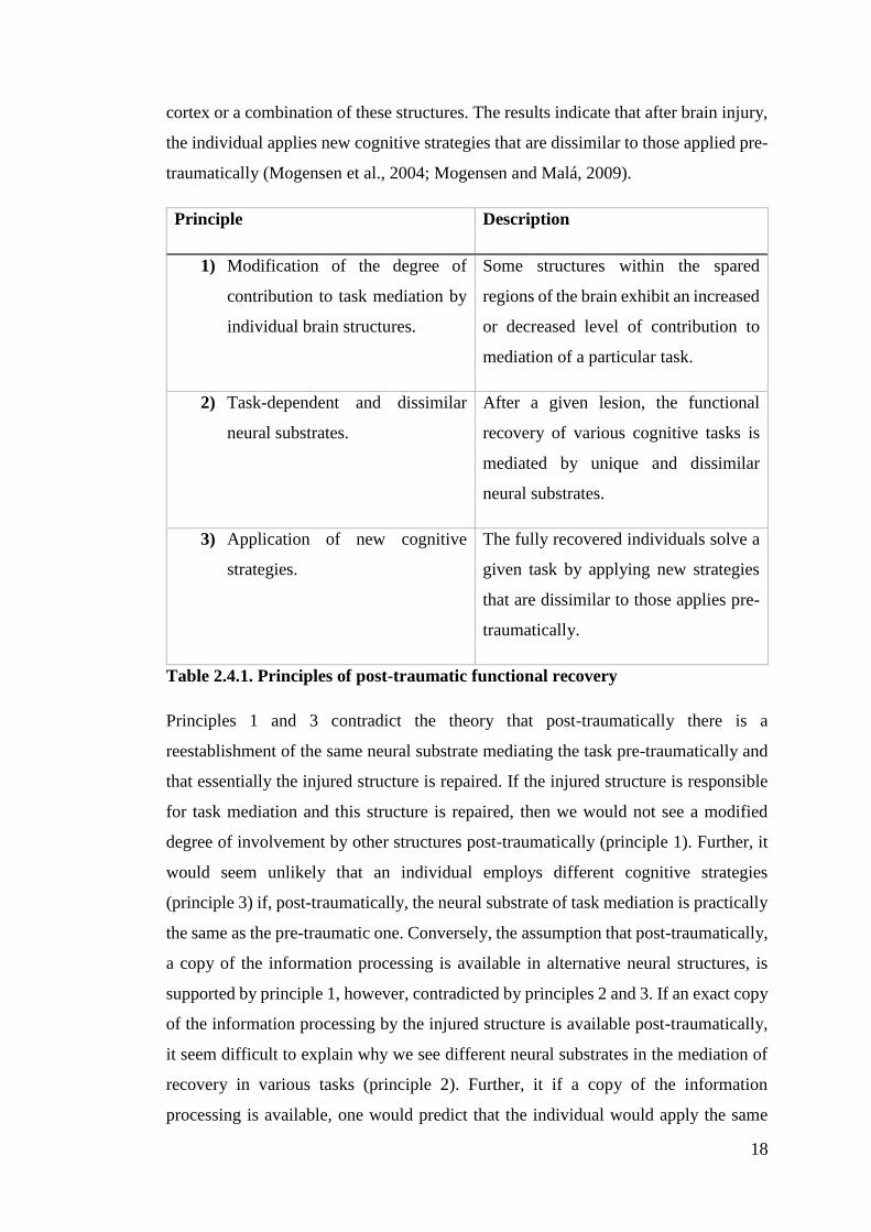
cortex or a combination of these structures. The results indicate that after brain injury,
the individual applies new cognitive strategies that are dissimilar to those applied pre-
traumatically (Mogensen et al., 2004; Mogensen and Malá, 2009).
Principle Description
1) Modification of the degree of
contribution to task mediation by
individual brain structures.
Some structures within the spared
regions of the brain exhibit an increased
or decreased level of contribution to
mediation of a particular task.
2) Task-dependent and dissimilar
neural substrates.
After a given lesion, the functional
recovery of various cognitive tasks is
mediated by unique and dissimilar
neural substrates.
3) Application of new cognitive
strategies.
The fully recovered individuals solve a
given task by applying new strategies
that are dissimilar to those applies pre-
traumatically.
Table 2.4.1. Principles of post-traumatic functional recovery
Principles 1 and 3 contradict the theory that post-traumatically there is a
reestablishment of the same neural substrate mediating the task pre-traumatically and
that essentially the injured structure is repaired. If the injured structure is responsible
for task mediation and this structure is repaired, then we would not see a modified
degree of involvement by other structures post-traumatically (principle 1). Further, it
would seem unlikely that an individual employs different cognitive strategies
(principle 3) if, post-traumatically, the neural substrate of task mediation is practically
the same as the pre-traumatic one. Conversely, the assumption that post-traumatically,
a copy of the information processing is available in alternative neural structures, is
supported by principle 1, however, contradicted by principles 2 and 3. If an exact copy
of the information processing by the injured structure is available post-traumatically,
it seem difficult to explain why we see different neural substrates in the mediation of
recovery in various tasks (principle 2). Further, it if a copy of the information
processing is available, one would predict that the individual would apply the same
19
cognitive strategies as it did pre-traumatically, which does not agree with principle 3
(Mogensen and Malá, 2009).
Thus, there is a need for a model of functional recovery that agrees with these three
principles, essentially explaining why individuals can exhibit full functional recovery
in spite of the fact that the structure mediating this particular function is permanently
lost (Mogensen, 2011a; Mogensen and Malá, 2009). This is the purpose of the REF-
model. In the following, I will account for the theoretical composition of the REF-
model, which is based on the empirical evidence accounted for above.
2.4.2. The REF-model
The REF-model consists of three levels of analysis, surface phenomena, algorithmic
strategies and elementary functions (Mogensen and Malá, 2009; Mogensen, 2011;
Mogensen, 2014). Clinically, the surface phenomena are the primary concern, since
these constitute the behavioral and cognitive deficits caused by brain injury. Further,
it is also at this level functional recovery can observed by the reestablishment of
behavioral and cognitive ability (Mogensen, 2014). The surface phenomena, therefore,
compose the top-most level of analysis. At the bottom-most level we find the
Elementary Functions (EFs). All traditionally defined structures, such as the
hippocampus or the prefrontal cortex, are comprised of the neural substrate of several
EFs, which means that when a specific brain region is lost to injury, so too are the EFs
mediated by that structure. The EFs are truly localized and at this level, functional
localization is indeed a reality (Mogensen, 2014). Evidently, the REF-model
accommodates functional recovery as well as functional localization, and, seemingly,
provides a model of functional modules within a connectionist network, something
which is demonstrated by the level of analysis between the levels of surface
phenomena and EFs, namely the level of Algorithmic Strategies (ASs). An AS is
constituted by several EFs, thus, the neural substrate of an AS consists of the neural
substrates of all the constituting EFs as well as the connections between them. ASs are
the mediators of the surface phenomena, and are not localized like the EFs. Rather,
they are distributed across many regions of the brain (Mogensen, 2014). Figure 2.4.2.
demonstrates the three levels of analysis and how every level relates to the others.

20
Fig. 2.4.2. REF-model, overview. Found in Mogensen, 2011.
Since the EFs are localized, damage to any given area, eliminates the neural substrates
of the EFs within this area and these EFs are lost forever (Mogensen, 2014), thus being
in accordance with the fact that after brain damage, the region lost to injury does not
grow back (Mogensen et al., 2007). Any AS that is composited of the EFs that are lost
to injury, is inevitably lost as well, since the neural substrates of these ASs are
constituted by neural substrates that are now lost. This does not mean, however, that
the surface phenomenon that is mediated by the lost AS is permanently lost as well.
Indeed, this would make the model entirely modular and unable to explain functional
recovery. In case of injury to the brain and the loss of EFs, and subsequent ASs, the
surface phenomenon that is pre-traumatically mediated by the lost AS is impaired.
However, as we know, full functional recovery is possible and does indeed happen,
which means that somehow the brain reorganizes and establishes an alternative AS
that mediates a surface phenomenon that resembles the one observed pre-traumatically
(Mogensen 2014). However, it is important to realize that even though an AS might
produce the same observable surface phenomenon as the one observed pre-
traumatically, the strategies are not identical, and might, quite possibly, not even be
identical to alternative strategies seen in other brain damaged individuals, as
demonstrated by Mogensen et al. (2004; 2007). Thus, essentially, functional recovery
is the process in which new ASs are formed by EFs that were not lost to injury,
21
exemplifying how this model is indeed a model of functional modules within a
connectionist network within the brain. As we know, confer the above, different types
of brain injury yield different cognitive strategies for solving cognitive tasks
(Mogensen et al., 2004; 2007; Mogensen and Malá, 2009), demonstrating that the brain
reorganizes in a manner that provides the reestablishment of the surface phenomenon
by utilization of the remaining structures. In this process, the brain seeks the most
efficient mediator of the surface phenomenon that was impaired by the brain damage.
This means that several new ASs may be constructed, however, only the AS producing
the most accurate surface phenomenon, and therefore the most efficient AS, is utilized
(Mogensen, 2014). This process depends on two mechanisms: The selector/evaluator
mechanism and the backpropagation mechanism, responsible for mediation of
successful ASs and problem solving, and the reorganization of the neural connectivity
between the underlying EFs, respectively. These mechanisms are highly dependent on
the environmental input, or the feedback, that is provided. An example of this is a
study by Wilms and Malá (2010) in which they modify the Prism Adaption Therapy,
typically applied on patients with visuospatial neglect, a syndrome caused by lesion to
the right-hemisphere parietal region, in which the patient is not aware of anything that
is presented in the left visual field (Spikman and van Zomeren, 2010). During training,
the patient wears the goggles, which shifts the visual field ten degrees to the right
(Mogensen, 2014), and is asked to point to targets appointed by the therapist without
being able to see his arm or where he is pointing. Feedback is provided to the patient
by revealing the pointing finger and to where it was pointing. In most cases, the patient
adapts to the visual shift provided by the goggles and this shift has been shown to
persist for a period of time even when the goggles have been removed (Frassinetti et
al., 2002). Wilms and Malá (2010) included a modified version of this procedure, in
which the feedback consisted of an X on a computer screen. In comparison with the
traditional procedure, this was not nearly as effective. In fact, the after-effect was
almost completely absent, which demonstrates that this after-effect depends heavily
on the situation in which training has been conducted, as well as the visual feedback
(Mogensen, 2014). Seemingly, the informational input has massive consequences for
the formation of new ASs as well as the modified connectivity of EFs (Mogensen,
2014).
22
In the light of this profound impact that the environment has on the functional
outcome, it seems pivotal that I address the consequences that this conceptualization
of the brain has for rehabilitative training after acquired brain damage.
2.4.3. Implementation of the REF-model
If the formation of an alternative AS does indeed depend on the environmental input,
the situation in which the rehabilitative cognitive training is conducted must,
inevitably, be highly significant to the functional outcome (Mogensen, 2011a; 2014).
Further, it is important to realize that rehabilitative training programs provide the basis
for the formation of ASs that mediate these specific tasks, since this formation process
is based solely on feedback in the specific situation. This, essentially, means that the
reestablishment of any given surface phenomenon relates to the training situation in
which it has been reestablished, not necessarily being generalizable to the patient’s
everyday life (Mogensen, 2011a; 2011c 2014; Mogensen and Malá, 2009). A patient
might demonstrate what seems to be full functional recovery, however, this might be
a very training-specific recovery. For example, the patient with visuospatial neglect
will typically not be able to solve the Line Bisection Test, in which the patient has to
mark the center point of horizontal lines on a piece of paper. The patient will, typically,
not mark the center of the line, but deviate towards the right pole of the line, not being
aware of the left half of the lines. With training, the patient can become aware of the
left part of the lines and solve the task correctly (Spikman and van Zomeren, 2010).
This, however, does not mean that they no longer have neglect or that they are cured.
It simply means that they have successfully established a cognitive strategy enabling
them to solve this particular task (Mogensen, 2011a; Mogensen, 2014). To
accommodate the shortcomings of this kind, rehabilitative training programs should
be constructed of training that resembles situations that the patient would encounter in
everyday life. In case of visuospatial neglect, entering a room can present a lot of
problems, since one risks bumping into things and hurting oneself. Training, therefore,
could consist of different situations in which the patient is reminded to scan the
surroundings, making sure that the visual information is provided to the non-
neglecting hemisphere and is therefore available to the patient.
In conclusion, it seems pivotal that we address these issues and try to modify the
treatment of our brain injured patients in order to optimize treatment, making it more
efficient and meeting environmental demands.

23
Since the majority of the above mentioned laboratory studies are conducted on
animals, and the experimental section of this paper, too, is based on an experiment
with rats, I will now move my focus to the utilization of animal models in laboratory
studies.
3. Animal models
The use of animals in research dates as far back as the 1600s and since then animal
studies have been the basis of many biomedical breakthroughs (Harding, Van Hoosier
Jr. and Grieder, 2011). Indeed, animal studies have provided us with knowledge that
help us understand the healthy, as well as the diseased organism, making it possible to
understand human diseases and how we treat them (Harding, Van Hoosier Jr. and
Grieder, 2011; Olsson, Robinson and Sandøe, 2011). For instance, the vaccine against
rabies was developed using animal research. In the 1800s, French scientist, Louis
Pasteur (1822-1895), adapted the rabies virus to laboratory animals and subsequently
developed the vaccine against a virus that up until then had a 100% mortality rate,
saving many lives (Harding, Van Hoosier Jr. and Grieder, 2011). More recently,
research on animals have helped scientists learn more about such neurodegenerative
diseases as Parkinson’s and Alzheimer’s diseases, prompting symptoms in animals
that mimic the symptoms of said diseases observed in humans and studying side effects
of drug regiments, existing and new ones. Further, studies on as diverse diseases as
Huntington’s disease, addiction and arthritis, as well as possible treatment strategies,
have been performed on animals as well (Harding, Van Hoosier Jr. and Grieder, 2011;
Olsson, Robinson and Sandøe, 2011). Evidently, the usefulness of animal research
applies to wide range of biomedical research and animal models have been proved to
be especially valuable in the field of neuroscience (Harding, Van Hoosier Jr. and
Grieder, 2011; Mogensen, 2011).
3.1. Usefulness of animal models
One might ask how we can compare an animal’s brain to that of our own and to answer
that question, one has to remember two things: First, that animal models are exactly
that – models. Models in which both symptoms and the cause of the condition in the
animal is identical to that of the human, homologous models, are extremely rare. Even
isomorphic models, in which the animal symptomology must be similar that of the
human, however, not necessarily provoked by the same event, are not very common.
24
Indeed, most animal models are partial models. These models are, obviously, neither
homologous nor isomorphic, but may still provide pivotal information about either the
disease in itself or the treatment thereof (Mogensen, 2011). Second, in reality, the
functional anatomy of the animal brain and the human brain is much more alike than
one might think, at least in some species. For instance, several studies have indicated
that rats have a hippocampus, the projections (e.g. van Groen and Wyss, 1990) and
functions (e.g. Morris, 1990) of which are similar to those found in humans. Further,
studies have demonstrated that the prefrontal cortex is not uniquely human, nor is it a
unique feature in “higher” mammals such as humans and non-human primates,
something that has long been the common understanding (Mogensen, 2011). In fact,
lower ranking mammals, such as rats, have similar prefrontal projections as those
found in the higher ranking mammals (Divac et al., 1978; Robertson and Murre, 1999).
There are even some indication that structures equivalent to the prefrontal cortex in
mammals, can be found in pigeons (Divac and Mogensen, 1985; Divac et al., 1985;
Mogensen and Divac, 1982).
Having established the basic grounds for interspecies comparisons, it seems pivotal
that I address the ethical issues involved in the conduction of animal studies, since all
scientific studies involving animals inevitably encounter essential ethical problems. Is
it ethically defendable to take advantage of our position as a higher species and fight
human disease by exploiting lower ranking animals? Are we exploiting the animals –
is that even possible? Questions like these divide scientists, as well as the general
population, in Western society (Olsson, Robinson and Sandøe, 2011; Cohen, 2007;
Rowlands, 1997; Foëx, 2007; Brom, 2002), and the answers depend on the ethical
theoretical stance. In the following, I will try to account for three of the most prominent
ethical positions, that all view animal studies differently and thus, all have different
implications for the conductions of (animal) research.
3.2. Ethical Considerations
The three views presented in the following are: 1) Contractarianism, 2) Utilitarianism
and 3) The Animal Rights View. Implications for animal research, as well as
comparison of the three views, will be ongoing.
3.2.1. Contractarianism
25
In this view animals have no inherent moral rights, since entering into a moral contract
with one another requires a certain level of linguistic and intellectual skill which,
according to contractarians, animals lack (Olsson, Robinson and Sandøe, 2011). In this
view, morality is perceived as precepts that determine the interaction of rational agents
in society, precepts that are defined by the very same rational agents. Thus, only
rational agents can be assigned direct moral rights and thereby receive moral
protection. Non-rational agents have no moral standing and, as such, are not morally
protected under these principles. Animals are regarded as non-rational agents and thus,
have no moral standing within the contractarian approach and possess no direct moral
rights (Rowlands, 1997), and as such, we should be free to utilize animals in research.
However, it is our right as possessors of direct moral rights to insist that animals we
care for, are not harmed. Thus, our moral responsibilities as human beings still provide
the animals with ethical protection in some way. In a contractarian view, harming an
animal that is under my moral protection is a violation of my moral rights, not that of
the animal, since it does not have any moral rights (Rowlands, 1997). However, this
view is not shared by all within the contractarian position. Some argue that moral
standing can be divided into two subcategories, primary and secondary moral standing
(Cohen, 2007). Indeed, the extreme contractarian view poses a problem for those in
society that do not meet the requirements of being a rational agent, which does not
only apply to animals, but also to not fully functioning humans. Consider, for example,
people suffering from dementia, the severely brain damaged, the intellectually
disabled (IQ under 70) and even infants. They do not meet the level of linguistic and
intellectual skill mentioned above and therefore cannot be considered rational agents
and as a direct result, possess no direct moral standing (Olsson, Robinson and Sandøe,
2011; Cohen, 2007; Rowlands, 1997). Though the extreme contractarian might insist
that the above is an over-interpretation, arguing that the equality argument, the notion
that inequalities are undeserved and therefore are arbitrary in the distribution of moral
shares (Rowlands, 1997), should be applied. I feel that the concept of secondary moral
standing deals with this issue more satisfyingly. Secondary moral standing is no less
moral than primary moral standing, as the only difference is the genesis of the standing
(Cohen, 2007). In this view, those who are not under the protection of direct, primary
moral rights can be appointed secondary moral rights by the possessors of the former,
thus giving rational agents and non-rational agents, such as the not fully functioning
humans, equal moral protection (Cohen, 2007). Secondary moral standing can be

26
acquired by animals as well, as long as a rational agent takes an interest in said animal’s
interests. Thus, since people generally care about animals and their welfare, and some
people even rely on animals, it makes us want to protect the animals and treat them
well, thus giving them some kind of moral status nonetheless (Olsson, Robinson and
Sandøe, 2011; Cohen, 2007; Brom, 2002).
This view relies on how people feel about animals, and therefore different species have
different values, since people generally care more about some animals than others. For
example, most people would probably say that they care more about cats or dogs than
they do about rats and mice, the latter typically being labeled vermin. This hierarchical
classification of animals, clearly positioning some animals as being more valuable than
others, is called the sociozoological scale (Olsson, Robinson and Sandøe, 2011), the
principles of which can be dated as far back as Aristotle (Foëx, 2007). This scale
perfectly frames the concept of contractarianism, because it solidifies that the value of
animals is defined by humans, a view that is highly criticized (Olsson, Robinson and
Sandøe, 2011; Cohen, 2007; Rowlands, 1997; Foëx, 2007; Brom, 2002), because what
happens, for example, if humans stop caring? Even though it seems unlikely that the
entire human race would suddenly stop caring about animals and see nothing wrong
in causing them pain and suffering, this concept composes one of the main critiques
of the contractarian view. If no one cared, humans would have carte blanche to do
whatever they wanted to animals – including harming them, because the animals are
only under our moral protection in as far as we extend our moral rights to them (Cohen,
2007; Rowlands 1997; Brom, 2002). This issue is dealt with in the utilitarian view,
which will be presented below.
3.2.2. Utilitarianism
The basic principle of utilitarianism is that any moral decision must be based on the
relationship between the positive outcome and the possible negative consequences of
the decision. This means that one must weigh the consequences and decide whether
the positive outcome exceeds the negative (Olsson, Robinson and Sandøe, 2011).
Utilitarians apply this rule to animal research as well. Within this view, animals are
extended the same moral rights as humans, since discrimination based on species is
considered just as wrong as discrimination based on gender or ethnicity (Foëx, 2007),
and the basic rule of morality is, simply, to always maximize the well-being of the
ones affected by your actions, be it humans or animals (Olsson, Robinson and Sandøe,
27
2011; Brom, 2002). Well-being can be defined as the absence of suffering, and
therefore requires sentience, the ability to feel, experience and perceive, all of which
can be attributed to most animals. According to utilitarianism, all creatures must be
treated as morally equal, meaning that the sociozoological scale is rejected, since one
cannot morally defend discriminating among animals in this way if they are to be
considered of equal value. Further, within the utilitarian view, humans cannot position
themselves above animals. Humans and animals are equal and as such, we cannot
defend using animals for research purposes (Olsson, Robinson and Sandøe, 2011).
However, this absolutist view seems impossible to implement in modern Western
society where the research performed on animals have so many clear-cut advantages.
Thus, moderate utilitarians accept that research performed on animals can result in the
curing of diseases, the alleviation of severely painful or inhibiting symptoms, and
ultimately saving human lives, all of which can be considered an overwhelming
justification of using animal in research (Foëx, 2007; Olsson, Robinson and Sandøe,
2011), in reality weighing positive and negative consequences as mentioned above.
Since animal research seems inevitable and, more importantly, extremely valuable,
utilitarianism adopts the principle of the three R’s, which basically advocates the 1)
replacement of existing animal experiments with alternatives whenever this is
possible, 2) reduction of the number of animals used in experiments, and 3) refinement
of experimental methods in order to ensure minimal animal suffering (Olsson,
Robinson and Sandøe, 2011). It is important to realize that this, however, contradicts
the basic principle of utilitarianism – that all sentient creatures are equal – since it can
be viewed as an expression of humans positioning themselves above animals. Even
research benefiting animals, for example veterinarian research, is difficult to justify
within the utilitarian position, because we then assume that the well-being of one
animal can be sacrificed in favor of the well-being of another, clearly accepting the
sociozoological scale, something an advocate of absolutist utilitarianism cannot
(Olsson, Robinson and Sandøe, 2011). However true, utilitarianism in its moderate
form does indeed embrace the principle of the three R’s and adopts a more pragmatic
attitude in which research on animals is inevitable, seeking justification in the
beneficial outcome for humans outweighing the possible suffering imposed on animals
(Olsson, Robinson and Sandøe, 2011; Foëx, 2007). In the eyes of the followers of the
animal rights movement, this is failing, which will be accounted for in the following.

28
3.2.3. Animal Rights View
Much like in utilitarianism, theorists of animal rights believe that animals have the
same moral rights as humans and that all sentient beings are equal. The lives of humans
and animals alike have inherent value and as a possessor of a valuable life, one has the
right to control one’s own life and no one else’s, which means that humans have no
right to exploit animals for their own benefit (Foëx, 2007). Believing that animals have
rights, means that animals cannot be treated as a means to an end, no matter how
glorifying that end might be. In this view, it does not matter if conducting an
experiment using animals could cure all the world’s worst diseases. The believer in
animal rights cares not about the possible advantages that this kind of experimentation
might have for the human species. The main idea is that it is morally wrong to utilize
animals even if the animals do not suffer. The animal rights advocate does not care. In
this view, animal research is a violation of animal rights and all animal
experimentation should be ceased, no matter the nature of it (Olsson, Robinson and
Sandøe, 2011; Foëx, 2007). This too, however, is quite an absolutist view and one
could imagine a more moderate animal rights view, advocating animals’ right not to
suffer, making it possible to conduct at least some animal research, for instance as seen
in a study by Brosnan and de Wall (2003), investigating the sense of fairness in the
capuchin monkey.
3.2.4. Conclusion
It seems clear that the three ethical positions accounted for above are highly
incompatible. The contractarian, in theory, would accept any kind of animal research,
no matter how much suffering the animal might be subjected to (though bearing public
concern in mind), whereas the utilitarian only accept experiments where the positive
outcome exceeds the negative that is constituted by animal suffering. The believer of
animal rights, on the other hand, will completely dismiss this kind of research and,
quite possibly, all research using animals (Olsson, Robinson and Sandøe, 2011). What
is the right thing to do, then? Well, the answer to that question depends on the moral
philosophy and personal beliefs of the one answering it (Foëx, 2007).
Thus, there is no unified answer to what is right and what is wrong when it comes to
conducting animal research. However, being part of a research unit that utilizes animal
models, inevitably, I position myself outside of the animal rights movement. Instead,
I take a stance that combines moderate utilitarianism and contractarianism. I feel that

29
animal research is pivotal if we are to answer the questions that we set out to answer.
Further, I feel that animal research is justifiable even though the outcome might not
cure diseases, as long as it provides us with knowledge about the healthy, as well as
the dysfunctional organism, aiding us to conduct other experiments that might indeed
cure diseases, alleviate painful symptoms and possibly save lives. I believe in
minimizing the number of animals used in experiments as well as maximizing the well-
being of laboratory animals, making sure they are not subjected to any more pain than
absolutely necessary, and feel that animal-free methods should replace animal research
whenever possible, which makes me a firm believer in the principle of the three R’s.
Further, I know that I am subjected to the ramifications of the sociozoological scale,
since I do feel more closely connected to such animals as cats and dogs than I do to
rats and mice, and as such there is indeed a hierarchy among animals in my mind. And
the fact that less than 1% of all animal research is conducted on non-human primates,
cats and dogs (Harding, Van Hoosier Jr. and Grieder, 2011), reveals that I am not alone
in this matter. However, I feel that choosing the right animal model is essential, which
means that in some instances, conduction of experiments on the higher ranking animals
is necessary, and in all cases I rely on the utilitarian principle of justice, feeling that
the positive outcome must outweigh the negative.
Having provided an ethical account, I now turn to different types of brain injury
models. The models presented below are rat models, since The Unit for Cognitive
Neuroscience (UCN), whom I have been working with, primarily uses rats in their
research. Further, the study I have been a part of, which will be presented later, was
also conducted using a brain injury model in the rat.
3.3. Brain Injury Models
The objective of an experimental model of traumatic brain injury is to reproduce
pathological conditions seen in human TBI (Cortez et al., 1989; McIntosh et al., 1989;
Thompson et al., 2005; Xiong et al., 2013). In the following, I will account for three
such models, two of the most frequently applied models, Fluid Percussion Injury (FPI)
and Controlled Cortical Impact (CCI), as well as one of the models presently applied
at UCN, transection of the fimbria-fornix fiber bundle, which is also the model applied
in the experiment presented later in this paper. As mentioned above, the focus of this
section will be on brain injury models in rats. Firstly, I will describe the FPI model and
account for positives and negatives of this particular model. Then, I will do the same

30
for the CCI model and the fimbria-fornix transection model. Comparison of the
models, as well as reflections about their usefulness, as well as ethical considerations
will be ongoing.
3.3.1. Fluid Percussion Injury
In FPI models, animals afflicted with brain injury exhibit symptoms comparable with
those observed in human closed head injury, including intracranial hemorrhage,
swelling of the brain and progressive grey matter damage (Cortez et al., 1989;
McIntosh et al., 1989; Xiong et al., 2013). Even though the symptoms are comparable
with cases of closed head injury, a craniotomy has to be performed on the animal,
through which the insult is inflicted, which obviously poses a contrast between the
animal model and the clinical setting. Figure 3.3.1. shows the setup. A pendulum
strikes the fluid-filled piston and generates a fluid pressure pulse causing a rapid
impact of fluid on the intact dura, producing injury (Thomson et al., 2005; Xiong et
al., 2013).
Fig. 3.3.1. Fluid Percussion Injury setup. Image downloaded 28th of August 2014
from http://www.uniklinikum-
saarland.de/de/einrichtungen/kliniken_institute/neurochirurgie/forschung/neurotraum
atologie/
The FPI models are mixed injury models, since they produce injuries with focal
cortical contusion characteristics, as well as diffuse neuronal injury (Thompson et al.,
2005; Xiong et al., 2013). The FPI models cause cognitive deficits comparable to those

31
observed in human TBI cases, for example memory deficits (Xiong et al., 2013),
affording them high construct validity (Thompson et al., 2005). The injuries seen using
FPI are highly reproducible and the simple mechanics of the experimental setup, the
height of the pendulum being the only adjustable mechanical parameter, ensures
precise and adjustable tuning of injury severity (Thompson et al., 2005). However, this
simplistic design also composes one of the main weaknesses of the FPI model, since
this is the only controllable factor (Xiong et al., 2013). This is not the only weakness.
In fact, there are several other notable weaknesses in the application of the FPI models.
Besides the high level of invasiveness due to the craniotomy, FPI models in general
have a very high mortality rate (Xiong et al., 2013), something which could give rise
to ethical dispute. Further, the fluid disperses across the dura in a manner that is
difficult to quantify (Dixon and Kline, 2009) and the highly diffuse character of
injuries caused by using FPI models, further, problematizes the quantification of the
extend of the injury (Dixon et al., 1991). In addition to this, the FPI models often cause
injury to the brainstem (Dixon et al., 1991; Xiong et al., 2013). This is especially true
for fluid percussion of high magnitudes, which poses a problem concerning the validity
of the models, since brainstem injury is not a primary feature in human TBI (Dixon et
al., 1991). However, not all FPI models affect the brainstem. FPI models can be
divided into three subcategories depending on the position of the craniotomy: Midline,
parasagittal, and lateral models, the latter of which is most commonly used, since it
primarily inflicts unilateral cortical damage and usually spares the brainstem,
something the midline and parasagittal models do not (Xiong et al., 2013). Lateral
fluid percussion injury model (LFPI) remains one of the most commonly used TBI
models, however, it does require careful ethical and methodological consideration.
3.3.2. Controlled Cortical Impact
The CCI model of TBI has the advantage that the mortality rate is low, compared to
that of FPI models (Dixon et al., 1991; Xiong et al., 2013). Further, the CCI model
poses a highly controllable alternative to FPI models, establishing a quantifiable
relationship between mechanical parameters and magnitude of damage (Dixon and
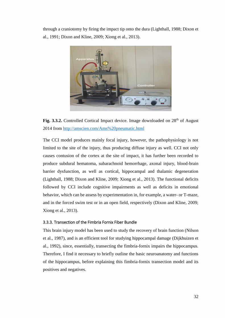
Kline, 2009; Xiong et al., 2013). Figure 3.3.2. shows the CCI device. The device is
either pneumatic, meaning that it is operated with controlled pressure of gases, or
electromagnetic. An impact tip is attached to the piston and the impact is produced
32
through a craniotomy by firing the impact tip onto the dura (Lighthall, 1988; Dixon et
al., 1991; Dixon and Kline, 2009; Xiong et al., 2013).
Fig. 3.3.2. Controlled Cortical Impact device. Image downloaded on 28th of August
2014 from http://amscien.com/Ams%20pneumatic.html
The CCI model produces mainly focal injury, however, the pathophysiology is not
limited to the site of the injury, thus producing diffuse injury as well. CCI not only
causes contusion of the cortex at the site of impact, it has further been recorded to
produce subdural hematoma, subarachnoid hemorrhage, axonal injury, blood-brain
barrier dysfunction, as well as cortical, hippocampal and thalamic degeneration
(Lighthall, 1988; Dixon and Kline, 2009; Xiong et al., 2013). The functional deficits
followed by CCI include cognitive impairments as well as deficits in emotional
behavior, which can be assess by experimentation in, for example, a water- or T-maze,
and in the forced swim test or in an open field, respectively (Dixon and Kline, 2009;
Xiong et al., 2013).
3.3.3. Transection of the Fimbria Fornix Fiber Bundle
This brain injury model has been used to study the recovery of brain function (Nilson
et al., 1987), and is an efficient tool for studying hippocampal damage (Dijkhuizen et
al., 1992), since, essentially, transecting the fimbria-fornix impairs the hippocampus.
Therefore, I find it necessary to briefly outline the basic neuroanatomy and functions
of the hippocampus, before explaining this fimbria-fornix transection model and its
positives and negatives.
33
3.3.3.1. Anatomy and functions of the hippocampus The hippocampal formation consists of the parahippocampal gyrus, dentate gyrus and
the hippocampus itself. This structure lies in the floor of the inferior horn of the lateral
ventricle, deep to the parahippocampal gyrus, and is part of the limbic system
(Crossman and Neary, 2010). It has been well documented that the hippocampus is
especially involved in the mediation of memory tasks (e.g. Scoville and Milner, 1957;
Henke, 2010; Duncan et al., 2014), and lesions to the hippocampus can have
devastating consequences. As was the case with H. M. (1926-2008), who is probably
the most famous patient with hippocampal damage or, in his case, removal, and he
represents one of the more severe cases. After a bilateral resection of the medial
temporal lobe, which includes the hippocampus, in order to alleviate his epileptic
symptoms, H. M. was no longer able to form new declarative memories – he suffered
from anterograde amnesia and was severely and permanently disabled (Scoville and
Milner, 1957).
The hippocampal formation receives afferents primarily from the inferior temporal
cortex through the entorhinal cortex, bilaterally. Further, it receives input from the
contralateral hippocampus via the fornix and the hippocampal commissure (Crossman
and Neary, 2010). In addition to this, the hippocampus is connected to the septum
(Dragoni et al., 1999). A major component of the septo-hippocampal projection
consists of cholinergic cells, residing in the medial septum and terminating in the
hippocampus (Meibach and Siegel, 1977; Dragoni et al., 1999), which means that the
hippocampus receives cholinergic input from the medial septum (Mogensen et al.,
2002). Septal input has been associated with hippocampal theta wave generation
(Dragoni, 1999), and hippocampal theta rhythm has been suggested to play a
significant role in memory formation (Vertes, 2005).
The main efferent pathway from the hippocampus is the fornix, which is a bundle of
fimbria fibers that connects the hippocampus with the hypothalamus. Essentially, these
efferent fibers converge on the ventricular surface of the hippocampus and passes
posteriorly to become continuous with the fornix, which curves forward beneath the
corpus callosum. The fibers then curve downwards and enter the mammillary body of
the hypothalamus (Crossman and Neary, 2010). Some fibers, however, split off in front
of the anterior commissure as the precommissural fornix, which ends in the septal
nuclei and the ventral striatum (Nolte, 2002).

34
3.3.3.2. Fimbria-fornix transection By transecting the fimbria-fornix bilaterally, one effectively impairs the hippocampus.
Since most hippocampal efferents go through the fimbria-fornix, transection of this
fiber bundle makes the hippocampus unable to send information to the rest of the brain.
Further, by transecting the fimbria-fornix, hippocampal theta rhythm is eliminated and
cholinergic input from the septum obstructed. Thus, even though the hippocampus
itself is not damaged as such, it is effectively disabled. This model produces an
extremely focal lesion that, if performed correctly, does not produce any diffuse
damage, and lesions are typically highly comparable.
The surgery is performed in a stereotaxic frame that allows a surgical procedure that
is minimally invasive, since the frame provides a three-dimensional coordinate system
of the brain, making it possible to locate areas within the brain (Swindle et al., 2011),
such as the fimbria-fornix. Fig. 3.3.3.2. shows the frame. The animal is fastened in the
frame and a craniotomy is performed at the correct coordinates and an encapsulated
wire-knife is inserted into the brain. When the encapsulated knife has been lowered
into the brain, the knife is extended and the fiber bundle is transected before extracting
the knife (e.g. Mogensen et al., 2004). The procedure is performed bilaterally to
completely impair the hippocampus.
Fig. 3.3.3.2. Stereotaxic frame. Image downloaded on 23rd of September from
http://products.narishige-group.com/group1/SR-5R/stereotaxic/english.html
35
A disadvantage of applying this model is that is bears very little resemblance to clinical
situations, since this type of lesion is extremely rare, if not absent, in humans.
However, if the intention is to study the reorganizational mechanisms involved in the
recovery after acquired brain damage, then a focal injury model has its clear
advantages, since, with a focal injury, one knows which structure is impaired.
Transection of the fimbria-fornix does indeed provide such a focal injury, impairing
the hippocampus. Further, this model has the advantage that lesions are similar across
individuals and therefor easily quantifiable, something the damage caused by the
models mentioned above is not. Therefore, transection of the fimbria-fornix might
provide us with information about the mechanisms involved in neural reorganization
after brain damage and, thus, the process of recovery.
As mentioned, transection of the fimbria-fornix produces similar dysfunctions as
damage to the hippocampus itself. Since this is the brain injury model applied in the
experimental study that will be presented later, I find it necessary to provide examples
of how one assesses impairments caused by damage to the hippocampus in humans as
well as animals.
3.3.3.3. Assessment of impairments caused by hippocampal damage The hippocampus has been identified to be highly involved in the mediation of several
functions related to memory, including spatial memory (Nilson et al., 1987), semantic
and episodic memory (Henke, 2010). Since the semantic and episodic memories are
declarative, they are typically assessed with test that require language, however, non-
language based tests of memory function do exist as well. An example of such a test
is the Rey-Östereich complex figure, a complex drawing that the patient has to
replicate. After a period of time, the patient is asked to draw the figure again, this time
from memory (Bradley and Kapur, 2010). If the patient has no problems producing
speech, one can assess the abovementioned memory deficits by having the patient try
to memorize a list of words or where in the room a couple of arbitrary objects are
placed, as well as interviews and spontaneously provided information by the patient or
with memory questionnaires. Typically, assessment of memory impairments caused
by brain injury is based on a test battery that includes several memory assessment tests
(Bradley and Kapur, 2010).
For obvious reasons, we cannot use these assessment tools in animal models. However,
the delayed alternation task in, for example, the T-maze has been established as a valid
36
assessment tool for several cognitive functions in rodents, including learning and
memory (Deacon & Rawlins, 2006), even though it is primarily associated with
functions of the frontal lobe (Zald et al., 2002). In the delayed alternation task in the
T-maze (see fig. 4.2.6.1. for an overview of the maze), the animal has to alternate
between left and right, always choosing the path it did not choose moments before,
which requires utilization of several distinct cognitive strategies. Initially, the animal
must exhibit goal-directed behavior, since the objective of the task cannot be acquired
if the animal is not driven towards anything. Goal-directed behavior is typically
mediated by the frontal lobe (Goldberg and Bougakov, 2005). Then, in order to solve
the delayed alternation task, the animal has to learn that there is an objective and what
that is, remember this objective in the following sessions, and, in every trial, remember
witch path it chose just before, indicating that this task involves learning and memory
mechanisms, specifically working memory and long-term memory mechanisms
(Deacon and Rawlins, 2006). However, before the delayed alternation testing can
commence, the animal is subjected to a series of shaping sessions, in which the only
objective is to enter either arm immediately in every trial, no matter which arm the
animal chose on the preceding trial. This definitely requires goal-directed behavior as
mentioned above. When the animal has reached the behavioral criterion for the shaping
sessions set by the research team, the delayed alternation testing can begin.
Thus, before the animal can even begin the above mentioned memory processes, it has
to be able to adapt to the new task, namely alternation, and this requires that the animal
exhibits a high level of cognitive flexibility, yet another function typically mediated
by the frontal lobe (Goldberg and Bougakov, 2005). Clearly, the delayed alternation
test is able to reveal abilities and disabilities of several cognitive functions, including
goal-directed behavior, cognitive flexibility, as well as learning, long-term memory
and working memory, which makes this test applicable in experiments with frontal
lobe dysfunction (Zald et al., 2002), as well as hippocampal damage (Deacon &
Rawlins, 2006).
Having provided information about the implications of transecting the fimbria-fornix
and how to assess the subsequent impairments, I will now introduce the experiment
that I have been working on at UCN, since in this particular experiment, test animals
had their fimbria-fornix transected, which impaired their hippocampus. We then
37
subjected them to the delayed alternation test in the T-maze presented above, thus
giving us the opportunity to study the mechanisms involved in functional recovery.
4. Experimental section
The following will include an introductory section, letting the reader know what the
experiment is about, followed by at methods section, describing the experiment and
presentation of results. Lastly, these results will be discussed.
The following consists of data that has not yet been published and, therefore, I cannot
include all of the data in my presentation and analysis. In agreement with the authors
of the article that will present this experiment, I will include a limited section of the
data and treat it as if it were the complete data set. Thus, the methods section, as well
as the results section, will be based on the limited data material. This means that the
experimental groups presented below are smaller than they are when one includes the
complete data material, which unfortunately affects my statistical analysis negatively.
Indeed, in some instances, my limited access to the data material and the smaller
experimental groups yield unreliable results that are not in accordance with the actual
results. Specifically, in several cases, I find no significant difference between the
experimental groups, when indeed there is one, thus making a type II statistical error,
failing to detect a difference that is indeed present. Further, in a single case, my limited
data material yields a significant difference when, in reality, there is none, thus making
a type I statistical error, detecting a difference that is not present (Field, 2013). I will
account for all of this in a concluding section after presentation of my pseudo-results.
To clarify, my presentation of the methodology of the experiment, as well as my report
and analysis of the results is based on a mere a fraction of the complete data material.
As such, these sections do not correspond to those based on the complete data material
and therefore, is not accordance with the actual methodology or results. Thus, these
sections are to be considered mere examples of how one could present an experiment
and how one could conduct statistical analysis for such an experiment. For an accurate
description of the experimental methodology, as well as a full exposition of the results
and analysis of the complete dataset, see M. G. Gram, L. Gade, E. Wogensen, J.
Mogensen and H. Malá (in preparation). The thought of discussing a pseudo-reality,
though, somehow pushes the boundaries of my integrity, since discussion of the
erroneous results seems wrong when I have knowledge of the actual results. Therefore,
38
the discussion will be based on the actual results, which as mentioned, will be
presented briefly following the analysis of the limited data material.
4.1. Introduction
As already mentioned, one typical symptom of TBI is profound memory deficits
(Johnson et al., 2013) and the inability to acquire new information seems especially
disabling, since returning to a normal lifestyle and workspace seems impossible if the
ability to learn and utilize new information is impaired or completely absent (Gade,
2010). Evidently, there is a need to investigate the mechanisms involved in recovering,
or compensating for, this particular function, and how to induce such mechanisms.
This is the focus of the study presented in the following.
Cognitive recovery in brain damaged rats, in this case hippocampal dysfunction by
transection of the fimbria-fornix fiber bundle, was assessed in a delayed alternation
test in a T-maze after having been subjected to different types of environmental
stimulation.
The standard definition of an enriched environment is “a combination of complex
inanimate and social stimulation” (Rosenzweig et al., 1978). In an enriched
environment the aim is to facilitate species-specific behavior (Abou-Ismail, 2011), in
this case rodent-specific behavior, which typically requires increased space for
exploration and exercise, sensory experience, and socialization (Sozda et al., 2010).
Therefore, animals living in enriched environments are usually kept in larger cages
and larger groups, which gives them the opportunity for more complex social
interaction compared to standard housing. The environment is complex and
stimulating, and consists of various nesting materials and toys, which are frequently
changed over the course of the experiment. Additionally, animals are often offered
voluntary exercise by placing running wheels in the environment (van Praag et al.,
2000). The typical enrichment paradigm therefore includes three components: 1)
Cognitive enrichment (general living conditions, toys, nesting materials etc.), 2) Social
enrichment (living in groups), and 3) Motoric enrichment (voluntary exercise).
The positive effects of environmental enrichment on brain injury seems well-
documented (e.g.: de Witt et al., 2011; Cheng 2012; Hamm et al., 1996). However,
since studies on typical environmental enrichment often offer physical exercise as well
as cognitive and social enrichment (Johnson et al, 2013; Leggio et al., 2005; Pang and
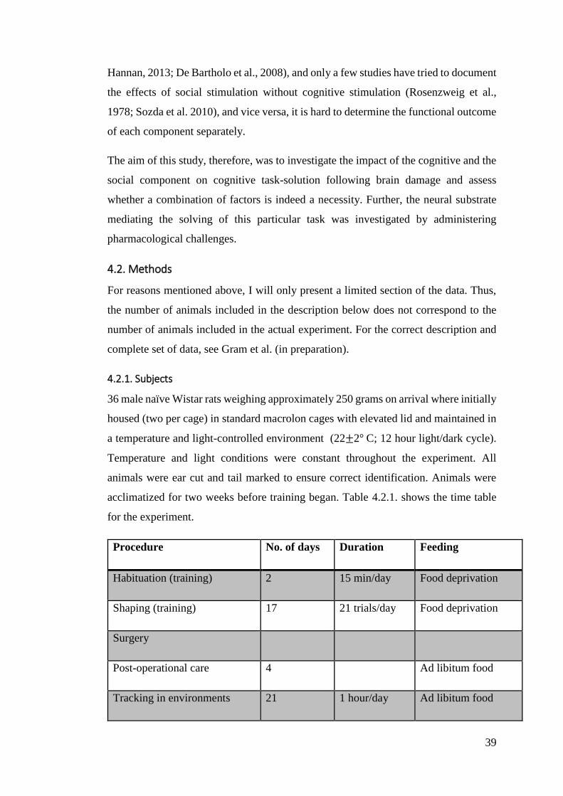
39
Hannan, 2013; De Bartholo et al., 2008), and only a few studies have tried to document
the effects of social stimulation without cognitive stimulation (Rosenzweig et al.,
1978; Sozda et al. 2010), and vice versa, it is hard to determine the functional outcome
of each component separately.
The aim of this study, therefore, was to investigate the impact of the cognitive and the
social component on cognitive task-solution following brain damage and assess
whether a combination of factors is indeed a necessity. Further, the neural substrate
mediating the solving of this particular task was investigated by administering
pharmacological challenges.
4.2. Methods
For reasons mentioned above, I will only present a limited section of the data. Thus,
the number of animals included in the description below does not correspond to the
number of animals included in the actual experiment. For the correct description and
complete set of data, see Gram et al. (in preparation).
4.2.1. Subjects
36 male naïve Wistar rats weighing approximately 250 grams on arrival where initially
housed (two per cage) in standard macrolon cages with elevated lid and maintained in
a temperature and light-controlled environment (22±2° C; 12 hour light/dark cycle).
Temperature and light conditions were constant throughout the experiment. All
animals were ear cut and tail marked to ensure correct identification. Animals were
acclimatized for two weeks before training began. Table 4.2.1. shows the time table
for the experiment.
Procedure No. of days Duration Feeding
Habituation (training) 2 15 min/day Food deprivation
Shaping (training) 17 21 trials/day Food deprivation
Surgery
Post-operational care 4 Ad libitum food
Tracking in environments 21 1 hour/day Ad libitum food
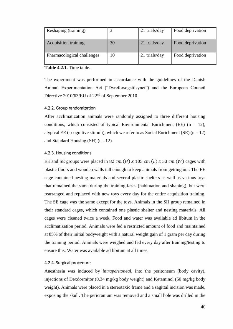
40
Reshaping (training) 3 21 trials/day Food deprivation
Acquisition training 30 21 trials/day Food deprivation
Pharmacological challenges 10 21 trials/day Food deprivation
Table 4.2.1. Time table.
The experiment was performed in accordance with the guidelines of the Danish
Animal Experimentation Act (“Dyreforsøgstilsynet”) and the European Council
Directive 2010/63/EU of 22nd of September 2010.
4.2.2. Group randomization
After acclimatization animals were randomly assigned to three different housing
conditions, which consisted of typical Environmental Enrichment (EE) (n = 12),
atypical EE (- cognitive stimuli), which we refer to as Social Enrichment (SE) (n = 12)
and Standard Housing (SH) (n =12).
4.2.3. Housing conditions
EE and SE groups were placed in 82 𝑐𝑚 (𝐻) 𝑥 105 𝑐𝑚 (𝐿) 𝑥 53 𝑐𝑚 (𝑊) cages with
plastic floors and wooden walls tall enough to keep animals from getting out. The EE
cage contained nesting materials and several plastic shelters as well as various toys
that remained the same during the training fazes (habituation and shaping), but were
rearranged and replaced with new toys every day for the entire acquisition training.
The SE cage was the same except for the toys. Animals in the SH group remained in
their standard cages, which contained one plastic shelter and nesting materials. All
cages were cleaned twice a week. Food and water was available ad libitum in the
acclimatization period. Animals were fed a restricted amount of food and maintained
at 85% of their initial bodyweight with a natural weight gain of 1 gram per day during
the training period. Animals were weighed and fed every day after training/testing to
ensure this. Water was available ad libitum at all times.
4.2.4. Surgical procedure
Anesthesia was induced by intraperitoneal, into the peritoneum (body cavity),
injections of Dexdormitor (0.34 mg/kg body weight) and Ketaminol (50 mg/kg body
weight). Animals were placed in a stereotaxic frame and a sagittal incision was made,
exposing the skull. The pericranium was removed and a small hole was drilled in the

41
skull at 1.1 mm posterior to bregma and 1.2 mm lateral to the sagittal suture, bilaterally.
The wire-knife, folded into a cannula, was lowered to a position 3.2 mm ventral to the
dura, and the knife was extended laterally to a length of 1.6 mm. The knife was then
lowered to a position 5.8 mm ventral to the dura and left in this position for one minute,
after which it was raised to its original position at 3.2 mm ventral to the dura. The knife
was drawn back into the cannula, and rotated 180𝑜. The knife was then re-extended to
a length of 1.6mm., and lowered to a position of 5.8 mm. ventral to the dura, where it
remained for one minute. Again, the knife was raised to a position 3.2 mm. ventral to
the dura, and the knife was drawn into the cannula, and withdrawn from the brain. The
same procedure was performed on both hemispheres, effectively transecting the
fimbria-fornix, bilaterally. Sham operated animals underwent analogous surgical
preparations, but no perforation of the skull was performed. Surgeries were performed
in clean, but non-sterile conditions. After the surgery, animals received saline
injections, and Buprenophine (Temgesic, 0.03 ml/kg body weight) was given as a post-
operative analgesic. Subsequently, saline injections and Buprenophene, either injected
or consumed orally, was administered every six to eight hours if necessary. All animals
were given a four-day post-surgery recovery period before being re-introduced to their
group housings.
The 36 animals were randomly divided into the following groups:
1) Sham operated animals in standard housing (Sham/SH) (n = 6)
2) Fimbria fornix transected animals in standard housing (FF/SH) (n = 6)
3) Sham operated animals living in the enriched environment (Sham/EE) (n = 6)
4) Fimbria fornix transected animals living in the enriched environment (FF/EE)
(n = 6)
5) Sham operated animals living in the social environment (Sham/SE) (n = 6)
6) Fimbria fornix transected animals living in the social environment (FF/SE)
(n = 6)
4.2.5. Ethovision
Tracking of the total distance travelled (m) by a total of 16 randomly picked animals
in groups EE and SE was made using Ethovision 3.1 (Noldus, Netherlands). Animals
were marked with different colors of Special Effects hair dye on the back of the body
in order to allow Ethovision to recognize each animal, thus tracking them separately.
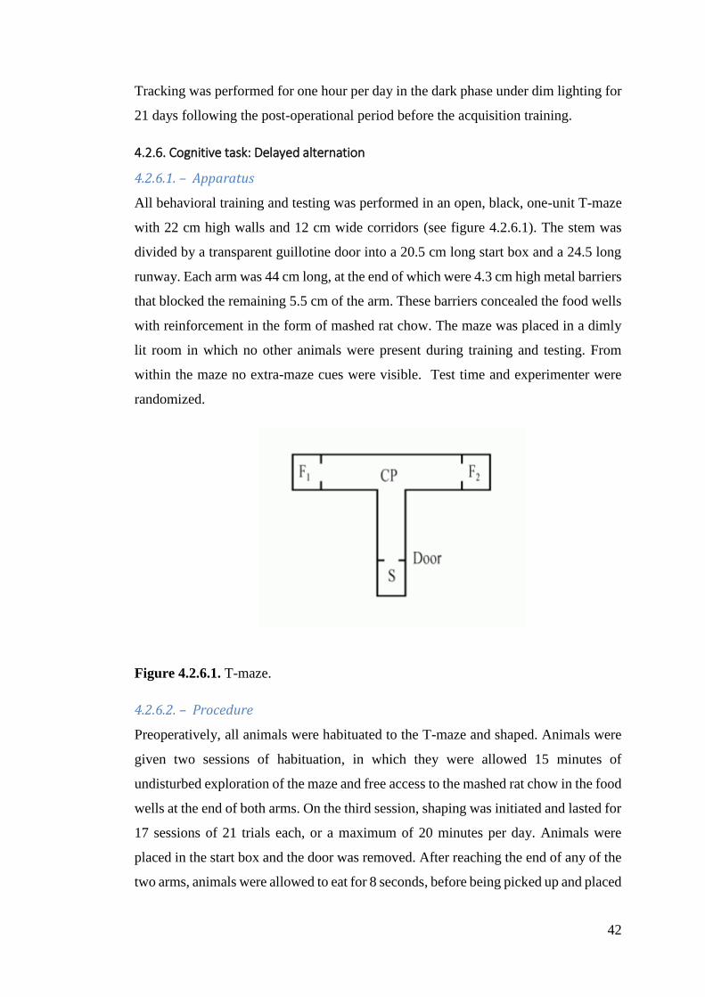
42
Tracking was performed for one hour per day in the dark phase under dim lighting for
21 days following the post-operational period before the acquisition training.
4.2.6. Cognitive task: Delayed alternation
4.2.6.1. – Apparatus
All behavioral training and testing was performed in an open, black, one-unit T-maze
with 22 cm high walls and 12 cm wide corridors (see figure 4.2.6.1). The stem was
divided by a transparent guillotine door into a 20.5 cm long start box and a 24.5 long
runway. Each arm was 44 cm long, at the end of which were 4.3 cm high metal barriers
that blocked the remaining 5.5 cm of the arm. These barriers concealed the food wells
with reinforcement in the form of mashed rat chow. The maze was placed in a dimly
lit room in which no other animals were present during training and testing. From
within the maze no extra-maze cues were visible. Test time and experimenter were
randomized.
Figure 4.2.6.1. T-maze.
4.2.6.2. – Procedure
Preoperatively, all animals were habituated to the T-maze and shaped. Animals were
given two sessions of habituation, in which they were allowed 15 minutes of
undisturbed exploration of the maze and free access to the mashed rat chow in the food
wells at the end of both arms. On the third session, shaping was initiated and lasted for
17 sessions of 21 trials each, or a maximum of 20 minutes per day. Animals were
placed in the start box and the door was removed. After reaching the end of any of the
two arms, animals were allowed to eat for 8 seconds, before being picked up and placed
43
in their holding cage for an additional 8 seconds, after which the next trial was initiated.
All trials were timed and the time recorded. If an animal had not chosen an arm within
5 minutes, it was removed from the maze and placed in the holding cage for 16
seconds. The shaping trained the animals to promptly enter either arm of the maze,
thus having animals reach the end of either arm in five seconds or less in every trial.
The animals then underwent surgery and were subsequently in four days of
postsurgical care, after which they received 21 days of intervention (EE, SE or SH) in
which they were tracked for one hour each day (see table 4.2.1.), before being reshaped
for three sessions. The reshaping procedure was exactly the same as the shaping
procedure. After reshaping, acquisition training was initiated. For 30 consecutive days,
animals received one daily session of training, consisting of 21 trials per session.
Animals were placed in the start box and released. On their first trial, animals could
enter either arm freely, but were subsequently expected to alternate. If an animal
alternated, and chose the opposite arm of the one chosen in the preceding trial, it was
allowed to eat for 8 seconds, before being returned to the holding cage, where it
remained for 8 seconds before commencing the next trial. If, on the other hand, an
animal did not alternate, and entered the same arm as it did in the preceding trial, it
was immediately removed from the maze and placed in the holding cage for 16
seconds. For each session, the number of errors, as well as the number of repetitive
errors, were recorded.
4.2.7. Pharmacological challenges
Animals were given a five day pause after acquisition training, before the
commencement of the pharmacological challenges, which lasted a total of ten days.
Same procedure as in the acquisition training was performed. The first two days,
animals were injected with saline, giving us a baseline. On the third day, animals were
injected with the muscarinic receptor antagonist scopolamine (0.5 mg/kg body weight,
dissolved in 0.9% saline) 20 minutes prior to testing. Day four was a wash out day.
Days five and six were saline days and on day seven, animals were administered
dopamine DA receptor antagonist SKF-85366 (0.1 mg/kg body weight, dissolved in
0.9% saline) 30 minutes prior to testing. Day eight was another wash out day and days
nine and ten were saline days. Performances on the saline days were pooled into three
averages. The average of days 1 and 2 was named “saline1”, average of days 5 and 6
was named “saline2”, and average days 9 and 10 was named “saline3”. These averages

44
were subsequently compared to performances on scopolamine and SKF-85366
challenge days.
4.2.8. Histology
After completion of behavioral training, all animals were anaesthetized by injection of
Dexdormitor and Ketaminol and transcardially perfused with sucrose followed by a
4% paraformaldehyde solution to fixate the brain tissue. After perfusion, the brains
were removed and kept at 4℃ in containers filled with a 4% paraformaldehyde
solution. The brains were cut horizontally at 50𝜇m using a vibrotome. The Cresyl-
stained sections were examined under a microscope (Leica DMD 108, Leica
Microsystems) to verify and quantify the lesions.
4.2.9. Statistical analysis
Statistical analyses were performed using IBM SPSS 19.0. Initially, all behavioral data
were analyzed using one-way analysis of variance (ANOVA) for the mean number of
total errors according to the group affiliation (surgery+living conditions). If the
analysis of variance revealed significant group differences, independent samples t-
tests were applied to determine differences between individual experimental groups.
Further, to test for perseverance, a one-way ANOVA was performed on the parameter
mean number of repetitive errors. In case this test revealed any significant differences,
independent samples t-tests were applied. Data from the pharmacological challenges
was compared to the data from the surrounding baseline saline injections days using a
repeated-measures ANOVA. Paired samples t-tests were subsequently applied to
account for group differences. Data gathered with Ethovision, tracking the physical
activity of animals in groups FF/EE and FF/SE, was analyzed using independent
samples t-tests. Lastly, differences in lesion size were analyzed using the Kruskal-
Wallis non-parametric test.
4.3. Results
This entire section is dominated by the miniscule possibilities the constricted data
material provides. With the reduced groups, it is simply not possible to attain a group
size that permits an actual analysis, which is why some of the following results will
contradict those represented by the complete data material. Further, data presented in
the sections on motoric activity and anatomy will reflect the full data material, since it
was not possible to provide individual data on these parameters, and as such data,

45
presented in these sections cannot be directly compared to the rest of the results
presented below. However, in order to show how the motoric activity parameter, as
well as the histological analysis, relates to the experiment, I will include them in the
analysis below, even though they are not directly comparable to my pseudo-results that
are only based on a fraction of the collected data. An analysis where some results are
based on the entire data material and some are not, clearly, will be distorted. Thus, I
will not present a complete analysis, but rather provide a mere presentation of
numbers. In a concluding section, I will try to, briefly, summarize the results yielded
by the complete data material, however, I must refer the reader to Gram et al. (in
preparation) for a full elaboration. The results presented in the following, is to be
perceived merely as a showcase of how to process experimental data.
4.3.1. Motor activity
No significant difference in motoric activity (distance moved) was found between the
enriched and the social group (𝑝 = 0.75) (Gram et al., in preparation). Fig. 4.3.1.
illustrates this.
EE SE
Mean
dis
tan
ce t
ravelled (
cm
)
0
2000
4000
6000
8000
10000
12000
14000
16000
18000
Fig. 4.3.1. Motor activity measured by the distance travelled (cm) within the enriched
environment (EE) and the socially enriched environment (SE), respectively.
46
4.3.2. Histology
Histological examination of the lesioned animals revealed a minimal amount of intact
fibres, leaving the fimbria fornix fibre bundle almost completely transected. In all
animals, the fimbria and dorsal fornix were damaged at the level of the ventral
hippocampal commissure and in some animals the damage extended ventrally into the
subfornical organ, dorsally into the ventral part of corpus callosum, and laterally into
the dorsomedial neostriatum. However, the Kruskal-Wallis test revealed that the size
of the lesion did not differ significantly between groups (𝑝 = 0.39) (Gram et al., in
preparation).
4.3.3. Behavioral data
4.3.3.1. – Mean number of errors
For the mean number of errors the one-way ANOVA revealed a significant difference
between groups. Mean number of errors (and standard deviations) for groups 1
(sham/SH), 2 (FF/SH), 3 (Sham/EE), 4 (FF/EE), 5 (Sham/SE) and 6 (FF/SE) were 3.59
(0.74), 10.32 (1.83), 3.31 (0.59), 7.72 (1.48), 3.58 (0.89) and 8.47 (2.16) respectively.
These means differed significantly, 𝐹 (5, 30) = 28.18, 𝑝 < 0.00. Figure 4.3.2.
illustrates the performance on individual sessions.
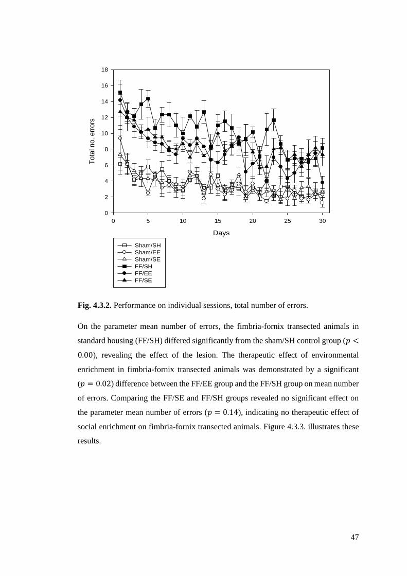
47
Days
0 5 10 15 20 25 30
To
tal n
o.
err
ors
0
2
4
6
8
10
12
14
16
18
Sham/SH
Sham/EE
Sham/SE
FF/SH
FF/EE
FF/SE
Fig. 4.3.2. Performance on individual sessions, total number of errors.
On the parameter mean number of errors, the fimbria-fornix transected animals in
standard housing (FF/SH) differed significantly from the sham/SH control group (𝑝 <
0.00), revealing the effect of the lesion. The therapeutic effect of environmental
enrichment in fimbria-fornix transected animals was demonstrated by a significant
(𝑝 = 0.02) difference between the FF/EE group and the FF/SH group on mean number
of errors. Comparing the FF/SE and FF/SH groups revealed no significant effect on
the parameter mean number of errors (𝑝 = 0.14), indicating no therapeutic effect of
social enrichment on fimbria-fornix transected animals. Figure 4.3.3. illustrates these
results.

48
Sham/SH Sham/EE Sham/SE FF/SH FF/EE FF/SE
Me
an
no
. o
f e
rro
rs
0
2
4
6
8
10
12
14
*
Fig. 4.3.3. ∗ 𝑝 < 0.05. Mean number of errors across sessions for each experimental
group. The results reveal a significant difference in performance between the FF/SH
and FF/EE groups.
4.3.3.2. – Mean number of repetitive errors
For the mean number of repetitive errors, the one-way ANOVA revealed a significant
difference between groups. Mean number of repetitive errors (and standard deviations)
for groups 1 (sham/SH), 2 (FF/SH), 3 (sham/EE), 4 (FF/EE), 5 (sham/SE) and 6
(FF/SE) were 0.39 (0.15), 5.63 (2.33), 0.37 (0.22), 3.34 (1.09), 0.35 (0.25) and 3.52
(1.58) respectively. These means differed significantly, 𝐹 = (5 , 30), 𝑝 = 0.00.
On the parameter mean number of repetitive errors the fimbria-fornix transected
animals in standard housing (FF/SH) differed significantly from the sham/SH control
group (𝑝 = 0.00), revealing the effect of the lesion. The therapeutic effect of
environmental enrichment (EE) in regards to perseverance in fimbria-fornix transected
animals was demonstrated by a significant (𝑝 = 0.05) difference between the FF/EE
group and the FF/SH group on mean number of repetitive errors. Comparing the FF/SE
and FF/SH groups revealed no significant effect on this parameter (𝑝 = 0.09),
indicating no therapeutic effect of social enrichment on fimbria-fornix transected
animals in regards to perseverance. Figure 4.3.4. illustrates these results.
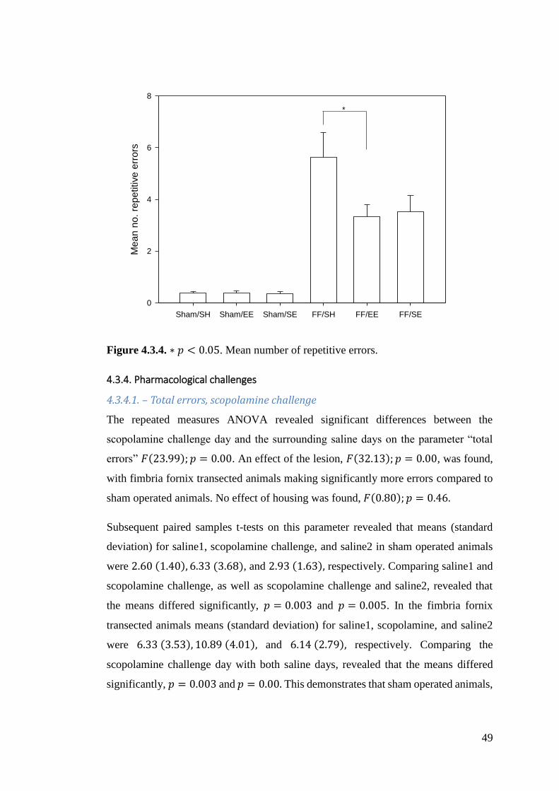
49
Sham/SH Sham/EE Sham/SE FF/SH FF/EE FF/SE
Me
an
no
. re
pe
titive
err
ors
0
2
4
6
8
*
Figure 4.3.4. ∗ 𝑝 < 0.05. Mean number of repetitive errors.
4.3.4. Pharmacological challenges
4.3.4.1. – Total errors, scopolamine challenge
The repeated measures ANOVA revealed significant differences between the
scopolamine challenge day and the surrounding saline days on the parameter “total
errors” 𝐹(23.99); 𝑝 = 0.00. An effect of the lesion, 𝐹(32.13); 𝑝 = 0.00, was found,
with fimbria fornix transected animals making significantly more errors compared to
sham operated animals. No effect of housing was found, 𝐹(0.80); 𝑝 = 0.46.
Subsequent paired samples t-tests on this parameter revealed that means (standard
deviation) for saline1, scopolamine challenge, and saline2 in sham operated animals
were 2.60 (1.40), 6.33 (3.68), and 2.93 (1.63), respectively. Comparing saline1 and
scopolamine challenge, as well as scopolamine challenge and saline2, revealed that
the means differed significantly, 𝑝 = 0.003 and 𝑝 = 0.005. In the fimbria fornix
transected animals means (standard deviation) for saline1, scopolamine, and saline2
were 6.33 (3.53), 10.89 (4.01), and 6.14 (2.79), respectively. Comparing the
scopolamine challenge day with both saline days, revealed that the means differed
significantly, 𝑝 = 0.003 and 𝑝 = 0.00. This demonstrates that sham operated animals,
50
as well as fimbria fornix transected ones, made significantly more errors on the
scopolamine challenge day compared to the surrounding saline days.
In sham operated animas, the results above can be attributed to the environmentally
enriched group (EE). Comparing scopolamine challenge day with saline1 and saline2,
respectively, reveals that scopolamine administration has a significant negative impact
on the performance in the delayed alternation test, 𝑝 = 0.00 and 𝑝 = 0.002. No such
impact was found in standard housed (SH) or socially enriched (SE) sham operated
animals, 𝑝 = 0.36 and 0.26 for the SH group, and 𝑝 = 0.33 and 𝑝 = 0.56 for the SE
group.
In fimbria fornix transected animals, differences can be attributed to the EE and SE
group. In these groups administering scopolamine significantly impacts the
performance negatively, which becomes evident when comparing the scopolamine
challenge day with saline1 and saline2, 𝑝 = 0.01 and 𝑝 = 0.01 for EE group, and 𝑝 =
0.02 and 𝑝 = 0.01 for the SE group. No significant differences between scopolamine
challenge and saline days were found in the SH group, 𝑝 = 0.49 and 𝑝 = 0.28. Figure
4.3.5. illustrates the above.

51
Sham/SH Sham/EE Sham/SE FF/SH FF/EE FF/SE
Me
an
no
. e
rro
rs
0
2
4
6
8
10
12
14
16
Saline1
Scopolamine
Saline2
Fig. 4.3.5. Mean number of errors on saline day 1, scopolamine challenge day and
saline day 2
4.3.4.2. – Repetitive errors, scopolamine challenge
The repeated measures ANOVA revealed significant differences between the
scopolamine challenge day and the surrounding saline days on the “repetitive errors”
𝐹(14.74); 𝑝 = 0.00. An effect of the lesion, 𝐹(28.16); 𝑝 = 0.00 , was found, with
fimbria fornix transected animals making significantly more repetitive errors
compared to sham operated animals. No effect of housing was found, 𝐹(1.31); 𝑝 =
0.29.
Subsequent paired samples t-tests on this parameter revealed that means (standard
deviation) for saline1, scopolamine challenge, and saline2 in sham operated animals
were 0.17 (0.41), 1.93 (2.25), and 0.27 (0.53), respectively. Comparing saline1 and
scopolamine challenge, as well as scopolamine challenge and saline2, revealed that
the means differed significantly, 𝑝 = 0.007 and 𝑝 = 0.02. In the fimbria fornix
52
transected animals means (standard deviation) for saline1, scopolamine, and saline2
were 2.64 (2.53), 6.56 (4.63), and 2.5 (1.98), respectively. Comparing the
scopolamine challenge day with both saline days, revealed that the means differed
significantly, 𝑝 = 0.01 and 𝑝 = 0.001. This demonstrates that sham operated animals,
as well as fimbria fornix transected ones, made significantly more repetitive errors on
the scopolamine challenge day compared to the surrounding saline days.
In sham operated animas, the results above can be attributed to the environmentally
enriched group (EE). Comparing the scopolamine challenge day with saline1 and
saline2, respectively, reveals that scopolamine administration has a significant
negative impact on the amount of repetitive errors, 𝑝 = 0.001 and 𝑝 = 0.002. No such
impact was found in standard housed (SH) or socially enriched (SE) sham operated
animals, 𝑝 = 0.24 and 0.19 for the SH group, and 𝑝 = 0.36 and 𝑝 = 0.58 for the SE
group.
In fimbria fornix transected animals, differences can be attributed to the EE and SE
groups. In these groups administering scopolamine significantly impacts the amount
of repetitive errors negatively, which becomes evident when comparing the
scopolamine challenge day with saline1 and saline2, 𝑝 = 0.02 and 𝑝 = 0.03 for EE
group, and 𝑝 = 0.04 and 𝑝 = 0.02 for the SE group. On this parameter, no significant
differences between scopolamine challenge and saline days were found in the SH
group, 𝑝 = 0.54 and 𝑝 = 0.19. Figure 4.3.6. illustrates this.

53
Sham/SH Sham/EE Sham/SE FF/SH FF/EE FF/SE
Me
an
no
. re
pe
tivite
err
ors
0
2
4
6
8
10
12
Saline1
Scopolamine
Saline2
Fig. 4.3.6. Mean number of repetitive errors on saline days 1, scopolamine challenge
day and saline day 2.
4.3.4.3. – Total errors, SKF-83566 challenge
The repeated measures ANOVA revealed a significant differences between the SKF-
83566 challenge day and the surrounding saline days on the parameter “total errors”
𝐹(4.37); 𝑝 = 0.02, with significantly less errors on the SKF-83566 challenge day
compared to the surrounding saline days. An effect of the lesion, 𝐹(46.48); 𝑝 = 0.00,
was found, with fimbria fornix transected animals making significantly more errors
compared to sham operated animals. No effect of housing was found, 𝐹(3.05); 𝑝 =
0.06.
Subsequent paired samples t-tests on this parameter revealed that means (standard
deviation) for saline2, SKF-83566 challenge, and saline3 in sham operated animals
were 2.93 (1.16), 1.80 (1.47), and 2.00 (1.43), respectively. Comparing saline1 and
SKF-83566 challenge, as well as SKF-83566 challenge and saline2, revealed that the
means differed significantly between saline2 and SKF-83566 challenge, 𝑝 = 0.005,
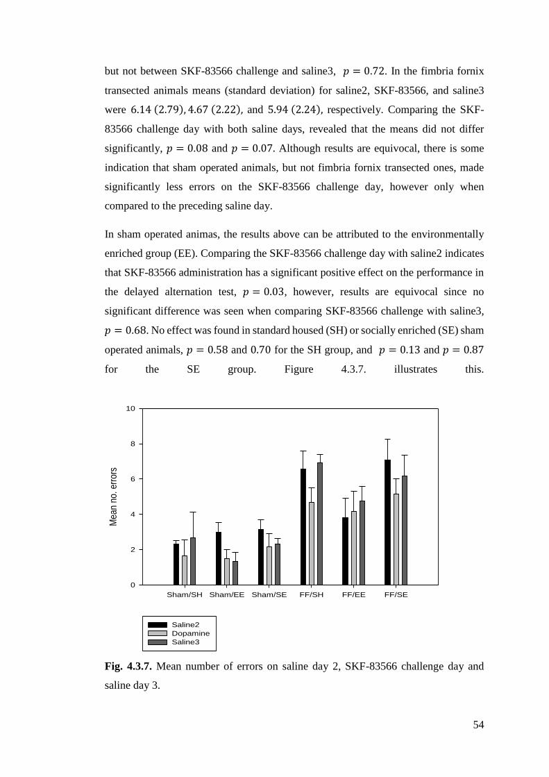
54
but not between SKF-83566 challenge and saline3, 𝑝 = 0.72. In the fimbria fornix
transected animals means (standard deviation) for saline2, SKF-83566, and saline3
were 6.14 (2.79), 4.67 (2.22), and 5.94 (2.24), respectively. Comparing the SKF-
83566 challenge day with both saline days, revealed that the means did not differ
significantly, 𝑝 = 0.08 and 𝑝 = 0.07. Although results are equivocal, there is some
indication that sham operated animals, but not fimbria fornix transected ones, made
significantly less errors on the SKF-83566 challenge day, however only when
compared to the preceding saline day.
In sham operated animas, the results above can be attributed to the environmentally
enriched group (EE). Comparing the SKF-83566 challenge day with saline2 indicates
that SKF-83566 administration has a significant positive effect on the performance in
the delayed alternation test, 𝑝 = 0.03, however, results are equivocal since no
significant difference was seen when comparing SKF-83566 challenge with saline3,
𝑝 = 0.68. No effect was found in standard housed (SH) or socially enriched (SE) sham
operated animals, 𝑝 = 0.58 and 0.70 for the SH group, and 𝑝 = 0.13 and 𝑝 = 0.87
for the SE group. Figure 4.3.7. illustrates this.
Sham/SH Sham/EE Sham/SE FF/SH FF/EE FF/SE
Mea
n no
. err
ors
0
2
4
6
8
10
Saline2
Dopamine
Saline3
Fig. 4.3.7. Mean number of errors on saline day 2, SKF-83566 challenge day and
saline day 3.
55
4.3.4.4. – Repetitive errors, SKF-83566 challenge
The repeated measures ANOVA revealed that there was no significant differences
between the SKF-83566 challenge day and the surrounding saline days on the
“repeated errors”, 𝐹(1.71); 𝑝 = 0.19. An effect of the lesion, 𝐹(31.99); 𝑝 = 0.00,
was found, with fimbria fornix transected animals making significantly more repetitive
errors compared to sham operated animals. No effect of housing was
found, 𝐹(3.33); 𝑝 = 0.06.
4.3.5. Summarizing main differences between the present data and the complete data
set
As mentioned above, some of the results presented here do not correspond to those
presented in the original article by Gram et al. (in preparation). In the following, I will
point out these differences and my discussion of results will reflect reality, not the
results above.
On the parameter “mean number of errors”, as well as on the parameter “mean
number of repetitive errors”, one will find that the lack of therapeutic effect of
social enrichment in fimbria fornix transected animals in the results presented
above, is the result of the constricted data material. In the original article, a
therapeutic effect of social enrichment was indeed present on both parameters,
𝑝 < 0.05 for mean number of errors and 𝑝 < 0.05 for mean number of
repetitive errors, revealing that FF/SE animals made fewer errors, as well as
fewer repetitive errors, than FF/SH animals (Gram et al., in preparation).
In the above presented results, no effect of scopolamine administration was
found in the sham/SE group, contrary to the actual results. In Gram et al. (in
preparation), one will find that sham/SE animals made significantly more
errors on the day of the scopolamine administration compared to the
surrounding saline days, 𝑝 < 0.05 and 𝑝 < 0.05, respectively (Gram et al., in
preparation).
In the above, only the sham operated animals in the EE group made
significantly more repetitive errors on the scopolamine challenge day
compared to the saline days. However, in the original article, this was also true
for the standard housed compared to the subsequent saline day (Gram et al., in
preparation).
56
In the results presented here, no effect of the SKF-83566 administration was
found in the fimbria fornix transected animals, contradicting reality. The
original article states that the animals in the FF groups made significantly less
errors, as well as repetitive errors, on the SKF-83566 challenge day than on the
surrounding saline days, 𝑝 = 0.001 and 𝑝 = 0.01, respectively. This was the
case in all housing groups (see Gram et al., in preparation).
In the original article, administration of SKF-83566 in sham operated animals
had no effect on neither the number of errors, nor number of repetitive errors,
when comparing the SKF-83566 challenge with the surrounding saline days
(Gram et al., in preparation), contradictory to what one might expect having
read the above presented results.
4.4. Discussion
In the following, I will be analyzing and discussing the results of the actual experiment,
no longer focusing on the limited data material. After this, a discussion of how these
results relate to the REF-model will follow.
4.4.1. Discussion of results
Firstly, the results of the behavioral data will be discussed. This section includes
perspectives on our experimental design and how we might optimize it to answer
further questions that might have arisen from this experiment. After this, a section
discussing the results of the pharmacological challenges will follow. This section will
include a brief exposition of the pharmacological agents and why administration of
these agents are relevant for our experimentation.
4.4.1.1. - Mean number of errors/repetitive errors
The behavioral data revealed, as it appears from figure 4.3.1., that all groups made
fewer errors on the last day of testing compared to the number of errors made in the
first days of acquisition training, which indicate that all animals learned the objective
of the task and progressively improved in performance over time. The progression
made by the sham operated control group (sham/SH) reveals the true learning curve
for this assignment and this group of animals outperformed all lesion groups, which
was to be expected. However, the fimbria-fornix transected living in enriched
environments, be it typical environmental enrichment (EE) or social enrichment (SE),
made significantly fewer errors over the course of the experiment compared to last

57
lesion group, standard housing (FF/SH), thus performing significantly better, revealing
the therapeutic effect of both environmental and social enrichment. Further, both of
our enriched lesion groups (FF/EE and FF/SE) showed a significantly lower degree of
perseverance, measured by number of repetitive errors, than animals in the standard
housed lesion group, indicating more cognitive flexibility in enriched animals.
Studying figure 4.3.1. reveals even more interesting information regarding the lesion
groups. Our enriched groups, FF/EE and FF/SE, only seem to outperform the standard
housed animals (FF/SH) in the early sessions (sessions 1-15), after which the groups
level out, even though the intervention groups perform significantly better overall. We
have learned that all groups improve their performance progressively, which means
that learning occurs in all cases. Could this mean that what we are affecting with our
interventions strategies is not the ability to learn, but how fast learning occurs?
Perhaps. While those are speculations, there might be another possibility: That our
intervention strategies could potentially have a greater effect if our time frame was
different. In regards to exercise as part of the treatment of TBI, there have been
speculations about a window of opportunity and evidence suggest that premature
exercise might indeed exacerbate symptoms and disrupt restorative processes
(Griesbach et al., 2004; Griesbach, 2011). Other intervention strategies work with time
windows as well, e.g. administration of Erythropoietin (EPO) (Xiong et al., 2011), so
to speculate that there might be some sort of time window for cognitive and/or social
stimulation might not be completely unwarranted. What if our results from the early
sessions actually tell us something about the potential beneficial effects of our
intervention strategies and that the results from the later sessions reveal that our timing
is off and that our treatment plans could actually be more efficient if we manage to
define the window of opportunity, if there indeed is one? Apparently, the basis of the
FF/EE and FF/SE groups is better than that of the FF/SH group, but over time all lesion
groups perform equally well (not compared to the sham group, of course), which might
indeed suggest something about the timing of our treatment. Not to say that our
treatment does not have an effect, it clearly does, otherwise we would not have
significant results. However, I do feel that the this gives grounds for speculations about
1) what kind of effects our interventions have, 2) whether our data from the early
sessions does indeed reveal the individual groups’ starting point and thereby the
potential benefits of treatment, and 3) whether this means that there might be a window
of opportunity for environmental and social intervention strategies.
58
As mentioned earlier, most studies on the effects of environmental enrichment
involves a combination of components and any effect seen in those studies might very
well be an additive one, which leaves questions unanswered. Indeed, the effect
produces by our environmental enrichment (EE group) might be just that – an additive
effect. See, our EE animals were socially enriched as well and as such, cognitive
stimulation on might not be enough to facilitate neural rehabilitation its own, and the
effect we see could be the result of the combination with another strategy, social
stimulation. How do we know that these two factors do not have an additive effect?
The answer is simple: We do not know. Indeed, they might. This particular design is
not complex enough for us to rule out the possibility that cognitive stimulation, and
the effects of it, might be reinforced by, or even contingent upon, social stimulation.
What we need to do from here, in order to establish the functional outcome of the
cognitive component of environmental enrichment on its own, is to control the other
factors (movement and social stimulation) and isolate the cognitive factor in a more
satisfying manner. In order to establish the functional outcome of cognitive stimulation
and its relative efficacy compared to motor stimulation (exercise) and a combination
of factors that might have an additive effect, we need to isolate and group factors
accordingly. In a design like the current one, the housing conditions, then, should look
like this:
1) Environmental Enrichment, typical (toys, social, running wheels)
2) Environmental Enrichment, no exercise (toys, social, no exercise
equipment provided, traditionally, meaning no running wheels)
3) Environmental Enrichment, no social stimulation (toys, running
wheels)
4) Environmental Enrichment, no toys (social, running wheels)
5) Cognitive stimulation (toys only)
6) Social stimulation (no toys, no running wheels)
7) Exercise (no toys, no social stimulation)
8) Standard housing, no intervention

59
With these groups and the same surgical procedure and cognitive task as the one used
in the present experiment, we should be able to see the effect of the three factors on
their own, as well as any additive effects across factors, assuming that movement
within the cage does not have an effect. Otherwise, we would have to experiment with
cognitive stimulation in smaller cages, where elaborate movement is not an issue, a
point I will get back to below. In a less elaborate setup, one could leave out the
combination groups (groups 1-4) and only focus on the effects of the three components
(cognitive, social, motor) on their own. However, I do feel that looking into additive
effects is highly relevant and since the effect of exercise on its own seems established
(e.g. Griesbach, 2004) and the current study found an effect of social stimulation alone,
groups 6 and 7 (social stimulation and exercise, respectively) could be omitted. Thus,
it seems more relevant to isolate the cognitive component and compare it to the
combination groups, to see whether cognitive stimulation on its own has an effect and
if the effects produced in the combination groups are indeed additive or just an
expression of the cognitive component's effect.
Let me return to the point about rearing in the cage being an issue when housing
animals in larger cages. In the present study, the animals did not have access to running
wheels and, as such, were not provided with the traditional tools for exercise, which
might lead one to suggest that exercise might not be a necessary part of the
intervention, even though it too is clearly beneficial (Griesbach et al., 2004). However,
we cannot completely rule out that movement within the cage contributed to the
cognitive improvements seen in our experiment. No significant difference in motoric
activity was found between the two enriched groups, EE and SE. However,
possibilities are that these animals exhibited more motoric activity than animals
housed in standard housing and as such, we cannot completely rule out that the
therapeutic effect of our enriched environments in this study can be attributed to
movement within the cage, or at least be the result of an additive effect. If we were to
find data that might lend themselves to this interpretation, we should have been able
to track the standard housed animals as well, thus making it possible to compare their
motoric activity with that of the enriched animals.
As mentioned earlier, one of my main concerns when it comes to exercise as a
treatment tool, is that some patients might not be able to exercise enough (if at all),
which is why it seems so important to establish whether we can alleviate these patients
60
of some of their symptoms by cognitive and social stimulation, and whether the effects
are equivalent to those of exercise.
To establish this, first, we would need to investigate how much exercise counts as
exercise. In other words, how much (or little!) exercise is enough to induce neural
plasticity and alleviate symptoms after TBI? An experimental setup placing lesioned,
as well as sham operated animals, in cages with build-in running wheel tracking
systems, tracking the daily distance each animal travels in the running wheel, could
give some indication. Assigning different distances that each animal is allowed to
travel each session and installing brakes to stop the wheel when the criteria is reached,
would give us the opportunity to control the motoric activity and possibly answer the
question above.
4.4.1.2. - Pharmacological challenges
Firstly, I feel the need to comment on why we perform pharmacological challenges
and what we hope to learn from them. By transecting the fimbria-fornix, we have
effectively stripped the animals of their use of the hippocampus, a structure we know
usually contributes to the mediation of the delayed alternation task. Still, we see
improvements in all lesion groups, some more than others, but improvements
nonetheless. Evidently, learning occurs at some level, even in animals that are
provided no other intervention than the cognitive training. This learning process can
apparently be reinforced with the application of more extensive intervention strategies,
such as the enriched environment, be it cognitive or social. The point is, that even
though we have eliminated the use of the hippocampus in these animals, they still
manage to learn the task and progressively improve their performance. Thus, when the
hippocampus is no longer an active part of the neural substrate used to mediate this
task, other parts of the brain must take over this mediation, since we know that that the
fimbria-fornix fiber bundle does not regrow and heal, it is lost (Mogensen et al., 2007).
Therefore, we must assume that the animals employ alternative neural substrates in the
execution of this task and the pharmacological challenges will help us determine which
neural substrate(s).
Administration of chemical compounds that inhibit a given neurotransmitter system,
will effectively, though temporarily, eliminate the use of structures that depend on this
particular neurotransmitter system, thus giving us the opportunity to record the

61
behavioral consequences and determine whether that particular system might
contribute to the mediation of a particular task (Mogensen and Malá, 2009).
Specifically, we administered scopolamine and SKF-83566 in order to determine
whether the alternative neural substrate utilized by our hippocampally lesioned rats in
the of solving the delayed alternation task might be mediated by structures that depend
on the neurotransmitters systems that we inhibited, the cholinergic and the
dopaminergic systems, respectively. Further, group comparisons of the challenge-
dependent consequences can reveal whether the involvement of these systems in the
mediation of the delayed alternation task is lesion-specific and even demonstrate
potential effects of utilizing environmental enrichment as a therapeutic tool.
There seems to be a functional overlap between the hippocampus and the prefrontal
cortex, since lesions within either structure impairs animals trying to solve various
cognitive tasks, for example allocentric place learning (Mogensen et al., 2004),
egocentric spatial orientation (Mogensen et al., 2005), as well as delayed alternation
(Mogensen et al., 2007), indicating that both of these structures are intricate parts of
the neural substrate involved in solving the aforementioned cognitive tasks. Thus,
manipulations that affect he prefrontal cortex might reveal whether the animals in the
present study employ a neural strategy that potentially up-regulate the prefrontal
mechanisms. Inhibition of the cholinergic and the dopaminergic systems, respectively,
might provide us with information regarding prefrontal involvement in solving the
delayed alternation task in the case of hippocampal lesions, since these systems project
to the hippocampus as well as to the prefrontal areas (Breedlove et al., 2010).
Scopolamine, being a muscarinic receptor antagonist, effectively inhibits the
cholinergic system (Mogensen and Malá, 2009; Breedlove et al., 2010), and previous
studies suggest that this system is highly involved in the solving of the delayed
alternation test (Mogensen et al., 2004). Thus, it would be expected that animals
perform significantly worse on the day of scopolamine administration. This hypothesis
was confirmed, as all animals in the present study performed significantly worse on
the day of the scopolamine injections in the dosage of 0.5 mg/kg body weight, making
more errors and more repetitive errors. All lesion groups, FF/EE, FF/SE, and FF/SH,
performed even worse than they usually did, suggesting that in cases of lesions to the
hippocampus, these animals still relied on cholinergic projections, indicating that even
when the use of the hippocampus is no longer possible, cholinergic projections are still
62
important mediators in the delayed alternation task. On the day of scopolamine
administration, the sham operated animals performed at a level equivalent to that of
the hippocamally lesioned animals during the regular acquisition training. Thus it
seems that inhibiting the cholinergic system of sham operated animals, heavily impairs
their ability to solve the delayed alternation task, which suggests that the cholinergic
system is a pivotal mediator in the solving of this task, even in non-lesioned animals.
In the present study, administration of the dopaminergic antagonist SKF-83566 in the
dosage of 0.1 mg/kg body weight had no effect on sham operated animals, suggesting
that in the mediation of the delayed alternation task, healthy animals that have acquired
this task are less dependent on dopaminergic-dependent structures than animals with
lesions to the hippocampus. Indeed, in the present study, SKF-83566 administration
had a significantly positive effect on the performance of the fimbria-fornix transected
animals, who all performed significantly better, making less errors and repetitive
errors, on the SKF-83566 challenge day compared to the surrounding saline
administration days. This group difference suggests that hippocampally lesioned
individuals employ a neural substrate in the mediation of the delayed alternation test
that is dissimilar to the neural substrate employed by non-lesioned individuals.
Apparently, there is an up-regulation of mechanisms in other structures when the
hippocampus is lost to injury. Mogensen, et al., (2004) demonstrated that this up-
regulation might be of prefrontal mechanisms. In this study, animals with fimbria-
fornix transection where heavily impaired in an allocentric place-learning task in a
watermaze when the chatecholaminernic system was rendered dysfunctional by
administration of d-amphetamine. Interestingly, the animals that had been subjected to
a combined lesion, fimbria-fornix transection and ablation of the prefrontal cortex,
were not affected by d-amphetamine administration (Mogensen et al., 2004),
demonstrating that these two lesion groups do not employ the same neural substrate in
mediating the task. When the animals had been subjected to the combined lesion, they
did not utilize structures dependent on the catecholaminerig system, contrary to the
isolated fibria-fornix transected animals. Seemingly, isolated lesions to the
hippocampus causes an up-regulation of mechanisms that might very well be
prefrontal (Mogensen et al., 2004). It is important to realize, though, that the cognitive
task utilized by Mogensen et al. (2004) differs from the one utilized in the present
study. However, another study by Mogensen et al. (2007) suggest that in mediating

63
the delayed alternation task, hippocampally lesioned animals might be up-regulating
prefrontal mechanisms as well, and further, that this up-regulation might not be the
most efficient strategy, something that supports the results of the present study.
Mogensen et al. (2007) subjected animals to either a transection of the fimbria-fornix
(FF), ablation of the prefrontal cortex (PFC) or a combination of both lesion types
(FF+PFC). Subsequently, animals were subjected to the delayed alternation task in a
T-maze, and errors as well as repetitive errors were recorded. When an animal made
three errors or less on two consecutive sessions it had reached the behavioral criterion
that had been established beforehand, and this was recorded. Initially, the two fimbria-
fornix transected groups, FF and FF+PFC, seemed equally impaired. Both groups
needed significantly more sessions before reaching the behavioral criterion and the
two groups did not differ significantly from one another. However, when the team
introduced a challenge, the results were quite different. On this challenge session, the
delay between each trial was prolonged. Thus, when the animal entered the opposite
arm as it did on the preceding trial, the delay was 24 seconds, and when the animal
made the mistake of entering the same arm as on the preceding trial, the delay was 32
seconds. In this session, the animals with the combined lesion, FF+PFC group,
performed on the same level as the animals with prefrontal ablation only, PFC group,
as well as sham operated animals. The animals that had only been subjected to fimbria-
fornix transection, FF group, however, made significantly more errors than the other
groups, suggesting that on this particular challenge, an intact prefrontal cortex is not
an advantage if the hippocampus, too, is dysfunctional (Mogensen et al., 2007).
Granted, the animals with the combined lesion had to perform all training without the
use of their hippocampus or their prefrontal cortex, thus forcing them to utilize
alternative strategies in the acquisition, differentiating them from the animals in our
study who did indeed have the use of their prefrontal cortex during acquisition training.
However, the results presented by Mogensen et al. (2007) do suggest that in some
instances of lesions to the hippocampus, the alternative mediators in the solving of the
delayed alternation test might be more efficient if other structures are inhibited as well.
And in fact, our study demonstrates that the neural substrate that is employed when
the dopaminergic system is inhibited is more efficient in the mediation of the delayed
alternation task than the one employed when the dopaminergic system is available.
Seemingly, hippocampally lesioned individuals become hyperdependent on
dopaminergic mechanisms, presumably located in the prefrontal cortex, and when

64
being forced to utilize other structures than the ones dependent on the dopaminergic
system, their neural strategy is more efficient. This indicates that the initial neural
reorganization in the animals in our study does in fact not yield the most efficient
strategy. In fact, the hippocampally lesioned animals in our study perform better when
we inhibit the dopaminergic system, indicating that structures within this system
potentially obstruct the optimal efficacy of reorganization.
4.4.2. Returning to the REF-model
It is important to realize that this particular study was not specifically designed to find
evidence that supports the REF-model. Rather, it was designed to find evidence that
might help us learn more about the possibilities for utilizing environmental enrichment
as a therapeutic tool. However, I do feel that it is possible to explain our results in
REF-model terms.
In terms of the REF-model, the aforementioned results reflect the construction of new
ASs after acquired brain damage. The animals in the present study that had been
subjected to transection of the fimbria-fornix, did not regrow the lost tissue, confer the
histological assessment. Therefore, the animals acquired task mediation without the
use of their hippocampus. Since the neural substrate mediating the task post-
traumatically is neither a reestablishment of the lost structure, nor a copy of the neural
substrate that mediated the task pre-traumatically, we must assume that the lesioned
animals in the present study utilized alternative neural substrates in the mediation of
the delayed alternation task, since all lesioned animals eventually learned to solve the
task.
The pharmacological challenges provide evidence that might lend itself to the
interpretation that in this particular case of lesions to the hippocampus of rats being
subjected to the delayed alternation task in the T-maze, the functional reorganization
is not the most efficient. Not to say that it is insufficient. All animals eventually reach
a performance level that resembles that of the sham operated animals, thus exhibiting
full functional recovery. However, the positive effect of administrating SKF-83566 on
the performance of the lesioned animals, suggests that there is an even more efficient
neural substrate available. However, this is not the neural substrate initially employed
by any of the lesion groups. This indicates that even though our intervention strategies
do have positive effects, seeing that both enriched groups (EE and SE) perform

65
significantly better overall, compared to the standard housed animals, it does not
induce the most efficient neural substrate. Evidently, the training and intervention
provided in the present experiment did not allow the animals to construct the most
efficient AS for mediation of the task presented to them. They obviously all formed
ASs that were able to mediate the task and as such the surface phenomenon was
reestablished in all animals, however, there seems to be some indication that yet
another alternative AS for task mediation was available, however, was not utilized.
Further studies might provide insight as to why that is.
I feel that this supports the notion that the functional outcome highly depends on the
training situation and the environmental input, one of the main arguments for
implementation of the REF-model. Earlier, I have argued that rehabilitative training
might be more efficient if we construct the training in a manner that resembles
situations that the patient might encounter in real life, since training in a specific test
might not be transferable to other situations, as is the case with the patient with neglect,
mentioned above. The question of generalizability, whether the effect of training in
one situation can be transferred to other situations, provides quite a challenge, both
clinically and in a laboratory setting. Since the present study was performed on
animals, I find it natural to focus on how generalizability of, for instance, therapeutic
effect can be studied. If we want to study whether the effects of rehabilitative training
has a positive effect on an everyday life situation, then we must conduct an experiment
in which animals are required to perform an everyday life task, for example gathering
food. The experiment could be conducted in an environment like the one seen in
Mogensen (1991), an environment that consists of several interconnected enclosures
with transparent walls, making it possible to observe the animals and test whether the
rehabilitative training provided to the animals affects their ability to solve the everyday
life task mentioned above – gathering food. Lesioned, as well as sham operated
animals, would be divided into groups that are either provided with rehabilitative
training or do not receive any other training than the testing itself. If the animals that
are provided with training are able to solve the everyday life task more efficiently than
the non-trained group, then this would support the interpretation that the animals are
able to generalize from the training situation to an everyday life situation.
In the present study, we inflicted the animals with a focal injury, which, as mentioned,
is typically an advantage when the aim of the experiment is to study the

66
reorganizational mechanisms involved in functional recovery. However, in our
experiment we especially wanted to study the effect of a specific type of treatment,
namely enrichment, both cognitive and social. When studying treatment effects it is
typically an advantage to use a TBI model that has more ecological validity, meaning
that it bears close resemblance to clinical situations, something the fimbria-fornix
transection models does not. However, as Mogensen (2011b; 2011c) argues, if we
want to learn more about functional recovery and rehabilitation, we need to study these
phenomena in a variety of ways. The shortcomings of one model or one test, might be
circumvented by another model or test, and vice versa. In the present study, the
infliction of a focal damage gave the advantage of knowing exactly which structure
was impaired, namely the hippocampus. Therefore, we have learned that enriched
environments have a positive effect on the recovery when the hippocampus is
impaired. Further, we have found evidence that has helped us learn more about the
hippocampus and its involvement in the mediation of delayed alternation. If we want
to learn more about the therapeutic effects of environmental enrichment, it would be
beneficial to study the effects of these treatment strategies in a variety of cognitive or
behavioral tests, and with other TBI models with more ecological validity, for instance
the CCI, the symptoms of which closely resembles the ones seen in humans that have
been in car accidents.
4.5. Conclusion
Over the thirty sessions of acquisition training in the delayed alternation task in the T-
maze, the therapeutic effects of environmental enrichment, as well as social
stimulation, was revealed by a significantly better performance on the delayed
alternation task by the two enriched lesion groups, making fewer errors and repetitive
errors compared to the standard housed lesion group. The revealed effect of the
cognitively enriched environment, though, might be an additive one. Animals housed
in the environmentally enriched cage where also socially enriched, which makes it
unwarrantable to make conclusions about the functional outcome of the cognitive
component on its own. Learning more about the therapeutic effects of cognitive
stimulation would require an experimental design in which the cognitive factor is
isolated more satisfyingly. Assuming that movement within the cage is not enough to
yield effects corresponding to those of exercise, we have evidence that exercise does
not necessarily need to be part of the enrichment treatment in order for it to have an

67
effect, since neither of the enriched groups were provided with tools for exercise.
However, it is crucial that one realizes that this does not prove that the motoric
component of environmental enrichment is not a necessity, since movement within the
cage could, quite possibly, still contribute the measured effects. What we need to
determine is whether the effect of enrichment without exercise is enough to alleviate
patients, unable to exercise, of some of their TBI symptoms. Further, it might be useful
to learn more about the additive effects of cognitive, social and motor stimulation. The
pharmacological testing confirmed the importance of the cholinergic system in
problem solving of this kind. Further, results from the SKF-83566 challenge might
lend the us the interpretation that the functional recovery seen in these animals might
not be yielded by the most efficient reorganization, since inhibiting the dopaminergic
system actually improves their performances.
A better understanding of the mechanisms involved in neural rehabilitation as well as
the mechanisms triggered by environmental enrichment and how these mechanisms
interact, will hopefully help us to better understand and utilize enrichment paradigms
as therapeutic tools.
Acknowledgements
A special thanks to Marie Gajhede Gram and Jesper Mogensen for letting me
participate in every aspect of this experiment. As a thesis student, I have not attended
the course licensing me to perform operations, anaesthetize or execute any other
invasive task on living animals, and of course I have not. I have been able to observe
all of these tasks, though, as well as ask questions about the procedures, and for that I
am grateful.
5. Concluding remarks
Since, worldwide, TBI is one of the leading causes of severe disability, research that
helps us learn more about the neural mechanisms involved in functional recovery,
research that investigates possibility for strengthening treatment or even presents
alternatives to existing treatment, is essential. However, in order for us to learn more
about functional recovery and improving the efficiency of rehabilitative training, we
need to understand how the brain functions in general. Applying an inadequate model
of brain functioning, can have dire consequences for the treatment we provide to brain
68
injured patients, since the way we conceptualize brain function directly affects the way
we conceive functional recovery.
Functional localization theorists argue that the brain consists of a series of functional
modules and that a specific module always processes the same informational input,
and therefore always mediates a specific function. In this view, any function that is
lost as a result of damage to the brain, is permanently lost, thus rendering rehabilitative
training completely irrelevant. However, numerous studies, both clinical and
laboratory, have produced evidence of complete functional recovery of an impaired
function after acquired brain injury. These instances, the extreme functional
localization theorists argue, are merely the result of incomplete lesions, where part of
the mediating module is preserved and regains function. Thus, conclusions about
functional localization should only be drawn from cases of complete lesions. However,
laboratory studies, in which animals have been inflicted with complete and localized
lesions, indicate that even in these cases, full functional recovery can occur. Therefore,
the brain cannot be conceived as a completely modular entity with each module being
the sole mediator of a specific function, since this does not explain functional recovery.
The connectionist model of the brain provides an alternative to the functional
localization theory and has a clear advantage, since this model has no problem
explaining functional recovery. In this view, the brain is constantly adapting to
environmental demands and functional recovery, it is argued, is merely a manifestation
of this adaption. This model suggests that input is distributed across the brain, thus
perceiving the brain as an intricate system, in which informational input is processed
by several neural networks, and, therefore, when an area of the brain is lost to injury,
another network can mediate the function that was initially impaired. Essentially, this
means that post-traumatically, a copy of the neural substrate that mediated the function
post-traumatically is available. However, even though functional recovery has been
demonstrated repeatedly, the brain does exhibit some level of functional localization
nonetheless, something connectionism cannot explain. Utilization of fMRI have
allowed the demonstration of correlations between activation of particular brain
regions and conducting specific tasks. Further, symptoms of lesions to a specific area
can quite accurately be predicted, for example the inability to produce fluent language
after a lesion to Broca’s area. Evidently, the brain cannot be conceived as being
completely modular or completely connectionist, and somehow functional localization
69
and functional recovery coexist. The REF-model explains this seemingly contradicting
statement and provides a more comprehensive model of the brain, a model that is
essentially a model of functional modules within a connectionist network. Basically,
this model rejects the idea that the damaged structure regrows or that preserved parts
of the damaged structure continues to mediate an impaired function after brain
damage. It also rejects the idea that, post-traumatically, a copy of the neural substrate
that mediated the impaired function, pre-traumatically, is available for mediation of
the impaired function. Instead, the REF-model, which is based on numerous laboratory
studies, argues that after acquired brain damage, the brain reorganizes and constructs
alternative neural substrates that mediate the impaired function. Within this view, any
function is mediated by an algorithmic strategy, which again is mediated by a series of
localized elementary functions and the connections between them. When an area is
lost to brain injury, so are the elementary functions within this area. Any algorithmic
strategy that was mediated by the lost elementary functions is lost as well and this
results in an impairment. However, through reorganization of elementary functions,
new algorithmic strategies are formed, and functional recovery can occur. This process
is highly dependent on environmental input, or the feedback that is provided, which
means that the situation in which rehabilitative training is conducted is has a significant
impact on recovery. Therefore, it is essential that we try to modify the treatment of
brain injury patients to make it more efficient and meet environmental demands.
In the experimental section of this paper, I presented a study in which we wanted to
learn more about the effects of environmental enrichment. Traditionally, an enriched
environment consists of three components, a cognitive, a social and a motoric
component, respectively. The positive effects of motoric activity have been
immensely covered, however, some patients might not be able to take advantage of
this effect, for instance if they are paralyzed. Therefore, we wanted to investigate the
functional outcome of the cognitive and the social component. The present study was
performed on rats that were housed in either an enriched environment that was both
cognitively and socially stimulating, a socially enriched environment or in a standard
cage with just one other rat. When conducting research on animals one inevitably
encounters ethical issues, since utilization of animals in research causes massive
dispute in the Western world. Some believe that an animal has no moral rights unless
a human being extends his moral rights to the animal and, therefore, humans have the
70
right to utilize animals for research purposes. Others believe that all sentient creatures
are equal and some even insist that all research on animals should be ceased. However,
there is no unified answer to what is right and wrong in this matter, since this depends
on one’s personal moral beliefs. In order for me to participate in the present study, I
had to decide for myself what I believe in. Essentially, I feel that research conducted
on animals can provide us with pivotal knowledge that might help us cure diseases,
alleviate symptoms, and save lives. I believe in minimizing the number of animals
used in research, as well as maximizing the well-being of the animals that we do use.
Further, I believe that whenever it is possible, we should replace animal research with
animal-free methods. The present study, I feel, reflects these considerations.
In the present study, the housing conditions mentioned above represented the
intervention strategies. Within all three housing conditions, animals were randomly
assigned either sham surgery or transection of the fimbria-fornix, thus dividing the
animals into six experimental groups: 1) Sham operated animals in standard housing,
2) Fimbria fornix transected animals in standard housing, 3) Sham operated animals
living in the enriched environment, 4) Fimbria fornix transected animals living in the
enriched environment, 5) Sham operated animals living in the social environment, and
6) Fimbria fornix transected animals living in the social environment. Transecting the
fimbria-fornix provides a focal lesion that is easily quantifiable and, further, gives us
the advantage of knowing that the hippocampus is heavily impaired, which enables us
to study the involvement of this structure in the mediation of a given task. We studied
its involvement in the mediation of the delayed alternation test. The acquisition of this
task was studied in a T-maze in the six groups mentioned above.
The acquisition training revealed the therapeutic effects of environmental enrichment,
as well as social stimulation. Animals in the two enriched groups performed
significantly better, making fewer errors, compared to the standard housed lesion
group. However, animals in the environmentally enriched group where also socially
enriched, which might make the revealed effect of the cognitively enriched
environment an additive one. Thus, at this point we cannot draw a conclusion about
the functional outcome of the cognitive component on its own. Since we were not able
to control rearing inside the cage, we cannot completely rule out that there might be
an effect of motoric activity as well, even though animals were not provided with tools
for exercise. Further research might help us determine whether the effects of

71
enrichment without exercise is enough to alleviate patients of their TBI symptoms.
Research on the additive effect of cognitive, social and motoric stimulation might be
beneficial as well.
Further, pharmacological testing provided results that indicate that the functional
recovery seen in the present study might not be yielded by the most efficient
reorganization. The lesioned animals all performed significantly better when we
inhibited their dopaminergic system, essentially benefitting from not being able to
utilize this system in the mediation of the delayed alternation task. In REF-model
terms, the animals all formed alternative algorithmic strategies, allowing them to
achieve full functional recovery, however, the applied strategy might not be the most
efficient strategy available. Evidently, the feedback provided to these animals was not
the feedback that yielded the most efficient neural substrate, which supports the notion
that environmental input is essential for the functional outcome.
In order for us to learn more about functional recovery and rehabilitation after acquired
brain damage, neural mechanisms as well as the possibilities for strengthening
treatment, possibly by applying intervention strategies like environmental enrichment,
we need a better understanding of the neural mechanisms involved in rehabilitation as
well as the mechanisms triggered by rehabilitative training and the situation in which
the training is conducted. Hopefully, studying different types of brain injury in a
variety of tests and with the utilization of several types of treatment will allow us to
learn more about treatment of TBI and help us alleviate TBI patients of their disabling
symptoms in the most efficient way possible. This type of testing, will help us
construct rehabilitative training in a fashion that provides the patients with the most
efficient alternative mediation of function and help them return to their everyday lives
and workplaces, overcoming the devastating effects of brain injury, and continue to
adapt to environmental demands in the most effective way possible.

72
6. References
Abou-Ismail, U. A. (2011). Are the effects of enrichment due to the presence of multiple
items or a particular item in the cages of laboratory rat? Applied Animal Behavior
Science, 134: 72-82.
Barrett, H. C. & Kurzban, R. (2006). Modularity in Cognition: Framing the Debate.
Psychological Review, 113 (3): 628-647.
Bradley, V. and Kapur, N. (2010). Neuropsychological assessment of memory disorders. .
In J. M. Gurd, U. Kischka and J. C. Marshall (Eds.) The Handbook of Clinical
Neuropsychology (2. ed.). New York, NY: Oxford University Press.

73
Breedlove, S. M., Watson, N. V. and Rosenzweig, M. R. (2010). Biological Psychology. An
Introduction to Behavioral, Cognitive, and Clinical Neuroscience (6th ed.).
Sunderland, Mass: Sinauer Associates, Inc. Publishers. Chapter 4.
Brom, F. W. (2002). Science and society: different bioethical approaches towards animal
experimentation. Atlex, 19 (2): 78-82.
Brosnan, S. F. and de Waal, F. B. M. (2003). Monkeys reject unequal pay. Nature, 425: 297-
299.
Buller, D. J. & Hardcastle, V. G. (2000). Evolutionary Psychology, Meet Developmental
Neurobiology: Against Promiscuous Modularity. Brain and Mind, 1: 307-325.
Bunzeck, N., Guitart-Masip, M., Dolan, R. J. and Duzel, E. (2014). Pharmacological
Dissociation of Novelty Responses in the Human Brain. Cerebral Cortex, 24: 1351-
1360.
Cheng, J. P., Shaw, K. E., Monaco, C. M., Hoffman, A. N., Sozda, C. N., Olsen, A. S. and
Kline, A. E. (2012). A Relatively Brief Exposure to Environmental Enrichment after
Experimental Traumatic Brain Injury Confers Long-Term Cognitive Benefits. Journal
of Neurotrauma, 29: 2684-2688.
Cohen, A. I. (2007). Contractarianism, Other-regarding Attitudes, and the Moral Standing
of Nonhuman Animals. Journal of Applied Philosophy, 24 (2): 188-201.
Cortez, S. C., McIntosh, T. K., and Noble, L. J. (1989). Experimental fluid percussion brain
injury: vascular disruption and neuronal and glial alterations. Brain Research, 482:
271-282.
Crossman, A. R. ans Neary, D. (2010). Neuroanatomy. An illustrated colour text, 4th ed.
Churchill Livingstone Elsevier. Chapers 12, 13 and 14.
De Bartholo, P., Leggio, M. G., Mandolesi, L., Foti, F, Gelfo, F., Ferlazzo, F. and Petrosini,
L. (2008). Environmental enrichment mitigates the effects of basal forebrain lesions
on cognitive flexibility. Neuroscience, 154: 444-453.
de Haan, E. H. E. & Cowey, A. (2011). On the Usefulness of 'What' and 'Where' Pathways
in Vision. Trends in Cognitive Neurosciences, vol. 15 (10).

74
Deacon, R. M. J. and Rawlins, J. N. P. (2006). T-maze alternation in the rodent. Nature
Protocols, 1 (1): 7-12.
Dijkhuizen, R. M., Muller, H.-J., Tamminga, K. S., van Doremalen, H. A. A. J., Spruijt, B.
M. and Nicolay, K. (1992). H-NMR Imaging of Frimbria Fornix Lesion in the Rat
Brain. Brain Topography, 5 (2): 147-151.
Divac, I., Kosmal, A., Björklund, A. and Lindvall, O. (1978). Subcortical projections to the
prefrontal cortex in the rat as revealed by the horseradish peroxidase technique.
Neuroscience, 3: 785-796.
Divac, I. and Mogensen, J. (1985). The prefrontal “cortex” in the pigeon. Catecholamine
histofluorescence. Neuroscience, 15: 677-682.
Divac, I., Mogensen, J. and Björklund, A. (1985). The prefrontal “cortex” in the pigeon.
Biochemical evidence. Brain Research, 332: 365-368.
Dixon, C. E., Clifton, G. L., Lighthall, J. W., Yaghami, A. A. and Hayes, R. L. (1991). A
controlled cortical impact model of traumatic brain injury in the rat. Journal of
Neuroscience Methods, 39: 253-262.
Dixon, C. E. and Kline, A. E. (2009). Controlled Cortical Impact Injury Model. In J. Chen,
Z. C. Xu, X. Xu, and J. H. Zhang (Eds.). Animal Models of Acute Neurological
Injuries. NJ, USA: Humana Press.
Dragoni, G., Carpi, D., Recce, M., Csicsvari, J. and Buzsáki, G. (1999). Interactions between
Hippocampus and Medial Septum during Sharp Waves and Theta Oscillation in the
Behaving Rat. The Journal of Neuroscience, 19 (14): 6191-6199.
Duncan, K., Tompary, A. and Davachi, L. (2014). Associative Encoding and Retrieval
Predicted by Functional Connectivity in Distinct Hippocampal Area CA1 Pathways.
Journal of Neuroscience, 34 (34): 11188-11198.
Ellison, A. & Cowey, A. (2006). TMS can Reveal Contrasting Functions of the Dorsal and
Ventral Visual Processing Streams. Experimental Brain Research, 175: 618-635.
Ellison, A. & Cowey, A. (2007). Time Course of the Involvement of the Ventral and Dorsal
Processing Streams in a Visuospatial Task. Neuropsychologia, 45: 3335-3339.

75
Field, A. (2013). Discovering Statistics using IBM SPSS statistics, 4th ed. London: Sage
Publications.
Flor, H. (2004). Remapping somatosensory cortex after injury. Advanced Neurology, 93,
195-204.
Fodor, J. (1983). The modularity of mind. Cambridge, MA: MIT Press.
Foëx, B. A. (2007). The Ethics of Animal Experimentation. Emergency Medicine Journal,
24 (11): 750-751.
Frassinetti, F., Angeli, V., Meneghello, F., Avanzi, S. and Ladavas, E. (2002). Long-lasting
amelioration of visuospatial neglect by prism adaption. Brain, 125: 608-623.
Gade, A. (2010). De kliniske amnesisyndromer. In A. Gade, C. Gerlach, R. Starrfelt and P.
M. Pedersen (Eds.). Klinisk neuropsykologi. København: Frydenlund.
Garcia, A. N., Shah, M. A., Dixon, C. E., Wagner, A. K. and Kline, A. E. (2011). Biologic
and plastic effects of experimental brain injury treatment paradigms and their
relevance to clinical rehabilitation. PubMed Central, 2 (6): 18-27.
Gerlach, C. & Marstrand, L. (2010). Visuelle perceptionsforstyrrelser og agnosi. In A. Gade,
C. Gerlach, R. Starrfelt og P. M. Pedersen (Eds.) Klinisk neuropsykologi. København:
Frydenlund.
Gerlach, C., Starrfelt, R., Gade, A. and Pedersen, P. M. (2010). Neuropsykologi.
Begrebernes og metodernes historie og udvikling. In A. Gade, C. Gerlach, R. Starrfelt
and P. M. Pedersen (Eds.). Klinisk neuropsykologi. København: Frydenlund.
Goldberg, E. and Bougakov, D. (2005). Neuropsychologic Assessment of Frontal Lobe
Dysfunction. Psychiatric Clinics of North America, 28: 567-580.
Gram, M. G., Gade, L., Wogensen, E., Mogensen, J. and Malá, H. (in preparation).
Enhancing cognitive performance equally with typical environmental and specific
social stimulation after fimbria-fornix transection.
Griesbach, G. S. (2011). Exercise After Traumatic Brain Injury: Is it a Double-Edged
Sword? Physical Medicine and Rehabilitation (3): 64-72.
76
Griesbach, G. S., Gomez-Pinilla & Hovda, D.A. (2004). The up-regulation of plasticity-
related proteins following TBI is disrupted with acute voluntary exercise. Brain
Research, 1016: 1564-162.
Griesbach, G. S., Hovda, D. A., Molteni, R., Wu, A. and Gomez-Pinilla, F. (2004).
Voluntary Exercise Following Traumatic Brain Injury: Brain-Derived Neurotrophic
Factor Upregulation and Recovery of Function. Neuroscience, 125: 129-139.
Hansen, N. and Manahan-Vaughan, D. (2014). Dopamine D1/D5 Receptors Mediate
Informational Saliency that Promotes Persistent Hippocampal Long-Term Plasticity.
Cerebral Cortex, 24: 845-858.
Harding, J. D., Van Hoosier Jr., G. L. & Grieder, F. B. (2011). The Contribution of
Laboratory Animals to Medical Progress – Past, Present, and Future. In: J. Hau and S.
J. Schapiro (Eds.) Handbook of Laboratory Animal Science, 3rd ed., volume I. Boca
Raton, FL.: CRC Press LLC.
Henke, K. (2010). A model for memory systems based on processing modes rather than
consciousness. Nature Reviews, 11,: 523-532.
Johansson, B. B. (2004). Brain plasticity in health and disease. Keio Journal of Medicine
53(4): 231-246.
Johnson, E. M., Traver, K. L., Hoffman, S. W. and Harrison, C. R. (2013). Environmental
enrichment protects against functional deficits caused by traumatic brain injury.
Frontiers in Behavioral Neuroscience, 7, 44.
Kartsonis, L. D. (2010). Assessment of Percetual Disorders. In J. M. Gurd, U. Kischka and
J. C. Marshall (Eds.) The Handbook of Clinical Neuropsychology (2. ed.). New York,
NY: Oxford University Press.
Kempermann, G., Kuhn, H. G. and Gage, F. H. (1997). More hippocampal neurons in adult
mice living in an enriched environment. Nature, 386 (3): 493-495.
Langlois, J. A., Ruthland-Brown, W. and Wald, M. M. (2006). The Epidemiology and
Impact of Traumatic Brain Injury: A Brief Overview. Head Trauma Rehabilitation, 21
(5): 375-378.

77
Leggio, M. G., Mandolesi, L., Federico, F., Spirito, F, Ricci, B. and Petrosini, L. (2005).
Environmental enrichment promotes improved spatial abilities and enhanced dendritic
growth in the rat. Behavioral Brain Research, 163: 78-90.
Lighthall, J. W. (1988). Controlled Cortical Impact: A New Experimental Brain Injury
Model. Journal of Neurotrauma, 5 (1): 1-15.
Mass, A. I. R., Stocchetti, N. and Bullock, R. (2008). Moderate and severe traumatic brain
injury in adults. Lancet Neurology, 7: 728-741.
McIntosh, T. K., Vink, R., Noble, L., Yamakami, I., Fernyak, S., Soares, H., and Faden, A.
L. (1989). Traumatic Brain Injury in the Rat: Characterization of a Lateral Fluid-
Percussion Model. Neuroscience, 28 (1): 233-244.
Milner, D. & Goodale, M. (1992). Seperate Pathways for Perception and Action. TINS, 15
(1)
Milner, D. & Goodale, M. (2006). The Visual Brain in Action (2nd ed.). New York, NY:
Oxford University Press.
Milner, D. & Goodale, M. (2008). Two Visual Systems Re-reviewed. Neuropsychologia,
46: (774-785).
Mishkin, M., Ungerleider, L., & Macko, K. (1983). Object Vision and Spatial Vision: Two
Cortical Pathways. TINS (October 1983).
Mogensen, J. (1991). Influences of the rearing conditions on functional properties of the rat’s
prefrontal system. Behavioral Brain Research, 42: 135-142.
Mogensen, J. (2011a). Almost unlimited potentials of a limited neural plasticity: levels of
plasticity in development and reorganization of the injured brain. Journal of
Consciousness Studies, 18: 13-45.
Mogensen, J. (2011b). Animal Models in Neuroscience. In: J. Hau and S. Schapiro (Eds.)
Handbook of Laboratory Animal Science, Volume II. Boca Raton, FL.: CRC Press
LLC.
Mogensen, J. (2011c). Reorganization in the injured brain: implications for studies of the
neural substrate of cognition. Frontiers in Psychology, 2:7.
78
Mogensen, J. (2014). Reorganization of Elementary Functions (REF) after Brain Injury and
in the Intact Brain: A Novel Understanding of Neurocognitive Organization and
Reorganization. In A. Costa and E. Villalba (Eds.) Horizons in Neuroscience
Research, volume 15. Nova Science Publishers, Inc.
Mogensen, J., Christensen, L. H., Johansson, A., Wörtwein, G., Bang, L. E. and Holm, S.
(2002). Place learning in scopolamine treated rats: the roles of distal cues and
catecholaminergic mediation. Neurobiology of Learning and Memory, 78: 139-166.
Mogensen, J. and Divac, I. (1982). The prefrontal “cortex” in the pigeon. Behavioral
evidence. Brain, Behavior and Evolution, 21: 60-66.
Mogensen, J., Hjortkjær, J., Ibervang, K. L., Stedal, K. and Malá, H. (2007). Prefrontal
cortex and Hippocampus in posttraumatic functional recovery: Spatial delayed
alternation by rats subjected to transection of the fimbria-fornix and/or ablation of the
prefrontal cortex. Brain Research Bulletin 73: 86-95.
Mogensen, J., Jørgensen, O. S. and Divac, I. (1982). Synaptic proteins in frontal and cortical
brain regions of rats after exposure to spatial problems. Behavioral Brain Research, 5:
375-386.
Mogensen, J. Lauritsen, K. T., Elvertorp, S., Hasman, A., Moustgaard, A. and Wörtwein, G.
(2004) Place learning and object recognition by rats subjected to transection of the
fimbria-fornix and/or ablation of the prefrontal cortex. Brain Research Bulletin, 63:
217-236.
Mogensen, J. and Malá, H. (2009). Post-traumatic functional recovery and reorganization in
animal models: A theoretical and methodological challenge. Scandinavian Journal of
Psychology, 50: 561-573.
Mogensen, J., Moustgaard, A., Khan, U., Wörtwein, G. and Nielsen, K. S. (2005).
Egocentric spatial orientation in a water maze by rats subjected to transection of the
fimbria-fornix and/or ablation of the prefrontal cortex. Brain Research Bulletin, 41:
41-58.
Morris, R. (1984). Development of a water-maze procedure for studying spatial learning in
the rat. Journal of Neuroscience Methods, 11: 47-60.
79
Morris, R. (1990). Toward a Representational Hypothesis of the Role of Hippocampal
Synaptic Plasticity in Spatial and Other Forms of Learning. Cold Spring Harbor
Symposia on Quantitative Biology, 55: 161-173.
Nilson, O. G., Shapiro, M. L., Gage, F. H. Olton, D. S. and Björklund, A. Spatial learning
memory following fimbria-fornix transection, grafting of fetal septal neurons to the
hippocampus. Experimental Brain Research, 67: 195-215.
Nolte, J. (2002). The Human Brain. An Introduction to Its Functional Anatomy. St. Louis,
Missouri: Mosby, Inc. Chapter 23.
Ogden, J. A. (2005). Fractured Minds (2nd ed.). New York, NY: Oxford University Press.
Olson, A. K., Eadie, B. D., Ernst, C. and Christie B. R. (2006). Environmental Enrichment
and Voluntary Exercise Massively Increase Neurogenesis in the Adult Hippocampus
via Dissociable Pathways. Hippocampus, 16: 250-260.
Olsson, I. A. S., Robinson, P. and Sandøe, P. (2011). Ethics of Animal Research. In J. Hau
and S. J. Schapiro (Eds.) Handbook of Laboratory Animal Science, 3rd ed., volume I.
Boca Raton, FL.: CRC Press LLC.
Pang, T. Y. C. and Hannan, A. J. (2012). Enhancement of cognitive function in models of
brain disease through environmental enrichment and physical activity.
Neuropharmacology, 64: 515-528.
Robertson, I. H. and Murre, J. M. J. (1999). Rehabilitation of Brain Damage: Brain Plasticity
and Principles of Guided Recovery. Psychological Bulletin, 125 (5): 544-575).
Rosenzweig, M. R., Bennet, E. L., Herbert, M. and Morimoto, H. (1978). Social grouping
cannot account for cerebral effects of enriched environments. Brain Research, 153:
563-576.
Rowlands, M. (1997). Contractarianism an Animal Rights. Journal of Applied Philosophy,
14 (3): 235-247.
Scoville, W. B. and Milner, B. (1957). Loss of recent memoryafter bilateral hippocampal
lesions. Journal of Neurological Surgery Psychiatry, 20: 11-21.
80
Sozda, C. N., Hoffman, A. N., Olsen, A. S., Cheng, J. P., Zafonte, R. D. and Kline, A. E.
(2010). Empirical Comparison of Typical and Atypical Environmental Enrichment
Paradigms on Functional and Histological Outcome after Experimental Traumatic
Brain Injury. Journal of Neurotrauma, 27: 1047-1057.
Spikman, J. and van Zomeren, E. (2010). Assessment of attention. . In J. M. Gurd, U.
Kischka and J. C. Marshall (Eds.) The Handbook of Clinical Neuropsychology (2. ed.).
New York, NY: Oxford University Press.
Swindle, M. M., Elliott, H. and Smith, A. C. (2011). Surgery. Basic Principles and
Procedures. In J. Hau and S. J. Schapiro (Eds.) Handbook of Laboratory Animal
Science, 3rd ed., volume I. Boca Raton, FL.: CRC Press LLC.
Thompson, H. J., Lifshitz, J., Marklund, N., Grady, M. S., Graham, D. I., Hovda, D. A., and
McIntosh, T. K. (2005). Lateral Fluid Percussion Brain Injury: A 15-year Review and
Evaluation. Journal of Neurotrauma, 22 (1): 42-75.
Tooby, J. and Cosmides, L. (1992). The psychological foundations of culture. In J. Barkow,
L. Cosmides, & J. Tooby (Eds.), The adapted mind: Evolutionary psychology and the
generation of culture. New York: Oxford University Press.
Van Groen, T. and Wyss, M. (1990). Extrinsic Projections From Area CA1 of the Rat
Hippocampus: Olfactory, Cortical, Subcortical, and Bilateral Hippocampal Formation
Projections. The Journal of Comparative Neurology, 302: 515-528.
van Praag, H., Kempermann, G. and Gage, F. H. (2000). Neural Consequences of
Environmental Enrichment. Nature Reviews, Neuroscience, 1: 191-198.
Vertes, R. P. (2005). Hippocampal Theta Rhythm: A Tag for Short-Term Memory.
Hippocampus, 15: 923-935.
Will, B., Galani, R., Kelche, C. and Rosenzweig, M. R. (2004). Recovery from brain injury
in animals: relative efficacy of environmental enrichment, physical exercise or formal
training (1990-2002). Progress in Neurobiology, 72: 167-182.
Wilms, I. and Malá, H. (2010). Indirect versus direct feedback in computer-based Prism
Adaption Therapy. Neuropsychological Rehabilitation, 20: 830-853.
81
Wilson, B. (2000). Compensating for Cognitive Deficits Following Brain Injury.
Neuropsychology Review, 10 (4): 233-243.
Wilson, B. (2002). Towards a comprehensive model of cognitive rehabilitation.
Neuropsychology Review, 12 (2), 97-110.
Xiong, Y., Mahmood, A. and Chopp, M. (2013). Animal models of traumatic brain injury.
Nature, 14: 128-142. [20 ns.]
Xiong, Y., Mahmood, A., Meng, Y., Zhang, Y., Qu, C., Schallert, T. and Chopp, M. (2011).
Delayed administration of erythropoietin reducing hippocampal cell-loss, enhancing
angiogenesis and neurogenesis, and improving functional outcome following
traumatic brain injury in rats: comparison treatment with single and triple dose.
Journal of Neurosurgery, 113: 598-608.
Zald, D. H., Clayton, C., Folley, B. S. and Pardo, J. V. (2002). Prefrontal Contributions to
Delayed Spatial and Object Alternation: A Positron Emission Tomography Study.
Neuropsychology, 16 (2): 182-189.