function of transmembrane domain ix in the na+/proline transporter putp
TRANSCRIPT

doi:10.1016/j.jmb.2008.07.070 J. Mol. Biol. (2008) 382, 884–893
Available online at www.sciencedirect.com
Function of Transmembrane Domain IX in theNa+/Proline Transporter PutP
Michael Raba1, Tobias Baumgartner1, Daniel Hilger1,2,Katrin Klempahn3, Tobias Härtel1, Kirsten Jung1,2
and Heinrich Jung1,3⁎
1LMU Munich, DepartmentBiology I, Microbiology,Grosshaderner Strasse 2-4,D-82152 Planegg-Martinsried,Germany2Munich Center for IntegratedProtein Science (CiPSM),Munich, Germany3Department of Microbiology,University of Osnabrück,D-49069 Osnabrück, GermanyReceived 25 June 2008;received in revised form23 July 2008;accepted 24 July 2008Available online30 July 2008
*Corresponding author. LMUMunicMartinsried, Germany. E-mail addrePresent addresses: K. Klempahn, D
T. Härtel, Max von Pettenkofer InstitAbbreviations used: AU, arbitr
N-ethylmaleimide; MTSET, methanPutP(ΔCys), engineered transporter dsymporter family; TM, transmembran
0022-2836/$ - see front matter © 2008 E
Selected residues of transmembrane domain (TM) IX were previouslyshown to play key roles in ligand binding and transport in members of theNa+/solute symporter family. Using the Na+/proline transporter PutP as amodel, a complete Cys scanning mutagenesis of TM IX (positions 324 to351) was performed here to further investigate the functional significance ofthe domain. G328, S332, Q345, and L346 were newly identified as importantfor Na+-coupled proline uptake. Placement of Cys at one of these positionsaltered Km(pro) (S332C and L346C, 3- and 21-fold decreased, respectively;Q345C, 38-fold increased), K0.5(Na+) (S332C, 13-fold decreased; Q345C, 19-fold increased), and/or Vmax [G328C, S332C, Q345C, and L346C, 3-, 22-, 2-,and 8-fold decreased compared to PutP(wild type), respectively]. Mem-brane-permeant N-ethylmaleimide inhibited proline uptake into cellscontaining PutP with Cys at distinct positions in the middle (T341C) andcytoplasmic half of TM IX (C344, L347C, V348C, and S351C) and had littleor no effect on all other single Cys PutP variants. The inhibition pattern wasin agreement with the pattern of labeling with fluorescein-5-maleimide. Inaddition, Cys placed into the cytoplasmic half of TM IX (C344, L347C,V348C, and S351C) was protected from fluorescein-5-maleimide labeling byproline while Na+ alone had no effect. Membrane-impermeantmethanethiosulfonate ethyltrimethylammonium modified Cys in themiddle (A337C and T341C) and periplasmic half (L331C) but not in thecytoplasmic half of TM IX in intact cells. Furthermore, Cys at the latterpositions was partially protected by Na+ but not by proline. Based on theseresults, a model is discussed according to which residues of TM IXparticipate in the formation of ligand-sensitive, hydrophilic cavities in theprotein that may reconstitute part of the Na+ and/or proline translocationpathway of PutP.
© 2008 Elsevier Ltd. All rights reserved.
Keywords: secondary transport; PutP; SSSF; sodium-coupled symport;membrane transport
Edited by I. B. Hollandh, Department Biology I, Microbiology, Grosshaderner Strasse 2-4, D-82152 Planegg-ss: [email protected] of Neurobiology, University of Osnabrück, D-49069 Osnabrück, Germany;ute for Hygiene and Medical Microbiology, 80336 Munich, Germany.ary units; BSA, bovine serum albumin; FM, fluorescein-5-maleimide; MalNEt,ethiosulfonate ethyltri-methylammonium; Mes, 4-morpholineethanesulfonic acid;evoid of all five native cysteine residues; smf, sodium motive force; SSSF, Na+/solutee domain.
lsevier Ltd. All rights reserved.
885Role of TM IX of PutP
Introduction
The Na+/solute symporter family (SSSF) (TC2A.21, SLC5) is an evolutionary related collectionof secondary transporters found in all three king-doms of life.1–3 Prokaryotic members of the familyutilize a sodium motive force (smf) to specificallyaccumulate, for example, proline (PutP, OpuE),galactose and glucose (SGLT), pantothenate (PanF),or phenylacetate (Ppa) in cells.4 Most of these trans-porters (PutP, SGLT, and Ppa) feed catabolic path-ways or play a role in cell adaptation to osmoticstress conditions (OpuE). PanF scavenges extracel-lular pantothenate for coenzyme A biosynthesis inthe cells.5 In eukaryotes, SSSF members are respon-sible for the transport of, for example, monosacchar-ides (SGLT), myoinositol (SMIT), vitamins (SMVT),iodide (NIS), and urea (DUR).6–8 So far, missinginformation on the three-dimensional structure ofSSSF members limits access to the molecular me-chanism of Na+-coupled substrate transport.We use PutP of Escherichia coli as a model to ob-
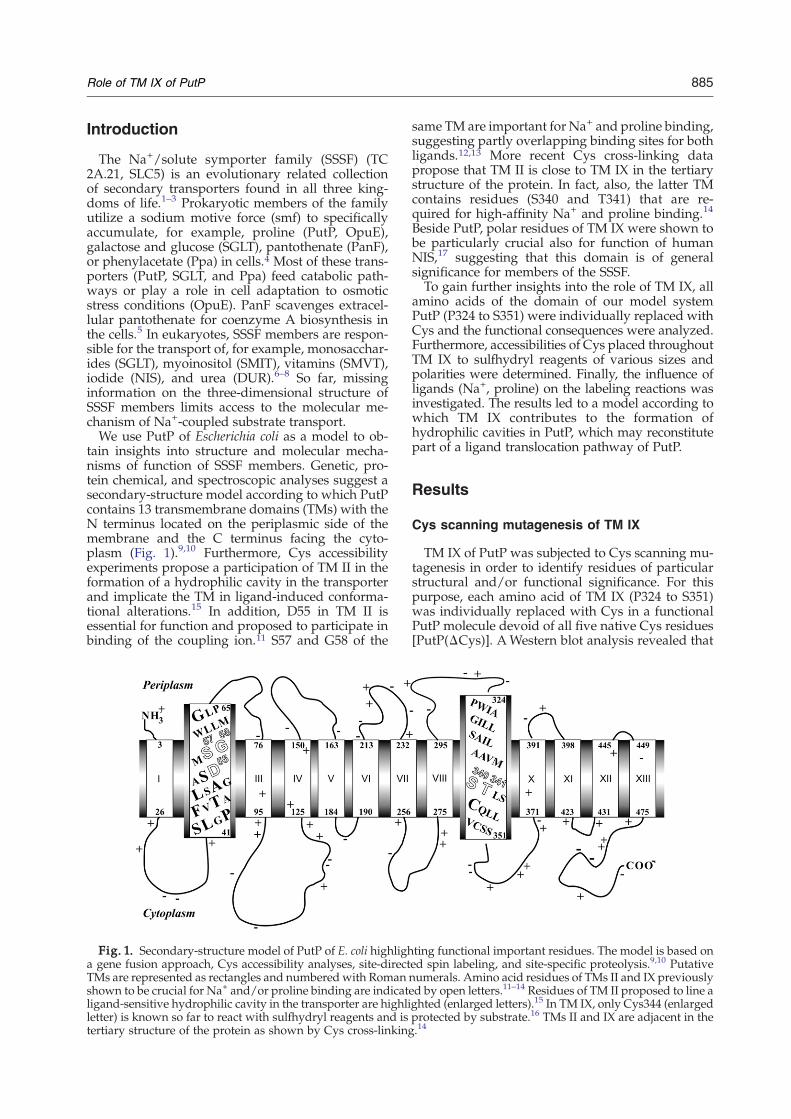
tain insights into structure and molecular mecha-nisms of function of SSSF members. Genetic, pro-tein chemical, and spectroscopic analyses suggest asecondary-structure model according to which PutPcontains 13 transmembrane domains (TMs) with theN terminus located on the periplasmic side of themembrane and the C terminus facing the cyto-plasm (Fig. 1).9,10 Furthermore, Cys accessibilityexperiments propose a participation of TM II in theformation of a hydrophilic cavity in the transporterand implicate the TM in ligand-induced conforma-tional alterations.15 In addition, D55 in TM II isessential for function and proposed to participate inbinding of the coupling ion.11 S57 and G58 of the
Fig. 1. Secondary-structure model of PutP of E. coli highligha gene fusion approach, Cys accessibility analyses, site-directTMs are represented as rectangles and numbered with Romanshown to be crucial for Na+ and/or proline binding are indicatligand-sensitive hydrophilic cavity in the transporter are highliletter) is known so far to react with sulfhydryl reagents and istertiary structure of the protein as shown by Cys cross-linking
same TM are important for Na+ and proline binding,suggesting partly overlapping binding sites for bothligands.12,13 More recent Cys cross-linking datapropose that TM II is close to TM IX in the tertiarystructure of the protein. In fact, also, the latter TMcontains residues (S340 and T341) that are re-quired for high-affinity Na+ and proline binding.14
Beside PutP, polar residues of TM IX were shown tobe particularly crucial also for function of humanNIS,17 suggesting that this domain is of generalsignificance for members of the SSSF.To gain further insights into the role of TM IX, all
amino acids of the domain of our model systemPutP (P324 to S351) were individually replaced withCys and the functional consequences were analyzed.Furthermore, accessibilities of Cys placed throughoutTM IX to sulfhydryl reagents of various sizes andpolarities were determined. Finally, the influence ofligands (Na+, proline) on the labeling reactions wasinvestigated. The results led to a model according towhich TM IX contributes to the formation ofhydrophilic cavities in PutP, which may reconstitutepart of a ligand translocation pathway of PutP.
Results
Cys scanning mutagenesis of TM IX
TM IX of PutP was subjected to Cys scanning mu-tagenesis in order to identify residues of particularstructural and/or functional significance. For thispurpose, each amino acid of TM IX (P324 to S351)was individually replaced with Cys in a functionalPutP molecule devoid of all five native Cys residues[PutP(ΔCys)]. AWestern blot analysis revealed that
ting functional important residues. The model is based oned spin labeling, and site-specific proteolysis.9,10 Putativenumerals. Amino acid residues of TMs II and IX previouslyed by open letters.11–14 Residues of TM II proposed to line aghted (enlarged letters).15 In TM IX, only Cys344 (enlargedprotected by substrate.16 TMs II and IX are adjacent in the.14

Fig. 3. Influence of amino acid replacements in TM IXon Na+-coupled proline uptake. Transport of L-[U-14C]proline (10 μM final concentration) into cells of E. coliWG170 was assayed in the presence of 50 mM NaCl and20 mM D-lactate (Na+ salt) as electron donor at 25 °C underaerobic conditions using a rapid filtrationmethod. (a) Initialrates of proline uptake. (b) Steady-state levels of prolineaccumulation. All values are expressed as percentage ofthe corresponding value of PutP(ΔCys) (initial rate, 23±5 nmol/min·mg; steady-state level of proline accumulation,24±3 nmol/mg). Standard deviations were calculated froma minimum of three independent experiments.
886 Role of TM IX of PutP
these replacements resulted in amounts of transpor-ter molecules in the membrane similar as observedfor PutP(ΔCys), indicating that possible differencesin proline uptake kinetics cannot be attributed todifferent amounts of transporter molecules in themembrane (Fig. 2). Active transport was then mea-sured under standard test conditions (70 mM Na+,10 μM proline) by using E. coli WG170 comple-mented with plasmid-encoded PutP variants. Underthese conditions, PutP(ΔCys) exhibited an initialrate of 23±5 nmol/min·mg [50% of PutP(wild type)]and a steady-state level of proline accumulation of24±3 nmol/mg [100% of PutP(wild type)].Initial analyses of rates and steady-state levels of
proline accumulation newly identified G328, S332,Q345, and L346 as important for PutP function(Fig. 3). Themost severe defect was detected for PutP(ΔCys)-S332C [2.6% and 15.8% of the initial rate andsteady-state level of proline accumulation of PutP(ΔCys)]. Intermediate inhibitory effects [30–40% ofthe initial rate and 70–100% of steady-state level ofproline accumulation of PutP(ΔCys)] were observedfor cells containing PutP(ΔCys)-G328C, -Q345C, or-L346C. In contrast, cells with PutP(ΔCys)-A337C or-C344 exhibited about twofold stimulated initialrates and unaltered steady-state levels of prolineaccumulation compared to PutP(ΔCys). It should benoted that placement of Cys at position 344 [Ser inPutP(ΔCys)] restored a native Cys residue. Allremaining substitutions had only little or no impacton transport kinetics (Fig. 3).
Effect of Cys replacements in TM IX of PutP(wildtype) on transport kinetics
Substitutions in PutP(ΔCys) causing alterations oftransport were also analyzed in the wild-typebackground. Cys in place of G328, S332, Q345, andL346 reduced the initial rate of transport to 30%, 6%,17%, and 28%, respectively, of the wild-type valueand, in the case of the three latter substitutions, alsoled to reduced maximum levels of proline accumu-lation (19–56% of the wild-type value). In contrast,replacement of A337 increased the initial rate oftransport by a factor of 1.8 with no significant effecton the steady-state level of accumulation (data notfurther illustrated). These results confirmed the ob-servations made in the PutP(ΔCys) background.A more detailed kinetic analysis of the func-
tional consequences of the Cys substitutions re-vealed diverse effects on Michaelis–Menten para-meters (Table 1). Replacement of G328 by Cys
Fig. 2. Immunological detection of PutP containing replacetotal membrane protein of each mutant was separated by 10%membrane (0.45 μm pore size) and probed with mouse antperoxidase. Detection was performed according to the enhanplasmid pT7-5 without putP served as negative control (NC).
affected Vmax (3-fold decreased) and left Km(pro)and K0.5(Na+) almost unaltered compared to PutP(wild type). PutP-S332C and -L346C exhibited dec-reased Km(pro) (3- and 21-fold, respectively) andVmaxvalues (22- and 8-fold, respectively). While K0.5(Na+)of PutP-L346C was only slightly altered, the valuecould not be reliably determined for PutP-S332C dueto the low transport activity (signal-to-noise ratio) inintact cells. Therefore, the analysis was performedwith proteoliposomes containing the purified trans-porter in an inside–out orientation. Under these
ments of amino acids in TM IX. Twenty-five micrograms ofSDS-PAGE. Proteins were transferred onto a nitrocellulosei-FLAG M2 monoclonal antibody linked to horseradishced chemiluminescence method. Cells transformed with

Fig. 4. Influence of sulfhydryl reagents on proline up-take into cells containing single Cys PutP variants of TMIX. Cells were incubated with 2 mM sulfhydryl reagent(MalNEt andMTSET) at room temperature for 15min. Thereaction was stopped by dilution with 100 mM Tris/Mesbuffer, pH 6.0, containing 0.1% BSA. Cells were washedand resuspended in the same buffer without BSA, andtransport was analyzed as described in the legend toFig. 3. Activities of MalNEt-treated (a) or MTSET-treated(b) cells are presented as percentage of the activity of therespective unlabeled PutP variant. Standard deviationswere calculated from a minimum of three measurements.
Table 1. Proline uptake kinetics of PutP bearing replace-ments of given amino acid residues
PutPKm(pro)(μM)
Vmax (nmol/min·mgof protein)
K0.5(Na+)(mM)
Wild type 1.3±0.2 44.6±1.6 0.038±0.007(0.7±0.09)a
G328C 1.5±0.5 16.7±1.0 0.018±0.004S332C 0.4±0.3 2.1±0.2 n.s.b
(0.055±0.014)a
A337C 1.7±0.5 72.5±3.8 0.117±0.025Q345C 49.3±1.0 23.0±1.8 0.705±0.088L346C 0.063±0.016 5.8±0.3 0.071±0.011
To determine Km(pro) and Vmax, we measured initial rates ofL-[U-14C]proline uptake by E. coli WG170 producing either PutP(wild type) or PutP with given replacements in the wild-typebackground in the presence of 50 mM NaCl and 20 mM D-lactate(Na+ salt) at proline concentrations from 0.2 to 500 μM. Fordetermination of the ion-specific parameters [K0.5(Na+)], transportof 10 μML-[U-14C]prolinewasmeasured in the presence of 0.005 to250 mMNaCl at 25 °C. The resulting data were plotted accordingto Eadie–Hofstee.
a Values in parentheses were obtained with proteoliposomescontaining the purified PutP variants.
b n.s., no stimulation of proline uptake into intact cells byincreasing concentrations of Na+.
887Role of TM IX of PutP
conditions, S332C exhibited a K0.5(Na+), which was13-fold reduced compared to the wild type inproteoliposomes (Table 1). In contrast to PutP-S332C and -L346C, placement of Cys at the positionof Q345 increased Km(pro) and K0.5(Na+) 38- and 19-fold, respectively, compared to PutP(wild type).Vmax was about 2-fold reduced (Table 1). The latterresults fit to previous analyses of substitutions forSer340 and Thr341, albeit the impact of alterations atthe latter positions on Km(pro) and K0.5(Na+) was evenstronger than that of the Gln345 replacement.14
Finally, placement of Cys at the position of A337,which was shown to stimulate proline uptake in theinitial analyses, slightly increased K0.5(Na+) and Vmax(3- and 1.6-fold) and left Km(pro) unaltered (Table 1).
Influence of Cys modification on transportactivity
Site-directed sulfhydryl modification of singleCys proteins in situ has been particularly useful forstudying both static and dynamic features oftransporters.18–20 Here, we used the method toprobe a potential participation of TM IX of PutP inthe formation of a hydrophilic cavity in the trans-porter. In a first approach, accessibility of Cys placedthroughout TM IX to membrane-permeant N-ethyl-maleimide (MalNEt) and membrane-impermeantmethanethiosulfonate ethyltrimethylammonium(MTSET) was analyzed via the impact of thereagents on proline uptake into intact cells. Onlyfreshly prepared cells were used for transport andadditional energization by D-lactate was omitted tominimize indirect inhibitory effects (e.g., caused byreaction of MalNEt or MTSET with components ofthe respiratory chain). In this way, MalNEt- orMTSET-treated cells containing PutP(ΔCys) main-tained a minimum of 80% and 96%, respectively, of
the activity of untreated cells. PutP(ΔCys) was notchemically modified as demonstrated below andelsewhere.14
MalNEt avidly reacted with Cys at distinct posi-tions in the cytoplasmic half of TM IX (C344, L347C,and S351C) as indicated by the highly reducedactivities of the respective MalNEt-treated cells (8%or less remaining activity compared to untreatedcells) (Figs. 4a and 5). Intermediate inhibitory effectsof MalNEt were observed with cells containingPutP(ΔCys)-T341C or -V348C (40% and 48% remain-ing activity, respectively). The activity of all othersingle Cys PutP variants was not or only slightlyaltered by MalNEt treatment. Here, either Cys wasnot accessible toMalNEt or labeling had only aminorimpact on transport activity. Since alkylation reagentsare known to react with sulfhydryl groups in polarbut not in apolar environments,21,22 the observedMalNEt inhibition pattern suggested that T341, C344,L347, V348, and S351 are in contact with a polar(hydrophilic) environment.
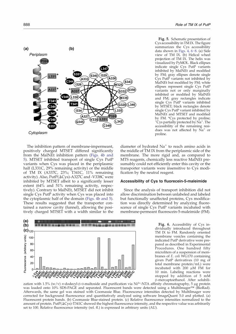
Fig. 5. Schematic presentation ofCys accessibility inTMIX. The figuresummarizes the Cys accessibilitydata shown in Figs. 4, 6−8. (a) Sideview of TM IX. (b) Helical wheelprojection of TM IX. The helix wasvisualized by PyMOL. Black ellipsesindicate single Cys PutP variantsinhibited by MalNEt and modifiedby FM; gray ellipses denote singleCys PutP variants not inhibited byMalNEt but modified by FM; whiteellipses represent single Cys PutPvariants not or only marginallyinhibited or modified by MalNEtand FM; gray rectangles indicatesingle Cys PutP variants inhibitedby MTSET; black rectangles denotesingle Cys PutP variant inhibited byMalNEt and MTSET and modifiedby FM. *Cys protected by proline;+Cys partially protected byNa+. Theaccessibility of the remaining resi-dues was not affected by Na+ orproline.
888 Role of TM IX of PutP
The inhibition pattern of membrane-impermeant,positively charged MTSET differed significantlyfrom the MalNEt inhibition pattern (Figs. 4b and5). MTSET inhibited transport of single Cys PutPvariants when Cys was placed in the periplasmichalf (L331C, 29% remaining activity) or the middleof TM IX (A337C, 23%; T341C, 11% remainingactivity). Also, PutP(ΔCys)-A327C and -V338Cwereinhibited by MTSET albeit to a significantly lesserextent (64% and 51% remaining activity, respec-tively). Contrary to MalNEt, MTSET did not inhibitsingle Cys PutP activity when Cys was placed intothe cytoplasmic half of the domain (Figs. 4b and 5).These results suggested that the transporter con-tained a narrow cavity (funnel), allowing the posi-tively charged MTSET with a width similar to the
zation with 1.5% (w/v) n-dodecyl-β-D-maltoside and purificawas loaded onto 10% SDS-PAGE and separated. FluorescenAfterwards, the same gel was stained with Coomassie Bluecorrected for background fluorescence and quantitatively anFluorescent protein bands. (b) Coomassie Blue-stained proteamount of protein. PutP(ΔCys)-T341C showed the highest fluoset to 100. Relative fluorescence intensity (rel. fl.) is expressed
diameter of hydrated Na+ to reach amino acids inthe middle of TM IX from the periplasmic side of themembrane. The more rigid and, as compared toMTS reagents, chemically less reactive MalNEt pre-sumably could not efficiently enter this cavity or thetransporter variants were insensitive to Cys modi-fication by the neutral reagent.
Accessibility of Cys to fluorecein-5-maleimide
Since the analysis of transport inhibition did notallow discrimination between unlabeled and labeledbut functionally unaffected proteins, Cys modifica-tion was directly determined by analyzing fluore-scence of single Cys PutP variants incubated withmembrane-permeant fluorescein-5-maleimide (FM).
Fig. 6. Accessibility of Cys in-dividually introduced throughoutTM IX to FM. Randomly orientedmembrane vesicles containing theindicated PutP derivative were pre-pared as described in ExperimentalProcedures. One hundred fiftymicroliters of a suspension of mem-branes of E. coli WG170 containinggiven PutP derivatives (10 mg oftotal membrane protein/mL) wereincubated with 200 μM FM for10 min. Labeling reactions werestopped by addition of 5 mMβ-mercaptoethanol. After solubili-
tion via Ni2+-NTA affinity chromatography, 5 μg proteint bands were detected using a MultiImager™ (BioRad).. Fluorescence intensities detected by MultiImager werealyzed using software ImageQuant 5.0 and plotted. (a)in. (c) Relative fluorescence intensities normalized to therescence intensity, and the respective value was arbitrarilyin arbitrary units (AU).
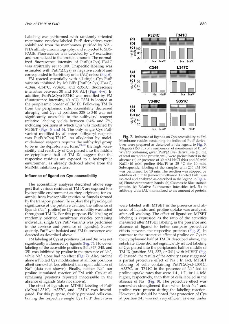
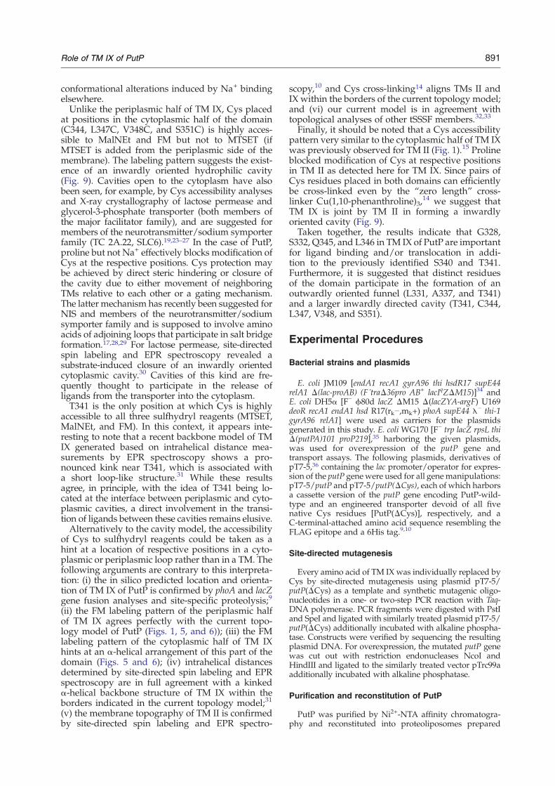
Fig. 7. Influence of ligands on Cys accessibility to FM.Membrane vesicles containing the indicated PutP deriva-tives were prepared as described in the legend to Fig. 5.Aliquots (150 μL) of a suspension of membranes of E. coliWG170 containing given PutP(ΔCys) derivatives (10 mgof total membrane protein/mL) were preincubated in theabsence (−) or presence of 30 mM NaCl (Na) and 30 mMNaCl/10 mM proline (Na/P) at 25 °C for 10 min.Subsequently, labeling of the samples with 200 μM FMwas performed for 10 min. The reaction was stopped byaddition of 5 mM β-mercaptoethanol. Labeled PutP wasisolated and analyzed as described in the legend to Fig. 4.(a) Fluorescent protein bands. (b) Coomassie Blue-stainedprotein. (c) Relative fluorescence intensities (rel. fl.) inarbitrary units (AU) normalized to the amount of protein.
889Role of TM IX of PutP
Labeling was performed with randomly orientedmembrane vesicles; labeled PutP derivatives weresolubilized from the membranes, purified by Ni2+-NTA affinity chromatography, and subjected to SDS-PAGE. Fluorescence was detected by UV excitationand normalized to the protein amount. The normal-ized fluorescence intensity of PutP(ΔCys)-T341Cwas arbitrarily set to 100. Unspecific labeling wasestimated with PutP(ΔCys) as negative control andcorresponded to 3 arbitrary units (AU) or less (Fig. 6).FM reacted essentially with all single Cys PutP
variants inhibited by MalNEt [PutP(ΔCys)-T341C,-C344, -L347C, -V348C, and -S351C; fluorescenceintensities between 30 and 100 AU] (Figs. 4−6). Inaddition, PutP(ΔCys)-P324C was modified by FM(fluorescence intensity, 40 AU). P324 is located atthe periplasmic border of TM IX. Following TM IXfrom the periplasmic side, accessibility decreasedabruptly, and Cys at positions 325 to 340 was notsignificantly accessible to the sulfhydryl reagent(relative labeling yields between 0.4% and 7%)including positions at which Cys was modified byMTSET (Figs. 5 and 6). The only single Cys PutPvariant modified by all three sulfhydryl reagentswas PutP(ΔCys)-T341C. As alkylation by malei-mide-based reagents requires the sulfhydryl groupto be in the deprotonated form,21,22 the high acces-sibility and reactivity of Cys placed into the middleor cytoplasmic half of TM IX suggest that therespective residues are exposed to a hydrophilicenvironment as already deduced above from theMalNEt inhibition pattern.
Influence of ligand on Cys accessibility
The accessibility analyses described above sug-gest that various residues of TM IX are exposed to ahydrophilic environment as they originate, for ex-ample, from hydrophilic cavities or funnels formedby the transport protein. To explore the physiologicalsignificance of the putative cavities, the influence ofligands (Na+, proline) on Cys accessibilitywas testedthroughout TM IX. For this purpose, FM labeling ofrandomly oriented membrane vesicles containingindividual single Cys PutP variants was performedin the absence and presence of ligand(s). Subse-quently, PutP was isolated and FM fluorescence wasdetected as described above.FM labeling of Cys at positions 324 and 341was not
significantly influenced by ligands (Fig. 7). However,labeling of the accessible positions 344, 347, 348, and351 was inhibited by proline in the presence of Na+,while Na+ alone had no effect (Fig. 7). Also, prolinealone inhibited Cys modification at all four positionsalbeit somewhat less efficient than upon addition ofNa+ (data not shown). Finally, neither Na+ norproline stimulated reaction of FM with Cys at allremaining positions reported inaccessible in theabsence of ligands (data not shown).The effect of ligands on MTSET labeling of PutP
(ΔCys)-L331C, -A337C, and -T341C was investi-gated. For this purpose, freshly prepared cells con-taining the respective single Cys PutP derivatives
were labeled with MTSET in the presence and ab-sence of ligands, and proline uptake was analyzedafter cell washing. The effect of ligand on MTSETlabeling is expressed as the ratio of the activitiesmeasured after MTSET labeling in the presence andabsence of ligand to better compare protectiveeffects between the respective proteins (Fig. 8). Incontrast to the protective effect of proline on Cys inthe cytoplasmic half of TM IX described above, thesubstrate alone did not significantly inhibit labelingof Cys placed into the periplasmic half or middle ofTM IX (position 331, 337, or 341) with MTSET (Fig.8). Instead, the results of the activity assay suggesteda partial protective effect of Na+. In fact, MTSETlabeling of cells containing PutP(ΔCys)-L331C,-A337C, or -T341C in the presence of Na+ led toproline uptake rates that were 1.4-, 1.7-, or 1.4-foldhigher, respectively, than that of cells labeled in theabsence of Na+ (Fig. 8). The protective effect wassomewhat strengthened than when both Na+ andproline were present during the labeling reaction.However, it should be noted that protection of Cysat position 341 was not very efficient as even under

Fig. 8. Influence of ligands on Cys accessibility toMTSET. Cells containing the respective single Cys variantwere preincubated in the absence (−) or presence of 30mMNaCl (Na), 10 mM proline, or 30 mM NaCl/10 mMproline (Na/P) at 25 °C for 10 min. Subsequently, 2 mMMTSET was added and incubation was continued for10 min. The reaction was stopped, cells were washed, andtransport was analyzed as described in the legend to Fig.4. The effect of ligand is shown as the ratio of the activitiesmeasured after MTSET labeling in the presence (v1) andabsence (v2) of ligand. Ratios N1 indicate inhibition,whereas ratios b1 denote stimulation of MTSET labeling.
890 Role of TM IX of PutP
conditions of maximum protection, uptake activitiesof MTSET-treated cells were reduced to 30% of thevalue of unlabeled cells. Cells containing PutP(ΔCys)-L331C or -A337C showed a maximum of60–70% of the activity of unlabeled cells if labelingwas performed in the presence of Na+ and proline.
Fig. 9. Model showing the participation of TMs II andIX in the formation of hydrophilic cavities in PutP. Themodel is based on the current analysis as well as onprevious investigations.11–15 Amino acids of TMs II and IXsupposed to line a hydrophilic cavity (funnel) are indi-cated. Residues shown to be important for ligand bindingand/or translocation are represented as open characters.There is no evidence so far that the periplasmic half of TMII participates in the formation of the outwardly orientedfunnel (drawn as dotted box) suggested to be lined byresidues of TM IX.
Discussion
S340 and T341 in TM IX of PutP have previouslybeen suggested to play key roles in ligand bindingand transport; thereby, the side chain of T341 maydirectly participate in Na+ binding.14 The currentanalysis of structure–function relationships in PutPnewly identified G328, S332, Q345, and L346 of thesame domain as important for Na+-coupled prolineuptake. In addition, the Cys accessibility pattern ofTM IX suggests the existence of hydrophilic cavitiesin the transporter that are affected by ligand binding.What are the roles of the newly identified struc-
tural and/or functional important residues? Whilenone of the four residues appears to be crucial formembrane insertion and stability of PutP, functionalanalyses of the effect of Cys replacements revealsignificant alterations of kinetic parameters oftransport. Km(pro) is most severely affected (morethan 1 order of magnitude) upon replacement ofQ345 or L346. Although the effects at both positionsare diametrical, the data suggest that ligand bindingis highly sensitive to alterations in this part of theprotein. In particular, the properties of PutP-Q345C[increased Km(pro) and K0.5(Na+), comparatively smallchanges of Vmax] are, to some extent, reminiscent ofthe results obtained upon replacement of S340 orT341.14 These observations further support the ideathat these polar residues of TM IX are located at or
close to a ligand binding site of PutP. Furthermore,as both Km(pro) and K0.5(Na+) are highly affected uponreplacement of S340, T341, or Q345, the idea thatbinding of coupling ion and substrate occurs in closeproximity is supported. Furthermore, the reducedVmax values observed with PutP-G328C, -S332C,and -L346C hint at defects in the transport cyclesubsequent to ligand binding, for example, inhibi-tion of conformational alterations or a hamperedrelease of ligands. The latter idea is, in principle,supported by the decreasedMichaelis–Menten para-meters found for PutP-S332C [Km(pro) and K0.5(Na+)]and PutP-Q346C [Km(pro)] (Table 1).If, in fact, residues of TM IX are located at or close
to a ligand binding site, they might be accessiblefrom one side and/or from the other side of mem-brane. First evidence for the existence of an out-wardly oriented cavity (funnel) in PutP comes fromlabeling of PutP-S340C and -T341C in right-side-outmembrane vesicles with various methane thiosulfo-nate compounds.14 Here, we present new evidencefor the existence of a funnel that may connect T341with the periplasmic surface of PutP. This evidencecomes from the observation that Na+-coupled pro-line uptake into intact cells containing PutP(ΔCys)-L331C, -A337C, or -T341C is efficiently inhibited byMTSET. Na+ but not proline inhibits the labelingreaction, although it cannot completely protect therespective Cys residues from modification possiblydue to the dynamics of the system. Considering theNa+-sensitive accessibility of these residues to posi-tively charged MTSET with a width similar to thediameter of hydrated Na+, as well as the previouslyreported particular significance of T341 for Na+
binding,14 it is tempting to speculate that the pro-posed funnel is used by Na+ as an entrance to a Na+
binding site in about the middle of the membrane(Fig. 9). Clearly, inhibition of MTSET labeling ofthe reported residues could also be achieved by
891Role of TM IX of PutP
conformational alterations induced by Na+ bindingelsewhere.Unlike the periplasmic half of TM IX, Cys placed
at positions in the cytoplasmic half of the domain(C344, L347C, V348C, and S351C) is highly acces-sible to MalNEt and FM but not to MTSET (ifMTSET is added from the periplasmic side of themembrane). The labeling pattern suggests the exist-ence of an inwardly oriented hydrophilic cavity(Fig. 9). Cavities open to the cytoplasm have alsobeen seen, for example, by Cys accessibility analysesand X-ray crystallography of lactose permease andglycerol-3-phosphate transporter (both members ofthe major facilitator family), and are suggested formembers of the neurotransmitter/sodium symporterfamily (TC 2A.22, SLC6).19,23–27 In the case of PutP,proline but not Na+ effectively blocks modification ofCys at the respective positions. Cys protection maybe achieved by direct steric hindering or closure ofthe cavity due to either movement of neighboringTMs relative to each other or a gating mechanism.The latter mechanism has recently been suggested forNIS and members of the neurotransmitter/sodiumsymporter family and is supposed to involve aminoacids of adjoining loops that participate in salt bridgeformation.17,28,29 For lactose permease, site-directedspin labeling and EPR spectroscopy revealed asubstrate-induced closure of an inwardly orientedcytoplasmic cavity.30 Cavities of this kind are fre-quently thought to participate in the release ofligands from the transporter into the cytoplasm.T341 is the only position at which Cys is highly
accessible to all three sulfhydryl reagents (MTSET,MalNEt, and FM). In this context, it appears inte-resting to note that a recent backbone model of TMIX generated based on intrahelical distance mea-surements by EPR spectroscopy shows a pro-nounced kink near T341, which is associated witha short loop-like structure.31 While these resultsagree, in principle, with the idea of T341 being lo-cated at the interface between periplasmic and cyto-plasmic cavities, a direct involvement in the transi-tion of ligands between these cavities remains elusive.Alternatively to the cavity model, the accessibility
of Cys to sulfhydryl reagents could be taken as ahint at a location of respective positions in a cyto-plasmic or periplasmic loop rather than in a TM. Thefollowing arguments are contrary to this interpreta-tion: (i) the in silico predicted location and orienta-tion of TM IX of PutP is confirmed by phoA and lacZgene fusion analyses and site-specific proteolysis;9
(ii) the FM labeling pattern of the periplasmic halfof TM IX agrees perfectly with the current topo-logy model of PutP (Figs. 1, 5, and 6)); (iii) the FMlabeling pattern of the cytoplasmic half of TM IXhints at an α-helical arrangement of this part of thedomain (Figs. 5 and 6); (iv) intrahelical distancesdetermined by site-directed spin labeling and EPRspectroscopy are in full agreement with a kinkedα-helical backbone structure of TM IX within theborders indicated in the current topology model;31
(v) the membrane topography of TM II is confirmedby site-directed spin labeling and EPR spectro-
scopy,10 and Cys cross-linking14 aligns TMs II andIX within the borders of the current topology model;and (vi) our current model is in agreement withtopological analyses of other tSSSF members.32,33
Finally, it should be noted that a Cys accessibilitypattern very similar to the cytoplasmic half of TM IXwas previously observed for TM II (Fig. 1).15 Prolineblocked modification of Cys at respective positionsin TM II as detected here for TM IX. Since pairs ofCys residues placed in both domains can efficientlybe cross-linked even by the “zero length” cross-linker Cu(1,10-phenanthroline)3,
14 we suggest thatTM IX is joint by TM II in forming a inwardlyoriented cavity (Fig. 9).Taken together, the results indicate that G328,
S332, Q345, and L346 in TM IX of PutP are importantfor ligand binding and/or translocation in addi-tion to the previously identified S340 and T341.Furthermore, it is suggested that distinct residuesof the domain participate in the formation of anoutwardly oriented funnel (L331, A337, and T341)and a larger inwardly directed cavity (T341, C344,L347, V348, and S351).
Experimental Procedures
Bacterial strains and plasmids
E. coli JM109 [endA1 recA1 gyrA96 thi hsdR17 supE44relA1 Δ(lac-proAB) (F´traΔ36pro AB+ lacIqZΔM15)]34 andE. coli DH5α [F− ϕ80d lacZ ΔM15 Δ(lacZYA-argF) U169deoR recA1 endA1 hsd R17(rk−,mk+) phoA supE44 λ− thi-1gyrA96 relA1] were used as carriers for the plasmidsgenerated in this study. E. coli WG170 [F− trp lacZ rpsL thiΔ(putPA)101 proP219],35 harboring the given plasmids,was used for overexpression of the putP gene andtransport assays. The following plasmids, derivatives ofpT7-5,36 containing the lac promoter/operator for expres-sion of the putP gene were used for all genemanipulations:pT7-5/putP and pT7-5/putP(ΔCys), each of which harborsa cassette version of the putP gene encoding PutP-wild-type and an engineered transporter devoid of all fivenative Cys residues [PutP(ΔCys)], respectively, and aC-terminal-attached amino acid sequence resembling theFLAG epitope and a 6His tag.9,10
Site-directed mutagenesis
Every amino acid of TM IX was individually replaced byCys by site-directed mutagenesis using plasmid pT7-5/putP(ΔCys) as a template and synthetic mutagenic oligo-nucleotides in a one- or two-step PCR reaction with Taq-DNA polymerase. PCR fragments were digested with PstIand SpeI and ligated with similarly treated plasmid pT7-5/putP(ΔCys) additionally incubated with alkaline phospha-tase. Constructs were verified by sequencing the resultingplasmid DNA. For overexpression, the mutated putP genewas cut out with restriction endonucleases NcoI andHindIII and ligated to the similarly treated vector pTrc99aadditionally incubated with alkaline phosphatase.
Purification and reconstitution of PutP
PutP was purified by Ni2+-NTA affinity chromatogra-phy and reconstituted into proteoliposomes prepared

892 Role of TM IX of PutP
from E. coli polar lipid extracts (Avanti Polar Lipids, Inc.,Alabaster, AL) at a lipid-to-protein ratio of 100:1 (w/w) asdescribed before.37
Proline transport assays
Transport was measured in E. coli WG170 transformedwith plasmid pT7-5/putP(ΔCys) or pT7-5/putP harboringthe given mutations. Cells were grown and prepared aspreviously described.12 Smf-driven proline uptake wasassayed under standard conditions with 10 μM L-[U-14C]proline (26 Ci/mol) in the presence of 20 mM D-lactate(Na+ salt) and 50 mM NaCl using the rapid filtrationmethod as described previously.38 Initial rates of transportwere calculated from the initial linear portion of the timecourse. Steady-state levels of proline accumulation weredetermined from time points after leveling out of theuptake curve. Kinetic parameters were determined byplotting initial rates and substrate concentrations accord-ing to Eadie-Hofstee. The resulting Km(pro), K0.5(Na+), andVmax values of pT7-5/putP-encoded PutP(wild type)closely match the parameters of the native, fully induced,chromosome-encoded PutP in the absence of PutA39,40
suggesting that deviations of kinetic parameters are solelydue to the site-specifically introduced amino acid replace-ments and not to overexpression artifacts.For transport inhibition analyses, cells containing the
respective single Cys variant were incubated with 2 mMsulfhydryl reagent (MalNEt or MTSET) at room tempe-rature for 15 min. The reaction was stopped by two-fold dilution of the reaction mixture with 100 mM Tris/4-morpholineethanesulfonic acid (Mes) buffer, pH 6.0,containing 0.1% bovine serum albumin (BSA). Cells werewashed and resuspended in the same buffer without BSA,and transport was analyzed as described above.For analysis of the effect of ligand on MTSET labeling,
cells containing the respective single Cys variant werewashed four times in sodium-free 100 mM Tris/Mesbuffer, pH 6.0, and preincubated in the absence or pre-sence of 30 mM NaCl, 10 mM proline, or 30 mM NaCl/10 mM proline at 25 °C for 10 min. Subsequently, 2 mMMTSET was added and incubation was continued for10 min. The reaction was stopped by twofold dilution ofthe reaction mixture with 100 mM Tris/Mes buffer,pH 6.0, containing 0.1% BSA, and washed two timeswith the same buffer without BSA. The transport wasanalyzed as described above.Smf-driven proline uptake into proteoliposomes was
analyzed as described before.37
Cys accessibility to FM
E. coli WG170 transformed with pTrc99a/putP(ΔCys)were grown, disrupted by sonification, and randomlyoriented membrane vesicles were prepared as previouslydescribed.15 For accessibility analyses, 150-μL aliquots ofthe membrane suspension containing 10 mg/mL totalprotein were incubated without or with Na+, K+, and/orL-proline at given concentrations at 25 °C for 10 min.Subsequently, 200 μM FM was added, and incubation wascontinued at 25 °C for an additional 10 min. Reactions werestopped by addition of 5 mM β-mercaptoethanol. Afterlabeling, PutP was solubilized with 1.5% (w/v) n-dodecyl-β-D-maltoside under stirring at 4 °C for 30 min. Then,samples were purified via Ni2+-NTA spin columns andeluted with 200 mM imidazole as described before.13 Afterprotein determination, equal amounts of protein weresubjected to 10% SDS-PAGE. Fluorescent bands of PutP
were visualized using the MultiImager™ device (BioRad,Munich) and quantified using the software ImageQuant 5.0.After analysis of fluorescent PutP bands, the gel was stainedwith Coomassie Blue to detect total amounts of protein.
Western blot analysis
Relative amounts of PutP with given amino acid replace-ments in membranes of E. coli WG170 were estimated byWestern blot analysis with horseradish peroxidase-linkedmouse anti-FLAG immunoglobulin G directed against theFLAG epitope at the C terminus of each PutP variant asdescribed before.12
Protein determination
Determination of protein was performed accordingto Bradford41 for purified protein and a modified Lowrymethod42 for total membrane protein with BSA asstandard.
Acknowledgements
We thank Maret Böhm for generating PutP-S332C.This work was financially supported by the Deut-sche Forschungsgemeinschaft (Ju333/3-2, Ju333/4-2, and Exc114-1).
References
1. Jung, H. (2002). The sodium/substrate symporterfamily: structural and functional features. FEBS Lett.529, 73–77.
2. Wright, E. M. & Turk, E. (2004). The sodium/glucosecotransport family SLC5. Pflugers Arch. 447, 510–518.
3. Reizer, J., Reizer, A. & Saier, M. H., Jr (1994). A func-tional superfamily of sodium/solute symporters.Biochim. Biophys. Acta, 1197, 133–166.
4. Jung, H. (2001). Towards the molecular mechanism ofNa+/solute symport in prokaryotes. Biochim. Biophys.Acta, 1505, 131–143.
5. Jackowski, S. & Alix, J. H. (1990). Cloning, sequence,and expression of the pantothenate permease (panF)gene of Escherichia coli. J. Bacteriol. 172, 3842–3848.
6. Dohan, O., De la Vieja, A. & Carrasco, N. (2006).Hydrocortisone and purinergic signaling stimulatesodium/iodide symporter (NIS)-mediated iodidetransport in breast cancer cells. Mol. Endocrinol. 20,1121–1137.
7. Wright, E. M., Hirayama, B. A. & Loo, D. F. (2007).Active sugar transport in health and disease. J. Intern.Med. 261, 32–43.
8. Kojima, S., Bohner, A., Gassert, B., Yuan, L. & vonWiren, N. (2007). AtDUR3 represents the majortransporter for high-affinity urea transport across theplasma membrane of nitrogen-deficient Arabidopsisroots. Plant J. 52, 30–40.
9. Jung, H., Rübenhagen, R., Tebbe, S., Leifker, K.,Tholema, N., Quick, M. & Schmid, R. (1998). Topologyof the Na+/proline transporter of Escherichia coli.J. Biol. Chem. 273, 26400–26407.
10. Wegener, C., Tebbe, S., Steinhoff, H. J. & Jung, H.(2000). Spin labeling analysis of structure and

893Role of TM IX of PutP
dynamics of the Na+/proline transporter of Escher-ichia coli. Biochemistry, 39, 4831–4837.
11. Quick, M. & Jung, H. (1997). Aspartate 55 in the Na+/proline permease of Escherichia coli is essential for Na+-coupled proline uptake. Biochemistry, 36, 4631–4636.
12. Quick, M., Tebbe, S. & Jung, H. (1996). Ser57 in theNa+/proline permease of Escherichia coli is criticalfor high-affinity proline uptake. Eur. J. Biochem. 239,732–736.
13. Pirch, T., Quick, M., Nietschke, M., Langkamp, M. &Jung, H. (2002). Sites important for Na+ and substratebinding in the Na+/proline transporter of Escherichiacoli, a member of the Na+/solute symporter family.J. Biol. Chem. 277, 8790–8796.
14. Hilger, D., Böhm,M., Hackmann, A. & Jung, H. (2008).Role of Ser-340 and Thr-341 in transmembranedomain IX of the Na+/proline transporter PutP ofEscherichia coli in ligand binding and transport. J. Biol.Chem. 283, 4921–4929.
15. Pirch, T., Landmeier, S. & Jung, H. (2003). Transmem-brane domain II of the Na+/proline transporter PutPof Escherichia coli forms part of a conformationallyflexible, cytoplasmic exposed aqueous cavity withinthe membrane. J. Biol. Chem. 278, 42942–42949.
16. Yamato, I. & Anraku, Y. (1993). Na+/substrate sym-port on prokaryotes. In Alkali Cation Transport Systemsin Prokaryotes (Bakker, E. P., ed), pp. 53–76, CRC Press,Boca Raton, FL.
17. De la Vieja, A., Reed, M. D., Ginter, C. S. & Carrasco,N. (2007). Amino acid residues in transmembranesegment IX of the Na+/I− symporter play a role in itsNa+ dependence and are critical for transport activity.J. Biol. Chem. 282, 25290–25298.
18. Guan, L. & Kaback, H. R. (2007). Site-directed alky-lation of cysteine to test solvent accessibility of mem-brane proteins. Nat. Protoc. 2, 2012–2017.
19. Kaback, H. R., Dunten, R., Frillingos, S., Venkatesan,P., Kwaw, I., Zhang, W. & Ermolova, N. (2007). Site-directed alkylation and the alternating access modelfor LacY. Proc. Natl Acad. Sci. USA, 104, 491–494.
20. Karlin, A. & Akabas, M. H. (1998). Substituted-cysteineaccessibility method. Methods Enzymol. 293, 123–145.
21. Zhou, J., Fazzio, R. T. & Blair, D. F. (1995). Membranetopology of the MotA protein of Escherichia coli. J. Mol.Biol. 251, 237–242.
22. Poelarends, G. & Konings, W. N. (2002). The trans-membrane domains of the ABC multidrug trans-porter LmrA form a cytoplasmic exposed, aqueouschamber within the membrane. J. Biol. Chem. 277,42891–42898.
23. Abramson, J., Smirnova, I., Kasho, V., Verner, G.,Kaback, H. R. & Iwata, S. (2003). Structure and me-chanism of the lactose permease of Escherichia coli.Science, 301, 610–615.
24. Huang, Y., Lemieux, M. J., Song, J., Auer, M. & Wang,D. N. (2003). Structure andmechanism of the glycerol-3-phosphate transporter from Escherichia coli. Science,301, 616–620.
25. Guan, L.,Mirza, O., Verner, G., Iwata, S. &Kaback, H. R.(2007). Structural determination of wild-type lactosepermease. Proc. Natl Acad. Sci. USA, 104, 15294–15298.
26. Quick, M., Yano, H., Goldberg, N. R., Duan, L.,Beuming, T., Shi, L. et al. (2006). State-dependent con-
formations of the translocation pathway in the tyro-sine transporter Tyt1, a novel neurotransmitter: so-dium symporter from Fusobacterium nucleatum. J. Biol.Chem. 281, 26444–26454.
27. Zhang, Y. W. & Rudnick, G. (2006). The cytoplasmicsubstrate permeation pathway of serotonin transpor-ter. J. Biol. Chem. 281, 36213–36220.
28. Yamashita, A., Singh, S. K., Kawate, T., Jin, Y. &Gouaux, E. (2005). Crystal structure of a bacterialhomologue of Na+/Cl− dependent neurotransmittertransporters. Nature, 437, 215–223.
29. Loland, C. J., Granas, C., Javitch, J. A. & Gether, U.(2004). Identification of intracellular residues in thedopamine transporter critical for regulation of trans-porter conformation and cocaine binding. J. Biol.Chem. 279, 3228–3238.
30. Smirnova, I., Kasho, V., Choe, J. Y., Altenbach, C.,Hubbell, W. L. & Kaback, H. R. (2007). Sugar bindinginduces an outward facing conformation of LacY. Proc.Natl Acad. Sci. USA, 104, 16504–16509.
31. Hilger, D., Polyhach, Y., Jung, H. & Jeschke, G. (2008).Backbone structure of transmembrane domain IX ofthe Na+/proline transporter PutP of Escherichia coli.Biophys. J. submitted.
32. Turk, E., Kerner, C. J., Lostao, M. P. & Wright, E. M.(1996). Membrane topology of the humanNa+/glucosecotransporter SGLT1. J. Biol. Chem. 271, 1925–1934.
33. Levy, O., De la Vieja, A., Ginter, C. S., Riedel, C., Dai,G. & Carrasco, N. (1998). N-linked glycosylation ofthe thyroid Na+/I− symporter (NIS). Implicationsfor its secondary structure model. J. Biol. Chem. 273,22657–22663.
34. Yanisch-Perron, C., Vieira, J. & Messing, J. (1985).ImprovedM13 phage cloning vectors and host strains:nucleotide sequences of the M13mp18 and pUC19vectors. Gene, 33, 103–119.
35. Stalmach, M. E., Grothe, S. & Wood, J. M. (1983). Twoproline porters in Escherichia coli K-12. J. Bacteriol. 156,481–486.
36. Tabor, S. & Richardson, C. C. (1985). A bacteriophageT7 RNA polymerase/promoter system for controlledexclusive expression of specific genes. Proc. Natl Acad.Sci. USA, 82, 1074–1078.
37. Jung, H., Tebbe, S., Schmid, R. & Jung, K. (1998).Unidirectional reconstitution and characterization ofpurified Na+/proline transporter of Escherichia coli.Biochemistry, 37, 11083–11088.
38. Chen, C. C., Tsuchiya, T., Yamane, Y., Wood, J. M. &Wilson, T. H. (1985). Na+ (Li+)-proline cotransport inEscherichia coli. J. Membr. Biol. 84, 157–164.
39. Wood, J.M.&Zadworny,D. (1979). Characterization ofan inducible porter required for L-proline catabolismby Escherichia coli K12. Can. J. Biochem. 57, 1191–1199.
40. Chen, C. C., Tsuchiya, T., Yamane, Y., Wood, J. M. &Wilson, T. H. (1985). Na+ (Li+)-proline cotransport inEscherichia coli. J. Membr. Biol. 84, 157–164.
41. Bradford, M. M. (1976). A rapid and sensitive methodfor the quantitation of microgram quantities of proteinutilizing the principle of protein–dye binding. Anal.Biochem. 72, 248–254.
42. Peterson, G. L. (1977). A simplification of the proteinassay method of Lowry et al. which is more generallyapplicable. Anal. Biochem. 83, 346–356.