fulminant jejuno-ileitis following ablation of enteric glia in adult
TRANSCRIPT

Cell, Vol. 93, 189–201, April 17, 1998, Copyright 1998 by Cell Press
Fulminant Jejuno-Ileitis following Ablationof Enteric Glia in Adult Transgenic Mice
identified, which target reporter gene expression specif-ically to astroglia, and have been used to express heter-ologous genes in transgenic mice (Toggas et al., 1994;
Toby G. Bush,1,2,7 Tor C. Savidge,1,3
Tom C. Freeman,4 Hilary J. Cox,1
Elizabeth A. Campbell,4 Lennart Mucke,5
Johnson et al., 1995). To achieve cellular ablation, weMartin H. Johnson,2 and Michael V. Sofroniew1,2,6
used the thymidine kinase gene of the herpes simplex1Medical Research Council Cambridge Centrevirus (HSV-Tk). Proliferating cells that express trans-for Brain Repairgene-derived HSV-TK metabolize the antiviral agentForvie Site, Robinson Wayganciclovir (GCV) to toxic nucleotide analogs, which dis-Cambridge CB2 2PYturb nucleic acid synthesis and induce cell death (Frank2Department of Anatomyet al., 1984; Borrelli et al., 1989; Heyman et al., 1989).University of CambridgeThus, cell ablation can be regulated in a regionally andDowning Streettemporally specific manner by where and when GCV isCambridge CB2 3DYadministered.3Institute of Child Health
We report here that in transgenic mice expressingUniversity of BirminghamHSV-TK from the mouse Gfap promoter, the pattern andBirmingham, BI6 8ETregulation of transgene-derived HSV-TK expression is4Sanger Centresimilar to that of endogenous GFAP, and that transgenicWellcome Trust Genome Campusastrocytes are vulnerable to GCV in vitro and after CNSHinxton CB10, 1SAinjury in vivo. During initial in vivo studies, we foundUnited Kingdomthat subcutaneous (s.c.) GCV delivery to uninjured adult5Gladstone Molecular Neurobiology Programtransgenic mice was invariably fatal within 19 days inand Department of Neurologythe absence of CNS pathology. We examined vital pe-University of Californiaripheral organs for expression of transgene-derivedSan Francisco, California 94141-9100HSV-TK and for pathological changes. RT-PCR screen-ing of transgenic mice revealed expression of Gfap andHSV-Tk in gut, heart, lung, liver, kidney, adrenal, andSummaryspleen. Histopathological examination of these organsin transgenic animals receiving GCV revealed majorTo investigate the roles of astroglial cells, we targetedchanges only in the gastrointestinal (GI) tract, whichtheir ablation genetically. Transgenic mice were gen-showed severe inflammation and necrosis of the jeju-erated expressing herpes simplex virus thymidine ki-num and ileum. Our investigations show that this pathol-nase from the mouse glial fibrillary acidic proteinogy is related to the ablation of GFAP-expressing enteric(GFAP) promoter. In adult transgenic mice, 2 weeksglia.of subcutaneous treatment with the antiviral agent
ganciclovir preferentially ablated transgene-express-Resultsing, GFAP-positive glia from the jejunum and ileum,
causing a fulminating and fatal jejuno-ileitis. This pa-Generation of HSV-TK Expressing Transgenic Micethology was independent of bacterial overgrowth andProgeny resulting from the pronuclear injection of awas characterized by increased myeloperoxidase ac-Gfap-HSV-Tk fusion gene construct were screened fortivity, moderate degeneration of myenteric neurons,successful integration of the transgene (Figures 1A andand intraluminal hemorrhage. These findings demon-1B). Transgenic lines were established from three founderstrate that enteric glia play an essential role in main-mice. Of these, line 7.1 showed the highest expressiontaining the integrity of the bowel and suggest thatof HSV-TK protein in thebrain. Densitometry of Southerntheir loss or dysfunction may contribute to the cellularblots estimated the number of copiesof transgene incor-mechanisms of inflammatory bowel disease.porated into the genome of transgenic progeny of line7.1 to be over 50. Males of line 7.1 were infertile inagreement with previous reports of male sterility inIntroductiontransgenic mice expressing HSV-TK from other promot-ers (Al-Shawi et al., 1988). All experimental animals wereAstroglia are among the most numerous cells in all neu-generated by mating heterozygous females of line 7.1ral systems. Many aspects of their functions are not wellwith wild-type males, followed by genotyping by South-understood. To investigate the roles of astrocytes andern blot and immunohistochemical demonstration ofrelated astroglia in the central (CNS) and peripheral ner-HSV-TK expression. Thus, transgenic and nontransgenicvous systems, we targeted their cell death geneticallycontrol animals had similar genetic backgrounds.using the promoter of glial fibrillary acidic protein (GFAP),
Production of HSV-TK protein by transgenic mice wasan intermediate filament protein (Eddleston and Mucke,demonstrated by Western blot of whole brain extracts1993). Mouse Gfap promoter sequences have been(Figure 1C) and immunohistochemistry. HSV-TK waspresent only in cells with the appearance of astrocytes6 To whom correspondence should be addressed.(Figures 1D and 1E). In double-labeling studies, HSV-7 Present address: Babraham Institute, Babraham, CB2 4AT, United
Kingdom. TK was always colocalized with GFAP in uninjured mice
Cell190
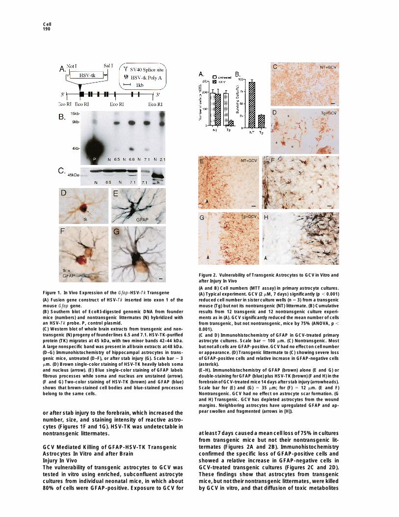
Figure 1. In Vivo Expression of the Gfap-HSV-Tk Transgene
(A) Fusion gene construct of HSV-Tk inserted into exon 1 of themouse Gfap gene.(B) Southern blot of EcoRI-digested genomic DNA from foundermice (numbers) and nontransgenic littermates (N) hybridized withan HSV-Tk probe. P, control plasmid.
Figure 2. Vulnerability of Transgenic Astrocytes to GCV in Vitro andafter Injury In Vivo
(A and B) Cell numbers (MTT assay) in primary astrocyte cultures.(A) Typical experiment. GCV (2 mM, 7 days) significantly (p , 0.001)reduced cell number in sister culture wells (n 5 3) from a transgenicmouse (Tg) but not its nontransgenic (NT) littermate. (B) Cumulativeresults from 12 transgenic and 12 nontransgenic culture experi-ments as in (A). GCV significantly reduced the mean number of cellsfrom transgenic, but not nontransgenic, mice by 75% (ANOVA, p ,
(C) Western blot of whole brain extracts from transgenic and non- 0.001).transgenic (N) progeny of founder lines 6.5 and 7.1. HSV-TK-purified (C and D) Immunohistochemistry of GFAP in GCV-treated primaryprotein (TK) migrates at 45 kDa, with two minor bands 42–44 kDa. astrocyte cultures. Scale bar 5 100 mm. (C) Nontransgenic. MostA large nonspecific band was present in all brain extracts at 48 kDa. but not all cells are GFAP-positive. GCV had no effect on cell number(D–G) Immunohistochemistry of hippocampal astrocytes in trans- or appearance. (D) Transgenic littermate to (C) showing severe lossgenic mice, untreated (D–F), or after stab injury (G). Scale bar 5 3 of GFAP-positive cells and relative increase in GFAP-negative cellsmm. (D) Brown single-color staining of HSV-TK heavily labels soma (asterisk).and nucleus (arrow). (E) Blue single-color staining of GFAP labels (E–H). Immunohistochemistry of GFAP (brown) alone (E and G) orfibrous processes while soma and nucleus are unstained (arrow). double-staining for GFAP (blue) plus HSV-TK (brown) (F and H) in the(F and G) Two-color staining of HSV-TK (brown) and GFAP (blue) forebrain of GCV-treated mice 14 days after stab injury (arrowheads).shows that brown-stained cell bodies and blue-stained processes Scale bar for (E) and (G) 5 35 mm; for (F) 5 12 mm. (E and F)belong to the same cells. Nontransgenic. GCV had no effect on astrocyte scar formation. (G
and H) Transgenic. GCV has depleted astrocytes from the woundmargins. Neighboring astrocytes have upregulated GFAP and ap-pear swollen and fragmented (arrows in [H]).or after stab injury to the forebrain, which increased the
number, size, and staining intensity of reactive astro-cytes (Figures 1F and 1G). HSV-TK was undetectable in
at least 7 days causeda mean cell lossof 75%in culturesnontransgenic littermates.from transgenic mice but not their nontransgenic lit-termates (Figures 2A and 2B). ImmunohistochemistryGCV Mediated Killing of GFAP-HSV-TK Transgenicconfirmed the specific loss of GFAP-positive cells andAstrocytes In Vitro and after Brainshowed a relative increase in GFAP-negative cells inInjury In VivoGCV-treated transgenic cultures (Figures 2C and 2D).The vulnerability of transgenic astrocytes to GCV wasThese findings show that astrocytes from transgenictested in vitro using enriched, subconfluent astrocytemice, but not their nontransgenic littermates, were killedcultures from individual neonatal mice, in which about
80% of cells were GFAP-positive. Exposure to GCV for by GCV in vitro, and that diffusion of toxic metabolites

Jejuno-Ileitis after Ablation of Enteric Glia191
of GCV produced by transgenic cells was not lethal toneighboring GFAP-negative cells.
The ability of s.c. GCV to kill astrocytes in vivo wastested after stab injury to the brain, a procedure knownto upregulate GFAP expression and cause astrocyteproliferation (Eddleston and Mucke, 1993). Because invitro experiments indicated that prolonged exposure toGCV was needed to achieve substantial ablation oftransgenic astrocytes, GCV was delivered continuouslyinvivo by s.c. osmoticminipump. The astrocytic reactionand glial scar induced by stab injury to the forebrain wererobust in GCV-treated nontransgenic mice or untreatedtransgenic or nontransgenic mice (n 5 3–6 mice pergroup) (Figures 2E and 2F). In contrast, transgenic micetreated with GCV for 7 or 14 days (n 5 6 per group)exhibited markedly fewer GFAP and HSV-TK immunore-active astrocytes adjacent to the wound (Figures 2Gand 2H). The pattern of immunohistochemical stainingat 7 and 14 days suggested that the GCV-induced deathof astrocytes next to the wound margin triggered anastrocytic reaction in adjacent tissue, where many astro-cytes looked fragmented, abnormal, and in an earlystage of cell death (Figures 2G and 2H).
Fatality of Chronic s.c. GCV Deliveryto Transgenic MiceWhen continuous s.c. GCV treatment was attempted forlonger times, transgenic mice became ill and died within Figure 3. Mortality, Body Weight, and Brain Histology of Transgenic19 days. This occurred whether or not they had received and Nontransgenic Mice Given Continuous s.c. GCVa brain injury, and in the absence of weight loss (Figures (A) Probability of survival was not altered by GCV in nontransgenic
(NT) mice but fell steeply in transgenic (Tg) mice after 13 days to3A and 3B).There was no evidence of ptosis, suggestingreach 0 by 19 days.normal function of the superior cervical ganglion and(B) Fourteen days of GCV caused less than 7% loss of initial bodysympathetic nervous system, a site of GFAP expressionweight in transgenic mice.in peripheral glial cells. The only external signs of illness(C–J) Cresyl violet staining and immunohistochemistry for GFAP
were reduced activity and scruffing of the fur. The illness show no difference in the appearance of neurons (C, E, G, and I)showed a fulminating course that was rapidly fatal. Mori- and astrocytes (D, F, H, and J) in the medulla of untreated (Unt)
nontransgenic (C–F) and moribund GCV-treated transgenic micebund animals were killed in accordance with animal wel-(G–J). Scale bar for (C), (D), (G), and (H) 5 80 mm; for (E), (F), (I), andfare regulations, and tissue collected for analysis.(J) 5 5 mm.Transgenic mice treated with GCV for 7 days and
discontinued (n 5 6) survived for 35 days after the startof GCV (the longest time monitored) and looked healthy. as well as in neural tissues such as brain and trigeminal
ganglion (Figure 4A). No gross or histopathological ab-Transgenic mice treated with GCV for 14 days and dis-continued (n 5 6) became terminally ill by day 18 after normalities were detected in any major organ in un-
treated transgenic mice (n 5 4 per group) or in non-the start of GCV, indicating that the lethal changes in-duced in these mice became irreversible between days transgenic mice given GCV for 14 days. Transgenic mice
given GCV for 14 days (n 5 8) also showed no obvious7 and 14. In contrast, s.c. GCV administered continu-ously for 28 days (the longest time studied) had no de- structural changes to lung, heart, pancreas, kidney,
liver, adrenal, spleen, stomach, or colon. However, thesetectable effects on nontransgenic animals (n 5 6) (Fig-ures 3A and 3B). mice exhibited pronounced macroscopic changes of
the small intestine in all cases (Figures 4C–4H), whichTo look for potential causes of death, we examinedthe CNS in detail, in particular vital centers in the hypo- correlated with a depletion of Gfap and HSV-Tk mRNA
from the small intestine but not other vital organs (Fig-thalamus and brainstem. Uninjured transgenic mice thatreceived s.c. GCV for 14–17 days (n 5 9) showed little ures 4A and 4B). The GI tract was therefore studied in
greater detail.or no increase in GFAP-immunoreactivity and no evi-dence of astrocyte loss or neuronal damage throughoutthe CNS, compared with similarly processed control Pronounced Inflammation and Necrosis of the
Jejunum and Ileum, Severe Blood Loss,mice (Figures 3C–3J).In the absence of an obvious cause of death due to and Positive Blood Cultures in
GCV-Treated Transgenic MiceCNS dysfunction, we looked for other sites that mightaccount for lethality. RT-PCR screening revealed ex- Macroscopic and microscopic examination of the GI
tract in untreated transgenic mice (n 5 6) or in non-pression of Gfap and HSV-Tk in major organs includingheart, lung, liver, spleen, adrenal, kidney, and GI tract, transgenic mice receiving GCV for 11 or 14 days (n 5
Cell192
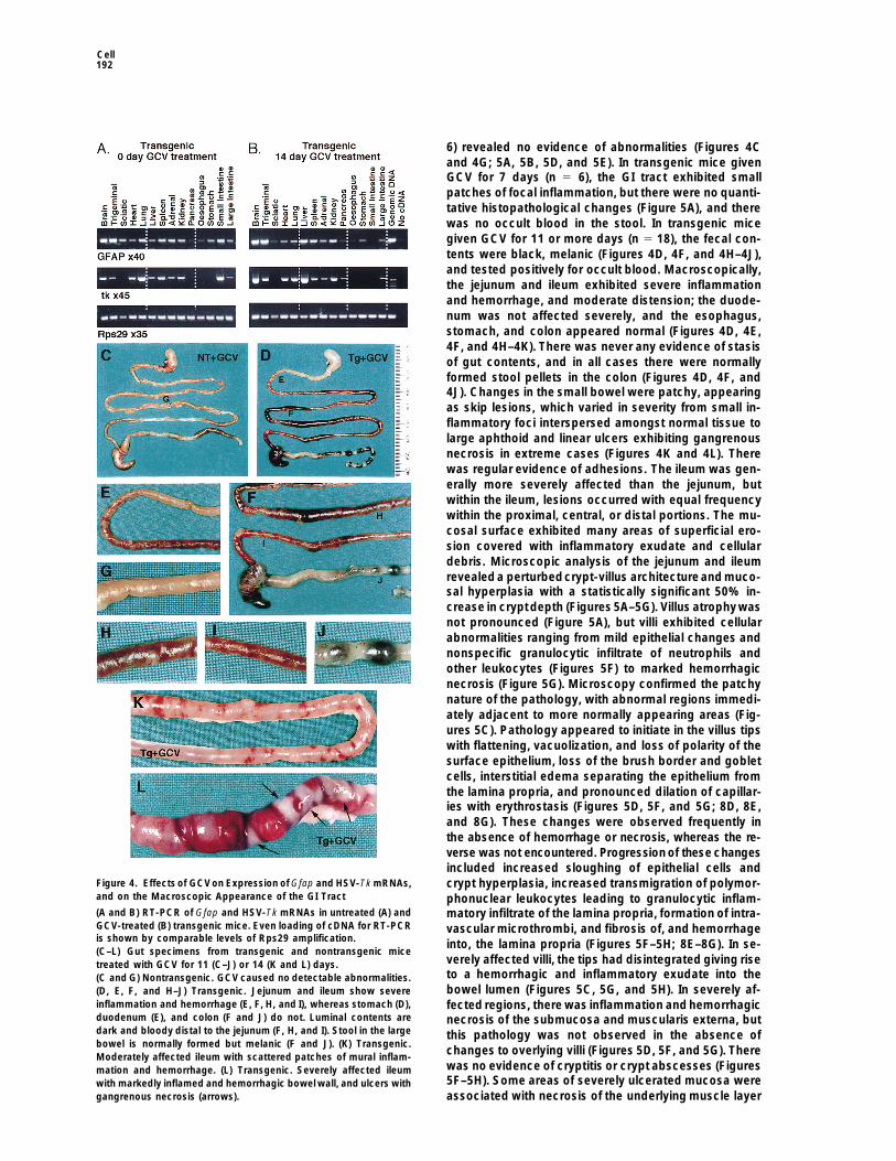
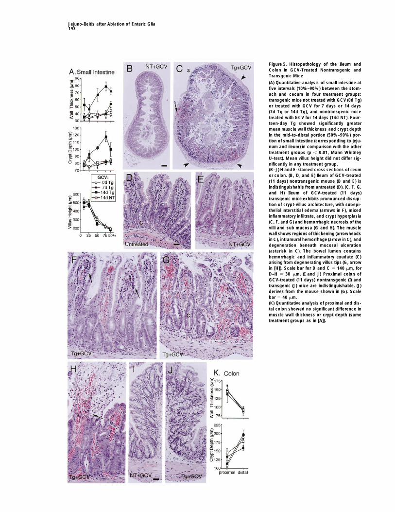
6) revealed no evidence of abnormalities (Figures 4Cand 4G; 5A, 5B, 5D, and 5E). In transgenic mice givenGCV for 7 days (n 5 6), the GI tract exhibited smallpatches of focal inflammation, but there were no quanti-tative histopathological changes (Figure 5A), and therewas no occult blood in the stool. In transgenic micegiven GCV for 11 or more days (n 5 18), the fecal con-tents were black, melanic (Figures 4D, 4F, and 4H–4J),and tested positively for occult blood. Macroscopically,the jejunum and ileum exhibited severe inflammationand hemorrhage, and moderate distension; the duode-num was not affected severely, and the esophagus,stomach, and colon appeared normal (Figures 4D, 4E,4F, and 4H–4K). There was never any evidence of stasisof gut contents, and in all cases there were normallyformed stool pellets in the colon (Figures 4D, 4F, and4J). Changes in the small bowel were patchy, appearingas skip lesions, which varied in severity from small in-flammatory foci interspersed amongst normal tissue tolarge aphthoid and linear ulcers exhibiting gangrenousnecrosis in extreme cases (Figures 4K and 4L). Therewas regular evidence of adhesions. The ileum was gen-erally more severely affected than the jejunum, butwithin the ileum, lesions occurred with equal frequencywithin the proximal, central, or distal portions. The mu-cosal surface exhibited many areas of superficial ero-sion covered with inflammatory exudate and cellulardebris. Microscopic analysis of the jejunum and ileumrevealeda perturbedcrypt-villus architectureand muco-sal hyperplasia with a statistically significant 50% in-crease in cryptdepth (Figures5A–5G). Villus atrophywasnot pronounced (Figure 5A), but villi exhibited cellularabnormalities ranging from mild epithelial changes andnonspecific granulocytic infiltrate of neutrophils andother leukocytes (Figures 5F) to marked hemorrhagicnecrosis (Figure 5G). Microscopy confirmed the patchynature of the pathology, with abnormal regions immedi-ately adjacent to more normally appearing areas (Fig-ures 5C). Pathology appeared to initiate in the villus tipswith flattening, vacuolization, and loss of polarity of thesurface epithelium, loss of the brush border and gobletcells, interstitial edema separating the epithelium fromthe lamina propria, and pronounced dilation of capillar-ies with erythrostasis (Figures 5D, 5F, and 5G; 8D, 8E,and 8G). These changes were observed frequently inthe absence of hemorrhage or necrosis, whereas the re-verse was not encountered. Progressionof these changesincluded increased sloughing of epithelial cells and
Figure 4. Effects of GCVon Expression of Gfap and HSV-Tk mRNAs, crypt hyperplasia, increased transmigration of polymor-and on the Macroscopic Appearance of the GI Tract phonuclear leukocytes leading to granulocytic inflam-(A and B) RT-PCR of Gfap and HSV-Tk mRNAs in untreated (A) and matory infiltrate of the lamina propria, formation of intra-GCV-treated (B) transgenic mice. Even loading of cDNA for RT-PCR vascular microthrombi, and fibrosis of, and hemorrhageis shown by comparable levels of Rps29 amplification. into, the lamina propria (Figures 5F–5H; 8E–8G). In se-(C–L) Gut specimens from transgenic and nontransgenic mice
verely affected villi, the tips had disintegrated giving risetreated with GCV for 11 (C–J) or 14 (K and L) days.to a hemorrhagic and inflammatory exudate into the(C and G) Nontransgenic. GCV caused no detectable abnormalities.bowel lumen (Figures 5C, 5G, and 5H). In severely af-(D, E, F, and H–J) Transgenic. Jejunum and ileum show severe
inflammation and hemorrhage (E, F, H, and I), whereas stomach (D), fected regions, there was inflammation and hemorrhagicduodenum (E), and colon (F and J) do not. Luminal contents are necrosis of the submucosa and muscularis externa, butdark and bloody distal to the jejunum (F, H, and I). Stool in the large this pathology was not observed in the absence ofbowel is normally formed but melanic (F and J). (K) Transgenic.
changes to overlying villi (Figures 5D, 5F, and 5G). ThereModerately affected ileum with scattered patches of mural inflam-was no evidence of cryptitis or crypt abscesses (Figuresmation and hemorrhage. (L) Transgenic. Severely affected ileum5F–5H). Some areas of severely ulcerated mucosa werewith markedly inflamed and hemorrhagic bowel wall, and ulcers with
gangrenous necrosis (arrows). associated with necrosis of the underlying muscle layer
Jejuno-Ileitis after Ablation of Enteric Glia193
Figure 5. Histopathology of the Ileum andColon in GCV-Treated Nontransgenic andTransgenic Mice
(A) Quantitative analysis of small intestine atfive intervals (10%–90%) between the stom-ach and cecum in four treatment groups:transgenic mice not treated with GCV (0d Tg)or treated with GCV for 7 days or 14 days(7d Tg or 14d Tg), and nontransgenic micetreated with GCV for 14 days (14d NT). Four-teen-day Tg showed significantly greatermean muscle wall thickness and crypt depthin the mid-to-distal portion (50%–90%) por-tion of small intestine (corresponding to jeju-num and ileum) in comparison with the othertreatment groups (p , 0.01, Mann WhitneyU-test). Mean villus height did not differ sig-nificantly in any treatment group.(B–J) H and E-stained cross sections of ileumor colon. (B, D, and E) Ileum of GCV-treated(11 days) nontransgenic mouse (B and E) isindistinguishable from untreated (D). (C, F, G,and H) Ileum of GCV-treated (11 days)transgenic mice exhibits pronounced disrup-tion of crypt-villus architecture, with subepi-thelial interstitial edema (arrows in F), mixedinflammatory infiltrate, and crypt hyperplasia(C, F, and G) and hemorrhagic necrosis of thevilli and sub mucosa (G and H). The musclewall shows regions of thickening (arrowheadsin C), intramural hemorrhage (arrow in C), anddegeneration beneath mucosal ulceration(asterisk in C). The bowel lumen containshemorrhagic and inflammatory exudate (C)arising from degenerating villus tips (G, arrowin [H]). Scale bar for B and C 5 140 mm, forD–H 5 30 mm. (I and J) Proximal colon ofGCV-treated (11 days) nontransgenic (I) andtransgenic (J) mice are indistinguishable. (J)derives from the mouse shown in (G). Scalebar 5 40 mm.(K) Quantitative analysis of proximal and dis-tal colon showed no significant difference inmuscle wall thickness or crypt depth (sametreatment groups as in [A]).
Cell194
and transmural perforation (Figures 5C). In general, how- (Figures 6E and 6F). Cross sections showed widespreadloss of GFAP-positive glial cells and their processesever, the smooth muscle wall exhibited a qualitatively
obvious and statistically significant 100% thickening from the submucosal regions and the lamina propria ofvilli throughout the jejunum and ileum (Figures 6I and(Figures 5A–5C). Other regions of the GI tract, in particu-
lar the colon, appeared microscopically normal by quali- 6J). All regions with histopathological abnormalities ex-hibited severe loss of glia (Figure 6J). In contrast, thetative and quantitative analysis (Figures 5I–5K).
Blood cell counts and serum chemistry were normal in colon of GCV-treated transgenic mice exhibited eithera small, or no obvious, loss of immunoreactive entericuntreated transgenic mice, in nontransgenic mice given
GCV for 14 days, and in transgenic mice given GCV for glia (Figures 6K and 6L). These findings indicate thatGCV treatment caused a highly selective, severe abla-7 days.Transgenic mice with mild signs of illness after14
days of GCV had red blood cell counts and hemoglobin tion of enteric glial cells from the jejunum and ileum oftransgenic mice, which correlated spatially and tempo-levels reduced to about 60% of normal, with up to 10-
fold elevations of nucleated red blood cells, up to 20- rally with the severe inflammation and hemorrhagic ne-crosis also induced by this treatment.fold elevations of neutrophils, and mild (about 30%) ele-
vations of serum urea. Moribund GCV-treated transgenicmice exhibited severe anemia with red blood cell counts Changes of Enteric Neurons in the Jejunum and
Ileum of GCV-Treated Transgenic Miceand hemoglobin levels reduced to less than 25% ofnormal, accompanied by high levels of nucleated red In the CNS, GFAP-positive astrocytes provide support-
ive and protective roles for nerve cells (Eddleston andcells and neutrophils relative to the number of red bloodcells, and markedly elevated (.3-fold) serum urea. Mean Mucke, 1993). We therefore looked for effects that abla-
tion of enteric glia might have on enteric neurons. Whole-corpuscular volume and hemoglobin, and the numberof thrombocytes were in the normal range in all animals. mount preparations of jejunum and ileum were double-
labeled for GFAP and cuprolinic blue, a specific markerThese findings ruled out bone marrow failure. Serumcreatinine and other serum chemistry values were also for enteric neurons (Holst and Powley, 1995; Karaosma-
noglu et al., 1996). In control mice (n 5 6), myentericnormal. High levels of serum urea combined with normalserum creatinine indicate normal kidney function com- neurons were completely enveloped by a dense network
of glial cell processes (Figure 7C). In transgenic micebined with GI bleeding. Bacterial cultures prepared fromblood, peritoneal fluid, and spleen showed no growth treatedwith GCV for 11 days or more (n 5 7), this network
was markedly depleted, with a patchy variation in sever-of gram-negative organisms in control mice whereastransgenic animals treated with GCV for 14 days showed ity. In areas where some glial cells remained, theneurons
appeared more or less normal and were denuded of.300 colonies per spleen or ml of blood or peritonealfluid. Together, these findings were compatible with se- enveloping glial processes to varying degrees (Figure
7D). Where glial cells and processes were largely absent,vere hemorrhage into the intestinal tract and perforationof the intestine accompanied by bacteremia as causes there was evidence of neuronal atrophy and loss (Figure
7E). To investigate neuronal changes quantitatively, weof acute decline and death in transgenic mice treatedwith GCV for 11 days or more. conducted unbiased computer-assisted stereological
analysis of whole-mount specimens prepared understandardized unstretched conditions, single-stained with
Localization of HSV-TK in GFAP-Positive Enteric cuprolinic blue. This analysis revealed statistically sig-Glia and Preferential Loss of These Cells nificant differences of 31% fewer myenteric neurons,from Jejunum and Ileum after GCV and a 27% smaller mean cross-sectional area of re-Treatment in Transgenic Mice maining neurons, in the ileum of GCV-treated transgenicTo investigate the cause of gut pathology in transgenic mice as compared with controls (Figures 7A and 7B).mice receiving GCV, the cellular sites of GFAP and HSV- No qualitative or statistically significant evidence of neu-TK expression were identified in the GI tract by immuno- ronal loss or atrophy was observed in the colon of GCV-histochemistry. In agreementwith previous studies (Jes- treated transgenic mice (Figures 7A and 7B). Immuno-sen and Mirsky, 1980; Gershon and Rothman, 1991), histochemistry revealed no detectable difference in thewe observed GFAP-positive enteric glia in an extensive appearance or density of substance P- or tyrosine hy-network throughout the myenteric and submucosal droxylase-immunoreactive fibers in the ileum (Figuresplexuses in whole-mount preparations and cross sec- 7F and 7G) of GCV-treated transgenic mice (n 5 6) astions (Figures 6A and 6G). In addition, we found many compared with controls (n 5 6). Together, these findingsGFAP-positive glia within the lamina propria, with pro- show that ablation of enteric glia caused a moderatecesses that closely embraced the crypts or extended and patchy, but statistically significant, degeneration ofto the distal-most tips of villi (Figures 6G and 6H). GFAP neurons intrinsic to the ileal myenteric plexus, withoutimmunoreactivity was not detected in other intestinal obvious structural changes to substance P or sympa-cell types. In transgenic mice, HSV-TK immunoreactivity thetic innervation.was present in the nuclei and somata of cells with themorphological appearance of enteric glia, and double Selective Decontamination of the Digestive Tractlabeling confirmed that HSV-TK was present only in (SDD) Prevents Bacterial Overgrowth but NotGFAP-positive cells (Figures 6B–6D). After 14 days of Pathological Changes and InflammationGCV treatment, there was a pronounced, but incom- in Jejunum and Ileum of GCV-Treatedplete, loss of GFAP- and HSV-TK-positive cells and pro- Transgenic Micecesses from the myenteric plexus in the jejunum and Degeneration of enteric neurons can disrupt gut motility
and cause bacterial overgrowth, which in extreme casesileum, and remaining glia had abnormal morphologies

Jejuno-Ileitis after Ablation of Enteric Glia195
Figure 6. Cellular Localization of GFAP andHSV-TK in Ileum of Transgenic Mice beforeand after GCV
(A–F) Immunohistochemistry for GFAP (blue)or HSV-TK (brown) in whole-mount prepara-tions of ileal myenteric plexus. Scale bar for(A) and (E) 5 55 mm; for (B), (C), and (F) 5 7mm; for (D) 5 4.5 mm.(A–D) Untreated transgenic. GFAP-positiveenteric glia and their processes extendthroughout the myenteric plexus (A). In indi-vidual plexi, cells stained positively for GFAP(B) or HSV-TK (C) show similar distributionsand morphologies. Double labeling showsthat HSV-TK is present only in GFAP-positivecells (D).(E and F). GCV-treated (14 days) transgenic.GFAP-positive cells and processes havebeen severely depleted (E), and surviving en-teric glia show abnormal morphologies (F).(G and H) Untreated nontransgenic. Immuno-histochemistry of cross and tangential sec-tions of ileum, showing GFAP-positive entericglia and their processes throughout the lam-ina propria, extending to the most distal villustips (G), and densely intertwining betweencrypts (H). Scale bar for G 5 30 mm, for H 5
20 mm.(I–L) Confocal microscopy of immunofluores-cence for GFAP (green) with propidium iodideas a nuclear counter stain (red). GFAP-immu-noreactivity in the ileum is unaltered by GCV(11 days) in a nontransgenic (I) but is ablatedfrom the lamina propria of a transgenic (J)mouse, although small amounts remain in themyenteric plexus (arrow). GFAP loss is asso-ciated with thickening of the muscle wall (m),crypt hyperplasia (c), and epithelial degener-ation. GFAP-immunoreactivity in the colonshows no obvious difference in GCV-treatednontransgenic (K) and transgenic (L) mice.Scale bar for (I) and (J) 5 25 mm; for (K) and(L) 5 40 mm.

Cell196
showed anaerobic and aerobic colony counts reducedsignificantly by more than 1 and 6 orders of magnitude,respectively, as compared with GCV-treated transgenicmice not receiving antibiotics (Figures 8Aand 8B). Func-tional decontamination of the bowel was further demon-strated by a statistically significant increase (p , 0.01,t test) of cecal weight relative to body weight in micewith SDD (2.8 6 0.5%) compared with untreated mice(1.5 6 0.4%). Aerobic bacterial colony counts were simi-larly reduced in GCV-treated transgenic mice after 5days of oral antibiotics (data not shown), indicating ef-fectiveness of SDD during the entire period of develop-ment of the intestinal pathology in these mice.
SDD did not reduce the severity of the illnessexhibitedby GCV-treated transgenic mice as judged by the ap-pearance of the mice, survival time, or severity in thereduction of hemoglobin levels. The small bowel of GCV-treated nontransgenic mice with SDD appeared normal;however, the small bowel of GCV-treated transgenicmice with SDD exhibited severe macro- and micro-scopic pathology with mucosal degeneration, capillarydilation, erythrostasis, granulocytic inflammatory infil-tration, and hemorrhagic necrosis similar to GCV-treatedtransgenic mice that did not receive antibiotics (Figures8C–8G). Standard microbiological screening and sero-logical tests showed no evidence of contagious patho-gens in these mice, such as Clostridium, Salmonella,Yersina, parasites, and various mouse viruses. Thesefindings demonstrate that the pathology induced intransgenic mice by GCV was not merely the result ofbacterial overgrowth in the small bowel, but they donot rule out a role for bacteria in the etiology of thispathology.
Myeloperoxidase (MPO) activity deriving from azuro-philic granules of polymorphonuclear neutrophils wasdetermined in bowel segments as a specific, quantita-tive measure of inflammatory response (Miller et al.,1993). GCV-treated transgenic mice with or without SDDexhibited substantial and statistically significant in-creases in MPO activity in ileum, but not duodenum orcolon, compared with GCV-treated nontransgenic micewith or without SDD (Figure 8C). These findings showthat the pathological changes of the small bowel in GCV-treated transgenic mice were associated with a pro-nounced inflammatory infiltrate, which was not pre-vented by SDD.
Discussion
Enteric glia form a large and widespread network ofcells at all levels of the GI tract. Their functions are not
Figure 7. Effects of Ablation of Enteric Glia on Myenteric Neurons
(A and B) Mean 6 SEM cell number (A) and cross-sectional area(B) of cuprolinic blue–stained myenteric neurons in whole-mountpreparations of ileum or colon from GCV-treated (11–14 days) non-transgenic (NT) and transgenic (Tg) mice (n 5 6 for all groups). Tgmice had significantly (p , 0.001) fewer neurons (A), and remainingneurons had a significantly (p , 0.001) smaller mean cross-sectionalarea (B). There were no significant differences in the colon.(C–E) Immunohistochemistry of GFAP-containing glia (brown) com-bined with cuprolinic blue staining of neurons inwhole-mount prepa-rations of the ileal myenteric plexus. Scale bar 5 10 mm.(C) Untreated transgenic. Processes from GFAP-positiveenteric gliaenvelope all myenteric neurons.(D and E) GCV-treated (14 days) transgenic. In areas where someglia remain but exhibit abnormal morphologies, the neurons appearnormal but are denuded of glial processes (D). In areas with nearcomplete loss of glia, there is neuronal loss and atrophy (E).(F and G) Immunohistochemistry of tyrosine hydroxylase (F) or sub-stance P (G) in whole-mount preparations of ileal myenteric plexusfrom a GCV-treated (14 days) transgenic, showing densities of fibersand terminals indistinguishable from untreated mice. Scale bar forF 5 35 mm, for G 5 15 mm.
may induce inflammatory pathology in the small intes-tine. We therefore quantitatively assessed bacterial lev- well understood. In this study, we found that ablationels in gut segments, and examined the effects of SDD of enteric glia in the jejunum and ileum of adult trans-achieved with oral antibiotics, on the intestinal pathol- genic mice led rapidly to a fulminating enteritis withogy triggered by GCV in transgenic mice. The ileum, severe inflammation and hemorrhagic necrosis of thesebut not duodenum or cecum, of GCV-treated transgenic organs. Ablation of enteric glia also caused degenerativemice showed statistically significant 10- to 100-fold in- changes in neural elements of the enteric nervous sys-creases in the number of anaerobic and aerobic bacte- tem, which may have contributed to the developmentrial colonies per gram tissue, respectively, as compared of the associated pathology. The macro- and histo-with untreated or GCV-treated nontransgenic mice (Fig- pathologic appearance of the small intestine was similarures 8A and 8B). The ileum of GCV-treated transgenic to that in experimental models of inflammatory bowelmice fed diet containing three oral antibiotics (broad disease (IBD) in rodents and in various human condi-
tions.spectrum, antianaerobic, and antifungal) for 14 days

Jejuno-Ileitis after Ablation of Enteric Glia197
s.c. GCV Treatment Preferentially and SpecificallyAblated Transgenic Glia in the Jejunumand IleumMost HSV-TK-expressing glial cells throughout the cen-tral, peripheral, and enteric nervous systems were notkilled in detectable numbers by the regimen of GCVdelivery used in this study. The reason for the preferen-tial ablation of HSV-TK-expressing glia in the small in-testine is uncertain. After phosphorylation of GCV bytransgene-derived HSV-TK, GCV-mediated cell death isthought to result from the formation of toxic intermedi-ates that disrupt DNA replication and kill proliferatingcells (Frank et al., 1984; Borrelli et al., 1989). In agreementwith these reports, we found that s.c. GCV treatmentkilled HSV-TK-expressing CNS astrocytes in vivo onlyadjacent to a stab injury, a procedure that induces prolif-eration of local reactive astrocytes. These observationssuggest that different rates of cell division amongstHSV-TK-expressing glia may account for variations intheir vulnerability to GCV, but other differences cannotbe ruled out.
In the small intestine, HSV-TK immunoreactivity waspresent only in GFAP-positive enteric glia, and not inother cell types. Enteric glia were clearly ablated in thejejunum and ileum by GCV treatment, and there is noevidence to suggest that the lethal metabolites of GCV,which they produced, had any effects on neighboringintestinal cells. In previous reports, GCV treatment oftransgenic mice had no toxic effects on non-HSV-TK-expressing cells that were intermingled with cells whichexpressed HSV-TK and died in the pituitary, thyroid,or stomach (Borrelli et al., 1989; Wallace et al., 1991;Canfield et al., 1996). In our in vitro experiments, GCVdid not kill GFAP- and HSV-TK-negative cells; instead,
Figure 8. Effects of SDD on the Inflammation and Pathology In-duced by GCV in Transgenic Mice
(A and B) Mean number 6 SEM of aerobic (A) and anaerobic (B)colony forming units per gram tissue. GCV-treated (14 days)transgenic mice (Tg 1 GCV) showed significantly higher counts ofboth aerobic and anaerobic bacteria in ileum, but not other gutregions, compared with untreated, or GCV-treated nontransgenic(NT 1 GCV), mice. Tg 1 GCV mice given antibiotics for 14 days(Tg 1 GCV 1 AB) showed significantly fewer aerobic counts in allthree gut regions, and significantly fewer anaerobic counts in ileumand cecum, compared with untreated or NT 1 GCV mice. (p , 0.01,ANOVA with Newman-Keuls post hoc pair-wise comparison).(C) Mean value 6 SEMof myeloperoxidase (MPO) activity expressedas units per 100 mg protein. Tg 1 GCV and Tg 1 GCV 1 AB miceshowed significantly higher values in ileum but not other gut regionscompared with NT 1 GCV mice or mice treated with NT 1 GCV 1
antibiotics (NT 1 GCV 1 AB). (p , 0.01, ANOVA with Newman-Keuls).(D–G) H and E-stained cross sections of villus tips in comparableregions of midileum. Scale bar for (D)–(F) 5 10 mm; for (G) 5 6.3mm. (D) NT 1 GCV 1 AB. Healthy columnar epithelium with a promi-nent brush border and small capillaries in lamina propria. (E) Tg 1
GCV 1 AB. Loss of brush border and epithelial polarity, increasedsloughing of epithelial cells (arrows), and pronounced dilation ofcapillaries (c). (F) Tg 1 GCV. Degeneration and vacuolization ofepithelium, sclerosis of lamina propria, formation of microthrombiin the capillaries (arrowheads), and extravasation of red and whiteblood cells. (G) Tg 1 GCV. Severe dilation of a capillary with erythro-stasis and numerous polymorphonuclear leukocytes including neu-trophils (n) and monocytes (m).

Cell198
these cells increased in number while neighboring GFAP- in the CNS, disruption of glutamate uptake by astrocytescauses excitotoxic neuronal death (Rothstein et al.,positive, HSV-TK-expressing astrocytes died. Together,
these findings indicate that lethal metabolites of GCV are 1996).Enteric glia may also influence neural fibers and pro-not released by HSV-TK-expressing cells in sufficient
quantities to kill neighboring cells, and that GCV specifi- jections from enteric neurons. The continuous turnoverof intestinal mucosa due to epithelial sloughing requirescally ablated enteric glia in the small intestine of our
transgenic mice. constant remodeling of enteric neural connections. Thisactivity is reflected in the constitutive expression byenteric neurons of GAP-43 (Sharkey et al., 1990). In the
Enteric Glia Play a Fundamental Role adult CNS, astrocytes support some types of sproutingin Bowel Function or regenerating axons (Kawaja and Gage, 1991). GFAP-Ablation of enteric glial cells in the jejunum and ileum positive enteric glia that envelop axon bundles (Gershonled rapidly to severe inflammation and necrosis. The and Rothman, 1991) may play a similar role, with thepersistence of the pathology in mice with SDD showed consequence that loss or dysfunction of enteric gliathat this subacute, necrotizing enteritis was not merely could lead to a failure of certain enteric neural projec-caused by bacterial overgrowth resulting from disturbed tions.gut motility due to neuronal degeneration. Thus, func- The widespread distribution of enteric glia and theirtional changes induced by the loss of enteric glia played processes throughout the intestinal mucosa, embracinga primary role in triggering the disease process. To our crypts and extending to the most distal villus tips, sug-knowledge, there are no previous reports presenting gests that molecules released from these cells couldevidence for, or postulating, a direct link between the influence many cell types. GFAP-positive glial cells inloss or dysfunction of enteric glia and pathological the CNS produce, and are affected by, many growthchanges in the bowel. The sequence of events appeared factors and cytokines and participate in inflammatoryto involve early breakdown of the epithelium, and micro- and immune responses (Eddleston and Mucke, 1993),vascular disturbances. Our observations are also com- a capacity that may be shared by GFAP-positive entericpatible with a role for normal gut constituents (food glia (Bar et al., 1997). CNS astrocytes also induce blood-proteins and bacterial flora) in the inflammatory process. brain barrier properties in endothelia (Janzer and Raff,
The rapid inductionof severe intestinal pathologyafter 1987), and some enteric glia display terminal swellingsthe ablation of enteric glia demonstrates that these cells resembling the endfeet of CNS astrocytes (Gershon andplay a fundamental role in maintaining the integrity of Rothman, 1991), which often anchor astrocytes to bloodthe bowel. Based on comparison with known functions vessels. In this context, it is interesting that ablation ofof related GFAP-expressing glia in other neural systems, enteric glia in this study caused microvascular distur-there are a number of ways in which enteric glia could bances and certain changes resembling ischemic bowel.exert powerful effects on bowel function, either by influ-encing enteric neurons or through direct interactions Changes Induced by Ablation of Enteric Gliawith nonneuronal cell types. Are Similar to IBD
In the CNS, fibrous processes of GFAP-positive astro- The pathological changes induced in our transgeniccytes surround and contact essentially all types of neu- mice by ablation of enteric glia, although subacute androns. Interactions between neurons and glia via these fulminating in nature, bear similarities to IBD in animalprocesses show considerable plasticity and play impor- models and various human conditions, in particulartant roles in regulating neuronal function (Theodosis and Crohn’s disease (Strober and Ehrhardt, 1993; SharkeyMacVicar, 1996). The denuding of myenteric and submu- and Parr, 1996). The features of IBD are of unknowncosal neurons of glial processes in GCV-treated trans- etiology in human disease (Goyal and Hirano, 1996) andgenic mice is likely to have had a pronounced effect on can be induced to varying degrees in experimental ani-their function. In other neural tissues, glia also provide mals either by treatment with certain chemicals (Sharkeyimportant protective and/or supportive functions for and Parr, 1996), the targeted deletion of genes for IL-2,neurons. In the Drosophila mutant drop dead, glial cell IL-10, or T cell receptors (Kuhn et al., 1993; Mombaertsdegeneration precedes, and is thought to cause, neu- et al., 1993; Sadlack et al., 1993), or the genetically tar-ronal degeneration (Buchanan and Benzer, 1993). In the geted disruption of epithelial adhesion (Hermiston andmammalian CNS, GFAP-positive astrocytes buffer neu- Gordon, 1995). While these animal models strongly im-rons from the accumulation of potentially toxic endoge- plicate immune and neural dysfunction in IBD, the cas-nous molecules and produce neurotrophic growth fac- cade of cellular events remains uncertain.tors (Eddleston and Mucke, 1993). Our observations Similarities in the appearance of end-stage pathologydemonstrate that loss or dysfunction of enteric glia leads in various bowel conditions suggests that the gut mayto substantial degeneration of myenteric neurons, which have limited ways of responding when it is defective.may be due to loss of trophic or neuroprotective func- For this reason, it is important to identify single eventstions. Enteric glia produce glial-derived neurotrophic that are able to trigger complex pathological cascades.factor (GDNF) (Bar et al., 1997), and embryonic mice In this study, loss or dysfunction of enteric glia played ahomozygous for deletion of the genes for GDNF or its primary role in triggering inflammatory bowel pathology,receptor fail to develop enteric neurons (Moore et al., suggesting that these cells may play a role in IBD. The1996; Pichel et al., 1996; Sanchez et al., 1996). Enteric enteric nervous system has been implicated in IBDneurons, like CNS neurons, are vulnerable to glutamate- throughpartial neural degeneration combined with pres-
ervation or increases in proinflammatory peptides suchmediated excitotoxicity (Kirchgessner et al., 1997), and

Jejuno-Ileitis after Ablation of Enteric Glia199
Surgical Proceduresas substance P and sympathetic innervation (Goyal andSurgical procedures were performed under anesthesia with AvertinHirano, 1996; Sharkey and Parr, 1996; McCafferty et al.,(0.015 ml/g) and halothane. Miniosmotic pumps were implanted1997). Our findings after ablation of enteric glia aresubcutaneously as recommended by the manufacturer (Alzet). Stab
similar. It is interesting that pronounced degenerative injuries to the forebrain were made with a number 11 scalpel bladechanges in the myenteric plexus alone do not precipitate using a rodent stereotaxic apparatus (Kopf, USA).IBD in experimental animals (Dahl et al., 1987) or in
GCV Administrationpatients with Hirschsprung’s disease, suggesting thatGCV (Roche, UK) was administered continuously at a rate of 100imbalance of neural regulation, rather than extensivemg/kg/day diluted in sterile physiological saline for 7, 14, or 28 daysneuronal degeneration, may be important. As discussedvia subcutaneously implanted osmotic minipumps (Alzet, models
above, the loss or dysfunction of enteric glia may have 1007D, 2001, 2002, or 2004). This dose was reported to kill thyro-effects on bowel functions other than, and in addition cytes expressing HSV-TK in transgenic mice (Wallace et al., 1991).to, effects on enteric neurons. We speculate that distur-
Hematology, Serum Chemistry, Bacteriological Analysisbances in enteric glia have the potential to represent aBlood was collected by cardiac puncture from animals under termi-link between immune, vascular, and neural dysfunctionsnal barbiturate anesthesia. Serum chemistry, hematological analy-in IBD. The pathological changes induced by ablationses, and guaiac tests for occult blood in stool were conducted
of enteric glia in the transgenic mice described here by the clinical laboratories of Addenbrooke’s hospital, Cambridge.provide a model for investigating both the etiology and Aerobic bacterial cultures of blood, peritoneal fluid, or organ speci-potential treatments of IBD and other forms of enteritis. mens (gut, liver, spleen, mesenteric lymph node) were prepared
using either brain heart infusion or blood agar plates. Aerobic andanaerobic bacteriological analysis of the bowel was performed byExperimental ProceduresBIBRA International (Surrey, UK). Segments of duodenum, ileum,and cecum in toto were homogenized in Brucella broth, plated ontoGfap-HSV-Tk Fusion Gene Constructblood agar plates, andgrown under anaerobic or aerobic conditions.Starting with a 15 kb Gfap-lacZ plasmid, the lacZ sequence was
replaced with HSV-Tk sequence (Figure 1A). A 1.7 kb XbaI–Bam HISDDfragment, including the HSV-Tk gene and its polyadenylation signal,SDD was achieved using three antibiotics incorporated into thewas inserted into a pBSII-KS vector (Stratagene). The insert wasdiet (Bioserve, Frenchtown, NJ, USA): a broad spectrum antibioticreexcised using Sal I and Not I and ligated into the first exon of a(ciprofloxacin, 0.5 mg/g), an antianaerobic agent (metronidazole,Gfap promoter cassette (clone 445) consisting of a modified murine0.25 mg/g), and an antifungal agent (fluconazole, 0.2 mg/g), whichGfap gene (Johnson et al., 1995). Integrity of the HSV-Tk insert andretain high concentrations in feces and are used to control bacterialflanking regions was confirmed by single-strand sequencing. Thenumber in the GI tracts of neutropenic patients. Mice consumedGfap-HSV-Tk fusion gene construct was excised by digestion with3–6 g per day.SfiI and purified by gel electrophoresis.
MPO AssayAnimals MPO activity from polymorphonuclear neutrophils was assayedMice werehoused in a 12 hr light/dark cycle with controlled tempera- (Miller et al., 1993) in bowel segments (100–250 mg), homogenized,ture and humidity, and allowed free access to food and water. and centrifuged at 20,000 g. Pellets were resuspended, pseudoper-Transgenic mice were produced using standard techniques (Hogan oxidase activity negated with hexadecyltrimethlammonium bro-et al., 1986). Approximately 2 pl of DNA (500 copies of linearized mide, followed by 3 cycles of sonication, freezing, and thawing.fusion gene construct) were pressure injected into the male pronu- Supernatant (10 ml) was mixed with 90 ml of potassium phosphatecleus of fertilized eggs from superovulated female C57BL/10 3 CBA buffer containing O-dianisidine dihydrochloride and H2O2 and absor-mice mated with CFLP males (Interfauna, UK). Two-cell stage eggs bence determined at 450 nm. One U of MPO activity was definedwere reimplanted into pseudopregnant foster mothers. DNA ob- as that required to degrade 1 mmol of H2O2 per min at 258C. Valuestained by tail biopsy from resulting mice was digested with EcoRI were expressed as MPO units per 100 mg total protein per sample.and screened by Southern blot using a 1.7 kb HSV-TK probe.
Tissue HarvestWestern Blot Mice were killed by terminal barbiturate anesthesia. Dissected tis-Frozen cerebral hemispheres were freeze/thawed, homogenized in sues were either frozen in liquid nitrogen and stored at 2708C untillysis buffer (20 mM HEPES [pH 7.5], 0.42 M NaCl, 1.5 mM MgCl2 PCR or sectioning, or fixed in 4% paraformaldehyde and processed0.2 mM EDTA, 0.5 mM PMSF, 0.25 mM DTT, 50 mM NaF, 1 mM for hematoxylin and eosin (H and E)-stained paraffin sections. Somesodium orthovanadate, and 20% glycerol [w/v]), and centrifuged at mice were fixed by transcardiac perfusion and frozen sections pre-10,000 g. Supernatant proteins were separated on SDS PAGE and pared.transferred to nitrocellulose membrane. Nonspecific binding siteswere blocked with 5% milk powder, followed by incubation in 1:250 RT-PCRrabbit anti-HSV-TK (P. Collins), 1:250 biotinylated anti-rabbit IgG Total RNA was extracted from frozen tissues and reverse tran-(Dako), streptavidin-biotin-horseradish peroxidase complex (ABC; scribed. PCR primers were: Gfap (accession number K01347, for-Vector), and diaminobenzidine (0.5 mg/ml). ward GTT GTG AAG GTC TAT TCC TGG C; reverse TCC CTT AGC
TTG GAG AGC AA); HSV-Tk (forward GGT CCC GGA TCC GGT GGTGG; reverse CGA GGC GGT GTT GTG TGG TGT); ribosomal proteinIn Vitro ExperimentsS29 (Rps29, accession number L31609, forward CTG ATC CGC AAAPrimary astrocyte cultures were prepared from brains of individualTAC GGG; reverse GCA TGA TCG GTT CCA CTT G). Primers wereneonatal (,48-hr-old) littermate pups (McCarthy and deVellis, 1980).used at 100 ng/reaction. Hot-lid PCR amplification of cDNA wasEach culture was genotyped by Southern blot of corresponding livercarried out in PCR buffer containing 0.5 mM dNTPs and 0.6 U Ampli-DNA. Astrocyte vulnerability to GCV was assessed using the MTTTaq DNA polymerase on PTC-225 thermal cyclers (Tetrad). PCR(3-[4,5-Dimethylthylthiazol-2-yl]-2,5-diphenyltetrazolium bromide) as-products were separated on agarose gel and stained with ethidiumsay (Hansen et al., 1989). Sister cultures were grown in triplicate inbromide.the presence or absence of 2 mM GCV for up to 7 days. For the
assay, medium was replaced by MTT solution (0.5 mg/ml in PBS)for 3 hr; cells were lysed with SDS, and OD measured at 570 nm. For Immunohistochemistry and Histology
Pieces of GI tract tissue, or 40 mm thick frozen forebrain sectionsimmunohistochemistry, cultures were fixed in 4% paraformaldehydeand stained as described below for tissue sections. were stained free-floating in primary antibody overnight, biotinylated

Cell200
secondary antibody for 2 hr, streptavidin-biotin-horseradish peroxi- Gershon, M.D., and Rothman, T.P. (1991). Enteric glia. Glia 4,195–204.dase complex (ABC, Vector) for 2 hr, and diaminobenzidine (DAB,
0.5 mg/ml). For double labeling, sections were stained sequentially Goyal, R.K., and Hirano, I. (1996). The enteric nervous system. N.using DAB as a brown chromagen followed by Vector SG (Vector) Engl. J. Med. 334, 1106–1115.to give a blue color. GFAP was also visualized using streptavidin- Gundersen, H.J.G., Bendtsen, T.F., Korbo, L., Marcussen, N., Moller,FITC (1:100) with propidium iodide (5 mg/ml) as a nuclear counter- A., Nielsen, K., Nyengaard, J.R., Pakkenberg, B., Sorensen, F.B.,stain and viewed by confocal laser scanning microscopy (Bio-Rad Vesterby, A., and West, M.J. (1988). Some new, simple and efficientMRC500). Rabbit primary antibody dilutions were: anti-HSV-TK steriological methods and their use in pathological research and1:25,000 (P. Collins), anti-GFAP 1:25,000 (Dako); anti-TH 1:25,000 diagnosis. APMIS (Copenhagen) 96, 379–394.(Dako); anti-substance P1:25,000 (Dako). Enteric neuronswere visu-
Hansen, M.B., Nielsen, S.E., and Berg, K. (1989). Re-examinationalized with cuprolinic blue alone or in combination with immunohis-and further development of a precise and rapid dye method for mea-tochemistry (Holst and Powley, 1995; Karaosmanoglu et al., 1996).suring cell growth/cell killing. J. Immunol. Methods 119, 203–210.
Hermiston, M.L., and Gordon, J.I. (1995). Inflammatory bowel dis-Morphometryease and adenomas in mice expressing a dominant negativeSmooth muscle wall thickness, crypt depth, and villus height wereN-cadherin. Science 270, 1203–1207.measured by image analysis (Seescan) in H and E-stained paraffinHeyman, R.A., Borrelli, E., Lesley, J., Anderson, D., Richman, D.D.,sections. Enteric neuron counts and cross-sectional area measure-and Baird, S.M. (1989). Thymidine kinase obliteration: creation ofments were performed in cuprolinic blue–stained whole-mounttransgenic mice with controlled immune deficiency. Proc. Natl.specimens using stereology and unbiased sampling (Gundersen etAcad. Sci. USA 86, 2698–2702.al., 1988). Tissue preparations were fixed unstretched in a standard-
ized manner. Care was taken not to introduce postmortem stretch Hogan, B., Constantini, E., and Lacy, E. (1986). Manipulating theartifacts, which might influence cell counts (Karaosmanoglu et al., Mouse Embryo: A Laboratory Manual (Cold Spring Harbor, New1996). Two counting frame fields (8564 mm2) per mm2 were selected York: Cold Spring Harbor Laboratory Press).at random using a computer-driven microscope stage (CAST, Olym- Holst, M.C., and Powley, T.L. (1995). Cuprolinic blue (quinolinicpus). Since whole-mount preparations are thin and unsectioned, all phthalocyanine) counterstaining of enteric neurons for peroxidaseneurons identifiable through the depth of the specimen were ana- imunocytochemistry. J. Neurosci. Methods 62, 121–127.lyzed per frame. At least 15 mm2 were analyzed, at least 200 neurons
Janzer, R.C., and Raff, M.C. (1987). Astrocytes induce blood-brainwere counted, and the cross-sectional area of at least 60 neuronsbarrier properties in endothelial cells. Nature 325, 253–256.was measured for each gut segment per animal.Jessen, K.R., and Mirsky, R. (1980). Glial cells in the enteric nervoussystem contain glial fibrillary acidic protein. Nature 286, 736–737.AcknowledgmentsJohnson, W.B., Ruppe, M.D., Rockenstein, E.M., Price, J., Sarthy,V.P., Vederber, L.C., and Mucke, L. (1995). Indicator expressionWe thank Dr. T. H. Rabbitts for the HSV-Tk plasmid; Dr. P. Collinsdirected by regulatory sequences of the glial fibrillary acidic proteinfor HSV-TK protein and antiserum; S. Jackson and Roche Products(GFAP) gene: in vitro comparison of distinct GFAP-lacZ transgenes.Ltd. for ganciclovir; Drs. S. Young, D. Wen-Lin, S. Murch, I. Sand-Glia 13, 174–184.erson, M. Kagnoff, and P. Sansonetti for helpful discussion; T.
Humby for statistics; and J. A. Bashford, A. P. Newman and I. Bolton Karaosmanoglu, T., Aygun, B., Wade, P.R., and Gershon, M.D.for photography. This work was supported by grants from The Well- (1996). Regional differences in the number of neurons in the myen-come Trust and Action Research to M. V. S. and M. H. J., and an teric plexus of the guinea pigsmall intestine and colon: an evaluationMRC studentship to T. G. B. of markers used to count neurons. Anat. Rec. 244, 470–480.
Kawaja, M.D., and Gage, F.H. (1991). Reactive astrocytes are sub-Received July 23, 1997; revised March 4, 1998. strates for the growth of adultCNS axons in the presence of elevated
levels of nerve growth factor. Neuron 7, 1019–1030.References Kirchgessner, A.L., Liu, M.-T., and Alcantara, F. (1997). Excitotoxicity
in the enteric nervous system. J. Neurosci. 17, 8804–8816.Al-Shawi, R., Burke, J., Jones, C.T., Simons, J.P., and Bishop, J.O. Kuhn, R., Lohler, J., Rennick, D., Rajewsky, K., and Muller, W. (1993).(1988). A Mup promoter-thymidine kinase reporter gene shows re- Interleukin-10-deficient mice develop chronic enterocoloitis. Cell 75,laxed tissue-specific expression and confers male sterility upon 263–274.transgenic mice. Mol. Cell. Biol. 8, 4821–4828. McCafferty, D.M., Wallace, J.L., and Sharkey, K.A. (1997). EffectsBar, K.J., Facer, P., Williams, N.S., Tam, P.K.H., and Anand, P. (1997). of chemical sympathectomy and sensory nerve ablation on experi-Glial-derived neurotrophic factor in human adult and fetal intestine mental colitis in the rat. Am. J. Physiol. 272, G272-G280.and in Hirschsprung’s disease. Gastroenterology 112, 1381–1385. McCarthy, K.D., and de Vellis, J. (1980). Preparation of separateBorrelli, E., Heyman, R.A., Arias, C., Sawchenko, P.E., and Evans, astroglial and oligodendroglial cell cultures from rat cerebral tissue.R.M. (1989). Transgenic mice with inducible dwarfism. Nature 339, J. Cell Biol. 85, 890–902.538–541. Miller, M.J.S., Sadowska-Krowicka, H., Chotinaruemol, S., Kakkis,Buchanan, R.L., and Benzer, S. (1993). Defective glia in the Drosoph- J.L., and Clark, D.A. (1993). Amelioration of chronic ileitis by nitricila brain degeneration mutant drop-dead. Neuron 10, 839–850. oxide synthase inhibition. J. Pharmacol. Exp. Ther. 264, 11–16.Canfield, V., West, A.B., Goldenring, J.R., and Levenson, R. (1996). Mombaerts, P., Mizoguchi, E., Grusby, M.J., Glimcher, L.H., Bhan,Genetic ablation of parietal cells in transgenic mice: a new model A.K., and Tonegawa, S. (1993). Spontaneous development of inflam-for analyzing cell lineage relationships in the gastric mucosa. Proc. matory bowel disease in T cell receptor mutant mice. Cell 75,Natl. Acad. Sci. USA 93, 2431–2435. 275–282.Dahl, J.L., Bloom, D.D., Epstein, M.L., Fox, D.A., and Bass, P. (1987). Moore, M.W., Klein, R.D., Farinas, I., Sauer, H., Armanini, M., Phillips,Effect of chemical ablation of myenteric neurons on neurotransmit- H., Reichardt, L.F., Ryan, A.M., Carver-Moore, K., and Rosenthal,ter levels in the rat jejunum. Gastroenterology 92, 338–344. A. (1996). Renal and neuronal abnormalities in mice lacking GDNF.
Nature 382, 76–79.Eddleston, M., and Mucke, L. (1993). Molecular profile of reactiveastrocytes—implications for their role in neurological disease. Neu- Pichel, J.G., Shen, L., Sheng, H.Z., Granholm, A.C., Drago, J., Grin-roscience 54, 15–36. berg, A., Lee, E.J., Huang, S.P., Saarma,M., Hoffer, B.J., et al. (1996).
Defects in enteric innervation and kidney development in mice lack-Frank, K.B., Chiou, J.-F., and Cheng, Y.-C. (1984). Interaction ofing GDNF. Nature 382, 73–76.herpes simplex virus–induced DNA polymerase with 9-(1,3-dihy-
droxy-2-propoxymethyl)guanine triphosphate. J. Biol. Chem. 259, Rothstein, J.D., Dykes-Hoberg, M., Pardo, C.A., Bristol, L.A., Jin, L.,Kunci, R.W., Kanai, Y., Hediger, M.A., Wang, Y., Schielke, J.P., and1566–1569.

Jejuno-Ileitis after Ablation of Enteric Glia201
Welty, D.F. (1996). Knockout of glutamate transporters reveals amajor role for astroglial transport in excitotoxicity and clearance ofglutamate. Neuron 16, 675–686.
Sadlack, B., Merz, H., Schorle, H., Schimpl, A., Feller, A.C., andHorak, I. (1993). Ulcerative colitis-like disease in mice with a dis-rupted interleukin-2 gene. Cell 75, 253–261.
Sanchez, M.P., Silos-Santiago, I., Frisen, J., He, B., Lira, S.A., andBarbacid, M. (1996). Renal agenesis and the absence of entericneurons in mice lacking GDNF. Nature 382, 70–73.
Sharkey, K.A., and Parr, E.J. (1996). The enteric nervous system inintestinal inflammation. Can. J. Gastroenterol. 10, 335–341.
Sharkey, K.A., Coggins, P.J., Tetzlaff, W., Zwiers, H., Bisby, M.A.,and Davison, J.S. (1990). Distribution of growth-associated proteinB-50 (GAP-43) in the mammalian enteric nervous system. Neurosci-ence 38, 13–20.
Strober, W., and Ehrhardt, R.O. (1993). Chronic intestinal inflamma-tion: an unexpected outcome in cytokine or T cell receptor mutantmice. Cell 75, 203–205.
Theodosis, D.T., and MacVicar, B. (1996). Neurone-glia interactionsin the hypothalamus and pituitary. Trends Neurosci. 19, 363–367.
Toggas, S.M., Masliah, E., Rockenstein, E.M., Rall, G.F., Abraham,C.R., and Mucke, L. (1994). Central nervous system damage pro-duced by expression of the HIV-1 coat protein gp120 in transgenicmice. Nature 367, 188–193.
Wallace, H., Ledent, C., Vassart, G., Bishop, J.O., and Al-Shawi, R.(1991). Specific ablation of thyroid follicle cells in adult transgenicmice. Endocrinology 129, 3217–3226.