the pathology and significance of ileitis
TRANSCRIPT

THE PATHOLOGY AND SIGNIFICANCE OF ILEITIS
K. Geboes, MD, PhD, AGAF, Dr. H.C. Cagliari
Belgium

Normal small intestineVariations
• The ileum constitutes 2/5ths of the small intestine
• The wall is thinner• Mesenteric fat is abundant• More vascular loops• More Goblet cells• Adult ileal mucosal stem cells might
be different from stem cells in other areas, for instance by inducing bile acid uptake and expression of the IBAT protein (Middendorp e.a. Stem cells 2014)
• Lymphoid tissue• Small intestinal tissue macrophages
are different from those in the colon
Duodenum
Ileum

Defensin5 expression in ileumDecreased expression of human defensins 5 & 6 in ileum in CD
Wehkamp et al Gut 2004; 53: 1658NOD2 expression in Paneth cells Gastroenterology 2003
Proliferativecpt

Peyer’s patches (lympho-epithelial complexes)Normal structure
First description 1677
Diffusely present along the smallintestine: antimesenteric
Numbers-24 weeks : +/-45-20 year : +/- 200-95 year : +/- 100
-CompositionEpithelial cells
M cellsFAE cells
Lymphoid componentsSubepithelial mixed zoneFollicles

NODULAR LYMPHOID HYPERPLASIA
Common in children and adolescents

Ileitis : IBD or not?
Clinical situations
Isolated ileitis
Lesions of colon and ileum
- Indications for biopsy : overview of historical and recentstudies
-The challenge of isolated ileitis- Histopathological features for the diagnosis of Crohn’s disease- Backwash ileitis- Other causes of ileitis : differential diagnostic issues- Miscellaneous

Studies concerning biopsies of terminal ileumHistory (1984-1995)

Is ileoscopy with biopsy worthwhile in patients presenting withsymptoms of IBD?
Geboes e.a. Am J Gastroenterol 1998; 93; 201

More recent studiesAsymptomatic ileitis, past present and future. Greaves ML, Pochapin M. J Clin Gastroenterol 2006; 40: 281
Etiologies of this phenomenon, include subclinical Crohn's disease, nonsteroidalanti-inflammatory drugs and spondylarthropaties
The diagnostic value of endoscopic terminal ileum biopsies. McHugh e.a. Am J Gastroenterol 2007; 102: 1084
– Biopsy of endoscopically normal mucosa is unlikely to yield diagnostically useful information, and is not encouraged as routine.
– However, when “ileitis,” ulcers, or erosions are identified, biopsies can be very helpful.
Ileitis when it is not Crohn’s disease. Dilauro e.a. Curr Gastroenterol Rep 2010; 12: 249
– Ileitis may be caused by a wide variety of other diseases.
– These include infectious diseases, spondyloarthropathies, vasculitides, ischemia, neoplasms, medication-induced, eosinophilic enteritis, and others.
– The diagnosis of the specific etiology is suggested by a detailed history and physical examination, laboratory testing, and ileocolonoscopy and/or radiologic data.

Conclusion
Ileoscopy with biopsy is useful in carefullyselected patients
These include : inflammatory diarrhea; presence of endoscopic lesions; anaemia…

Isolated active ileitis (IAI)
• Typical CD in 8/28 pts (27%)– Goldstein Am J Surg Pathol 2006
• 60 patients with IAI (O’Donnell et al 2013)– Repeat endoscopy– Serum analysis for ANCA, anti-OmpC, ASCA IgA, ASCA IgG, anti-Cbir– Results
• No significant difference in the prevalence of antibodies between IAI cases and healthy controls
• Endoscopy follow up in 43 pts– 6/43 (14%) : definite Crohn’s disease– 18/43 (42%) : normal– 11/43 (26%) : persistent IAI
• 40 pts : no lesions in a median follow up of 3.2 yrs (82% NSAIDs)– Lengeling e.a Clin Gastroenterol Hepatol. 2003;

Isolated ileitisChallenges
• NSAIDs ulc
• Adhesion
• Vascular diseases
• Infections
• Tumors Mass lesions :– neuroendocrine tumor of
ileum– Metastasis
• Elderly patients; with a history of joint lesions
• Abdominal surgical history• General symptoms/
systemic disease• General symptoms
• No features of – Age of the patient– No malabsorption in clinical
chemistry– Short history

Histopathological features for the diagnosis of Crohn’sdisease
- Based on multiple samples- Heterogeneity of villousarchitecture
Early lesions
Diagnostic lesions
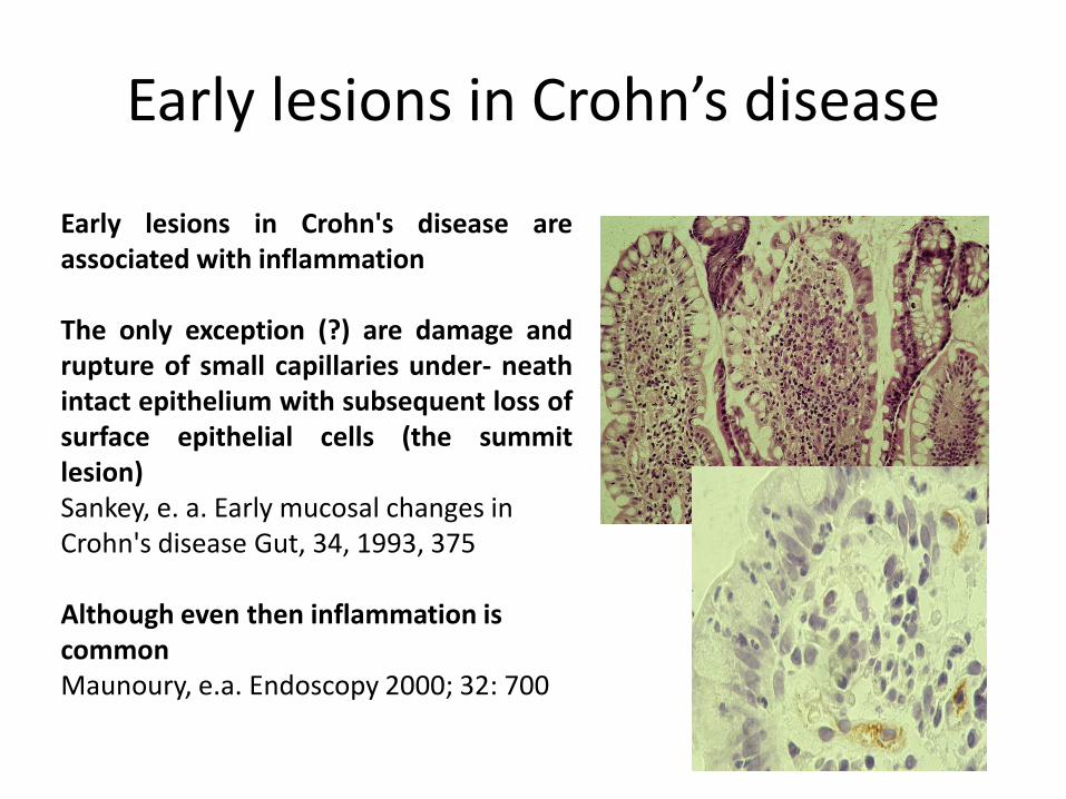
Early lesions in Crohn's disease areassociated with inflammation
The only exception (?) are damage andrupture of small capillaries under- neathintact epithelium with subsequent loss ofsurface epithelial cells (the summitlesion)Sankey, e. a. Early mucosal changes in Crohn's disease Gut, 34, 1993, 375
Although even then inflammation is commonMaunoury, e.a. Endoscopy 2000; 32: 700
Early lesions in Crohn’s disease
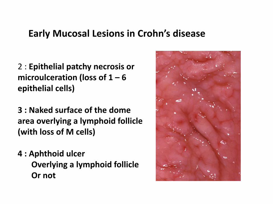
2 : Epithelial patchy necrosis ormicroulceration (loss of 1 – 6 epithelial cells)
3 : Naked surface of the domearea overlying a lymphoid follicle(with loss of M cells)
4 : Aphthoid ulcerOverlying a lymphoid follicleOr not
Early Mucosal Lesions in Crohn’s disease

Diagnostic ! lesions

Mucoid metaplasiaNot specificStatiscally most common in Crohn’sdisease

OTHER FEATURESGranulomas
- Not specific- Diagnosis of Crohn’s disease in association with
other lesion- Frequency of finding : 3 – 56% for endoscopic
samples- Highest frequenty : children

Lymphatics and Crohn’s disease
Dilated mucosal lymphatics
Increased numbersLymphagiogenesis

Active inflammation – chronic inflammation -dilated lymphatics

Active inflammation(relation with treatment)

Histopathology and relapseInflammation (early postoperative lesions)
EosinophilsEosinophilic infiltration may occur in theneoterminal ileum within a few weeks ofresection.Rutgeerts et al Gut 1984; 25: 665Mucosal expression of interleukin 5 (IL-5) animportant eosinophilic activating factor isincreased (in association with prominenteosinophilic infiltration) in early recurrence.Dubucquoi et al Gut 1995; 37: 242

Hypercrinia – Mucin preservation and relapse
Ileum – Distinctive mucosal featuresIncreased proportion of goblet cells within theepithelium (Segal & Petras, in : Histology for Pathologists, 1992, p547-)
Ratio Goblet cells/absorptive enterocytes1/1
HypercriniaIncreased number of goblet cells
Endoscopic recurrence18/22 endoscopic recurrence / 55.6% hypercrinia
5 pts ratio goblet cells/enterocytes > 50%5 pts ratio > 75%
10/12 recurrence / 60% hypercrinia31/37 recurrence / 67.7% hypercrinia

Terminal Ileitis & Ulcerative colitisBackwash ileitis ?
Definition (historical)
- Backwash : reflux of contents due to inflammation-inducedmalfunction of ileocecalvalve
- Associated withpancolitis
Terminal ileitis in UC withmildly active disease!?
But

Terminal - Backwash ileitisGoldstein & Dulsi Am J Clin Pathol 2006; 126:365
• Ileal lesions in continuitywith colonic lesions
• Histology
– Diffuse inflammation
– Regular shortening of villi
• Correlation with extent of disease
• Disease activity correlateswith level of cecal disease
• Frequency decreases
• Pathogenesis?
– Terminology dates frombarium enemas, whenileocecal valve was opened
– Primary manifestation of the disease (would explainterminal ileitis in patients with mildly active disease

DIFFERENTIAL DIAGNOSTIC ISSUES
Other infectionsSelf-limited infections
Viral gastroenteritis occurs especially in the pediatric agegroup.
Bacterial pathogens are Shigella, Salmonella, Campylobacter, Yersinia, Escherichia coli, Clostridium difficile
Chronic infectionsMycobacteria
Mimics of IBDNSAIDSOther DRUGSIleitis and spondylarthropatyTumor associated lesions
PrimaryMetastatic

GranulomasNot specificDiagnosis of Crohn’s disease in association with other lesion
Yersinia


Tuberculosis

MIMICS OF IBDNSAIDS
OTHER DRUGSILEITIS AND SPONDYLARTHROPATYTUMOR ASSOCIATED LESIONS
PRIMARYMETASTATIC
VASCULITIS
NSAIDs
Clinical history
Tumor associated lesions
Patients are usually older
Spondylarthropathy
Associated lesions

Other Drugs
• Olmesartan medoxomil, an angiotensin II receptor antagonist – 25 cases out of a series of 12.935 or 0.19%. – Lesions are observed usually one to two years after the start of
the medication. – Woman are slightly more affected. – Increased collagen deposition can be noted.
• Ipilimumab, a humanized monoclonal antibody developed to reduce and overcome cytotoxic T-lymphocyte antigen 4 (iatrogenic autoimmune enteropathy)
• Imatinib mesylate (treatment of GIST)• Mycophenolate mofetil (MMF)

Olmesartan

INFLAMMATION & SPONDYLARTHROPATHY
Histopathology of intestinal inflammation relatedto reactive arthritis Cuvelier e.a. Gut 1987
65% reactive arthritis; 57% ankylosing spondylitis(n = 232)
Long-term evolution of gut inflammation in patients with spondylarthropathy De Vos e.a. Gastroenterology 1996
Evolution towards CD : 7% (n = 49)

Female patient°1944
Clinical HistoryStenosis of a renal arteryand the celiac trunkArterial hypertensionMigraineTreatment : Cafergot, omeprazole, tiberal, plavix(clodipogrel)Current complaints : headache and diarrhea
Endoscopy : Ischemia? > normal aspect

Microscopic colitisHistology – Small Intestine
Duodenal abormalitiesin up to 70% (7% antiendomysialantibodies)Ileal abnormalities in up to 15%Primary Ileal villousatrophy

Endometriosis

Crohn’s disease and endometriosisCraninx e.a. Eur J Gastroenterol Hepatol 2000; 12: 217
• In Crohn’s diseaseendometriosis of the terminal ileum seemsmore common
• Endometriosis canmimic Crohn’s disease
• Endometriosis canoccur simultaneously
• 8 female pts : surgeryfor Crohn’s disease of terminal ileum (n=7) or colon (n=1)
• Intestinal endometriosisof the ileum (n=6); colon (n=2)

Miscellaneous
Particularly in macrophages associatedwith Peyer’s patches (situated in the base) in the small intestineIn stromaAppearance : dark brown or black (pigment rich in aluminium, silicon and titanium)Frequency 34/42 (over 6 yrs of age) (Shepherd e a Hum Pathol 1987; 18: 50)Sampling through M cellsPowell e.a. Gut 1996; 38: 390
Athmospheric/food additives dust

MiscellaneousIleum – Deposition of iron

Miscellaneous
Bile pigment(ileum)

MiscellaneousWaldenström’s MacroglobulinemiaStaining for kappa light chain
Systemic mastocytosis

Behçet diseaseCan involve Ileum (& colon)
Crohn
Behçet- Ulcers on opposite side of the mesentery- Unrelated to the site of M-cells orlymphoid tissue- Changes limited to mucosa adjacent to ulcers
Endoscopic biopsiescan hardly
differentiate

Ulceration in small bowel
• Isolated non-specific ulcer– Rare (40/100,000 pts)– Mainly ileum (at 100 cm of the valve)– Male preponderance
• Cryptogenic multifocal ulcerous stenosing enteritis (CMUSE)
• Idiopathic chronic ulcerative enteritis (ICUE)• Chronic non-specific multiple ulcers of the intestine
– Four candidate mutations in the solute carrier organic anion transporter family, member 2A1 (SLCO2A1) gene, encoding a prostaglandin transporter, were identified (Hosoe e.a. J Crohn Colitis 2017)

Conclusions
• Ileal biopsies can provide information in patients withendoscopic features of ileitis and clinical symptoms of (inflammatory diarrhea)
• Isolated active ileitis is not always Crohn’s disease
• So-called backwash ileitis is not yet well understood
• Various conditions may induce either isolated ileitis or ileitis in association with colitis
• These include infections, drugs but also less commonconditions (mass lesions…)

Iatrogenic autoimmune-like enteritisPathophysioloy
• CTLA-4 is expressed on regulatory T cells and patients receiving treatment with anti-CTLA-4 show abnormal numbers of regulatory T cells in intestinal biopsies
• Enterocytes can express MHC class II (like normal antigen presenting cells) but lack expression of the costimulatorymolecules (CD80 and CD86) needed to activate naïve T-cell– Yet, under certain conditions, they can express other
costimulatory markers such as PD-L1 (programmed cell death)– Blockade of PD-L1, the ligand of PD-1, leads to the development
of autoimmune enteritis.
• AIE-like enteritis can develop following severe depletion of gut microbiota from antibiotic therapy, consistent with the idea that commensal microorganisms play an important role in regulating gut immunity