full paper
TRANSCRIPT
Polyhedron 117 (2016) 404–414
Contents lists available at ScienceDirect
Polyhedron
journal homepage: www.elsevier .com/locate /poly
Synthesis, structural, DFT calculations and biological studies of rhodiumand iridium complexes containing azine Schiff-base ligands
http://dx.doi.org/10.1016/j.poly.2016.06.0010277-5387/� 2016 Elsevier Ltd. All rights reserved.
⇑ Corresponding author. Tel.: +91 364 2722620; fax: +91 364 2550076.E-mail address: [email protected] (K.M. Rao).
Sanjay Adhikari a, Dipankar Sutradhar a, Samantha L. Shepherd b, Roger M. Phillips b, Asit K. Chandra a,K. Mohan Rao a,⇑aCentre for Advanced Studies in Chemistry, North-Eastern Hill University, Shillong 793 022, IndiabDepartment of Pharmacy, School of Applied Sciences, University of Huddersfield, Huddersfield HD1 3DH, UK
a r t i c l e i n f o
Article history:Received 4 May 2016Accepted 1 June 2016Available online 5 June 2016
Keywords:RhodiumIridiumAzinesCytotoxicityTDDFT
a b s t r a c t
The reaction of [Cp*MCl2]2 (M = Rh/Ir) with N–N0 azine Schiff-base ligands (L1–L4) leads to the formationof mononuclear cationic half-sandwich complexes having the general formula [Cp*M(L)Cl]+ (1–8),(M = Rh/Ir and L = (2-hydroxy-4-methoxybenzylidene)2-pyridylamidrazone (L1), (2-hydroxybenzyli-dene)2-pyridylamidrazone (L2), (1-(2-hydroxyphenyl)ethylidene)2-pyridylamidrazone (L3) and(1-phenylethylidene)2-pyridylamidrazone (L4). All these complexes were isolated as their hexafluo-rophosphate salts and fully characterized by spectroscopic and analytical techniques. The molecularstructure of complexes (1), (3), (4), (7) and (8) have been determined by single crystal X-ray crystallo-graphic studies which displayed the coordination of the ligand to the metal in a bidentate N\N fashionthrough nitrogen atom of pyridine and one azine nitrogen. The chemo-sensitivity activities of the complexeswere evaluated against HT-29 (human colorectal cancer) cell line and non-cancer cell line ARPE-19 (humanretinal epithelial cells) which revealed that the complexes are moderately cytotoxic to cancer cells overhuman cells although complex 5was the most potent among all the compounds. Theoretical studies carriedout using DFT and TD-DFT at B3LYP level shows good agreement with the experimental results.
� 2016 Elsevier Ltd. All rights reserved.
1. Introduction
The chemistry of half-sandwich organometallic complexes hasevolved as a versatile subject of research during the past few dec-ades due to its wide application in biological and medicinal fields[1–4]. Organometallic half-sandwich compounds of the generalformula [Cp*MCl(LL0)] (M = Rh, Ir and LL0 = N,N or N,O donorligands) have been extensively studied for their cytostatic activity,DNA binding, cellular uptake and as DNA intercelators [5–9]. Rho-dium and iridium complexes have also been investigated as analternative to platinum based drugs mainly because of their watersolubility and lability towards ligand exchange [10,11]. RecentlyTherrien et al reported dinuclear dithiolato bridged rhodium andiridium complexes which exhibit cytotoxicity against human ovar-ian cancer cells lines (A2780 and A2780cisR) [12]. CAH activatedcyclometalated Rh(III) and Ir(III) complexes can effectively bindto DNA and protein through electrostatic and hydrophobic interac-tions [13]. Iridium complexes of dihydroxybipyridine are activecatalysts for homogenous water oxidation under mild reaction
conditions [14]. Rh(III) and Ir(III) polypyridyl complexes exhibitsstrong antiproliferative activity towards human cancer cell linesand are also capable of binding to DNA [15]. A number of half-sandwich Ir(III) complexes have been reported by Sadler et al. withchelating C, N and pyridine ligands and N, N donor ligands whichshowed strong antiproliferative activity [16,17].
Pyridyl azines represent an important class of organic com-pounds with interesting properties having wide applications invarious areas [18]. Open chain diazine Schiff base ligands linkedby a single N–N bond are of great interest due to its rotational flex-ibility around the N–N bond and potential donor sites which cangive rise to a rich variety of coordination compounds with differentbinding modes [19]. The N–N bridging ligand plays a crucial role incommunicating the metal centers to form mononuclear, dinuclearor polynuclear complexes [20]. The diazine ligand has beenemployed into several transition metal azido and thiocyanato sys-tems namely Mn(II)-azido, Cd(II)-NCS to obtain several 1D, 2D and3D polymers which exhibit interesting magnetic properties[21,22]. Dinuclear transition metal complexes of Cu, Zn, Mn andNi have been reported with bridging N–N diazine ligandswhich give rise to strong ferromagnetic and antiferromagneticcoupling [23]. In the recent years our group has reported many

S. Adhikari et al. / Polyhedron 117 (2016) 404–414 405
half-sandwich Ru(II), Rh(III) and Ir(III) complexes with azineligands [24,25]. In continuation with our interest of these ligandsherein we report four new azine Schiff base ligands derived from2-pyridylamidrazone and its corresponding rhodium and iridiumhalf-sandwich metal complexes. The complexes were tested fortheir cytotoxic property to selectively kill HT-29 cancer cell lineagainst normal ARPE-19 cells.
2. Experimental
2.1. Physical methods and materials
All the reagents were purchased from commercial sources andused as received. Starting materials RhCl3�nH2O, IrCl3�nH2O werepurchased from Arora Matthey limited. 2-cyanopyridine,2-hydroxybenzaldehyde, 2-hydroxyacetophenone, were obtainedfrom Aldrich, acetophenone and 2-hydroxy-4-methoxybenzalde-hyde were obtained from Alfa-Aesar. The solvents were purifiedand dried according to standard procedures [26]. All the reactionswere carried out under normal conditions. The starting precursormetal complexes [Cp*MCl2]2 (M = Rh/Ir) were prepared accordingto the literature methods [27]. Infrared spectra were recorded ona Perkin-Elmer 983 spectrophotometer by using KBr pellets inthe range of 400–4000 cm�1. 1H NMR spectra were recorded on aBruker Avance II 400 MHz spectrometer using DMSO-d6 and CDCl3as solvents. Absorption spectra were recorded on a Perkin-ElmerLambda 25 UV–Vis spectrophotometer in the range of 200–800 nm at room temperature in acetonitrile. Elemental analysesof the complexes were performed on a Perkin-Elmer 2400 CHN/Sanalyzer. Mass spectra were recorded using Q-Tof APCI-MS instru-ment (model HAB 273). All these mononuclear metal complexeswere synthesized and characterized by using FT-IR, 1H NMR, UV–Vis, and Single-crystal X-ray diffraction techniques.
2.2. Single-crystal X-ray structures analyses
The orange crystals of complexes (1), (3), (7) and (8) wereobtained by slow diffusion of hexane into acetone or DCM solutionand yellow crystals of complex (4) was obtained by diffusing hex-ane into DCM solution. Single crystal X-ray diffraction data for allthe complexes (1), (3) (4), (7) and (8) were collected on a OxfordDiffraction Xcalibur Eos Gemini diffractometer at 293 K using gra-phite monochromated Mo Ka radiation (k = 0.71073 Å). The strat-egy for the data collection was evaluated using the CrysAlisProCCD software. Crystal data were collected by standard ‘‘phi–omegascan” techniques and were scaled and reduced using CrysAlisProRED software. The structures were solved by direct methods using
SHELXS-97 and refined by full-matrix least squares with SHELXL-97refining on F2 [28,29]. The positions of all the atoms were obtainedby direct methods. Metal atoms in the complex were located fromthe E-maps and non-hydrogen atoms were refined anisotropically.The hydrogen atoms bound to the carbon were placed in geomet-rically constrained positions and refined with isotropic tempera-ture factors, generally 1.2 Ueq of their parent atoms.Crystallographic and structure refinement details for the com-plexes are summarized in Table 1, and selected bond lengths andbond angles are presented in Table S1. Figs. 1–3 were drawn with
ORTEP3 program. Fig. 4 and Figs. S3–S6 were drawn with MER-CURY3.6 program [30].
2.3. Biological studies
All complexes (1–8) were dissolved in DMSO at 100 mM andstored at �20 �C until needed. The complexes were testedagainst cancer cell line HT-29 (human colorectal cancer), and one
non-cancer cell line ARPE-19 (human retinal epithelial cells). Cellswere seeded into 96 well plates at 1 � 103 cells per well andincubated at 37 �C in a CO2 enriched (5%), humidified atmosphereovernight to adhere. The cells were exposed to a range of drugconcentrations in the range of 0–100 lM for four days before cellsurvival was determined using the MTT assay [31]. To each wellMTT (0.5 mg/ml) was added and was further incubated at 37 �Cfor 4 h. After this the MTT was removed from each well and theformazan crystals formed were dissolved in 150 lM DMSO. Theabsorbance of the resulting solution was recorded at 550 nm usingan ELISA spectrophotometer. The percentage of cell inhibition wascalculated by dividing the absorbance of treated cell by the controlvalue absorbance (exposed to 0.1% DMSO). The results wereexpressed in terms of IC50 values (concentration required to kill50% cell) and all studies were performed in triplicate. The resultswere also expressed in terms of a ‘selectivity index’ defined as theIC50 of the non-cancer cell line ARPE divided by the IC50 of cancercell lines [32]. Values greater than 1 demonstrate that the com-pound is preferentially active against tumor cell compared to nor-mal cell lines.
2.4. Computational methodology
All the electronic structure calculations of the metal complexes(1–8) were carried out using the Gaussian 09 suite of program [33].The geometries of the rhodium and iridium complexes were opti-mized in the gas phase employing the DFT-based B3LYP methodwith 6-31G⁄⁄ basis set for (H, C, N, O, Cl, F and P atoms andLANL2DZ [34,35] for (Rh and Ir) atoms. Harmonic frequency calcu-lations were carried out at the same level of theory to ensure thatthe optimized geometries were true minima on the potentialenergy surface (PES). Natural Bond Orbital (NBO) analysis [36]was used to obtain the charge distribution on individual atomsand the d-orbital occupations of the metal present in the com-plexes. Time dependent-Density Functional Theory (TD-DFT) [37]has been employed to evaluate the absorption spectra and theelectronic transitions of the metal complexes. In order to incorpo-rate the effect of the solvent around the molecule, the PolarizableContinuum Model (PCM) [38] was used in TD-DFT calculations.The percentage contribution of molecular orbital analysis was car-ried out using Chemissian software package [39].
2.5. General procedure for preparation of ligands 1–4
2.5.1. The azine Schiff base ligands (L1–L4) were prepared by two stepprocedure
In the first step 2-pyridylamidrazone was prepared, by follow-ing a reported procedure [40]. 2-cyanopyridine and hydrazinehydrate were dissolved and stirred in absolute ethanol overnightto give 2-pyridylamidrazone as yellow crystalline solid whichwas used in the next step without further purification (Scheme 1).In the second step (5 mmol) of aldehyde or ketone and 2-pyridy-lamidrazone (5 mmol) was refluxed in 10 ml ethanol for 5 h(Scheme 2). The products obtained after cooling the solution werefiltered off washed with cold methanol and diethyl ether and driedin vacuum.
2.5.2. (2-hydroxy-4-methoxybenzylidene)2-pyridylamidrazone (L1)Color: Yellow needles; Yield: 88%; IR (KBr, cm�1): 3487(s), 3380
(s), 3333(m), 2964(m), 1627(s), 1587(m), 1566(m), 1394(m), 1340(s); 1H NMR (400 MHz, CDCl3): d = 11.82 (s, 1H, OH), 8.60 (s, 1H,CH(imine)), 8.57 (d, 1H, J = 4.0 Hz, CH(py)), 8.34 (d, 1H, J = 8.0 Hz,CH(py)), 7.76 (t, 1H, CH(py)), 7.35 (t, 1H, CH(py)), 7.20 (d, 2H,J = 8.0 Hz, CH(Ar)), 6.46–6.50 (m, 3H, NH2, CH(Ar)), 3.80 (s, 3H,OMe); HRMS-APCI (m/z): 271.11 [M+H]+; UV–Vis {Acetonitrile,kmax, nm (e/10�4 M�1 cm�1)}: 218 (0.84), 314 (0.68), 342 (0.92),

Table 1Crystal structure data and refinement parameters of complexes.
Complexes [1] PF6 [3] PF6 [4] PF6 [7] PF6 [8] PF6 Cl
Empirical formula C24H29ClN4O2F6PRh C23H27ClF6N4OPRh C23H27ClF6N4OPIr C24H29ClF6N4PRh C24H29Cl2F6N4PIrFormula weight 688.84 658.82 748.11 656.84 781.58T (K) 298(2) 293(2) 293(2) 293(2) 293(2)k (Å) 0.71073 0.71073 0.71073 0.71073 0.71073Crystal system triclinic monoclinic monoclinic monoclinic triclinicSpace group P�1 P21/c P21/c P21/c P�1a (Å)/a (�) 8.3893(7)/89.370(6) 10.6710(6)/90 10.7019(5)/90 38.850(5)/90 7.9976(4)/87.496b (Å)/b (�) 10.5533(7)/86.439(6) 17.0730(8)/92.708(4). 17.0860(9)/93.062(4) 7.9488(5)/98.027(4) 12.4774(4)/82.086(4)c (Å)/c (�) 16.6554(11)/71.182(7) 14.5390(8)/90 14.6118(9)/90 28.562(4)/90 14.6442(6)/72.596(4)V (Å3) 1393.00(18) 2645.8(2) 2668.0(2) 1344.83(10) 1381.15(10)Z 2 4 4 2 2Dcalc (Mg m�3) 1.642 1.654 1.862 1.622 1.879Absorption coefficient (l)
(mm�1)0.836 0.874 5.231 0.857 5.148
F(000) 696 1328 1456 664 762Crystal size (mm3) 0.23 � 0.21 � 0.21 0.21 � 0.19 � 0.04 0.23 � 0.23 � 0.21 0.22 � 0.20 � 0.120 0.19 � 0.12 � 0.09h range for data collection
(�)3.174–28.654 3.33–26.73 3.31–26.37 3.386–28.842 3.23–26.37
Index ranges �11 6 h 6 10,�12 6 k 6 13,�22 6 l 6 20
�13 6 h 6 10,�10 6 k 6 21,�12 6 l 6 18
�13 6 h 6 7,�21 6 k 6 19,�16 6 l 6 18
�9 6 h 6 9,�12 6 k 6 22,�14 6 l < 8
�8 6 h 6 9,�15 6 i 6 15,�17 6 l < 18
Reflections collected 10811 9506 10081 5614 7889Independent reflections 6286 [Rint = 0.0717] 5375 [Rint = 0.0268] 5422 [Rint = 0.0277] 4000 [Rint = 0.0268] 5335 [Rint = 0.0296]Completeness to h = 25.00� 99.57% 99.5% 99.2% 99.2% 94.8%Absorption correction semi-empirical from
equivalentssemi-empirical fromequivalents
semi-empirical fromequivalents
semi-empirical fromequivalents
semi-empirical fromequivalents
Refinement method full-matrix least-squareson F2
full-matrix least-squareson F2
full-matrix least-squareson F2
full-matrix least-squares on F2
full-matrix least-squares on F2
Data/restraints/parameters
6286/0/362 2375/0/330 5422/0/340 4000/1/340 5335/0/349
Goodness-of-fit (GOF) onF2
1.197 1.063 1.026 1.041 1.046
Final R indices [I > 2r(I)] R1 = 0.0703, wR2 = 0.1706 R1 = 0.0440, wR2 = 0.0895 R1 = 0.0340, wR2 = 0.0630 R1 = 0.0394,wR2 = 0.0822
R1 = 0.0368,wR2 = 0.0875
R indices (all data) R1 = 0.0855, wR2 = 0.1772 R1 = 0.0592, wR2 = 0.0968 R1 = 0.0500, wR2 = 0.0683 R1 = 0.0462,wR2 = 0.0858
R1 = 0.0431,wR2 = 0.0912
Largest difference in peakand hole (e �3)
0.583 and �0.461 0.520 and �0.543 1.102 and �1.143 0.512 and �0.478 1.828 and �1.071
CCDC No. 1477976 1477977 1477978 1477979 1477980
Structures were refined on F02: wR2 = [R[w(F02 � Fc
2)2]/Rw(F02)2]1/2, where w�1 = [R(F02)+(aP)2 + bP] and P = [max(F02, 0) + 2Fc2]/3.
Fig. 1. ORTEP diagram of complex [Cp*RhCl(L1)Cl]PF6 (1) with 50% probability thermal ellipsoids. Hydrogen atoms and counter ions are omitted for clarity.
406 S. Adhikari et al. / Polyhedron 117 (2016) 404–414
355 (0.94); Anal. Calc. for C14H14N4O2 (270.29): C, 62.21; H, 5.22; N,20.73. Found: C, 62.36; H, 5.35; N, 20.86%.
2.5.3. (2-hydroxybenzylidene)2-pyridylamidrazone (L2)Color: Yellow needles; Yield: 92%; IR (KBr, cm�1): 3477(s), 3363
(s), 3340(s), 3043(m), 1626(s), 1576(m), 1567(m), 1473(m), 1337
(m); 1H NMR (400 MHz, CDCl3): d = 11.61 (s, 1H, OH), 8.59 (s, 1H,CH(imine)), 8.54 (d, 1H, J = 4.0 Hz, CH(py)), 8.28 (d, 1H, J = 8.0 Hz,CH(py)), 7.74 (t, 1H, CH(py)), 7.33 (t, 1H, CH(py)), 7.24–7.27 (m, 3H,NH2, CH(Ar)), 6.96 (d, 1H, J = 8.0 Hz, CH(Ar)), 6.87 (t, 2H, CH(Ar));HRMS-APCI (m/z): 241.10 [M+H]+; UV–Vis {Acetonitrile, kmax, nm(e/10�4 M�1 cm�1)}: 219 (0.84), 247 (0.55), 349 (1.30), 361
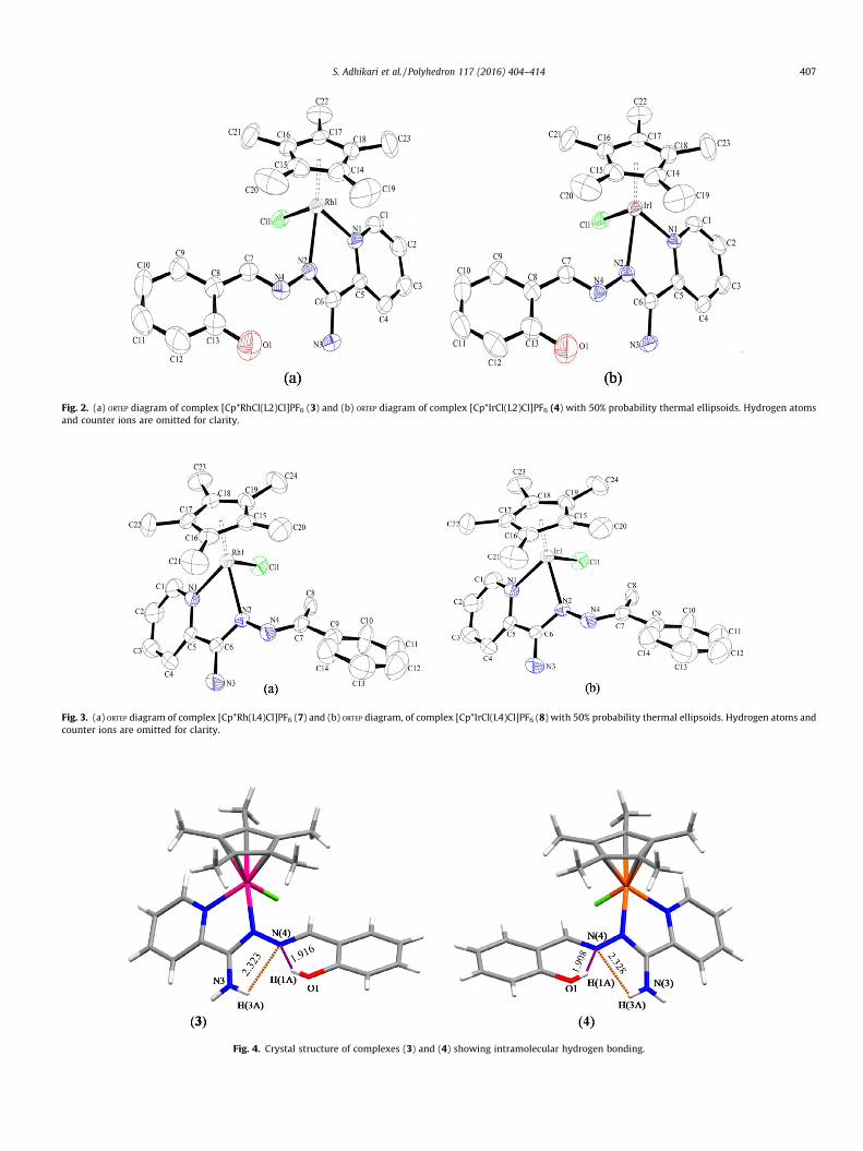
Fig. 2. (a) ORTEP diagram of complex [Cp*RhCl(L2)Cl]PF6 (3) and (b) ORTEP diagram of complex [Cp*IrCl(L2)Cl]PF6 (4) with 50% probability thermal ellipsoids. Hydrogen atomsand counter ions are omitted for clarity.
Fig. 3. (a) ORTEP diagram of complex [Cp*Rh(L4)Cl]PF6 (7) and (b) ORTEP diagram, of complex [Cp*IrCl(L4)Cl]PF6 (8) with 50% probability thermal ellipsoids. Hydrogen atoms andcounter ions are omitted for clarity.
Fig. 4. Crystal structure of complexes (3) and (4) showing intramolecular hydrogen bonding.
S. Adhikari et al. / Polyhedron 117 (2016) 404–414 407

Scheme 1. Synthesis of 2-pyridylamidrazone.
408 S. Adhikari et al. / Polyhedron 117 (2016) 404–414
(1.29); Anal. Calc. for C13H12N4O (240.26): C, 64.99; H, 5.03; N,23.32. Found: C, 65.12; H, 5.18; N, 23.44%.
2.5.4. (1-(2-hydroxyphenyl)ethylidene)2-pyridylamidrazone (L3)Color: Yellow crystalline solid; Yield: 95%; IR (KBr, cm�1): 3482
(s), 3339(s), 3056(m), 3003(m), 1615(s), 1562(m), 1507(m), 1300(m); 1H NMR (400 MHz, CDCl3): d = 13.73 (s, 1H, OH), 8.59 (d,1H, J = 4.0 Hz, CH(py)), 8.36 (d, 1H, J = 8.0 Hz, CH(py)), 7.79 (t, 1H,CH(py)), 7.58 (t, 1H, CH(py)), 7.21–7.28 (m, 3H, NH2, CH(Ar)), 6.98(d, 2H, J = 8.0 Hz, CH(Ar)), 6.89 (t, 1H, CH(Ar)), 2.62 (s, 3H, CH3);HRMS-APCI (m/z): 255.12 [M+H]+; UV–Vis {Acetonitrile, kmax, nm(e/10�4 M�1 cm�1)}: 217 (1.21), 303 (0.74), 344 (0.95); Anal. Calc.for C14H14N4O (254.29): C, 66.13; H, 5.55; N, 22.03. Found: C,66.25; H, 5.68; N, 22.21%.
2.5.5. (1-phenylethylidene)2-pyridylamidrazone (L4)Color: Yellow crystalline solid; Yield: 92%; IR (KBr, cm�1): 3450
(s), 3331(s), 3056(m), 3009(m), 1604(s), 1568(m), 1445(m), 1362(m); 1H NMR (400 MHz, CDCl3): d = 8.58 (d, 1H, J = 4.0 Hz, CH(py)),8.23 (d, 1H, J = 8.0 Hz, CH(py)), 7.71 (t, 1H, CH(py)), 7.30 (t, 1H,CH(py)), 7.21–7.28 (m, 3H, NH2, CH(Ar)), 6.93 (m, 3H, CH(Ar)), 6.89(t, 1H, CH(Ar)), 2.39 (s, 3H, CH3); HRMS-APCI (m/z): 239.13 [M+H]+; UV–Vis {Acetonitrile, kmax, nm (e/10�4 M�1 cm�1)}: 225(0.21), 327 (0.29); Anal. Calc. for C14H14N4 (238.29): C, 70.57; H,5.92; N, 23.51. Found: C, 70.72; H, 6.03; N, 23.62%.
2.6. General procedure for preparation of metal complexes (1–8)
Amixture of metal precursor [Cp*MCl2]2 (M = Rh/Ir) (0.1 mmol),azine Schiff-base ligands (L1-L4) (0.2 mmol) and 2.5 equivalents ofNH4PF6 in dry methanol (10 ml) was stirred at room temperaturefor 8 h (Scheme 3). The solvent was evaporated under reducedpressure, and the residue was dissolved in dichloromethane andfiltered over celite to remove excess salt. The filtrate was reducedto 2 ml and diethyl ether was added to induce precipitation. Theyellow colored precipitate, which formed, was filtered and washedwith diethyl ether and dried in vacuum.
2.6.1. [Cp*Rh(L1)Cl]PF6 (1)Yield: 56 mg (40%); IR (KBr, cm�1): 3460(m), 3237(m), 2926(w),
1630(s), 1595(m), 1296(m), 846(s); 1H NMR (400 MHz, CDCl3):d = 10.5 (s, 1H, OH), 9.02 (s, 1H, CH(imine)), 8.76 (d, 1H, J = 4.0 Hz,CH(py)), 8.54 (d, 1H, J = 4.0 Hz, CH(py)), 8.13 (t, 1H, CH(py)), 7.76 (t,
Scheme 2. Preparation
1H, CH(py)), 7.41 (d, 1H, J = 8.0 Hz, CH(Ar)), 7.38 (s, 2H, NH2), 6.53(d, 1H, J = 8.0 Hz, CH(Ar)), 6.50 (s, 1H, CH(Ar)), 3.81 (s, 3H, OMe),1.58 (s, 15H, CH(Cp*)); HRMS-APCI (m/z): 507.12 [M�PF6�HCl]+;UV–Vis {Acetonitrile, kmax, nm (e/10�4 M�1 cm�1)}: 233 (0.98),277 (0.57), 352 (0.42); Anal. Calc. for C24H29ClF6N4O2PRh(688.84): C, 41.85; H, 4.24; N, 8.13. Found: C, 41.96; H, 4.16; N,8.23%.
2.6.2. [Cp*Ir(L1)Cl]PF6 (2)Yield: 70 mg (45%); IR (KBr, cm�1): 3447(m), 3241(m), 2925(m),
1630(s), 1610(m), 1293(m), 846(s); 1H NMR (400 MHz, CDCl3):d = 10.4 (s, 1H, OH), 9.02 (s, 1H, CH(imine)), 8.77 (d, 1H, J = 4.0 Hz,CH(py)), 8.51 (d, 1H, J = 4.0 Hz, CH(py)), 8.17 (t, 1H, CH(py)), 7.78 (t,1H, CH(py)), 7.42 (d, 1H, J = 8.0 Hz, CH(Ar)), 7.39 (s, 2H, NH2), 6.56(d, 1H, J = 8.0 Hz, CH(Ar)), 6.54 (s, 1H, CH(Ar)), 3.87 (s, 3H, OMe),1.62 (s, 15H, CH(Cp*)); HRMS-APCI (m/z): 597.18 [M�PF6�HCl]+;UV–Vis {Acetonitrile, kmax, nm (e/10�4 M�1 cm�1)}: 266 (0.36),347 (0.29); Anal. Calc. for C24H29ClF6N4O2PIr (778.14): C, 37.04;H, 3.76; N, 7.20. Found: C, 37.19; H, 3.89; N, 7.31%.
2.6.3. [Cp*Rh(L2)Cl]PF6 (3)Yield: 52 mg (39%); IR (KBr, cm�1): 3422(m), 3310(w), 2923(w),
1636(s), 1603(m), 1457(m), 845(s); 1H NMR (400 MHz, CDCl3):d = 10.1 (s, 1H, OH), 9.11 (s, 1H, CH(imine)), 8.78 (d, 1H, J = 4.0 Hz,CH(py)), 8.49 (d, 1H, J = 8.0 Hz, CH(py)), 8.14 (t, 2H, CH(py)), 7.78 (t,1H, CH(Ar)), 7.60 (d, 1H, J = 8.0 Hz, CH(Ar)), 7.38 (s, 2H, NH2), 6.92–7.01 (m, 2H, CH(Ar)), 1.58 (s, 15H, CH(Cp*)); HRMS-APCI (m/z):477.12 [M�PF6�HCl]+; UV–Vis {Acetonitrile, kmax, nm(e/10�4 M�1 cm�1)}: 235 (1.55), 283 (0.79), 348 (1.00); Anal. Calc.for C23H27ClF6N4OPRh (658.81): C, 41.93; H, 4.13; N, 8.50. Found:C, 42.08; H, 4.25; N, 8.68%.
2.6.4. [Cp*Ir(L2)Cl]PF6 (4)Yield: 52 mg (34%); IR (KBr, cm�1): 3479(s), 3329(s), 2924(w),
1642(s), 1618(m), 1602(m), 842(s); 1H NMR (400 MHz, CDCl3):d = 10.1 (s, 1H, OH), 9.13 (s, 1H, CH(imine)), 8.80 (d, 1H, J = 4.0 Hz,CH(py)), 8.56 (d, 1H, J = 8.0 Hz, CH(py)), 8.18 (t, 2H, CH(py)), 7.80 (t,1H, CH(Ar)), 7.64 (d, 1H, J = 8.0 Hz, CH(Ar)), 7.40 (s, 2H, NH2), 6.99–7.35 (m, 2H, CH(Ar)), 1.63 (s, 15H, CH(Cp*)); HRMS-APCI (m/z):567.17 [M�PF6�HCl]+; UV–Vis {Acetonitrile, kmax, nm(e/10�4 M�1 cm�1)}: 291 (0.62), 344 (0.78); Anal. Calc. forC23H27ClF6N4OPIr (748.12): C, 36.93; H, 3.64; N, 7.49. Found:C, 37.11; H, 3.83; N, 7.62%.
2.6.5. [(Cp*Rh(L3)Cl]PF6 (5)Yield: 58 mg (43%); IR (KBr, cm�1): 3452(s), 3318(s), 2924(m),
1648(s), 1600(m), 1566(m), 1489(m), 842(s); 1H NMR (400 MHz,DMSO-d6): d = 12.5 (s, 1H, OH), 8.96 (d, 1H, J = 4.0 Hz, CH(py)),8.33–8.38 (m, 3H, CH(py)), 7.91 (t, 1H, CH(Ar)), 7.86 (d, 1H,J = 8.0 Hz, CH(Ar)), 7.48 (t, 1H, J = 8.0 Hz, CH(Ar)), 7.01–7.06 (m, 3H,NH2, CH(Ar)), 2.48 (s, 3H, CH3), 1.59 (s, 15H, CH(Cp*)); HRMS-APCI
of ligands (L1–L4).

Scheme 3. Preparation of metal complexes (1–8).
S. Adhikari et al. / Polyhedron 117 (2016) 404–414 409
(m/z): 491.14 [M�PF6�HCl]+; UV–Vis {Acetonitrile, kmax, nm(e/10�4 M�1 cm�1)}: 229 (0.95), 268 (0.59), 332 (0.32); Anal. Calc.for C24H29ClF6N4OPRh (672.84): C, 42.84; H, 4.34; N, 8.33. Found:C, 42.98; H, 4.26; N, 8.48%.
2.6.6. [Cp*Ir(L3)Cl]PF6 (6)Yield: 65 mg (42%); IR (KBr, cm�1): 3460(m), 3237(m), 2926(w),
1630(s), 1595(m), 1296(m), 846(s), 3456(m), 3369(m), 2925(m),1649(s), 1618(m), 1598(m), 1306(m), 845(s); 1H NMR (400 MHz,DMSO-d6): d = 12.3 (s, 1H, OH), 8.94 (d, 1H, J = 4.0 Hz, CH(py)),8.44 (d, 1H, J = 4.0 Hz, CH(py)), 8.35 (t, 2H, CH(py)), 7.90 (t, 1H,CH(Ar)), 7.86 (d, 1H, J = 8.0 Hz, CH(Ar)), 7.48 (t, 1H, CH(Ar)), 7.01–7.06 (m, 3H, NH2, CH(Ar)), 2.46 (s, 3H, CH3), 1.58 (s, 15H, CH(Cp*));HRMS-APCI (m/z): 581.19 [M�PF6�HCl]+; UV–Vis {Acetonitrile,kmax, nm (e/10�4 M�1 cm�1)}: 209 (1.27), 263 (0.66), 330 (0.36);Anal. Calc. for C24H29ClF6N4OPIr (762.15): C, 37.82; H, 3.84; N,7.35. Found: C, 37.96; H, 3.96; N, 7.44%.
2.6.7. [(Cp*Rh(L4)Cl]PF6 (7)Yield: 54 mg (41%); IR (KBr, cm�1): 3441(s), 3137(m), 2961(w),
1640 (s), 1593(m), 1464(m), 841(s); 1H NMR (400 MHz, DMSO-d6):d = 8.96 (d, 1H, J = 4.0 Hz, CH(py)), 8.33–8.37 (m, 2H, CH(py)), 7.88 (t,1H, CH(py)), 7.81 (d, 1H, J = 8.0 Hz, CH(Ar)), 7.46 (t, 1H, CH(Ar)), 7.23–7.28 (m, 2H, CH(Ar)), 6.97–7.02 (m, 3H, NH2, CH(Ar)), 2.47 (s, 3H,CH3), 1.59 (s, 15H, CH(Cp*)); HRMS-APCI (m/z): 511.12 [M-PF6]+;UV–Vis {Acetonitrile, kmax, nm (e/10�4 M�1 cm�1)}: 229 (1.37),265 (0.37), 400 (0.22); Anal. Calc. for C24H29ClF6N4PRh (656.84):C, 43.89; H, 4.45; N, 8.53. Found: C, 44.02; H, 4.39; N, 8.61%.
2.6.8. [Cp*Ir(L4)Cl]PF6 (8)Yield: 65 mg (43%); IR (KBr, cm�1): 3458(s), 3383(s), 2922(m),
1643(s), 1603(m), 1567(m), 1447(m), 844(s); 1H NMR (400 MHz,DMSO-d6): d = 8.97 (d, 1H, J = 4.0 Hz, CH(py)), 8.31–8.34 (m, 2H,CH(py)), 7.85 (t, 1H, CH(py)), 7.79 (d, 1H, J = 8.0 Hz, CH(Ar)), 7.44 (t,1H, CH(Ar)), 7.19–7.23 (m, 2H, CH(Ar)), 6.99–7.03 (m, 3H, NH2,CH(Ar)), 2.46 (s, 3H, CH3), 1.59 (s, 15H, CH(Cp*)); HRMS-APCI(m/z): 601.17 [M-PF6]+; UV–Vis {Acetonitrile, kmax, nm(e/10�4 M�1 cm�1)}: 256 (0.53), 361 (0.20); Anal. Calc. forC24H29ClF6N4PIr (746.15): C, 38.63; H, 3.92; N, 7.51. Found:C, 38.74; H, 4.03; N, 7.63%.
3. Results and discussion
3.1. Synthesis of ligands and complexes
The azine Schiff-base ligands (L1–L4) were prepared by thereaction of 2-pyridylamidrazone and the respective aldehyde or
ketone in absolute ethanol medium. The complexes (1–8) weresynthesized by the reaction of Rh/Ir metal precursors with theazine Schiff-base ligands. The cationic complexes were isolatedwith PF6 counter ion. All these metal complexes were obtained ingood yields and are yellow in color. They are stable in air as wellas in solid state, and are non-hygroscopic. These complexes are sol-uble in common organic solvents such as dichloromethane, ace-tonitrile and acetone but insoluble in diethyl ether and hexane.All the synthesized ligands and complexes were fully characterizedby spectroscopic techniques.
3.2. Spectroscopic characterization of ligands
The infrared spectra of the free ligand shows characteristicstretching frequencies for NH2, OH, C@N and C@C groups. TheNH2 and OH stretching frequencies for the azine ligand appearedin the range of 3300–3500 cm�1. The C@C and C@N stretching fre-quencies were observed in the range of 1550–1626 cm�1. The pro-ton NMR spectra of the ligands displayed signals in the range of7.30–8.57 ppm assignable to the protons of the pyridine ring. Theimine protons for L1 and L2 are located at 8.60 and 8.59 ppmrespectively. The methoxy proton signal was observed as a singletfor L1 at 3.80 ppm. The methyl protons of L3 and L4 were observedas a singlet at 2.62 and 2.39 ppm respectively. The hydroxyl protonresonance for the ligands appeared in the range of 11.5–11.9 ppm.The aromatic protons of the ligand appeared as doublet, triplet andmultiplet in the range of 6.21–7.29 ppm. The [M+H]+ molecular ionpeak for the ligands are shown in the experimental section whichare found to be in good agreement with the expected range. Theelectronic spectra of the free ligands are shown in (Fig. S1). Theelectronic spectra of the free ligands show absorption bands inthe range of 210–360 nm. The band in the range of 210–250 nmcan be assigned as p–p⁄ and n–p⁄ transition. The band around300–370 nm is due to the intermolecular charge transfer transitionwithin the whole molecule [41].
3.3. Spectroscopic characterization of complexes
The IR spectra of the complexes show sharp bands around842–846 cm�1 due to the P-F stretching frequency of the counterion [42]. The OH and NH2 stretching vibrations in the complexeswere found around 3300–3500 cm�1. The retaining of the OHand NH2 stretching frequencies indicates that they are not involvedin bonding to the metal center. The strong absorption band for mC@N
around 1630–1650 cm�1 at higher wave numbers as compared tothe free ligand around 1615–1626 cm�1 suggest that the coordina-tion to the metal occurs through the imine and pyridine nitrogen.

Table 2Selected hydrogen bonding distances (Å) and angles (�) of complexes 3 and 4.
Complexes DAH� � �A H� � �A(Å)
D� � �A(Å)
D� � �H(Å)
\D–H� � �A(�)
3 O(1)AH(1A)� � �N(4)
1.916 2.638 0.820 146.39
N(3)AH(3A)� � �N(4)
2.323 2.624 0.860 100.76
4 O(1)AH(1A)� � �N(4)
1.908 2.634 0.820 146.90
N(3)AH(3A)� � �N(4)
2.328 2.629 0.860 100.78
Fig. 5. Response of HT-29 (human colorectal cancer) to compounds (1–8) andcisplatin. Cell were exposed to compounds (1–8) for 96 h. Each value represents themean ± standard deviation from three independent experiments.
410 S. Adhikari et al. / Polyhedron 117 (2016) 404–414
The proton NMR spectra of the metal complexes show that theligand resonance signals are shifted downfield as compared to thatof the free ligand. These signals are shifted downfield because ofthe ligand coordination to the metal atom. The imine proton signalwas observed in the range of 9.0–9.13 ppm for complexes (1–4).The hydroxyl proton resonance for the complexes appeared inthe range of 10.1–12.5 ppm respectively. The appearance of thehydroxyl proton signal indicates that the hydroxyl group is notinvolved in bonding to the metal atom. The pyridine ring protonsalso showed downfield signals comprising of doublet and tripletin the range of 7.75–8.96 ppm. The NH2 protons were observedas a singlet for complexes (1–4) in the range of 7.35–7.37 ppmrespectively. The methoxy proton resonance for complexes (1and 2) appeared as a singlet at 3.81 and 3.83 ppm. The aromaticproton signals for complexes appeared in the range of 6.50–7.86 ppm as doublet, triplet and multiplet. The methyl protonsignal for complexes (5–8) appeared as a singlet around2.46–2.48 ppm respectively. In addition to the signals for theligand protons, a sharp singlet was observed for all the complexesbetween 1.58 and 1.63 ppm respectively corresponding to themethyl protons of the Cp⁄ ring. In the mass spectra of the com-plexes (1–6) the peaks at m/z: 507.12, m/z: 597.18, m/z: 477.12,m/z: 567.17, m/z: 491.13 and m/z: 581.20 can be assigned as[M�PF6�HCl]+ ion peaks respectively. Whereas, the mass spectraof the complex 7 and 8 displayed molecular ion peaks at m/z:511.12 and 601.17 which corresponds to the [M-PF6]+ ion.
The electronic spectra of the complexes were recorded in ace-tonitrile at 10�4 M concentration at room temperature and the plotis shown in (Fig. S2). The electronic spectra of complexes displaytwo absorption band in the higher energy region around 210–330 nm. The bands in the higher energy UV region can be assignedas ligand centered or intra ligand p–p⁄ and n–p⁄ transition. The Rh(III) and Ir(III) complexes provides filled dp (t2g) orbitals which caninteract with low lying p⁄ orbitals (C@N) of the ligand. The band inthe lower energy region around 345–405 nm can be assigned as Rh(dp) or Ir (dp) to p⁄ ligand metal to ligand charge transfer (MLCT)transition [43].
3.4. Molecular structures of complexes
The molecular structures of some of the respective complexeshave been elucidated by single crystal X-ray analysis. Suitable sin-gle crystals were attached to a glass fiber and transferred into theOxford Diffraction Xcalibur Eos Gemini diffractometer. The crystal-lographic details and structure refinement details are summarizedin Table 1. The geometrical parameters around the metal atominvolving ring centroid are listed in Table S1. In all these complexesthe ligand is coordinated to the metal atom in a similar mannerwith N\N binding mode. Complex (1) and (8) crystallized in tri-clinic system with space group P1. Complex (8) crystallized withone PF6 and one chloride counter ion. Complex (3) and (4) crystal-lized in monoclinic system with space group P21/c whereas com-plex (7) crystallized in monoclinic system with space group P21.
All these complexes display a typical three-legged piano stoolgeometry around the metal center with coordination sites occu-pied by one chloride group, two r bonded nitrogen atoms fromchelating azine ligand and the pentamethylcyclopentadienyl(Cp⁄) ring in g5 manner. The metal atom in all these complexes issituated in a pseudo-octahedral arrangement with the azine ligandcoordinating through the pyridine and azine nitrogen atoms form-ing a five membered metallocycle. In complexes (1), (3), (4) and (7)the M-N bond length {2.088(5), 2.099(3), 2.098(4) and 2.102(4) Å}from pyridine is comparatively shorter than the azine nitrogen-metal distances {2.135(5), 2.116(3), 2.105(4) and 2.159(4) Å},which are similar to those, reported with similar complexes[24,44]. However in complex (8) the metal-nitrogen distance from
pyridine {2.102(5) Å} is comparatively larger than azine nitrogen-metal distance, which is {2.096(5) Å}. The C@N bond length ofthe coordinated nitrogen in complex (1), (3), (4) and (8) is longerthan that of the uncoordinated C@N (Table S1) which could bedue to the back bonding of electron from metal (dp) to p⁄ orbitalof the ligand. But in complex (7), a reverse pattern has beenobserved where the C@N bond length of the coordinated nitrogen{1.346(7) Å} is shorter than uncoordinated C@N {1.358(7) Å} bond.The average M-C distances are {2.159 (1), 2.1534 (3), 2.1616 (4),2.1528 (7) and 2.1726 (8) Å} while the distance between the metalto Cp⁄ centroid ring is in the range of 1.758–1.793 Å respectively.The MACl bond lengths {2.3976(15) (1), 2.4172(9) (3), 2.4190(12) (4), 2.4242(16) (7) and 2.4220(17) (8) shows no significant dif-ferences and is comparable to previously reported values (Table 1)[45–48]. The bite angle N(1)-Rh(1)-N(2) values are 75.10(19) (1),75.09(11) (3), and 75.44(17) (7) whereas in complex (4) and (8)the bite angle values are N(1)-Ir(1)-N(2) values are 74.99(14) (4)and 75.26(18) respectively which probably indicates an inwardbending of the coordinated pyridyl and azine group [49]. The bondangles N(1)-M-Cl(1) and N(2)-M-Cl(1) in complexes are compara-ble to the piano stool arrangement about the metal atom and iscomparable to reported values for closely related systems[50–52]. Further the crystal packing in complex (1) is stabilizedby weak intermolecular hydrogen bonding CAH� � �O (2.702 Å)between the hydrogen atom from methoxy group and oxygenatom of the hydroxyl group and CAH� � �Cl (2.793 Å) interactionbetween CH3 group of Cp⁄ and chloride atom (Fig. S3). These inter-actions play a significant role in the formation of supramolecularmotifs.
On the other hand in the crystal structure of complex (3) and (4)two types of intramolecular hydrogen bonding has been observed;the first one between the uncoordinated nitrogen atom of the azinelinkage with the hydrogen atom of the hydroxyl group OAH� � �N
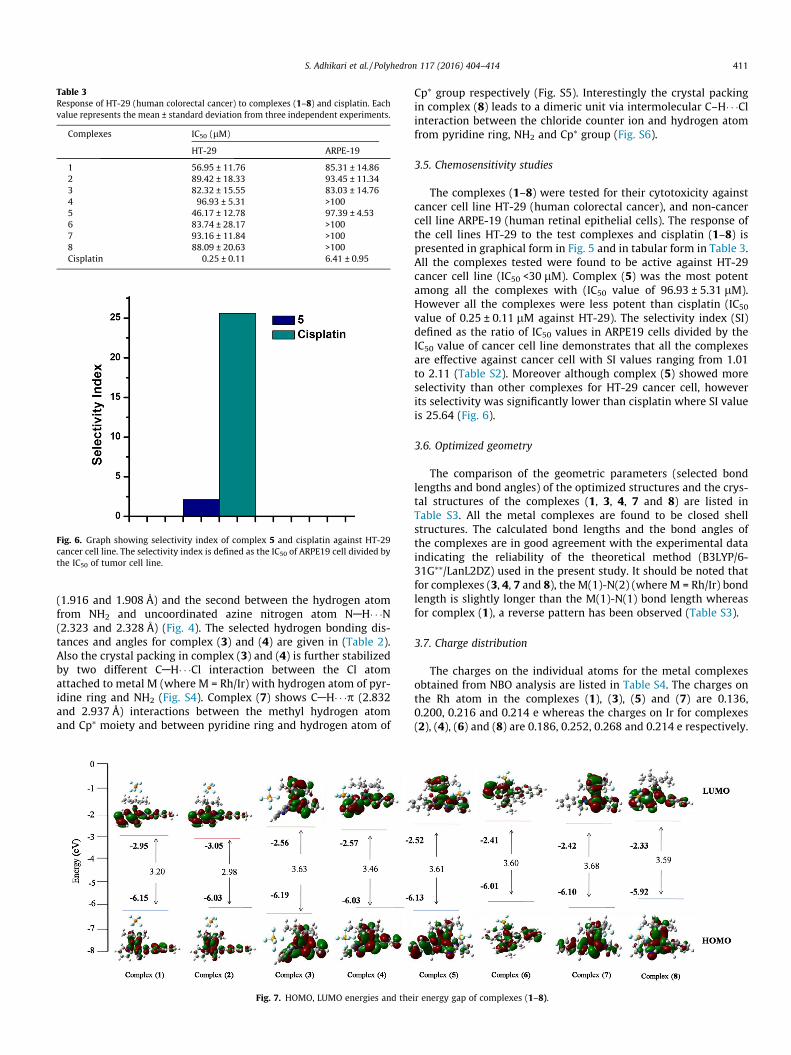
Table 3Response of HT-29 (human colorectal cancer) to complexes (1–8) and cisplatin. Eachvalue represents the mean ± standard deviation from three independent experiments.
Complexes IC50 (lM)
HT-29 ARPE-19
1 56.95 ± 11.76 85.31 ± 14.862 89.42 ± 18.33 93.45 ± 11.343 82.32 ± 15.55 83.03 ± 14.764 96.93 ± 5.31 >1005 46.17 ± 12.78 97.39 ± 4.536 83.74 ± 28.17 >1007 93.16 ± 11.84 >1008 88.09 ± 20.63 >100Cisplatin 0.25 ± 0.11 6.41 ± 0.95
Fig. 6. Graph showing selectivity index of complex 5 and cisplatin against HT-29cancer cell line. The selectivity index is defined as the IC50 of ARPE19 cell divided bythe IC50 of tumor cell line.
S. Adhikari et al. / Polyhedron 117 (2016) 404–414 411
(1.916 and 1.908 Å) and the second between the hydrogen atomfrom NH2 and uncoordinated azine nitrogen atom NAH� � �N(2.323 and 2.328 Å) (Fig. 4). The selected hydrogen bonding dis-tances and angles for complex (3) and (4) are given in (Table 2).Also the crystal packing in complex (3) and (4) is further stabilizedby two different CAH� � �Cl interaction between the Cl atomattached to metal M (where M = Rh/Ir) with hydrogen atom of pyr-idine ring and NH2 (Fig. S4). Complex (7) shows CAH� � �p (2.832and 2.937 Å) interactions between the methyl hydrogen atomand Cp⁄ moiety and between pyridine ring and hydrogen atom of
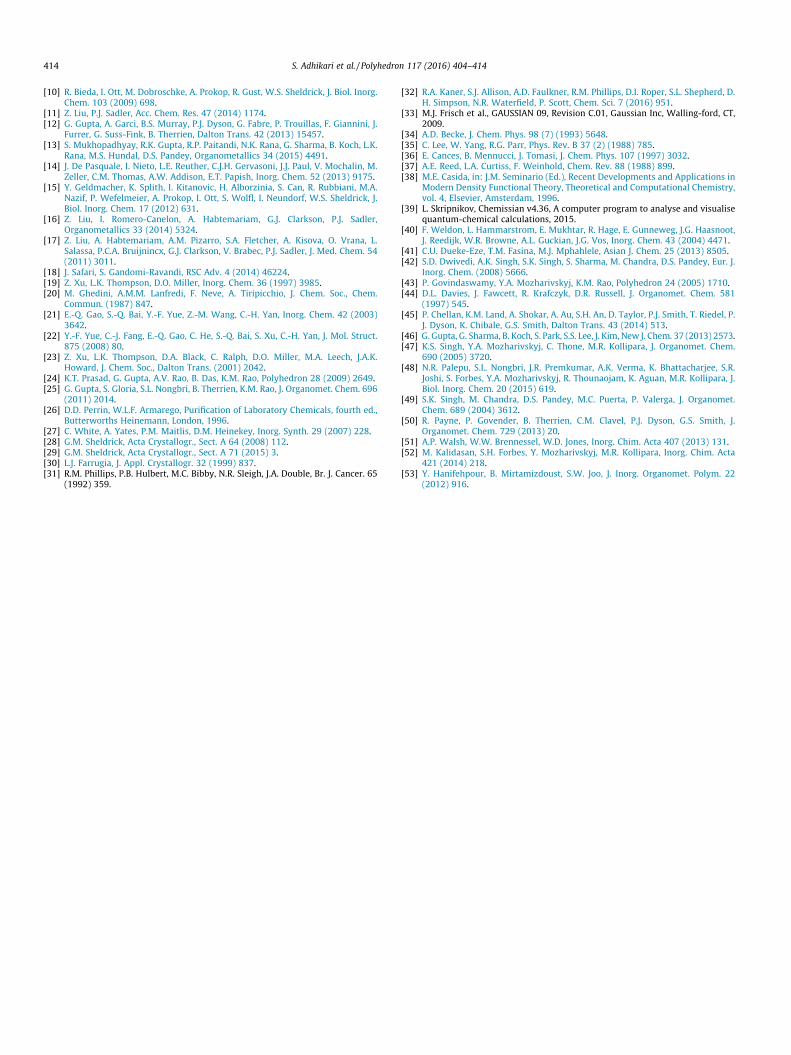
Fig. 7. HOMO, LUMO energies and the
Cp⁄ group respectively (Fig. S5). Interestingly the crystal packingin complex (8) leads to a dimeric unit via intermolecular C–H� � �Clinteraction between the chloride counter ion and hydrogen atomfrom pyridine ring, NH2 and Cp⁄ group (Fig. S6).
3.5. Chemosensitivity studies
The complexes (1–8) were tested for their cytotoxicity againstcancer cell line HT-29 (human colorectal cancer), and non-cancercell line ARPE-19 (human retinal epithelial cells). The response ofthe cell lines HT-29 to the test complexes and cisplatin (1–8) ispresented in graphical form in Fig. 5 and in tabular form in Table 3.All the complexes tested were found to be active against HT-29cancer cell line (IC50 <30 lM). Complex (5) was the most potentamong all the complexes with (IC50 value of 96.93 ± 5.31 lM).However all the complexes were less potent than cisplatin (IC50
value of 0.25 ± 0.11 lM against HT-29). The selectivity index (SI)defined as the ratio of IC50 values in ARPE19 cells divided by theIC50 value of cancer cell line demonstrates that all the complexesare effective against cancer cell with SI values ranging from 1.01to 2.11 (Table S2). Moreover although complex (5) showed moreselectivity than other complexes for HT-29 cancer cell, howeverits selectivity was significantly lower than cisplatin where SI valueis 25.64 (Fig. 6).
3.6. Optimized geometry
The comparison of the geometric parameters (selected bondlengths and bond angles) of the optimized structures and the crys-tal structures of the complexes (1, 3, 4, 7 and 8) are listed inTable S3. All the metal complexes are found to be closed shellstructures. The calculated bond lengths and the bond angles ofthe complexes are in good agreement with the experimental dataindicating the reliability of the theoretical method (B3LYP/6-31G⁄⁄/LanL2DZ) used in the present study. It should be noted thatfor complexes (3, 4, 7 and 8), the M(1)-N(2) (where M = Rh/Ir) bondlength is slightly longer than the M(1)-N(1) bond length whereasfor complex (1), a reverse pattern has been observed (Table S3).
3.7. Charge distribution
The charges on the individual atoms for the metal complexesobtained from NBO analysis are listed in Table S4. The charges onthe Rh atom in the complexes (1), (3), (5) and (7) are 0.136,0.200, 0.216 and 0.214 e whereas the charges on Ir for complexes(2), (4), (6) and (8) are 0.186, 0.252, 0.268 and 0.214 e respectively.
ir energy gap of complexes (1–8).

Table 4The energy gap, theoretical and experimental absorption bands, electronic transitions and dominant excitation character for various singlet states of the complexes (1–8)calculated with TD-DFT method.
The most important orbital excitations Calculated k (nm) Energy gap E (eV) Oscillator strength (f) Dominant excitation character Experimental k (nm)
Complex (1)H? L 417.16 3.20 0.2051 L1? L1(ILCT)H-2? L 359.64 3.53 0.0542 Cl? L1(LLCT) 352.21H? L + 2 355.41 4.03 0.0120 L1? L1(ILCT)H-4? L + 2 338.40 4.89 0.0139 L1? L1(ILCT)H-1? L + 4 278.91 4.73 0.0248 L1? L1(ILCT) 276.0H-6? L 282.81 4.54 0.0073 L1? L1(ILCT)H-6? L + 2 275.41 5.37 0.0050 L1? L1(ILCT)H-11? L 235.62 5.08 0.0216 Rh? L1(MLCT) 233.3H-5? L + 4 233.62 5.74 0.0480 Cl? L1(LLCT)H-6? L + 3 232.46 5.46 0.0105 L1? L1(ILCT)
Complex (2)H? L 462.76 2.98 0.0644 Ir? L1(MLCT)H? L + 3 358.38 4.64 0.0191 Ir? Cp⁄(MLCT) 347.0H-4? L 340.55 4.01 0.0075 L1? L1(ILCT)H-5? L + 1 273.73 5.11 0.0470 Cp⁄ ? L1(LLCT) 266.0H-2? L + 4 266.79 5.33 0.1540 L1? Ir(LMCT)
Complex (3)H? L 532.04 3.63 0.0087 L2? Rh(LMCT)H-2? L + 1 348.95 4.11 0.0369 L2? L2(ILCT) 344.10H? L + 2 345.72 3.84 0.0128 L2? Rh(LMCT)H-1? L + 2 344.68 4.10 0.0089 Cl + L2? Rh(LMCT)H-3? L 336.07 4.24 0.0047 Cl? Rh(LMCT)H? L + 4 289.42 4.85 0.0063 L2? L2(ILCT) 286.1H-6? L 285.32 5.14 0.0163 Cl + L2? Rh(LMCT)H-5? L + 1 282.59 4.98 0.0391 L2? L2(ILCT)H-4? L + 4 237.16 5.74 0.0422 L2? L2(ILCT) 234.30H-5? L + 3 234.58 5.74 0.0161 L2? L2(ILCT)
Complex (4)H? L 444.09 3.46 0.0355 L2? L2(ILCT)H-2? L 362.56 3.91 0.0077 L2? L2(ILCT) 346.1H-1? L + 1 332.29 3.80 0.0076 Rh + L2? L2(MLCT/ILCT)H? L + 3 329.46 4.48 0.0468 L2? L2(ILCT)H-4? L 324.35 4.53 0.0265 Cl + Cp⁄ ? L2(LLCT)H-4? L + 2 294.35 5.47 0.0164 Cl + Cp⁄ ? L2(LLCT) 292.21H-1? L + 2 288.71 4.74 0.0048 Rh + L2? Rh + L2H-8? L + 3 212.19 6.47 0.0387 L2? L2(ILCT) 210.9H-6? L + 4 210.71 6.56 0.0399 Cl? L2(LLCT)H-2? L + 5 210.22 6.26 0.0669 L2? L2(ILCT)
Complex (5)H? L 519.01 3.61 0.0076 L3? Rh(LMCT)H-2? L + 2 338.46 4.30 0.0120 Cl? L3(LLCT) 332.0H-4? L + 1 326.16 4.56 0.0052 L3? Rh(LMCT)H-1? L + 4 271.36 5.01 0.265 L3? L3(ILCT) 268.0H-2? L + 4 267.30 5.21 0.0177 Cl? L3(LLCT)H-5? L + 4 232.84 5.83 0.0498 L3? L3(LLCT) 229.0H-10? L + 2 229.44 6.14 0.0127 Rh + L3? L3(MLCT/ILCT)
Complex (6)H? L 441.76 3.60 0.0160 L3? L3(ILCT)H-2? L + 1 334.33 4.45 0.0109 Ir? L3(MLCT) 330.0H-1? L + 1 328.24 4.33 0.1160 L3? L3(ILCT)H-7? L 268.58 5.42 0.0159 Ir? L3(MLCT) 256.0H-6? L 264.64 5.25 0.0304 Cl? L3(LLCT)H-1? L + 4 260.56 5.17 0.0350 L3? L3(ILCT)
Complex (7)H? L 518.07 3.68 0.0071 L4? Rh(LMCT)H-1? L 448.90 4.05 0.0132 Cl? Rh(LMCT) 400.0H-1? L + 1 397.25 4.09 0.0148 Cl? Rh(LMCT)H-1? L + 4 269.94 5.23 0.0297 Cl? L4(LLCT) 265.0H-2? L + 3 262.52 5.20 0.0465 Cl? L4(LLCT)H? L + 5 231.04 5.66 0.0547 L4? L4(ILCT) 229.0
Complex (8)H? L 439.65 3.59 0.0196 Ir? L4(MLCT)H-1? L + 2 371.55 4.79 0.0381 Ir? L4(MLCT) 361.0H-1? L 358.22 4.0 0.0349 Ir? L4(MLCT)H-2? L + 3 257.57 5.29 0.0359 Cl? Cp⁄(LLCT) 256.0H-4? L + 3 255.65 5.61 0.0142 L4? Cp⁄(LLCT)
412 S. Adhikari et al. / Polyhedron 117 (2016) 404–414
These NBO charges on Rh and Ir are comparatively lower than theirformal charge of +3 which suggests that the ligand transfers their
negative charge to the respective rhodium and iridium metal oncomplex formation. In metal complexes (1–8), the charge on Cl

S. Adhikari et al. / Polyhedron 117 (2016) 404–414 413
ranges between �0.439 e (Complex-1) and �0.394 e (Complex-4).In isolated ligands, the charge on N(1) ranges between �0.416and �0.417 e whereas for N(2) it ranges between �0.324 e and�0.348 e. It should be noted that for isolated ligands as well asfor complexes (1–8), the negative charges on N(1) (�0.385,�0.381, �0.372, �0.373, �0.369, �0.398, �0.368 and �0.373 e)are slightly higher than the charges on N(2) (�0.258, �0.253,�0.284, �0.283, �0.305, �0.297, �0.311 and �0.305 e). On com-plex formation, the negative charge on the N(1) and N(2) reducesslightly giving an indication of the charge transfer on Rh and Irin metal complexes. The population of the 4d (4dxy, 4dxz, 4dyz,4dx2�y2 and 4dz
2) orbital of Rh complexes and 5d orbital of Ir com-plexes are shown in Table S5. The orbital occupations of each orbi-tal (ndxy, ndxz, ndyz, ndx2�y2 and ndz
2) for all the complexes arecomparatively higher in rhodium complexes than iridium com-plexes. In free Rh(III) and Ir(III) state, the population of ndxy, ndxz
and ndyz are 2.0, 2.0 and 2.0 e and the other two orbitals remainvacant. But on complex formation, the population on ndxy, ndxz
and ndyz orbital gets reduced whereas the ndx2�y2 and ndz2 orbitals
gain some population as indicated in Table S5. For most of thecomplexes, the population of 4d and 5d orbital containing thesame ligand follow similar pattern of filling, except for the com-plexes containing ligand L1 where the ndxz orbital population isslightly lower and ndx2�y2 is higher as compared to the othercomplexes.
3.8. Frontier molecular orbitals and absorption spectra
The molecular orbital representation of the complexes alongwith their HOMO, LUMO energies and HOMO–LUMO energy gapsare shown in Fig. 7. The HOMO–LUMO energy gap can be used asan important parameter in analyzing the chemical reactivity andkinetic stability of a molecule. This energy gap is also related tothe hardness/softness of a chemical species [53]. The lowerHOMO–LUMO energy gap is a suitable condition where a moleculecan be excited easily and thereby increasing its reactivity anddecreasing its kinetic stability whereas higher energy gap can leadto more kinetic stability but less reactivity. The HOMO–LUMOenergy gaps for all the complexes (1–8) are found to be 3.20,2.98, 3.63, 3.46, 3.61, 3.60, 3.68 and 3.59 eV respectively. The gapis slightly lower for the iridium complexes as compared to rho-dium complexes containing the same ligand indicating the reactiv-ity of Ir complexes over the complexes containing Rh metal. The %contribution of molecular orbital analysis as shown in Table S6,predicts that the most percentage of HOMO is located on the liganditself except for complex (2) and (8) where as it is mostly presenton the Ir metal. On the other hand, LUMO is located on the ligandfor complexes (1) (about 97%), (2) (91%), (4) (89%), (6) (92%) and (8)(69%) whereas for complexes (3) (40%), (5) (35%) and (7) (38%), it islocated on the Rh metal.
The electronic absorption spectra were calculated using theTD-DFT method in acetonitrile solvent employing PCM model.The calculated and the experimental absorption data, HOMO–LUMO energy gaps, and the character of electronic transitions arelisted in Table 4. The H? L transitions for complexes (1), (4) and(6) occurring at 417, 444 and 441 nm corresponds to ILCT charac-ter, for complexes (2) and (8) at 463 and 440 nm corresponds toMLCT character whereas for complexes (3), (5) and (7) at 532,519 and 518 nm corresponds to LMCT character. These MLCTcharacter can be assigned for dp(M)? p⁄(L) transitions whereasthe ILCT character are for p? p⁄ transitions. It should be notedthat all LMCT transitions are occurring at higher wavelengthregions (i.e. >500 nm). In good agreement with the experimentaldata, the TD-DFT calculations shows few MLCT transitions at358 nm complex (2), 332 nm, complex (4), 334 nm complex (6)
and 372, 358 nm complex (8). However, in the range between340 and 400 nm, few LMCT, ILCT and LLCT transitions have alsobeen observed (Table 4).
4. Conclusion
In summary, we have synthesized four new azine Schiff-baseligands and its rhodium and iridium half-sandwich complexes.All these complexes and ligands were full characterized by variousspectroscopic techniques. The ligands under study preferably bindto the metal in a bidentate N\N fashion using pyridine and oneazine nitrogen atom. Our attempt to synthesize dinuclear rhodiumand iridium complexes with NN0 and NO bonding was howeverunsuccessful irrespective of molar ratio of metal to ligand whereas in the presence of base, it leads to decomposition of the reaction.These complexes possess some important intramolecular andintermolecular hydrogen bonding and also possess some weaknon-covalent interactions, particularly CAH� � �Cl and CAH� � �pinteractions. Chemosensitivity activity of the complexes againstHT-29 cancer cell demonstrates that the complexes are activehowever complex (5) was found to be the most potent among allother complexes. Theoretical studies reveal that the HOMO–LUMOenergy gap is lower for iridium complexes indicating better reac-tivity over the rhodium complexes. The charge distribution analy-sis (using NBO analysis) of these complexes predicts how thecharges on nitrogen atom (which are coordinating to the metal)are delocalized on complex formation. Especially, the NBO charges,on rhodium and iridium confirm that the ligands transfer theirnegative charge to the respective metal on complex formation.TD-DFT calculations were carried out in order to evaluate the elec-tronic transitions occurring in the metal complexes, which are ingood agreement with the experimental results.
Acknowledgements
Sanjay Adhikari and Dipankar Sutradhar thanks UGC, NewDelhi, India for providing financial assistance in the form of univer-sity fellowship (UGC-Non-Net). We thank DST-PURSE SCXRD,NEHU-SAIF, Shillong, India for providing Single crystal X-ray anal-ysis and other spectral studies. AKC thanks Computer centre,NEHU, for computational facilities.
Appendix A. Supplementary data
CCDC 1477976 (1), 1477977 (3), 1477978 (4), 1477979 (7) and1477980 (8) contains the supplementary crystallographic data forthis paper. These data can be obtained free of charge via http://www.ccdc.cam.ac.uk/conts/retrieving.html, or from the CambridgeCrystallographic Data Centre, 12 Union Road, Cambridge CB2 1EZ,UK; fax: (+44) 1223-336-033; or e-mail: [email protected] data associated with this article can be found, inthe online version, at http://dx.doi.org/10.1016/j.poly.2016.06.001.
References
[1] G. Gasser, I. Ott, N. Metzler-Nolte, J. Med. Chem. 54 (2011) 3.[2] L. Ronconi, P.J. Sadler, Coord. Chem. Rev. 251 (2007) 1633.[3] Y.K. Yan, M. Melchart, A. Habtemariam, P.J. Sadler, Chem. Commun. (2005)
4764.[4] B. Therrien, Coord. Chem. Rev. 253 (2009) 493.[5] U. Sliwinska, F.P. Pruchnik, S. Ulaszewski, M. Latocha, D. Nawrocka-Musial,
Polyhedron 29 (2010) 1653.[6] M.A. Scharwitz, I. Ott, Y. Geldmacher, R. Gust, W.S. Sheldrick, J. Organomet.
Chem. 693 (2008) 2299.[7] M.A. Nazif, J. Amade Bangert, I. Ott, R. Gust, R. Stoll, W.S. Sheldrick, J. Biol. Inorg.
Chem. 103 (2009) 1405.[8] M. Gras, B. Therrien, G. Suss-Fink, A. Casini, F. Edafe, P.J. Dyson, J. Organomet.
Chem. 695 (2010) 1119.[9] Y. Geldmacher, M. Oleszak, W.S. Sheldrick, Inorg. Chim. Acta 393 (2012) 84.
414 S. Adhikari et al. / Polyhedron 117 (2016) 404–414
[10] R. Bieda, I. Ott, M. Dobroschke, A. Prokop, R. Gust, W.S. Sheldrick, J. Biol. Inorg.Chem. 103 (2009) 698.
[11] Z. Liu, P.J. Sadler, Acc. Chem. Res. 47 (2014) 1174.[12] G. Gupta, A. Garci, B.S. Murray, P.J. Dyson, G. Fabre, P. Trouillas, F. Giannini, J.
Furrer, G. Suss-Fink, B. Therrien, Dalton Trans. 42 (2013) 15457.[13] S. Mukhopadhyay, R.K. Gupta, R.P. Paitandi, N.K. Rana, G. Sharma, B. Koch, L.K.
Rana, M.S. Hundal, D.S. Pandey, Organometallics 34 (2015) 4491.[14] J. De Pasquale, I. Nieto, L.E. Reuther, C.J.H. Gervasoni, J.J. Paul, V. Mochalin, M.
Zeller, C.M. Thomas, A.W. Addison, E.T. Papish, Inorg. Chem. 52 (2013) 9175.[15] Y. Geldmacher, K. Splith, I. Kitanovic, H. Alborzinia, S. Can, R. Rubbiani, M.A.
Nazif, P. Wefelmeier, A. Prokop, I. Ott, S. Wolfl, I. Neundorf, W.S. Sheldrick, J.Biol. Inorg. Chem. 17 (2012) 631.
[16] Z. Liu, I. Romero-Canelon, A. Habtemariam, G.J. Clarkson, P.J. Sadler,Organometallics 33 (2014) 5324.
[17] Z. Liu, A. Habtemariam, A.M. Pizarro, S.A. Fletcher, A. Kisova, O. Vrana, L.Salassa, P.C.A. Bruijnincx, G.J. Clarkson, V. Brabec, P.J. Sadler, J. Med. Chem. 54(2011) 3011.
[18] J. Safari, S. Gandomi-Ravandi, RSC Adv. 4 (2014) 46224.[19] Z. Xu, L.K. Thompson, D.O. Miller, Inorg. Chem. 36 (1997) 3985.[20] M. Ghedini, A.M.M. Lanfredi, F. Neve, A. Tiripicchio, J. Chem. Soc., Chem.
Commun. (1987) 847.[21] E.-Q. Gao, S.-Q. Bai, Y.-F. Yue, Z.-M. Wang, C.-H. Yan, Inorg. Chem. 42 (2003)
3642.[22] Y.-F. Yue, C.-J. Fang, E.-Q. Gao, C. He, S.-Q. Bai, S. Xu, C.-H. Yan, J. Mol. Struct.
875 (2008) 80.[23] Z. Xu, L.K. Thompson, D.A. Black, C. Ralph, D.O. Miller, M.A. Leech, J.A.K.
Howard, J. Chem. Soc., Dalton Trans. (2001) 2042.[24] K.T. Prasad, G. Gupta, A.V. Rao, B. Das, K.M. Rao, Polyhedron 28 (2009) 2649.[25] G. Gupta, S. Gloria, S.L. Nongbri, B. Therrien, K.M. Rao, J. Organomet. Chem. 696
(2011) 2014.[26] D.D. Perrin, W.L.F. Armarego, Purification of Laboratory Chemicals, fourth ed.,
Butterworths Heinemann, London, 1996.[27] C. White, A. Yates, P.M. Maitlis, D.M. Heinekey, Inorg. Synth. 29 (2007) 228.[28] G.M. Sheldrick, Acta Crystallogr., Sect. A 64 (2008) 112.[29] G.M. Sheldrick, Acta Crystallogr., Sect. A 71 (2015) 3.[30] L.J. Farrugia, J. Appl. Crystallogr. 32 (1999) 837.[31] R.M. Phillips, P.B. Hulbert, M.C. Bibby, N.R. Sleigh, J.A. Double, Br. J. Cancer. 65
(1992) 359.
[32] R.A. Kaner, S.J. Allison, A.D. Faulkner, R.M. Phillips, D.I. Roper, S.L. Shepherd, D.H. Simpson, N.R. Waterfield, P. Scott, Chem. Sci. 7 (2016) 951.
[33] M.J. Frisch et al., GAUSSIAN 09, Revision C.01, Gaussian Inc, Walling-ford, CT,2009.
[34] A.D. Becke, J. Chem. Phys. 98 (7) (1993) 5648.[35] C. Lee, W. Yang, R.G. Parr, Phys. Rev. B 37 (2) (1988) 785.[36] E. Cances, B. Mennucci, J. Tomasi, J. Chem. Phys. 107 (1997) 3032.[37] A.E. Reed, L.A. Curtiss, F. Weinhold, Chem. Rev. 88 (1988) 899.[38] M.E. Casida, in: J.M. Seminario (Ed.), Recent Developments and Applications in
Modern Density Functional Theory, Theoretical and Computational Chemistry,vol. 4, Elsevier, Amsterdam, 1996.
[39] L. Skripnikov, Chemissian v4.36, A computer program to analyse and visualisequantum-chemical calculations, 2015.
[40] F. Weldon, L. Hammarstrom, E. Mukhtar, R. Hage, E. Gunneweg, J.G. Haasnoot,J. Reedijk, W.R. Browne, A.L. Guckian, J.G. Vos, Inorg. Chem. 43 (2004) 4471.
[41] C.U. Dueke-Eze, T.M. Fasina, M.J. Mphahlele, Asian J. Chem. 25 (2013) 8505.[42] S.D. Dwivedi, A.K. Singh, S.K. Singh, S. Sharma, M. Chandra, D.S. Pandey, Eur. J.
Inorg. Chem. (2008) 5666.[43] P. Govindaswamy, Y.A. Mozharivskyj, K.M. Rao, Polyhedron 24 (2005) 1710.[44] D.L. Davies, J. Fawcett, R. Krafczyk, D.R. Russell, J. Organomet. Chem. 581
(1997) 545.[45] P. Chellan, K.M. Land, A. Shokar, A. Au, S.H. An, D. Taylor, P.J. Smith, T. Riedel, P.
J. Dyson, K. Chibale, G.S. Smith, Dalton Trans. 43 (2014) 513.[46] G. Gupta, G. Sharma, B. Koch, S. Park, S.S. Lee, J. Kim, New J. Chem. 37 (2013) 2573.[47] K.S. Singh, Y.A. Mozharivskyj, C. Thone, M.R. Kollipara, J. Organomet. Chem.
690 (2005) 3720.[48] N.R. Palepu, S.L. Nongbri, J.R. Premkumar, A.K. Verma, K. Bhattacharjee, S.R.
Joshi, S. Forbes, Y.A. Mozharivskyj, R. Thounaojam, K. Aguan, M.R. Kollipara, J.Biol. Inorg. Chem. 20 (2015) 619.
[49] S.K. Singh, M. Chandra, D.S. Pandey, M.C. Puerta, P. Valerga, J. Organomet.Chem. 689 (2004) 3612.
[50] R. Payne, P. Govender, B. Therrien, C.M. Clavel, P.J. Dyson, G.S. Smith, J.Organomet. Chem. 729 (2013) 20.
[51] A.P. Walsh, W.W. Brennessel, W.D. Jones, Inorg. Chim. Acta 407 (2013) 131.[52] M. Kalidasan, S.H. Forbes, Y. Mozharivskyj, M.R. Kollipara, Inorg. Chim. Acta
421 (2014) 218.[53] Y. Hanifehpour, B. Mirtamizdoust, S.W. Joo, J. Inorg. Organomet. Polym. 22
(2012) 916.