friocourt g, chafey p, billuart p, koulakoff a, vinet mc, schaar b
TRANSCRIPT

CD
C
di
Molecular and Cellular Neuroscience 18, 307–319 (2001)
doi:10.1006/mcne.2001.1022, available online at http://www.idealibrary.com on MCN
A
Doublecortin Interacts with m Subunits oflathrin Adaptor Complexes in theeveloping Nervous System
Gaelle Friocourt,* Philippe Chafey,* Pierre Billuart,*Annette Koulakoff,† Marie-Claude Vinet,* Bruce T. Schaar,‡
Susan K. McConnell,‡ Fiona Francis,* and Jamel Chelly* ,1
*Laboratoire de Genetique et Physiopathologie des retards mentaux, ICGM, INSERM, CHUochin, 24, rue du Faubourg Saint Jacques, 75014 Paris, France; †Laboratoire de Biochimie
cellulaire, CNRS UPR 9065, College de France, 11 Place M. Berthelot, 75005 Paris, France;and ‡Department of Biological Sciences, Stanford University, Stanford, California 94305
edrsnoc
Pa1aatdcmwcF1t
Doublecortin is a microtubule-associated protein re-quired for normal corticogenesis in the developing brain.We carried out a yeast two-hybrid screen to identify in-teracting proteins. One of the isolated clones encodes them1 subunit of the adaptor complex AP-1 involved in clath-rin-dependent protein sorting. We found that Doublecor-tin also interacts in yeast with m2 from the AP-2 complex.Mutagenesis and pull-down experiments showed thatthese interactions were mediated through a tyrosine-based sorting signal (YLPL) in the C-terminal part of Dou-blecortin. The functional relevance of these interactionswas suggested by the coimmunoprecipitation of Dou-blecortin with AP-1 and AP-2 from mouse brain extracts.This interaction was further supported by RNA in situhybridization and immunofluorescence studies. Taken to-gether these data indicate that a certain proportion ofDoublecortin interacts with AP-1 and/or AP-2 in vivo andare consistent with a potential involvement of Doublecor-tin in protein sorting or vesicular trafficking.
INTRODUCTION
During embryonic development of the mammalianbrain, neuronal migration is one of the key processesthat leads to the formation of the cerebral cortex. Neu-rons migrate long distances from the neuroepithelium,where they arise, to the cortical plate where they formthe complex laminar structures of the cortex. Postmi-
1 To whom correspondence and reprint requests should be ad-
MAressed. Fax: (33) 1 44 41 24 21. E-mail: [email protected].
1044-7431/01 $35.00Copyright © 2001 by Academic Press
ll rights of reproduction in any form reserved.
totic neurons exit from the neuroepithelium in waves,then migrate tangentially, or radially, along glial fibresto the cortical plate. They settle in six layers, which areformed sequentially from the deepest to the most su-perficial (“inside-out”) so that each new wave of neu-rons migrates past the previously formed ones (Mc-Connell, 1995; Caviness et al., 1997). The molecularvents that drive migrating neuronal cells to their finalestinations as well as the environmental cues thategulate their spatial position are not yet fully under-tood. However, the genetic analysis of mammalianeuronal migration disorders suggests the involvementf several pathways, which can be more and moreonsidered as complementary.
Mutations in LISI (Reiner et al., 1993), doublecortin (desortes et al., 1998; Gleeson et al., 1998), cdk5 (Oshima etl., 1996), p35 (Chae et al., 1997), filamin 1 (Fox et al.,998), reelin (D’Arcangelo et al., 1995), mdab1 (Howell etl., 1997; Ware et al., 1997; Sheldon et al., 1997), VLDLRnd ApoER2 (Trommsdorff et al., 1999) have been foundo lead to specific phenotypes in which corticogenesis isisrupted. The strikingly similar phenotype of lissen-ephaly (Pinard et al., 1994) between humans with LIS1utations and those with doublecortin mutations alongith findings demonstrating that both proteins are mi-
rotubule-associated proteins (MAPs) (Sapir et al., 1997;rancis et al., 1999; Gleeson et al., 1999; Horesh et al.,999) suggest that LIS1 and Doublecortin may functionhrough similar mechanisms in neuronal migration.
oreover, recent studies showed that reelin, mDab1,poER2, and VLDLR are potentially involved in the
307

pD(ihda s fouk
308 Friocourt et al.
control of the final position of neurons and functionthrough a linear pathway (D’Arcangelo et al., 1999;Hiesberger et al., 1999; Trommsdorff et al., 1999). Thus,further studies are required to investigate the linksbetween this pathway, the MAPs and the other mole-cules involved in neuronal migration.
Toward this aim, we carried out a yeast two-hybridscreen to identify proteins interacting with Doublecortin.One of the clones isolated encodes the m1 subunit of theadaptor complex AP-1 implicated in clathrin-dependentprotein sorting from the trans-Golgi network (TGN) to theendosomes and lysosomes. Three other adaptor com-plexes, AP-2, AP-3, and AP-4, have also been character-
FIG. 1. Two-hybrid analysis. (A) Schematic representation of diffeimplicated in the Doublecortin-m1 interaction was found to reside wi
ositions in Doublecortin sequence. MT, microtubule; Ser-Pro, serinoublecortin interaction with m1. Mating was performed between L
indicated on left-side). Replica plating was carried out on plates lacknteracting constructs were able to grow without histidine. F1, F2, anduman m1 not containing the first 20 amino acids of the protein. RASbcn [GLPL], mutated Doublecortin. Wild type but not mutated Doubnd m3B were tested in comparison to F1. m1, but not m3A and m3B, wanown to interact with m1 and m2 (Berlioz-Torrent et al., 1999).
ized, and additional complexes may similarly exist. AP-2is involved in clathrin-mediated endocytosis and receptor
recycling at the plasma membrane (reviewed by Hirst etal., 1998), whereas AP-3 and AP-4 seem to play a role inthe endosome-lysosome or TGN-lysosome pathways(Simpson et al., 1996; Dell’Angelica et al., 1997, 1999; Hirstet al., 1999). Very few studies have been performed toassess the roles of these adaptor complexes in neurons ofthe developing nervous system, although both AP-2 andAP-3 have been implicated in synaptic vesicle recycling inmature neurons (Gonzalez-Gaitan et al., 1997; Shi et al.,1998; Faundez et al., 1998). Each complex is a heterotet-ramer composed of two large subunits or adaptins, i.e., gand b1 (AP-1), a and b (AP-2), d and b3 (AP-3), and « andb4 (AP-4), a medium-sized subunit (m1, m2, m3, or m4) and
parts of Doublecortin tested in the two-hybrid system. The domainthe C-terminus of Doublecortin. Numbers correspond to amino acidline-rich region. (B) Effects of the site-directed mutagenesis on the
ansformants (indicated above the plates) and AMR70 transformantsryptophan, leucine and histidine. Only the transformants expressingare the 3 clones isolated in the two-hybrid screen. F1 corresponds to: positive interaction controls. dbcn [YLPL]: wild-type Doublecortin.tin interacts with m1. (C) Study of homologous proteins. m1, m2, m3A,nd to clearly interact with Doublecortin. HIV-1 was used as a control,
rentthine–pro40 tring tF14
-RAFlecor
a small subunit (s1, s2, s3, or s4). The m chains have beenshown to mediate the capture of specific proteins (often

Ow
c
At
309Doublecortin Interacts with m Subunits
integral membrane proteins) in clathrin-coated vesicles bybinding to tyrosine-based sorting signals and possiblydileucine signals present in the protein (Ohno et al., 1998).
ur data described here show that Doublecortin interactsith both m1 and m2 subunits in the two-hybrid system
and in in vitro binding studies, through a tyrosine-basedsorting signal present in its C-terminus. RNA in situ hy-bridizations show a strong expression of both m1 and m2in the developing nervous system where doublecortin isexpressed. In migrating and differentiating neurons, Dou-blecortin immunofluorescence staining partially over-lapped with both g (AP-1) and a (AP-2). In addition, bothg and a subunits were coimmunoprecipitated from em-bryonic brain extracts using anti-Doublecortin antibodies.These observations strongly suggest an association withthe AP-1 and AP-2 complexes in vivo.
RESULTS
Doublecortin Interacts with the m1 Subunit of theAP-1 Complex in Yeast
Using the full-length cDNA of Doublecortin as bait,we screened a human foetal brain cDNA library using
FIG. 2. In vitro binding assay. 35S-labeled in vitro-translated m1 andm2 were incubated with 10 mg of the indicated GST fusion proteins.Wild-type Doublecortin (dbcn [YLPL]) was compared with Dou-blecortin mutated at the tyrosine-based sorting signal (dbcn [GLPL])and GST. (A) The autoradiography shows binding of m1 specificallyto wild-type Doublecortin and a significant difference between m2/wild-type Doublecortin and m2/mutated Doublecortin, which is inagreement with an interaction between m1/m2 and Doublecortin. (B)
Coomassie blue-stained gel showed that similar amounts of wildype and mutated Doublecortin were used in this assay.
the two-hybrid system (Fields et al., 1989). Seventeenlones, of 106 screened, scored positive for both reporter
genes as they grew on selective medium without histi-dine, and were positive in the lacZ test. To assess thespecificity of the interaction we used RAS, RAF, andlamin C proteins as controls. Only 3 of the 17 clonesshowed a consistent specific interaction with Dou-blecortin. Clone F1 shared 90% nucleotide identity witha mouse cDNA encoding the m1 subunit of the adaptorcomplex AP-1 (Genbank Accession No. M62419) and isthus likely to be the human ortholog. Sequencingshowed that the first 20 amino acids of the protein werenot contained in this clone.
In order to determine which region of Doublecortininteracts with m1, we tested two different fusion pro-teins containing either the N-terminal part (amino acids1 to 110) or the last two-thirds (amino acids 98 to 361) ofDoublecortin, respectively. Using a two-hybrid assay, aspecific interaction with m1 was detected only with thefusion protein containing the C-terminal part of Dou-blecortin, which indicates that the interacting domainresides within this region (Fig. 1A).
Recent studies have reported that the m subunits ofadaptor complexes interact with either a tyrosine-basedsorting signal, YXXF (where F is an amino acid with abulky hydrophobic side-chain like leucine, isoleucine,phenylalanine, methionine and valine (Ohno et al.,1998)), and/or a dileucine signal present in their vari-ous target proteins. This prompted us to search forthese motifs in the sequence of Doublecortin: a YLPLmotif (amino acids 345 to 348) was found in its C-terminal end. To further confirm the involvement ofthis potential tyrosine-based sorting signal in the inter-action with m1, site-directed mutagenesis of the tyrosineto a glycine residue was performed and the mutatedfull-length Doublecortin was assessed in a two-hybridassay. Lack of growth in histidine-free medium as wellas a negative lacZ test indicated a loss of interactionbetween m1 and mutated Doublecortin, whereas themutation did not affect the interaction with two differ-ent clones F2 and F14 isolated in the two-hybrid screen(Fig. 1B). These data strongly suggest that m1 interactsspecifically with Doublecortin via its tyrosine-basedsorting signal.
Because of the high similarity between m1, m2, and m3subunits, we also tested for a possible interaction be-tween Doublecortin and m2 and m3. Two forms of m3,m3A, and m3B, were tested, which represent ubiquitousand neuronal-specific forms, respectively. The resultspresented in Fig. 1C show an interaction between Dou-blecortin and m1, but none with either m3A or m3B. Thissuggests a specific interaction, and not simply a non-
specific binding of the tyrosine-based sorting signal.Cotransformation of Doublecortin and m2 showed a
f
ftmFcCtasio
s
E
Dfim
sll
elz(swehohsoAoctpetp
b
Aii(dpdaiA
310 Friocourt et al.
very small amount of growth on histidine-lacking me-dium and the LacZ gene was activated, which maysuggest a faint interaction (Fig. 1C). The cytoplasmicdomain of the transmembrane envelope protein of thehuman immunodeficiency virus type 1 (HIV-1), used asa control, interacted strongly with m1 and m2 and moreaintly with m3B (Berlioz-Torrent et al., 1999).
Doublecortin is homologous to another geneKIAA0369 (Omori et al., 1998) that has numerous iso-orms produced by alternative splicing. One mouseranscript, A18108, shares 75% amino-acid identity to
ouse Doublecortin and has a similar gene structure (F.rancis and J. Chelly, unpublished observations). It alsoontains a conserved tyrosine-based sorting signal in its-terminus: YRPL (amino acids 348 to 351). We thus
ested this protein with the m subunits of AP-1, AP-2,nd AP-3 using a two-hybrid assay (Fig. 1C). The re-ults were similar to those obtained with Doublecortin,.e., A18108 interacts strongly with m1, but not with m3Ar m3B in the yeast system. The results of the lacZ test
suggest that m2 interacts more strongly with A18108than with Doublecortin.
In Vitro Binding Assay
To confirm the two-hybrid results concerning a directinteraction between the YLPL motif in Doublecortinand the m subunits, an in vitro binding assay was per-formed. m1 and m2 proteins were translated and labeledwith [35S]methionine using a rabbit reticulocyte lysatesystem. The labeled proteins were incubated with GST-Doublecortin, mutated GST-Doublecortin (GLPL) andGST control, and bound proteins were purified usingglutathione-sepharose 4B. Figure 2 shows an interactionbetween Doublecortin and m1, which is lost when thetyrosine-based sorting signal of Doublecortin is mu-tated. An interaction between Doublecortin and m2 isalso suggested by the significant difference between thesignals corresponding to wild-type and mutated Dou-blecortin. There is still a faint band with mutated Dou-blecortin but it is clear that the mutation strongly de-creases the affinity of m2 for Doublecortin. These resultsthus suggest that the YLPL signal of Doublecortin isrequired for an interaction with both the m1 and m2ubunits.
xpression of AP-1, AP-2, and Doublecortin
In order to compare the patterns of expression ofoublecortin and the AP-1 and AP-2 complexes, we
rst performed in situ hybridization experiments onouse sections at different stages using m1 and m2 RNAprobes. In every section examined, m1 and m2 showedimilar expression patterns (Fig. 3). The results areikely to be specific since the sense probes gave noabelling. m1 and m2 were ubiquitous at early stages
(E10.5), but at E12.5 and E15.5, mRNA appeared moreabundant in the developing nervous system (Figs. 3A–3D). This was obvious in the developing brain (Figs.3I–3N), the eye, the developing spinal cord (Figs. 3Qand 3R), the dorsal root ganglia (Figs. 3O and 3P) andthe trigeminal and inferior glossopharyngeal ganglia.We focused on the lateral ventricle where the cortexbegins to form between E11 and E17; m1 and m2 werexpressed in the ventricular zone, a region of cell pro-iferation, in the migrating neurons of the intermediateone and in the cortical plate where the neurons settleFigs. 3I–3N). In neonatal brain, both m1 and m2 weretill widely expressed in every region (Figs. 3F and 3G),hereas in adult brain they displayed more restricted
xpression patterns (Fig. 3S): they showed labelling ofigher intensity in the cerebellum (grey matter), thelfactory bulb and trunk and the dentate gyrus in theippocampus. These data were compared to the expres-ion of doublecortin, which has previously been thor-ughly studied in our laboratory (Francis et al., 1999).t E14, in situ hybridization shows a strong expressionf doublecortin in all brain regions, the developing spinalord, the dorsal root ganglia and the retina (Fig. 3E). Inhe developing cortex, although doublecortin is notresent in the cells of the ventricular zone, it is highlyxpressed in neurons of the intermediate zone and inhe cortical plate. Thus, there is a large overlap in ex-ression during development with both m1 and m2
most strongly expressed in regions containing dou-lecortin.
To examine the subcellular distribution of AP-1,P-2, and Doublecortin, we analyzed their expression
n primary neuronal cultures from fetal mouse brain bymmunofluorescence (Fig. 4). As previously observedFrancis et al., 1999), Doublecortin appeared more abun-ant at the end of certain neurites and in the proximalart of growth cones (Figs. 4A, 4D, and 4J). In order toetect AP-1 and AP-2 complexes, we used anti-g andnti-a, respectively, since anti-m1 and anti-m2 antibod-es do not work in immunofluorescence experiments.nti-g showed a punctate labeling around the nucleus,
which is consistent with its known localization in theTGN and endosomes (Fig. 4E). In most neurons, thestaining was polarised and seemed to enter preferen-tially into one neurite (Fig. 4B). Nevertheless, a lightpunctate staining was observed along the length of
some neurites as well as in growth cones. The pattern ofg-adaptin staining in growth cones was usually distal to
(
IZ, i
311Doublecortin Interacts with m Subunits
Doublecortin, but with some partial overlap (Fig. 4C).Anti-a-adaptin gave a punctate labelling of the plasmamembrane of both the soma and the neurites as seenwith confocal microscopy (Fig 4K). This latter staininglikely corresponds to endosomes and is consistent withthe role of AP-2 in clathrin-mediated endocytosis. Thus,double-labeling experiments show that doublecortinand the AP complexes are expressed in some commonregions of differentiating neurons (Figs. 4C, 4F, and 4L).
To determine whether the Doublecortin-AP-1 inter-action depends on the ARF GTPase, we treated neurons
FIG. 3. Localization of m1 and m2 in mouse sections. Sagittal sectionS) or with m2 RNA (B, D, G, J, L, N, P, and R) probes. Their localizatioA, B), E14 (E), E15.5 (C, D), neonatal brain (F–H, H, sense probe), a
developing cortex (I, J: dark field; K–N, bright field), the dorsal root glabeling is shown as deposited small black grains. CP, cortical plate;
in culture with brefeldin A. This drug is known to blockthe exchange of GTP to GDP, which results in the
constitutive activation of the ARF GTPase (Robinson etal., 1992), and under these conditions, AP-1 is dissoci-ated from the membrane. Immunofluorescence datashowed that, whereas g staining became more diffuse,Doublecortin labelling was identical to the one obtainedin untreated cells: this observation suggests that theDoublecortin localisation is not ARF-dependent (Figs.4G and 4H). We also treated the cells with taxol ornocodazole. Whereas the taxol did not seem to have anymajor effect on g or a localisation, the g signal appearedless polarised in the presence of nocodazole (Fig. 4I).
m mouse were hybridised with m1 RNA (A, C, F, I, K, M, O, Q, ands compared with doublecortin (E). Different stages were studied: E12.5ult brain (S). At E15.5, m1 and m2 showed higher expression in the
ia (O, P) and in the developing spinal cord (Q, R). In bright field, thentermediate zone; VZ, ventricular zone.
s fron wand adangl
This latter observation highlights the relationship be-tween the AP-1 complex and microtubules.

b atmeteract
m
312 Friocourt et al.
To study AP-1 and AP-2 localization in migrating
FIG. 4. Localization of AP-1 and AP-2 in primary cultures of neuronanti-N-term (Doublecortin) (C, D, G) and monoclonal anti-g-adaptinthe adaptins in red, except for C, in which Doublecortin is in red an
etween Doublecortin and g (C, F) and Doublecortin and a (L). The treon Doublecortin labeling (G), which suggests that Doublecortin-g in
ore diffuse (I).
neurons, we performed immunohistochemistry usingmouse E15.5 sections. To detect AP-1 and AP-2 com-
plexes we used anti-g and anti-a or anti-b2, respec-
ression in neurons was detected with anti-dbcn pep (A, J) or purified, E, H, I) or anti-a-adaptin (K). Doublecortin is labeled in green andin green. Double-labeling experiments show a partial colocalizationnt with brefeldin A induced a relocalization of g (H) but had no effection is ARF-independent. In the presence of nocodazole, g appeared
s. Exp(B, Cd g
tively. The regional distributions of g and a or b2examined in this way were found to be very similar to
v
e aininrlaps.
313Doublecortin Interacts with m Subunits
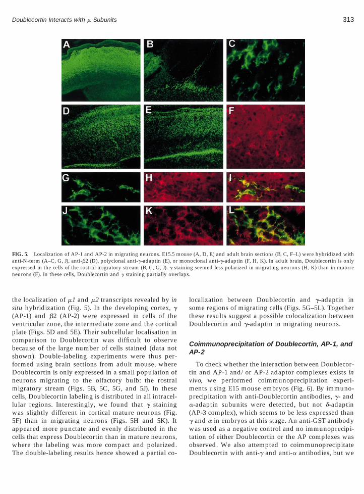
the localization of m1 and m2 transcripts revealed by insitu hybridization (Fig. 5). In the developing cortex, g(AP-1) and b2 (AP-2) were expressed in cells of theventricular zone, the intermediate zone and the corticalplate (Figs. 5D and 5E). Their subcellular localisation incomparison to Doublecortin was difficult to observebecause of the large number of cells stained (data notshown). Double-labeling experiments were thus per-formed using brain sections from adult mouse, whereDoublecortin is only expressed in a small population ofneurons migrating to the olfactory bulb: the rostralmigratory stream (Figs. 5B, 5C, 5G, and 5J). In thesecells, Doublecortin labeling is distributed in all intracel-lular regions. Interestingly, we found that g stainingwas slightly different in cortical mature neurons (Fig.5F) than in migrating neurons (Figs. 5H and 5K). Itappeared more punctate and evenly distributed in thecells that express Doublecortin than in mature neurons,
FIG. 5. Localization of AP-1 and AP-2 in migrating neurons. E15.5 manti-N-term (A–C, G, J), anti-b2 (D), polyclonal anti-g-adaptin (E), orxpressed in the cells of the rostral migratory stream (B, C, G, J). g st
neurons (F). In these cells, Doublecortin and g staining partially ove
where the labeling was more compact and polarized.The double-labeling results hence showed a partial co-
localization between Doublecortin and g-adaptin insome regions of migrating cells (Figs. 5G–5L). Togetherthese results suggest a possible colocalization betweenDoublecortin and g-adaptin in migrating neurons.
Coimmunoprecipitation of Doublecortin, AP-1, andAP-2
To check whether the interaction between Doublecor-tin and AP-1 and/or AP-2 adaptor complexes exists inivo, we performed coimmunoprecipitation experi-
ments using E15 mouse embryos (Fig. 6). By immuno-precipitation with anti-Doublecortin antibodies, g- anda-adaptin subunits were detected, but not d-adaptin(AP-3 complex), which seems to be less expressed thang and a in embryos at this stage. An anti-GST antibodywas used as a negative control and no immunoprecipi-tation of either Doublecortin or the AP complexes was
e (A, D, E) and adult brain sections (B, C, F–L) were hybridized withoclonal anti-g-adaptin (F, H, K). In adult brain, Doublecortin is onlyg seemed less polarized in migrating neurons (H, K) than in mature
ousmon
observed. We also attempted to coimmunoprecipitateDoublecortin with anti-g and anti-a antibodies, but we

te
fd
314 Friocourt et al.
were unable to detect convincing bands by Western blotanalysis. This may be due to steric hindrance or to theinvolvement in this interaction of only a small percent-age of each of the adaptor complexes. Nevertheless, ourimmunoprecipitation data using anti-Doublecortin an-tibodies confirm the results obtained using the two-hybrid system and in vitro binding assays and suggestthat the Doublecortin–adaptor complex interaction canoccur in vivo.
DISCUSSION
In this study, we searched for proteins interactingwith Doublecortin using the two-hybrid system to bet-ter understand its role in neuronal migration. One clonewas found to encode the human m1 subunit of theadaptor complex AP-1, known to play a role in clathrin-dependent protein sorting. We characterized the Dou-blecortin–m1 interaction and further tested for an inter-action with the similar m2 and m3 subunits of the AP-2
FIG. 6. Coimmunoprecipitation experiments. Extracts from E15mouse embryos were immunoprecipitated with either anti-N-term oranti-GST antibodies. After transfer to nitro cellulose membrane andprotein immunoblotting, blots were detected with anti-N-term (Dou-blecortin), anti-g (AP-1), anti-a (AP-2), and anti-d (AP-3). g and asubunits were found to coimmunoprecipitate with Doublecortin. TheAP-3 complex seemed to be less abundant than AP-1 and AP-2 in E15mouse embryos.
and AP-3 complexes. These data showed a potentialinteraction between Doublecortin and m1, and possibly
mr
m2, but we found no evidence of an equivalent interac-tion with m3. The spatial and temporal expression pat-terns of Doublecortin were compared with those ofAP-1 and AP-2 adaptor complexes, including an exam-ination of their subcellular localizations in primary cul-tures of neurons as well as in neurons of the developingcortex and in adult brain. Coimmunoprecipitation ex-periments from embryonic mouse brain were also usedto verify the interactions. These combined data supportthe existence of an association in vivo between Dou-blecortin and m1 and m2. The interaction betweenA18108 and m1/m2 observed in the two-hybrid systemis another interesting aspect. Indeed, this protein shares75% homology with Doublecortin and contains a ty-rosine-based sorting signal. It is also associated withmicrotubules but has a less restrictive pattern of expres-sion than Doublecortin as it is also expressed in glialcells and in mature neurons. Thus, it is possible thatA18108 replaces Doublecortin function in these cells.We are generating antibodies against this protein tobetter address these issues.
We previously demonstrated that Doublecortin iswidely expressed in the developing nervous systemfrom mouse embryonic day 11 until neonatal stages; butit is greatly down regulated after birth and is onlyexpressed in the rostral migratory stream in the adultbrain. In order to check that m1 and m2 were expressedin the same cells during development, we first per-formed in situ hybridization experiments using anti-sense RNA probes. Interestingly, although m1 and m2were largely ubiquitous, a more prominent expressionof both genes in the developing nervous system wasobvious, and overlapped with Doublecortin expression.This may indicate that higher levels of AP-1 and AP-2are required in young migrating and differentiatingneurons over certain stages of development. Indeed,since neurons develop long leading processes and ax-ons and dendrites, it may be expected that proteintransport mechanisms play an important role withinthe cell. An enrichment of m2 in the Drosophila devel-oping nervous system has been previously documented(Zhang et al., 1999); moreover, AP-2 and AP-3 com-plexes are known to be involved in mature nerve cellfunction (Gonzalez-Gaitan et al., 1997; Shi et al., 1998;Faundez et al., 1998). Nevertheless, to our knowledge,here has been no previous report concerning AP-1nrichment in the developing nervous system.
We performed immunofluorescence experiments tourther study protein localisations in migrating andifferentiating neurons of the developing and adult
ouse brain. In agreement with in situ hybridizationesults, AP-1 and AP-2 displayed ubiquitous expression

i
so
aDaD
attm
it
315Doublecortin Interacts with m Subunits
patterns including the cells expressing Doublecortin. Inprimary neuronal cultures, Doublecortin and g label-ling partially overlapped in the proximal regions ofgrowth cones. The growth cone is known to be animportant region for endocytosis and recycling of celladhesion molecules, as shown for L1 (Kamiguchi et al.,1998, 2000). Altogether, these data suggest that the cel-lular and subcellular compartments where Doublecor-tin is present, i.e., migrating neuronal cells and parts ofthe growth cones of differentiating neurons, are placeswhere highly dynamic vesicular trafficking occurs.
It is known that there is a close relationship betweenmicrotubules and the Golgi network (reviewed by Thy-berg et al., 1999). Since Doublecortin is a MAP, annteraction with m1 and/or m2 may indicate a microtu-
bule-dependent role in vesicle or tubule transport (Fig.7). These data are consistent with a recent study thatshowed a direct interaction between the kinesin KIF13Aand the AP-1 complex, which mediates the transport ofmannose-6-phosphate receptors from the TGN to theplasma membrane. These results demonstrate that theAP complexes can function as motor adaptors in vesic-ular transport (Nakagawa et al., 2000) and may furtherupport an interaction between the AP complexes andther MAPs.Both two-hybrid experiments and in vitro binding
ssays showed the importance of the C-terminus ofoublecortin for the interaction. This result may be in
greement with a microtubule-associated role of theoublecortin-m interaction since several reports have
highlighted the important part played by the N-termi-nal part of Doublecortin in the interaction with micro-tubules (Horesh et al., 1999; Sapir et al., 2000; Taylor etal., 2000). Tyrosine-based sorting signals, recognised bym subunits, have previously been reported to be impor-tant for the recruitment of integral membrane proteinsinto clathrin-coated vesicles (Ohno et al., 1995). Weidentified such a motif in the Doublecortin sequenceand mutagenesis experiments confirmed its involve-ment in the interaction with both m1 and m2. Neverthe-less, Doublecortin is not an integral membrane protein,which rules out that it is recruited into vesicle mem-branes in this way. However, several cytosolic proteinshave been shown to act as connector molecules betweenadaptor complexes and receptors, and hence to influ-ence endocytic and exocytic processes. Two well-char-acterized examples are Nef (Le Gall et al., 1998; Piguet etl., 1998) and the b-arrestins (Laporte et al., 1999). Onehus could assume that Doublecortin is such a connec-
or molecule, recruited from the cytosol or attached toicrotubules, which influences the trafficking of other
ntegral membrane proteins by its interaction with ei-her m1 or m2.
The importance of VLDLR, APOE-R2, cadherin-relatedneuronal receptors (CNR) and a3b1 integrin receptor fornormal corticogenesis in the mouse has been previouslyhighlighted (Trommsdorff et al., 1999; D’Arcangelo et al.,1999; Hiesberger et al., 1999; Senzaki et al., 1999; Dulabonet al., 2000). In addition, another protein involved in neu-ronal migration, DabI has been shown to interact with asorting signal NPxY present in the cytoplasmic domainsof the VLDLR and APOE-R2 receptors (Trommsdorff etal., 1999). This motif is also implicated in the internalisa-tion and recycling of VLDLR and APOE-R2 via an inter-action with either m1 or m2. Indeed, we have shown aninteraction between m1 and m2 and both receptors usingthe two-hybrid system (unpublished data). Doublecortinmay in some way influence this process by competingwith the receptors for an interaction with the m subunits(Fig. 7). It is also of interest that AP-2 complexes have beensuggested to work as dimers (Owen et al., 1998), whichmight allow simultaneous binding to different partners.These and other hypotheses remain to be elaborated, butmay provide a link between the Reelin signalling path-way, vesicle transport and the microtubular network. In-terestingly, LIS-1 has also been shown to interact withdynein, which is involved in cargo transport from theendoplasmic reticulum to the Golgi, further emphasisinga role for vesicular trafficking in neuronal migration(Smith et al., 2000; Liu et al., 2000; Faulkner et al., 2000).
Besides its known function as a MAP, the data de-scribed here point to a novel biological role for Dou-blecortin in protein transport or receptor recyclingwithin migrating neurons. These findings provide newinsights into understanding the physiopathology un-derlying cortical dysgenesis associated with the loss offunction of Doublecortin.
EXPERIMENTAL METHODS
Yeast Cultures and Two-Hybrid Analysis
All the cloning procedures were carried out as de-scribed by Sambrook et al. (1989). The full-length dou-blecortin cDNA was cloned in pBTM116 downstream ofthe LexA binding domain (Bartel et al., 1993). The hu-man foetal brain cDNA library cloned in pACT2, waspurchased from CLONTECH laboratories (MATCH-MAKER) and screened according to manufacturer’s in-
structions. This library was prepared from 19- to 22-week old embryos by a combination of oligo-dT and
gat
it
tb volv
316 Friocourt et al.
random-priming, followed by orientated cloning. TheSaccharomyces cerevisiae L40 strain (MATa, trp1, leu2,his3, LYS2::lexA-HIS3, URA3::lexA-lacZ) was cotrans-formed with the plasmids using the lithium acetateprocedure (Bartel et al., 1995). Transformants were
rown at 30°C on plates lacking tryptophan, leucinend histidine. His1 colonies were screened for activa-ion of the lacZ reporter gene by testing b-galactosidase
activity using replica plating (lacZ test). Library plas-mids from positive clones were rescued in Escherichiacoli HB101 by plating on leucine-free medium. Eachpositive clone was then sequenced using an ABI 373machine and a DyeDeoxy terminator cycle sequencingkit (Applied Biosystems) with vector specific primers.
Sequenced plasmids were transformed into the yeaststrain AMR70 (MATa) to check the specificity of the
FIG. 7. A model for the role of the Doublecortin–AP interaction. Douhe internalisation or the recycling of VLDLR and APOER2 receptorsetween AP-1 and Doublecortin may occur in vesicular trafficking in
nteraction. The resulting cells were mated with L40ransformed with the original and other unrelated baits
(lamin C and RAS) cloned in pBTM116. Mating wascarried out by replica plating on YPD medium, thendiploids were selected on medium lacking tryptophanand leucine. Histidine and lacZ tests were performed asabove.
To compare interactions between Doublecortin, a ho-mologous protein A18108 and the m subunits, the full-length A18108 cDNA (lacking the first 12 amino acids)was cloned in pBTM116. pSG m1, m2 and pACT2 m1, m2(mouse cDNA), m3A and m3B (rat cDNA) constructswere kindly provided by S. Benarous and were origi-nally a gift from J. S. Bonifacino (Ohno et al., 1995). Theplasmid pLex-HIV-1 LAI construct was described byBerlioz-Torrent et al. (1999). RAS and RAF, respectively,cloned in pBTM116 and pGAD424 were used as con-trols.
rtin may regulate the Reelin–Dab1 signalling pathway by influencingernatively, although Doublecortin is not a motor protein, interactioning microtubules.
bleco. Alt
Site-directed mutagenesis was performed using theQuik Change Mutagenesis kit (Stratagene).

brwr
I
i
R(
A
b1pdP
317Doublecortin Interacts with m Subunits
In Vitro Binding Assay
GST fusion proteins were expressed in E. coli BL21and purified according to the manufacturer’s instruc-tions. In vitro transcription and translation of PSG m1and m2 were performed using the Promega TNT/T7rabbit reticulocyte lysate system with [35S]methioninefor 90 min at 30°C. Eight microliters of radioactivelabeled product were incubated with 10 ml of gluta-thione–Sepharose 4B beads coated with 10 mg of GSTfusion protein in 200 ml of buffer (50 mM Tris–HCl pH7.5, 350 mM NaCl, 1% Triton X-100) for 2 h at 4°C. The
eads were washed five times with the same buffer andesuspended in 40 ml of SDS sample buffer. Samplesere separated by SDS/PAGE followed by autoradiog-
aphy.
n Situ Hybridization
m1 and m2 cDNA were cloned in pBluescript andsense and antisense probes were generated by in vitrotranscription. Mouse embryos and adult brains werefixed in 2–4% paraformaldehyde, cryoprotected with30% sucrose in phosphate buffer, frozen with isopen-tane, and cut into ;20-mm-thick sections. Slides werencubated with a35S-labeled RNA probes in a 50% for-
mamide solution at 65°C. Washes were performed insuccessively stringent SSC solutions, with a final washat 0.13 SSC at 60°C. Slides were then dipped in dilutedKodak NTB2 emulsion and exposed for 24–48 h. Emul-sions were developed and sections counterstained withtoluidine blue, mounted in Eukitt and examined underlight microscopy. In situ hybridizations for doublecortin
NA probes were performed as previously describedFrancis et al., 1999).
ntibodies
The production of polyclonal antibodies against Dou-lecortin has been reported elsewhere (Francis et al.,999). We used anti-dbcn pep produced from syntheticeptides and the affinity-purified anti-N-term antibodyirected against the N-terminal part of the protein.olyclonal antibodies against d, g, and b2 were kindly
provided by M. S. Robinson. Monoclonal antibodies:anti-a-adaptin (AC1-M11) and anti-g-adaptin (A36120)were respectively purchased from Alexis Corporationand Transduction Laboratories.
Cell Cultures and Immunofluorescence Staining
Primary neuronal cultures were prepared from foetalmouse brain at 15 days of gestation as previously de-
scribed (Berwald-Netter et al., 1981). In some experi-ments, neurons were treated for 15 min at 37°C withbrefeldin A (10–40 mg/ml, Sigma) or for 4 h with taxol(10 mg/ml) or nocodazole (3.5 or 8 mM). Neuronsgrown on coverslips were fixed for 10 min at 220°C inmethanol/acetone. After four washes in phosphate-buffered saline (PBS), the cells were prehybridized for1 h with TBST (10 mM Tris–HCl pH 8, 150 mM NaCl,0.05% Triton X-100) containing 2% goat serum, and thenincubated overnight at 4°C with the primary antibody(anti-a, 1:100, monoclonal anti-g, 1:100, anti-Doublecor-tin pep, 1:300, affinity-purified anti-N-term, 1:200). Thecells were washed three times in TBST and incubatedfor 1 h at room temperature with affinity-purified sec-ondary antibodies (FITC conjugated goat anti-rabbit Ig[Biosys., Compiegne, France] or Texas red-conjugatedgoat anti-mouse Ig [Jackson ImmunoResearch, WestGrove, PA]) used at 1:200 dilution. After three washes,coverslips were mounted in Mowiol and examinedwith a confocal microscope MRC-1000 (Bio-Rad)equipped with epifluorescence illumination.
Immunohistochemistry
Mouse embryos and adult brains were cryoprotectedwith 30% sucrose in phosphate buffer, frozen with iso-pentane and sectioned (;15–20 mm thick). Sectionswere fixed for 10 min in methanol at 220°C and washedwith phosphate buffered saline (PBS). They were pre-hybridized for 1 h on ice with TBST containing 2% goatserum, washed in TBST and incubated overnight at 4°Cwith the primary antibody (polyclonal anti-g, 1:100,monoclonal anti-g, 1:100, anti-a, 1:100, anti-b2, 1:100,affinity-purified anti-N-term, 1:200). The followingsteps were done as previously described for immuno-cytochemistry. Sections were examined with a Zeissmicroscope equipped with epifluorescence illumina-tion.
Coimmunoprecipitation
Mouse brains taken from E15 embryos were crushedin liquid nitrogen and resuspended in 10 volumes of IPbuffer (50 mM Tris–HCl pH8, 150 mM NaCl, 1% NP-40and protease inhibitors). After a centrifugation at50,000g for 30 min at 4°C, 2-ml aliquots of supernatantwere incubated with protein A-agarose (Sigma) for 2 hat 4°C and spun at 10,000g for 5 min at 4°C to eliminatethe immunoglobulins. Aliquots of the resulting super-natant were incubated at 4°C with the antibodies of
interest (affinity-purified anti-N-term or affinity-puri-fied anti-GST, 1:10). Two hours later, protein A–agarose
D
D
D
F
F
F
G
G
G
H
H
H
H
318 Friocourt et al.
was added and samples were incubated overnight at4°C. Immunoprecipitates were washed approximately10 times with IP buffer, then proteins were separated onSDS–polyacrylamide gels. Transfer to nitrocellulosemembrane and protein immunoblotting were carriedout following standard protocols (Sambrook et al.,1989). For Western blot detection, polyclonal anti-g-adaptin was used at 1:500, anti-a at 1:200, anti-d at1:200, and nonpurified anti-N-term at 1:5000.
ACKNOWLEDGMENTS
We thank Jenny Hirst, Margaret Robinson, Jacques Camonis, Clar-isse Berlioz-Torrent, and Serge Benichou for the kind gifts of reagents,Isabelle Bouchaert for confocal microscopy analysis, Odette Godardfor her logistic support and Peter Schu, Cherif Beldjord, Alain Carrie,Thierry Bienvenu, Philippe Couvert, Fabien Fauchereau, RamziZemni, Jean-Marie Genevard, Nathalie McDonnell, and Denis Tempefor helpful discussions. This work was supported in part by grantsfrom the Institut National de la Sante et de la Recherche Medicale(INSERM), the Fondation pour la Recherche Medicale, the Associa-tion Francaise contre les myopathies (AFM), the Fondation JeromeLejeune, the European Commission, and the Human Frontier ScienceProgram (RG0283/1999-B). G.F. is supported by a Ph.D. fellowshipfrom the Ministere de la Recherche, and F.F. was supported by aEuropean Molecular Biology Organisation (EMBO) fellowship.
REFERENCES
Bartel, P. L., Chien, C.-T., Sternglanz, R., and Fields, S. (1993). CellularInteractions in Development: A Practical Approach (D. A. Hartley, Ed.),p. 153. Oxford Univ. Press, Oxford.
Bartel, P. L., and Fields, S. (1995). Analyzing protein-protein interac-tions using two-hybrid system. Methods Enzymol. 254: 241–263.
Berlioz-Torrent, C., Shacklett, B. L., Erdtmann, L., Delamarre, L.,Bouchaert, I., Sonigo, P., Dokhelar, M. C., and Benarous, R. (1999).Interactions of the cytoplasmic domains of human and simianretroviral transmembrane proteins with components of the clathrinadaptor complexes modulate intracellular and cell surface expres-sion of envelope glycoproteins. J. Virol. 73: 1350–1361.
Berwald-Netter, Y., Martin-Moutot, N., Koulakoff, A., and Couraud,F. (1981). Na1-channel-associated scorpion toxin receptor sites asprobes for neuronal evolution in vivo and in vitro. Proc. Natl. Acad.Sci. USA 78: 1245–1249.
Caviness, V. S., Jr., Takahashi, T., and Nowakowski, R. S. (1997).Normal and Abnormal Development of the Cortex (A. M. Galaburdaand Y. Christen, Eds.), pp. 1–24. Springer-Verlag, Berlin/Heidel-berg.
Chae, T., Kwon, Y. T., Bronson, R., Dikkes, P., Li, E., and Tsai, L. H.(1997). Mice lacking p35, a neuronal specific activator of cdk5,display cortical lamination defects, seizures, and adult lethality.Neuron 18: 29–42.
D’Arcangelo, G., Miao, G. G., Chen, S. C., Soares, H. D., Morgan, J. I.,and Curran, T. (1995). A protein related to extracellular matrix
proteins deleted in the mouse mutant reeler. Nature 374: 719–723.D’Arcangelo, G., Homayouni, R., Keshvara, L., Rice, D. S., Sheldon, H
M., and Curran, T. (1999). Reelin is a ligand for lipoprotein recep-tors. Neuron 24: 471–479.
Dell’Angelica, E. C., Ohno, H., Ooi, C. E., Rabinovich, E., Roche,K. W., and Bonifacino, J. S. (1997). AP-3: An adaptor-like proteincomplex with ubiquitous expression. EMBO J. 16: 917–928.ell’Angelica, E. C., Mullins, C., and Bonifacino, J. S. (1999). AP-4, anovel protein complex related to clathrin adaptors. J. Biol. Chem.274: 7278–7285.es Portes, V., Pinard, J. M., Billuart, P., Vinet, M. C., Koulakoff, A.,Carrie, A., Gelot, A., Dupuis, E., Motte, J., Berwald-Netter, Y.,Catala, M., Kahn, A., Beldjord, C., and Chelly, J. (1998). A novelCNS gene required for neuronal migration and involved in X-linked subcortical laminar heterotopia and lissencephaly syn-drome. Cell 92: 51–61.ulabon L., Olson, E. C., Taglienti, M. G., Eisenhuth, S., McGrath, B.,Walsh, C. A., Kreidberg, J. A., and Anton, E. S. (2000). Reelin bindsa3b1 integrin and inhibits neuronal migration. Neuron 27: 33–44.
Faulkner, N. E., Dujardin, D. L., Tai, C. Y., Vaughan, K. T., O’Connell,C. B., Wang, Y. L., and Vallee, R. B. (2000). A role for the lissen-cephaly gene LISI in mitosis and cytoplasmic dynein function.Nature Cell Biol. 2: 784–791.
Faundez, V., Horng, J. T., and Kelly, R. B. (1998). A Function for theAP3 coat complex in synaptic vesicle formation from endosomes.Cell 93: 423–432.
ields, S., and Song, O. (1989). A novel genetic system to detectprotein–protein interactions. Nature 340: 245–246.
ox, J. W., Lamperti, E. D., Eksioglu, Y. Z., Hong, S. E., Feng, Y.,Graham, D. A., Scheffer, I. E., Dobyns, W. B., Hirsch, B. A., Radtke,R. A., Berkovic, S. F., Huttenlocher, P. R., and Walsh, C. A. (1998).Mutations in filamin 1 prevent migration of cerebral cortical neu-rons in human periventricular heterotopia. Neuron 21: 1315–1325.
rancis, F., Koulakoff, A., Boucher, D., Chafey, P., Schaar B., Vinet,M. C., Friocourt, G., McDonnell, N., Reiner, O., Kahn, A., McCon-nell, S. K., Berwald-Netter, Y., Denoulet, P., and Chelly, J. (1999).Doublecortin is a developmentally regulated, microtubule-associ-ated protein expressed in migrating and differentiating neurons.Neuron 23: 247–256.leeson, J. G., Allen, K. M., Fox, J. W., Lamperti, E. D., Berkovic, S.,Scheffer, I., Cooper, E. C., Dobyns, W. B., Minnerath, S. R., Ross,M. E., and Walsh, C. A. (1998). Doublecortin, a brain-specific genemutated in human X-linked lissencephaly and double cortex syn-drome, encodes a putative signaling protein. Cell 92: 63–72.leeson, J. G., Lin, P. T., Flanagan, L. A., and Walsh, C. A. (1999).Doublecortin is a microtubule-associated protein and is expressedwidely by migrating neurons. Neuron 23: 257–271.onzalez-Gaitan, M., and Jackle, H. (1997). Role of Drosophilaa-Adaptin in presynaptic vesicle recycling. Cell 88: 767–776.iesberger, T., Trommsdorff, M., Howell, B. W., Goffinet, A., Mumby,M. C., Cooper, J. A., and Herz, J. (1999). Direct binding of Reelin toVLDL receptor and ApoE receptor 2 induces tyrosine phosphory-lation of disabled-1 and modulates tau phosphorylation. Neuron 24:481–489.irst, J., and Robinson, M. S. (1998). Clathrin and adaptors. Biochim.Biophys. Acta 1404: 173–193.irst, J., Bright, N. A., Rous, B., and Robinson, M. S. (1999). Charac-terization of a fourth adaptor-related protein complex. Mol. Biol.Cell 10: 2787–2802.oresh, D., Sapir, T., Francis, F., Wolf, S. G., Caspi, M., Elbaum, M.,Chelly, J., and Reiner, O. (1999). Doublecortin, a stabilizer of mi-
crotubules. Hum. Mol. Genet. 8: 1599–1610.owell, B. W., Hawkes, R., Soriano, P., and Cooper, J. A. (1997).
M
N
O
O
O
O
O
P
P
R
R
S
S
S
S
S
S
S
S
T
T
T
W
Z
319Doublecortin Interacts with m Subunits
Neuronal position in the developing brain is regulated by mousedisabled-1. Nature 389: 733–737.
Kamiguchi, H., Long, K. E., Pendergast, M., Schaefer, A. W., Rap-oport, I., Kirchhausen, T., and Lemmon, V. (1998). The neural celladhesion molecule L1 interacts with the AP-2 adaptor and is endo-cytosed via the clathrin-mediated pathway. J. Neurosci. 18: 5311–5321.
Kamiguchi, H., and Lemmon, V. (2000). Recycling of the cell adhesionmolecule L1 in axonal growth cones. J. Neurosci. 20: 3676–3686.
Laporte, S. A., Oakley, R. H., Zhang, J., Holt, J. A., Ferguson, S. S.,Caron, M. G., and Barak, L. S. (1999). The b2-adrenergic receptor/barrestin complex recruits the clathrin adaptor AP-2 during endo-cytosis. Proc. Natl. Acad. Sci. USA 96: 3712–3717.
Le Gall, S., Erdtmann, L., Benichou, S., Berlioz-Torrent, C., Liu, L.,Benarous, R., Heard, J. M., and Schwartz, O. (1998). Nef interactswith the m subunit of clathrin adaptor complexes and reveals acryptic sorting signal in MHC I molecules. Immunity 8: 483–495.
Liu, Z., Steward, R., and Luo, L. (2000). Drosophila Lis1 is required forneuroblast proliferation, dendritic elaboration and axonal trans-port. Nature Cell Biol. 2: 776–783.cConnell, S. K. (1995). Constructing the cerebral cortex: Neurogen-esis and fate determination. Neuron 15: 761–768.akagawa, T., Setou, M., Seog, D. H., Ogasawara, K., Dohmae, N.,Takio, K., and Hirokawa, N. (2000). A novel motor, KIF13A, trans-ports mannose-6-phosphate receptor to plasma membrane throughdirect interaction with AP-1 complex. Cell 103: 569–581.hno, H., Stewart, J., Fournier, M. C., Bosshart, H., Rhee, I., Miyatake,S., Saito, T., Gallusser, A., Kirchhausen, T., and Bonifacino, J. S.(1995). Interaction of tyrosine-based sorting signals with clathrin-associated proteins. Science 269: 1872–1875.hno, H., Aguilar, R. C., Yeh, D., Taura, D., Saito, T., and Bonifacino,J. S. (1998). The medium subunits of adaptor complexes recognizedistinct but overlapping sets of tyrosine-based sorting signals.J. Biol. Chem. 273: 25915–25921.mori, Y., Suzuki, M., Ozaki, K., Harada, Y., Nakamura, Y., Taka-hashi, E., and Fujiwara, T. (1998). Expression and chromosomallocalization of KIAA0369, a putative kinase structurally related toDoublecortin. J. Hum. Genet. 43: 169–177.shima, T., Ward, J. M., Huh, C. G., Longenecker, G., Veeranna, Pant,H. C, Brady, R. O., Martin, L. J., and Kulkarni, A. B. (1996). Targeteddisruption of the cyclin-dependent kinase 5 gene results in abnor-mal corticogenesis, neuronal pathology and perinatal death. Proc.Natl. Acad. Sci. USA 93: 11173–11178.wen, D. J., and Evans, P. R. (1998). A structural explanation for therecognition of tyrosine-based endocytotic signals. Science 282: 1327–1332.
iguet, V., Chen, Y. L., Mangasarian, A., Foti, M., Carpentier, J. L., andTrono, D. (1998) Mechanism of Nef-induced CD4 endocytosis: Nefconnects CD4 with the m chain of adaptor complexes. EMBO J. 17:2472–2481.
inard, J. M., Motte, J., Chiron, C., Brian, R., Andermann, E., andDulac, O. (1994). Subcortical laminar heterotopia and lissencephaly
in two families: A single X linked dominant gene. J. Neurol. Neuro-surg. Psychiatry 57: 914–920.
einer, O., Carrozzo, R., Shen, Y., Wehnert, M., Faustinella, F.,Dobyns, W. B., Caskey, C. T., and Ledbetter, D. H. (1993). Isolationof a Miller–Dieker lissencephaly gene containing G protein b-sub-unit-like repeats. Nature 364: 717–721.
obinson, M. S., and Kreis, T. E. (1992). Recruitment of coat proteinsonto Golgi membranes in intact and permeabilized cells: effects ofbrefeldin A and G protein activators. Cell 69: 129–138.
ambrook, J., Fritsch, E. F., and Maniatis, T. (1989). Molecular Cloning:A Laboratory Manual, 2nd ed. Cold Spring Harbor Laboratory, ColdSpring Harbor, NY.
apir, T., Elbaum, M., and Reiner, O. (1997). Reduction of microtubulecatastrophe events by LIS1, platelet-activating factor acetylhydro-lase subunit. EMBO J. 16: 6977–84.
apir, T., Horesh, D., Caspi, M., Atlas, R., Burgess, H. A., Wolf, S. G.,Francis, F., Chelly, J., Elbaum, M., Pietrokovski, S., and Reiner, O.(2000). Doublecortin mutations cluster in evolutionarily conservedfunctional domains. Hum. Mol. Genet. 9: 703–712.
enzaki, K., Ogawa, M., and Yagi, T. (1999). Proteins of the CNRfamily are multiple receptors for reelin. Cell 99: 635–647.
heldon, M., Rice, D. S., D’Arcangelo, G., Yoneshima, H., Nakajima,K., Mikoshiba, K., Howell, B. W., Cooper, J. A., Goldowitz, D., andCurran, T. (1997). Scrambler and Yotari disrupt the disabled geneand produce a reeler-like phenotype in mice. Nature 389: 730–733.
hi, G., Faundez, V., Roos, J., Dell’Angelica, E. C., and Kelly, R. B.(1998). Neuroendocrine synaptic vesicles are formed in vitro byboth clathrin-dependent and clathrin-independent pathways. J. CellBiol. 143: 947–955.
impson, F., Bright, N. A., West, M. A., Newman, L. S., Darnell, R. B.,and Robinson, M. S. (1996). A novel adaptor-related protein com-plex. J. Cell Biol. 133: 749–760.
mith, D. S., Niethammer, M., Ayala, R., Zhou, Y., Gambello, M. J,Wynshaw-Boris, A., and Tsai, L. H. (2000). Regulation of cytoplas-mic dynein behaviour and microtubule organization by mamma-lian Lis1. Nature Cell Biol. 2: 767–775.
aylor, K. R., Holzer, A. K., Bazan, J. F., Walsh, C. A., and Gleeson,J. G. (2000). Patient mutations in Doublecortin define a repeatedtubulin-binding domain. J. Biol. Chem. 275: 34442–34450.
hyberg, J., and Moskalewski, S. (1999). Role of microtubules in theorganization of the Golgi complex. Exp. Cell Res. 246: 263–279.
rommsdorff, M., Gotthardt, M., Hiesberger, T., Shelton, J., Stock-inger, W., Nimpf, J., Hammer, R. E., Richardson, J. A., and Herz, J.(1999). Reeler/Disabled-like disruption of neuronal migration inknockout mice lacking the VLDL receptor and ApoE receptor 2. Cell97: 689–701.are, M. L., Fox, J. W., Gonzalez, J. L., Davis, N. M., Lambert deRouvroit, C., Russo, C. J., Chua, S. C., Jr., Goffinet, A. M., andWalsh, C. A. (1997). Aberrant splicing of a mouse disabled ho-molog, mdabl, in the scrambler mouse. Neuron 19: 239–249.
hang, Y. Q., and Broadie, K. (1999). Cloning, mapping and tissue-
specific expression of Drosophila clathrin-associated protein AP50gene. Gene 233: 171–179.Received April 3, 2001Revised June 18, 2001
Accepted June 28, 2001