fracture resistance of human cortical bone across … resistance of human cortical bone across...
TRANSCRIPT

lable at ScienceDirect
Biomaterials 35 (2014) 5472e5481
Contents lists avai
Biomaterials
journal homepage: www.elsevier .com/locate/biomateria ls
Fracture resistance of human cortical bone across multiplelength-scales at physiological strain rates
Elizabeth A. Zimmermann a, Bernd Gludovatz a, Eric Schaible b, Björn Busse a,c,Robert O. Ritchie a,d,*
aMaterials Sciences Division, Lawrence Berkeley National Laboratory, Berkeley, CA, USAb Experimental Systems Group, Lawrence Berkeley National Laboratory, Berkeley, CA, USAcDepartment of Osteology and Biomechanics, University Medical Center, Hamburg, GermanydDepartment of Materials Science and Engineering, University of California, Berkeley, CA, USA
a r t i c l e i n f o
Article history:Received 23 January 2014Accepted 22 March 2014Available online 13 April 2014
Keywords:BoneStrain rateFracture toughnessPlasticityX-ray diffraction
* Corresponding author. Department of Materials Sversity of California, Berkeley, CA, USA. Tel.: þ1 510 48
E-mail address: [email protected] (R.O. Ritchie).1 The fracture toughness can be expressed as the critic
for unstable fracture in the presence of a pre-existing cYsapp(pa)½¼ KIc,wheresapp is theapplied stress, a is the(oforderunity)ofcrack sizeandgeometry.Alternatively,as a critical value of the strain-energy release rate,Gc, defienergyperunit increase in crack area, or the nonlinear el
http://dx.doi.org/10.1016/j.biomaterials.2014.03.0660142-9612/Published by Elsevier Ltd.
a b s t r a c t
Whilemost fracture-mechanics investigations on bone have beenperformed at low strain rates, physiologicalfractures invariably occur at higher loading rates. Here, at strain rates from 10�5 to 10�1 s�1, we investigatedeformation and fracture in bone at small length-scales using in situ small-angle x-ray scattering (SAXS) tostudy deformation in the mineralized collagen fibrils and at the microstructural level via fracture-mechanicsexperiments to study tougheningmechanisms generating toughness through crack-tip shielding. Our resultsshow diminished bone toughness at increasing strain rates as cracks penetrate through the osteons at higherstrain rates instead of deflecting at the cement lines, which is a prime toughening mechanism in bone at lowstrain rates. Theabsenceofcrackdeflectionmechanismsathigher strain rates isconsistentwith lower intrinsicbone matrix toughness. In the SAXS experiments, higher fibrillar strains at higher strain rates suggest lessinelastic deformation and thus support a lower intrinsic toughness. The increased incidence of fractureinduced by high strain rates can be associated with a loss in toughness in the matrix caused by a strain rateinduced stiffening of the fibril ductility, i.e., a “locking-up” of the viscous sliding and sacrificial bondingmechanisms, which are the origin of inelastic deformation (and toughness) in bone at small length-scales.
Published by Elsevier Ltd.
1. Introduction namely, intrinsic toughening mechanisms that promote “plas-
Traumatic injuries, such as falls, often can lead to bone fractures.This fragility is especially significant in theelderly,wherebrokenbonescan be associated with a further deterioration in health [1]. As thesetraumatic injuries invariably result fromloadingovershort time-scales,it is necessary to understand how the structural framework of thehuman body resists fracture at such physiologically high strain rates.
The fracture resistance of human cortical bone is a direct result ofits hierarchically assembled structure of collagen and hydroxyapa-tite (HA) mineral, which spans multiple length-scales from molec-ular to near-macroscopic dimensions (Fig. 1) [2,3]. Basically, thereare two major contributions to the fracture toughness of bone,1
cience and Engineering, Uni-6 5798; fax: þ1 510 643 5792.
al value of the stress intensityKrack, i.e., in mode I when K ¼crack length, andY is a functionthetoughness canbeexpressedned as the change in potential
astic version ofG, the J-integral.
ticity”, i.e., ductility in the mineralized tissue, and extrinsic tough-ening mechanisms that act to “shield” a growing crack from theglobal stresses and strains.2 The intrinsic toughness represents theinherent fracture resistance of thematerial and is developed at small(sub-micron) length-scales; here, the fibril can elastically stretchthrough cooperative deformation between themineral and collagen[8,9] as well as absorb further deformation through inelasticmechanisms, such as intra/interfibrillar sliding, breaking/reformingof sacrificial bonds, and even through the opening of dilatationalbands at the mineral/collagen interface [8,10e12]. The extrinsictoughness of bone, conversely, is a primary function of how themicrostructure can inhibit the growth of a crack; essentially, as a
2 Fracture resistance can be considered as a mutual competition between twoclasses of mechanisms: intrinsic mechanisms, which are microstructural damagemechanisms that operate ahead of the crack tip to promote cracking, and extrinsicmechanisms, which operate principally in the wake of the crack tip to inhibitcracking by “shielding” the crack from the applied driving force [4e7]. Whereasintrinsic toughening mechanisms, principally plastic deformation, act in general toresist intrinsic microstructural damage and thus are effective in inhibiting both theinitiation and growth of cracks, extrinsic toughening mechanisms, e.g., crackbridging, are only effective in inhibiting crack growth [6].

Fig. 1. The structure of human cortical bone spans multiple size-scales, which allows it to develop strength and resistance to fracture. At the smallest length-scales, human corticalbone is composed of an array of collagen molecules embedded with hydroxyapatite (HA) mineral crystals. The collagen and mineral comprises an array of mineralized collagenfibrils with various types of cross-links stabilizing the array and the individual fibrils. At higher length-scales, secondary osteons are the main motif at the microstructural scale. Theosteons have a central vascular cavity (the Haversian canal) that is concentrically surrounded by lamellae, which are composed of collagen fibers. At the outer boundary of theosteon is a boundary called the cement line, which has a higher mineralization relative to the surrounding bone matrix. Adapted from Ref. [25].
E.A. Zimmermann et al. / Biomaterials 35 (2014) 5472e5481 5473
crack begins to grow, the interaction of the crack path with themicrostructure can lead tomechanisms such as crack deflection andbridging, which “shield” the crack tip from the full stress intensity,thereby increasing the bone toughness [6]. Because most cracks areon the micron-scale, these extrinsic toughening mechanisms aremost effective when they interact with structural length-scales ofcomparable dimensions, i.e., w10se100s mm, specifically with theosteonal systems throughwhichbone remodels. The osteons consistof circumferential lamellar structures surrounding the Haversiancanals (Fig. 1) and with an outer boundary separating the osteonfrom the interstitial matrix called the cement line, which is thoughttohavea relativelyhighermineralization than the surroundingbonetissue3 [14,16]. As a large population of the microcracks created inbone formwithin the interstitial bone (i.e., the bonematrix betweenthe osteons) [17], a growing crack which impinges on the osteonalborders (i.e., the cement lines) is invariably subject to crack deflec-tion and/or twisting,4 often causing delamination along the cementlines; furthermore, the resulting intact material left between themicrocracks can lead to so-called “uncracked ligament” bridgesspanning themain crack wake, which further enhance the crack-tipshielding [19,20]. In thismanner, through a combination of intrinsic“plasticity” mechanisms at small (sub-micron) length-scales andcrack-tip shielding mechanisms at larger length-scales, healthyhuman cortical bone develops numerous potent mechanisms thatcan act to resist bone fracture.
Manystudies on the strength and toughness properties of humancortical bone have shown succinctly that the complex hierarchical
3 The composition of the cement lines has been a matter of debate in the liter-ature [13e16]. However, the general consensus is that the cement lines in healthybone represent regions of high mineralization relative to the surrounding bonematrix or a collagen deficient feature in the bone microstructure [14,16].
4 The deflection of a crack from a path of maximum tangential stress, essentiallythe path of maximum strain-energy release rate G, can lead to significant re-ductions in the crack-driving force experienced locally at the crack tip. Typically, anin-plane crack deflection of w90� can reduce the stress intensity K at the crack tipby almost a factor of two; if out-of-plane twisting of the crack path occurs, thereduction in the crack-tip K can be even higher [18].
bone-matrix structure at both small and large length-scales is pro-ficient in resisting the initiation and propagation of themajor cracksthat can cause bone fractures [19,21e26].However, the reality is thatmost of these studies have been conducted at low strain rates on theorder of 10�4 s�1 where it is easier to observe and collect data,whereas most physiological bone fractures are generally associatedwith much higher strain rates. For example, in vivo loading ratemeasurements on bone suggest strain rates of w0.007e0.013 s�1
during walking or running and strain rates as high as w0.02 s�1
during sprinting or downhill running [27e29]; other studies simu-lating a fall have shown that it takesw6e10ms for a falling femur toreach the peak load once it has begun to make contact with theground [30], with an upper bound for these high strain rates to bew25 s�1 for very high impacts [31]. As these physiologically realisticsituations represent strain rates some four or more orders ofmagnitude higher than those used in most bone fracture experi-ments in the laboratory, characterizing and understanding the roleof loading rate in influencing the multi-scale mechanisms by whichbone resists fracture is clearly pertinent.
Previous studies have characterized the strength and toughness ofbone at a wide range of strain rates [32e42]. The majority of studiespoint towards a ductile to brittle transition in bone, where there is aprogressive decrease in the amount of post-yield ductility as well asincrease in strength andmodulus [34e37,40e42]. Notched toughnesstests have also been performed at various strain rates and generallyshow a decrease in toughness at higher strain rates with a corre-sponding decrease in the accumulation of damage [32,33,38,39].
Consequently, we analyze here the mechanical response ofbone over multiple length-scales at physiological strain rates ofw10�5e10�1 s�1. Using in situ synchrotron small- and wide-anglex-ray scattering/diffraction (SAXS/WAXD) during uniaxial tensiletesting and fracture-mechanics-based fracture toughness analyses,we examine the specific roles of plasticity on intrinsic toughnessat sub-micron dimensions and the role of crack-tip shielding onextrinsic toughness at the scale of w1e100s mm to investigatewhether the salient mechanisms of toughening in bone are still aseffective in resisting bone fractures at physiologically high strainrates.

5 To determine the contribution of scattering from the background, SAXS andWAXD data were also acquired with no sample present; the background includedscattering from the air, hydration cell as well as HBSS and was taken at the cor-responding exposure time. The background data were normalized by the x-rayphoton counts and radially integrated over a 10� sector parallel to the loadingdirection.
E.A. Zimmermann et al. / Biomaterials 35 (2014) 5472e54815474
2. Materials and methods
2.1. Materials
The cortical bone from the femur of a 52-year-old male was used for all me-chanical testing. The posterior side of the diaphysis/shaft was used in the x-rayscattering experiments, while the samples for toughness testing were taken fromthe lateral side. Following harvesting, all samples were kept frozen prior to testing,wherein they were machined and then immersed in Hanks’ Balanced Salt Solution(HBSS) for at least 12 h.
2.2. Small- and wide-angle x-ray scattering/diffraction
By performing uniaxial tensile tests on small bone samples and subjectingthem to real time SAXS and WAXD in the synchrotron light source, we can measurethe macroscopic strain in the bone tissue sample, and then partition this strain todetermine the individual strains in the fibril (from the SAXS spectra) and HAmineral (from the WAXD spectra) constituents of the bone, based on the pro-cedures initially devised by Gupta et al. [43]. Rectangular samples of humancortical bone in the transverse orientation were sectioned to a thickness of 0.5 mmby using a water-irrigated low-speed saw with a diamond-coated blade. Thesamples were oriented such that the long axis of the samples was parallel to thelong axis of the bone. The samples were then polished with water-irrigated 800grit silicon carbide paper to final dimensions of roughly 15 mm � 1 mm � 250 mm.Silicon carbide paper was glued to the ends of the samples with cyanoacrylate glueto form frictional surfaces to grip during testing, and then soaked in HBSS for 24 hprior to testing.
The samples were loaded in tension in a custom-made mechanical testingdevice, with the sample held between two grips, one of which was stationaryand the other connected to a displacement stage (model UTMPP1HL, Newport,Irvine, CA) and a 34-kgf load cell (LC703-75, Omega, Stamford, CT). The rig waspositioned in beamline 7.3.3 at the Advanced Light Source (ALS) synchrotronradiation facility (Lawrence Berkeley National Laboratory, Berkeley, CA) [44],such that SAXS and WAXD data collection could be recorded simultaneously withmechanical loading. The testing was performed at room temperature withsamples hydrated throughout the experiment by means of a hydration cellcomprised of a strip of cellophane held to the sample through capillary actionwith a few drops of HBSS.
The samples were randomly split up into four groups, which were tested atone of four displacement rates. The displacement rates used were 10 mm/s(N ¼ 12), 1 mm/s (N ¼ 3), 0.1 mm/s (N ¼ 7) and 0.001 mm/s (N ¼ 10), whichproduced strain rates on the order of magnitude of 10�1, 10�2, 10�3 and 10�5 s�1,respectively. As discussed above, a strain rate of 1 s�1 corresponds physiologi-cally to a fall whereas strain rates of 10�3e10�2 s�1 pertain to that experiencedwhen running or walking; for comparison, a strain rate of 10�5 s�1 was alsoexamined as strain rates near this magnitude have been used in previous labo-ratory studies.
A high-speed 100k Pilatus detector (Dectris, Baden, Switzerland) was used tocollect the SAXS data, while a Pilatus 300K-W detector was used to collect theWAXD data. The SAXS detector was located at the largest allowable distancefrom the sample (w4100 mm) to detect fine changes in the collagen peak’sposition, whereas the WAXD detector was placed w120 mm from the sample atan angle of w18� .
The highest strain rate tested the limits of data collection from the detectors.To measure deformation in the collagen and mineral at the highest strain rate, aTTL pulse was sent to the SAXS and WAXD detectors to trigger a burst of 300images. During this period, the sample was exposed to x-rays for 3 ms (i.e.,exposure time) and the data were subsequently read out by the detector for2.5 ms (limit of the detector). Thus, at the fastest strain rate, the SAXS, WAXD,and tissue strain data were acquired every 5.5 ms (i.e., frame interval). For the10�2 s�1 strain rate, an exposure time of 30 ms was used with a frame interval of35 ms. For the strain rate of 10�3 s�1, an exposure time of 40 ms was used with a50-ms frame interval, and for the lowest strain rate of 10�5 s�1 tests, an exposuretime of 500 ms and a frame interval of 10 s were used. The load data weredigitized at 2 kHz with a data acquisition card (National Instruments). As radi-ation damage can affect the mechanical properties of the bone, the total x-rayirradiation dose did not exceed 30 kGy [45].
The strain applied to the bulk sample (i.e., tissue strain) was measured bymarking the sample with two sets of horizontal lines. A CCD camera imaged thesample as the loads were applied and the macroscopic tissue strain in the samplewas determined from the change in spacing during testing of the horizontal lineson the sample. The tissue strain, εt, applied to the samples can then be simplycalculated as εt ¼ Dl/lo, where Dl ¼ li � lo is the change in length between thelines on the sample, and li and lo are, respectively, their instantaneous and initialseparations.
2.3. X-ray scattering/diffraction data analysis
The analysis software IGOR Pro (Wavemetrics, Portland, OR) was used inconjunction with the custom macro NIKA [46] to convert the 2-D SAXS data to 1-D.First, the sample-to-detector distance and beam center were calibrated with the 2-D
scattering pattern of a silver behenate standard. A mask was created that removedthe beamstop, the detector module gaps, and all hot pixels from the 2-D data. The 2-D scattering data were normalized by the x-ray photon counts measured by an ionchamber downstream of the sample. Next, the 2-D data were converted to 1-D databy radially integrating over a 10� sector oriented parallel to the direction of loading.To increase the statistics for the 10�1, 10�2, and 10�3 s�1 strain rates, the 1-D in-tegrations of the 10� sector above and below the beam center were joined andscattering from the background was subtracted.5 The location of the first-ordercollagen peak was found by fitting the 1-D SAXS data with a combination of alinear function and an exponentially modified Gaussian. The strain in the collagenfibrils was measured as the change in position of the first-order collagen peak’scenter divided by its location at zero load.
The WAXD data used to determine the strain in the mineral were calibratedusing an aluminum standard to find the sample-to-detector distance and beamcenter, as above. The 2-D datasets for the background and bone were normalized bythe photon counts and then converted to a 1-D dataset by taking a 4� sector that wasaligned with the loading axis. The background was subtracted from each set of bonedata and the (0002) peak of the mineral was fit with a Gaussian and linear function.The strain in the mineral was defined as the change in position of the (0002) peakdivided by the location at zero load.
2.4. Strength measurements
The fracture surfaces of the samples used in the SAXS testing were imagedwith back-scattered electrons in a variable pressure scanning electron micro-scope (S-4300SE/N SEM, Hitachi America, Brisbane, CA) at a pressure of 35 Paand an accelerating voltage of 25 kV; the porosity of these fracture surfaces wasdetermined with the image analysis software Fiji [47]. To construct the uniaxialstressestrain curves, the stress during the tensile tests was calculatedby normalizing the load values measured during testing by the cross-sectionalarea of each sample’s fracture surface, which excluded porosity and structuralvoids.
2.5. Fracture toughness testing
15-mm long rectangular bend specimens, with a width of W w 3 mm andthickness of B w 1.5 mm, were cut by using a water-irrigated low-speed saw with adiamond-coated blade. The samples were oriented in the transverse orientation,such that the direction of the long axis of the bone, and hence that of the osteons,was parallel to the sample length. The samples were thenmicro-notched for fracturetoughness testing by using a water-irrigated low-speed saw to produce an initialnotch. Then, the notch tip was subsequently sharpened by polishing the initial notchwith a razor blade irrigated with 1-mm diamond solution to give a crack length ofaw 1.5mm and a consistent notch root radius ofw3e5 mm. Prior to testing, all of thenotched samples were given a final polish in a 0.05 mm diamond suspension andsoaked in ambient HBSS for w24 h. For the highest and lowest displacement rates,four samples were tested per group, while the moderate displacement rate grouphad five samples.
The samples were tested in vitro in three-point bending on a mechanicaltesting machine (ElectroForce, Bose, Eden Prairie, MN) in Hanks’ Balanced SaltSolution (HBSS) at 37 �C and the loadedisplacement curve was reported. To attainthe appropriate strain rates corresponding to the uniaxial tensile tests, thedisplacement rates for the toughness tests were calculated based on the defor-mation of an unnotched sample with the same dimensions as the notched sample.Fracture toughness measurements were carried out in general accordance withASTM Standard E1820 [48] for single-edge notched bend specimens, usingnonlinear elastic J-integral measurements to incorporate the role of plasticdeformation in the determination of the fracture toughness. Specifically, the Jintegral was computed as the sum of elastic, Jel, and plastic components, Jpl, suchthat at any point on the loadedisplacement curve the J-integral can be written asfollows:
J ¼ K2.E0 þ Jpl;
where E’ ¼ E, Young’s modulus, in-plane stress and E/(1 � y2) in-plane strain, y isPoisson’s ratio (for bone, y w 0.3), and K is the linear-elastic stress intensity calcu-lated as follows:
K ¼ PSBW3=2 f ða=WÞ

E.A. Zimmermann et al. / Biomaterials 35 (2014) 5472e5481 5475
where P is the applied load, S is the major (three-point) loading span, and f(a/W) is ageometry dependent function of the crack length to width ratio provided in ASTMStandard E1820 [48]. The plastic component of J is calculated from the followingequation:
Jpl ¼ hApl
Bb;
where h¼ 1.9, Apl is the plastic area underneath the loadedisplacement curve, and bis the uncracked ligament width (i.e., b¼W� a). Using this formulation, the value ofJ can be determined at any point along the loadedisplacement curve. For thisanalysis, the point of fracture on the loadedisplacement curve was used to calculatethe critical mode I value, JIc.
Fracture toughness values expressed in terms of the stress intensity were thencomputed using the standard JeK equivalence (mode I) relationship KJIc ¼ (E0 JIc)1/2,where JIc is the fracture toughness measured in terms of J and E0 is the plane-strainelastic modulus. Values of E for the cortical bone have previously been determinedusing nano-indentation in young and aged human femoral samples [49]. The resultsshowed a true elastic modulus of approximately 15.70� 3.5 GPa for young bone and15.85 � 3.3 GPa for aged bone.
For all fracture toughness tests conducted, conditions for J-dominance, asspecified by ASTM Standard E1820 [48], were met at the high and moderate strainrates, i.e., b, B >> 10 (J/sy), where sy is the flow stress. This latter criterion ensuresthat the critical JIc (and calculated KJIc) values represent valid fracture toughnessvalues. However, a single value toughness test is not optimal for low strain rateconditions, where the toughness increases with crack growth and is best capturedthrough a crack-growth resistance curve, or R-curve. In terms of ASTM Standard1820 [48], all single-value toughness measurements at low strain rates werewithin the maximum J-integral capacity of the samples (equivalent to a stressintensity of 16.4 MPa.m1/2).
Fracture surfaces and crackepath profiles were imaged in the variable pressurescanning electron microscope, which was operated at 25 kV accelerating voltage inthe secondary electronmode at high vacuum and in back-scattered electronmode ata pressure of 35 Pa.
2.6. Statistics
To test for differences between the strain rates in the fracture-mechanics data,a univariate ANOVA with post-hoc tests was performed under Bonferronicorrection for multiple comparisons. p-Values less than 0.05 were consideredsignificant.
Fig. 2. The mechanical properties of human cortical bone were tested at three stress-inapproaching that of a fall (w1 s�1), the moderate strain rate to running (w10�2 s�1), and the lThe corresponding fracture toughness, KJIc, is plotted as a function of the stress-intensity rstrain rate (p ¼ 0.008) consistent with the observation that the boneematrix structure is ablecortical bone are consistent with prior studies on bovine [32] and equine [39] bone.
3. Results
3.1. Fracture toughness
Fracture toughness testswere performed at three different strainrates. The corresponding critical values of the fracture toughness ofthe human bone at each strain rate are shown in Fig. 2, where sig-nificant overall differences were found between the study groups(p ¼ 0.009). Additionally, post hoc tests indicate a significantdecrease in toughness by 33% from the lowest to highest strain rates(p¼ 0.008). A similar trend has been observed by Kulin et al. [38,39]for equinebone andAdharapurapuet al. [32] for bovine bone (Fig. 2).
The decrease in toughness values at the higher loading rates isconsistent with observations of the crack paths and their effect onthe magnitude of the extrinsic toughening mechanisms. Observa-tions of the crack path following testing (Fig. 3) indicate that theextent of crack deflection, particularly as the cracks encounteredthe osteons, was progressively diminished at increasing strainrates. At low strain rates, cracks tend to deflect macroscopicallywith a corresponding torturous crack path (Fig. 3a); the deflectedand twisted crack path can be associated with the crack followingthe cement lines, as observed at higher magnifications by theincidence of either osteons protruding from the surface or osteonssurrounded by a circular shell rising above them (Fig. 3b). Incontrast to the complex path of the crack around osteons at lowstrain rates, the incidence and extent of such deflectionwas far lessapparent at the highest strain rates, where relatively smootherfracture surfaces are observed (Fig. 3c). This behavior is alsomanifested in the nature andmorphology of the fracture surfaces athigher magnifications (Fig. 3d), where the crack path progressestransversely across the face of the osteons with little evidence ofcrack deflection at the cement lines. Clearly, fractures occur bymore tortuous paths at low strain rates as cracks tend to follow the
tensity rates ( _K) corresponding to physiological strain rates: the highest strain rateowest strain rate typical of most mechanical tests on bone in the literature (w10�4 s�1).ate. Bone clearly displays a significantly higher toughness (w33% higher) at the lowerto absorb more energy during slower deformation. The data presented here for human

Fig. 3. The fracture surfaces of the toughness samples were observed in the scanning electron microscope following fracture toughness measurements. For the low loadingrate tests corresponding to lower strain rates, (a) cracks follow an expected deflected path through the microstructure, consistent with the rough fracture surfaces whichindicate a tortuous fracture path with numerous twists and deflections. (b) A closer examination at high magnification shows whole osteons either protruding from thesurface or containing a circular shell rising above them suggesting that the crack deflected at, and then followed, the cement lines as it advanced through the boneematrixstructure. Such highly twisted and deflected crack paths provide a major contribution to the extrinsic toughness of bone. For the toughness tests at the highest loading ratecorresponding to the highest strain rate, (c) cracks in the bone followed a distinctively straighter path, with a smoother and less tortuous fracture profile (i.e., less crackdeflection and twist). (d) The corresponding high-resolution SEM imaging revealed that the crack grew across the osteons and did not take a circuitous route around thecement lines. Thus, as the strain rate increases, the toughening mechanism of crack deflection along the brittle cement lines appears to be less effective. The crack is growingfrom right to left in all images.
E.A. Zimmermann et al. / Biomaterials 35 (2014) 5472e54815476
cement lines that act as preferential locations for microcrackingdue to their higher mineralization in comparison to the sur-rounding bone tissue, thereby promoting toughening via crackdeflection and twist; this critical extrinsic toughening mechanism,however, is significantly curtailed at the higher strain rates.
3.2. Strength
The strengthof bone samples froma single donorwasmeasuredin uniaxial tension during the SAXS/WAXD tests in the x-ray syn-chrotron. The resulting stressestrain curves are shown in Fig. 4a forthe four strain rates tested ranging from 10�1 to 10�5 s�1. Fromthese results, it is apparent that bone strength actually increases athigher strain rates whereas the ductility of the bone, as measuredby the post-yield plastic deformation, is diminished,6 which is inagreement with previous mechanical tests [31,35,50].
6 The observation that the bone ductility is lower as the strain rate is increased issomewhat difficult to assess because the current SAXS/WAXD setup was not opti-mized to acquire a precise value of maximum strain. Specifically, we collect datapoints at fixed intervals to avoid over-irradiating the bone at slow strain rates andto allow data read-out at high strain rates. If the sample breaks in between datacollection points, then precise data for the maximum strain will not be collected.This error, however, is relatively small, and we believe that the results still clearlyshow that the extent of plasticity, as measured by the post-yield strain, is definitelyreduced with progressively increasing strain rates.
3.3. Small- and wide-angle x-ray scattering/diffraction
Results from the in situ SAXS analysis of the uniaxial tensile testsat the four strain rates, shown in Fig. 4b, indicate the individualstrain in themineralized collagen fibril as a function of macroscopictissue strain in the bone sample. Despite the difficulty in obtainingprecise data at the highest strain rate of 10�1 s�1 due to the shortexposure times (see Appendix Fig. A1), there is a trend towards ahigher proportion of strain being transferred to the fibril withincreasing strain rate, implying a distinct strain-rate dependentchange in the deformationmechanisms in the bone at small length-scales. A rate-dependency in the mineral strain was not observed.
4. Discussion
The question posed here is whether the strength andtoughening mechanisms found at lower strain rates are aseffective in inhibiting bone fractures at the high strain ratesassociated with physiological activity and fracture incidents. Toaddress this, we have investigated the toughness of bone at bothsmall length-scales, using SAXS/WAXD experiments to examineits strength and plasticity, and at larger length-scales, using afracture-mechanics characterization of the toughness and cor-responding crack trajectories, at strain rates between 10�1 and10�5 s�1.
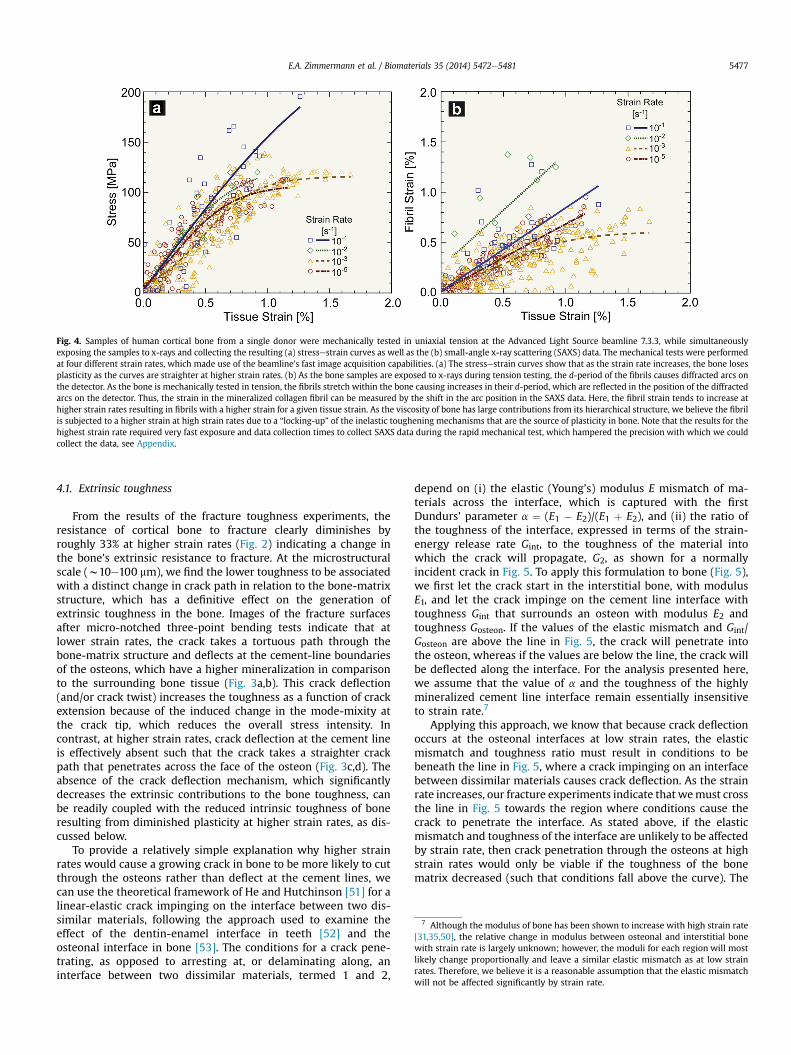
Fig. 4. Samples of human cortical bone from a single donor were mechanically tested in uniaxial tension at the Advanced Light Source beamline 7.3.3, while simultaneouslyexposing the samples to x-rays and collecting the resulting (a) stressestrain curves as well as the (b) small-angle x-ray scattering (SAXS) data. The mechanical tests were performedat four different strain rates, which made use of the beamline’s fast image acquisition capabilities. (a) The stressestrain curves show that as the strain rate increases, the bone losesplasticity as the curves are straighter at higher strain rates. (b) As the bone samples are exposed to x-rays during tension testing, the d-period of the fibrils causes diffracted arcs onthe detector. As the bone is mechanically tested in tension, the fibrils stretch within the bone causing increases in their d-period, which are reflected in the position of the diffractedarcs on the detector. Thus, the strain in the mineralized collagen fibril can be measured by the shift in the arc position in the SAXS data. Here, the fibril strain tends to increase athigher strain rates resulting in fibrils with a higher strain for a given tissue strain. As the viscosity of bone has large contributions from its hierarchical structure, we believe the fibrilis subjected to a higher strain at high strain rates due to a “locking-up” of the inelastic toughening mechanisms that are the source of plasticity in bone. Note that the results for thehighest strain rate required very fast exposure and data collection times to collect SAXS data during the rapid mechanical test, which hampered the precision with which we couldcollect the data, see Appendix.
7 Although the modulus of bone has been shown to increase with high strain rate[31,35,50], the relative change in modulus between osteonal and interstitial bonewith strain rate is largely unknown; however, the moduli for each region will mostlikely change proportionally and leave a similar elastic mismatch as at low strainrates. Therefore, we believe it is a reasonable assumption that the elastic mismatchwill not be affected significantly by strain rate.
E.A. Zimmermann et al. / Biomaterials 35 (2014) 5472e5481 5477
4.1. Extrinsic toughness
From the results of the fracture toughness experiments, theresistance of cortical bone to fracture clearly diminishes byroughly 33% at higher strain rates (Fig. 2) indicating a change inthe bone’s extrinsic resistance to fracture. At the microstructuralscale (w10e100 mm), we find the lower toughness to be associatedwith a distinct change in crack path in relation to the bone-matrixstructure, which has a definitive effect on the generation ofextrinsic toughness in the bone. Images of the fracture surfacesafter micro-notched three-point bending tests indicate that atlower strain rates, the crack takes a tortuous path through thebone-matrix structure and deflects at the cement-line boundariesof the osteons, which have a higher mineralization in comparisonto the surrounding bone tissue (Fig. 3a,b). This crack deflection(and/or crack twist) increases the toughness as a function of crackextension because of the induced change in the mode-mixity atthe crack tip, which reduces the overall stress intensity. Incontrast, at higher strain rates, crack deflection at the cement lineis effectively absent such that the crack takes a straighter crackpath that penetrates across the face of the osteon (Fig. 3c,d). Theabsence of the crack deflection mechanism, which significantlydecreases the extrinsic contributions to the bone toughness, canbe readily coupled with the reduced intrinsic toughness of boneresulting from diminished plasticity at higher strain rates, as dis-cussed below.
To provide a relatively simple explanation why higher strainrates would cause a growing crack in bone to be more likely to cutthrough the osteons rather than deflect at the cement lines, wecan use the theoretical framework of He and Hutchinson [51] for alinear-elastic crack impinging on the interface between two dis-similar materials, following the approach used to examine theeffect of the dentin-enamel interface in teeth [52] and theosteonal interface in bone [53]. The conditions for a crack pene-trating, as opposed to arresting at, or delaminating along, aninterface between two dissimilar materials, termed 1 and 2,
depend on (i) the elastic (Young’s) modulus E mismatch of ma-terials across the interface, which is captured with the firstDundurs’ parameter a ¼ (E1 � E2)/(E1 þ E2), and (ii) the ratio ofthe toughness of the interface, expressed in terms of the strain-energy release rate Gint, to the toughness of the material intowhich the crack will propagate, G2, as shown for a normallyincident crack in Fig. 5. To apply this formulation to bone (Fig. 5),we first let the crack start in the interstitial bone, with modulusE1, and let the crack impinge on the cement line interface withtoughness Gint that surrounds an osteon with modulus E2 andtoughness Gosteon. If the values of the elastic mismatch and Gint/Gosteon are above the line in Fig. 5, the crack will penetrate intothe osteon, whereas if the values are below the line, the crack willbe deflected along the interface. For the analysis presented here,we assume that the value of a and the toughness of the highlymineralized cement line interface remain essentially insensitiveto strain rate.7
Applying this approach, we know that because crack deflectionoccurs at the osteonal interfaces at low strain rates, the elasticmismatch and toughness ratio must result in conditions to bebeneath the line in Fig. 5, where a crack impinging on an interfacebetween dissimilar materials causes crack deflection. As the strainrate increases, our fracture experiments indicate that wemust crossthe line in Fig. 5 towards the region where conditions cause thecrack to penetrate the interface. As stated above, if the elasticmismatch and toughness of the interface are unlikely to be affectedby strain rate, then crack penetration through the osteons at highstrain rates would only be viable if the toughness of the bonematrix decreased (such that conditions fall above the curve). The

Fig. 5. To theoretically determine whether a crack at an interface between two dissimilar materials will deflect at the interface or penetrate through the interface, the He andHutchinson [51] framework is applied, shown here for a normally incident crack. Based on the ratio of the interfacial toughness and the toughness of the osteonal bone, bothexpressed in terms of the strain-energy release rate G, as well as Dundurs’ parameter, a (in this case, the elastic mismatch between interstitial and osteonal bone), we can determinewhether the point lies above or below the critical condition. Based on the assumption that the toughness of the cement line remains constant and that the relative elastic pa-rameters change relatively with strain rate, conditions would change from the crack deflecting at the interstitial bone/osteon (cement line) interface at low strain rates to one athigh strain rates where an incident crack would penetrate the interface, if the toughness of the osteon decreases at such high rates.
E.A. Zimmermann et al. / Biomaterials 35 (2014) 5472e54815478
implication of this result is that when loading is applied to bone at ahigher rate, the bone-matrix structure ahead of a growing crack hasthe effect of appearing to be less ductile, i.e., displaying less plas-ticity or toughness, and as a result, the cement lines are no longer asource of extrinsic toughening through the generation of deflectedcrack trajectories.8
4.2. Intrinsic toughness
The intrinsic toughness of the bone describes its inherentresistance to fracture, which primarily originates through plasticitymechanisms at small length-scales. As the fracture toughness re-sults suggest that the bone matrix toughness diminishes at higherstrain rates, we used SAXS/WAXD to study the intrinsic tougheningmechanisms at the fibrillar scale.
Previous experimental and molecular dynamics studies onunmineralized and mineralized collagen fibrils indicate thatduring elastic (recoverable) straining, fibrillar deformation oc-curs through stretching of the fibrils [8,9,11,25]. At the onset ofinelastic deformation, slippage and/or sliding mechanisms be-tween the collagen and mineral components as well as betweenfibrils have been proposed and may include the breaking/reforming of sacrificial bonds and/or the formation of dilatationalbands [10e12,25,56]. These deformation mechanisms allow thebone to absorb deformation energy during inelastic (post-yield)straining.
Through small-angle x-ray scattering, we investigated fibrillardeformation at low and high strain rates. At slower strain rates,we found that the fibrillar strain linearly increases with tissuestrain (i.e., applied strain) in the elastic region implyingstretching of the fibril (Fig. 4b). As the applied strain increases, aplateau is reached. At this point, essentially the maximum strainof the fibril has been reached and further deformation is
8 Comparable effects can occur in bone with aging, irradiation damage and dis-ease where abnormal mineralization, and/or cross-linking profiles within the ma-trix can reduce the relative inhomogeneity between the bone matrix and thecement lines, again contributing to less deflected crack paths [25,45,54,55].
proposed to occur through inelastic mechanisms, such as slidingbetween fibrils, or possibly microcracking at higher length-scales. This inelastic deformation allows the bone to developintrinsic toughness because the higher length-scales can deformrather than induce complete fracture of the fibrils [25].
At higher strain rates, the fibril strain vs. tissue strain has ahigher slope (Fig. 4b), especially for the two highest strain rates.The higher slope at higher strain rates implies that for a giventissue strain, more strain or deformation occurs within the fibril.The fact that more strain occurs within the fibril implies thatmechanisms responsible for bone’s inelasticity (i.e., fibrillarsliding, sacrificial debonding, etc.) are constrained becausedeformation is not dissipated through these inelastic mecha-nisms but directly results in fibril stretching. We believe that thisbehavior is related to the viscosity or time-dependent nature ofthe deformation of the whole mineralized collagen fibrilstructure.
To study this in further detail, we need to focus on the originsof inelastic deformation within the bone. A common assertion inthe literature has been that bone develops its toughness from itscollagen constituents, which naturally have viscoelastic materialbehavior in comparison to the whole bone or the mineral.However, recent experimental and computational studies havefound that the whole fibril (in this case, unmineralized) is moreviscous than a single collagen molecule [9,57]. This plays on theidea that while strength comes from stretching of the fibril (i.e.,composite of collagen and mineral), toughness comes from thehierarchical architecture, which allows damage tolerance [58].Thus, the viscous or inelastic nature of bone may not originatecompletely from the collagen constituent but from the multilength-scale architecture, in this case of the fibril. Indeed, thearrangement of collagen and mineral within the fibril as well asthe relative assembly of fibrils allows sliding within and betweenfibrils, sacrificial length-scales, and the opening of dilatationalbands or microcracks, which all allow bone to deforminelastically.
With respect to the SAXS results, we see more strain in the fibrilbecause the viscous mechanisms essentially “lock-up” at the higher
E.A. Zimmermann et al. / Biomaterials 35 (2014) 5472e5481 5479
strain rates, i.e., strain-rate stiffening akin to the behavior of adashpot at high rates of deformation. However, instead ofrestricting stretching (as a higher cross-link profile would do), theloss in viscosity allows the fibril to stretch further. Thus, at slowerstrain rates, the fibrils have a certain amount of plasticity and en-ergy absorption through sliding mechanisms within and betweenfibrils, while at higher strain rates, the sliding mechanisms lock-upcausing the fibrils themselves to stretch further, accounting for lessoverall plasticity, but higher strength, in the macroscopicproperties.
Studies at low strain rates provide a pathway towards un-derstanding the complexities of deformation in human bone andthe mechanisms at multiple size-scales that resist bone fracture.However, studies such as this that simulate physiological con-ditions reinforce the necessity to incorporate the physiologicalconditions of environment and disease in order to make progresstowards decreasing the incidence of bone fracture.
Finally, the shortcomings of this study should be noted in thattesting at the physiological strain rates required us to test thelimits of the experimental equipment and the flux limits of athird-generation synchrotron. Specifically at high strain rates,short exposure times and fast data collection times wererequired to make multiple measurements along the stressestraincurve for each sample. Both of these factors reduce the numberof scattering events captured on the SAXS detector, which in turnreduces the precision with which we can measure the fibrillarand mineral strains at the highest strain rate. The precision of thestrain measurement is given in Appendix Fig. A1 and should beregarded as a limitation of this study. An additional limitation isthe use of a single human donor for the experiments. Whiletesting the effects of strain rate on a single donor eliminates theconfounding factors of inter-individual variability, only using asingle donor does pose a limitation to broadly applying the re-sults to a larger population.
5. Summary and conclusions
Human cortical bone is a complex hierarchical composite thatallows deformation at numerous length-scales throughout itsstructure. At low strain rates, bone resists fracture through thestretching of the mineralized fibrils followed by the generation ofplasticity through inelastic mechanisms, such as intra-/interfibrillarsliding, sacrificial bonding, and dilatational band formation. Addi-tionally, at the micron length-scale, the microstructure resists crackgrowth through the mechanisms of crack deflection along cementlines and uncracked-ligament bridging that both increase thebone’s extrinsic toughness.
The results of this study indicate that these tougheningmechanisms may change as bone is loaded at higher strain rates.First, at large length-scales, the bone has a lower toughnessassociated with straighter crack paths across osteons, in com-parison to the tortuous crack path taken along the cement linesat low strain rates. The loss in the extrinsic crack deflectionmechanism implies that the toughness of the bone matrix mustbe lower at higher strain rates. SAXS experiments support alower matrix toughness by indicating that a greater proportion ofthe applied strain is transferred to the fibrils at high strain rates,which may be due to the “locking-up” of the viscous mechanismspromoting inelastic or intrinsic toughness.
Specifically, we conclude that:
1. Strength tests reveal a progressive loss in plasticity as strain rateincreases, which indicates that structural mechanisms promot-ing inelastic deformation change with strain rate.
2. As the strain rate (or stress-intensity rate) increases (by somefour orders of magnitude), the fracture toughness decreasesby some 33%. Cracks propagate across the osteonal matrixtaking a straighter path than at lower strain rates, where thetoughness increases as cracks deflect and follow the cementlines. The absence of crack deflection at the osteonal in-terfaces represents a significant loss in extrinsic toughness ofthe bone.
3. The fact that the cracks do not deflect at the osteonal interfacesat higher strain rates can be qualitatively explained through aHe and Hutchinson analysis. Essentially, if the bone matrixtoughness (i.e., the energy absorption capability) decreases inrelation to the toughness of the cement line, then the crack willpenetrate the cement line instead of arresting or deflectingalong it.
4. SAXS/WAXD allows us to investigate deformation at the fibrillarandmineral length-scales. As the strain rate increases, the fibrilscarry a higher proportion of strain. Indeed, the viscous nature ofthe deformationmechanisms in bone is deemed to originate notonly from the collagen molecules but also from the hierarchicalstructure of the collagen fibrils and fibers. Inelastic deformationwithin the fibrils essentially “locks-up” at increasing strain rate;consequently the fibrils are subjected to an increasing stretchwith less contributions from the viscous toughening mecha-nisms, which may include intra- and interfibrillar sliding, dila-tational band formation, microcracking and breaking/reformingof sacrificial bonds.
Acknowledgments
This work was supported by the National Institute of Health(NIH/NIDCR) under grant no. 5R01 DE015633 to the LawrenceBerkeley National Laboratory (LBNL). BB was supported by theEmmy Noether program of the German Research Foundation (DFG)under grant number BU 2562/2-1. We acknowledge the use of thex-ray synchrotron beamline 7.3.3 (SAXS/WAXD) at the AdvancedLight Source (ALS) at LBNL, which is funded by the Office of Scienceof the U.S. Department of Energy under contract no. DE-AC02-05CH11231. The authors wish to thank Dr. Tony Tomsia at LBNLfor his support.
Appendix. Interpretation of SAXS data at high strain rates
Small-angle x-ray scattering duringmechanical tension tests is acommonly used technique to investigate the strain at the fibrillength-scale in relation to the deformation applied at the wholebone level [25,43,59]. However, even at low strain rates (such as10�5 to 10�3 s�1), small variations in data are to be expected. Thesevariations can be caused by the heterogeneity of the bone structure,associated with the inherent variability in composition and struc-ture at the local scale. The consequent scatter in measured me-chanical properties of the collagen fibrils is not uncommon and hasalso been reported with other techniques, such as atomic forcemicroscopy [60,61].
The SAXS technique does pose further limitations at fast strainrates. The experimental limitations of the equipment specificallyapply to the fastest strain rate of 10�1 s�1 where the fast datacollection times reduce the number of scattering events that can becaptured (see Fig. A1a,b). The reduced number of scattering eventscan then lower the precision of the fibril d-spacing measurement.Suchfibril strainmeasurements at fast strain rates are thus affectedby the precision of data measurement and by local variations instructure.
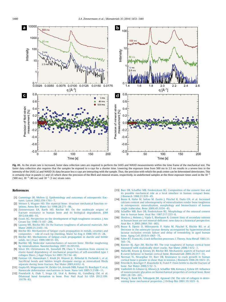
Fig. A1. As the strain rate is increased, faster data collection rates are required to perform the SAXS and WAXD measurements within the time frame of the mechanical test. Thefaster data collection also requires that the samples be exposed to x-rays for a shorter time. Lowering the exposure time from 500 ms to 2.5 ms results in a severe loss in theintensity of the SAXS (a) andWAXD (b) data because less x-rays are interacting with the sample. Thus, the precisionwith which the peak center can be determined deteriorates. Thisis certainly clear in panels (c) and (d) which show the precision of the fibril and mineral strains, respectively, in undeformed samples at the three exposure times used in the 10�5
(500 ms), 10�3 (40 ms) and 10�1 (3 ms) strain rates.
E.A. Zimmermann et al. / Biomaterials 35 (2014) 5472e54815480
References
[1] Cummings SR, Melton LJ. Epidemiology and outcomes of osteoporotic frac-tures. Lancet 2002;359:1761e7.
[2] Weiner S, Wagner HD. The material bone: structure mechanical function re-lations. Annu Rev Mater Sci 1998;28:271e98.
[3] Zimmermann EA, Barth HD, Ritchie RO. On the multiscale origins offracture resistance in human bone and its biological degradation. JOM2012;64:486e93.
[4] Evans AG. Perspective on the development of high-toughness ceramics. J AmCeram Soc 1990;73:187e206.
[5] Launey ME, Ritchie RO. On the fracture toughness of advanced materials. AdvMater 2009;21:2103e10.
[6] Ritchie RO. Mechanisms of fatigue crack-propagation in metals, ceramics andcomposites e role of crack tip shielding. Mater Sci Eng A 1988;103:15e28.
[7] Ritchie RO. Mechanisms of fatigue-crack propagation in ductile and brittlesolids. Int J Fract 1999;100:55e83.
[8] Buehler MJ. Molecular nanomechanics of nascent bone: fibrillar tougheningby mineralization. Nanotechnology 2007;18:295102.
[9] Silver FH, Christiansen DL, Snowhill PB, Chen Y. Transition from viscous toelastic-based dependency of mechanical properties of self-assembled type Icollagen fibers. J Appl Polym Sci 2001;79:134e42.
[10] Fantner GE, Hassenkam T, Kindt JH, Weaver JC, Birkedal H, Pechenik L, et al.Sacrificial bonds and hidden length dissipate energy as mineralized fibrilsseparate during bone fracture. Nat Mater 2005;4:612e6.
[11] Gupta HS, Wagermaier W, Zickler GA, Aroush DRB, Funari SS, Roschger P, et al.Nanoscale deformation mechanisms in bone. Nano Lett 2005;5:2108e11.
[12] Poundarik A, Diab T, Sroga GE, Ural A, Boskey AL, Gundberg CM, et al.Dilational band formation in bone. Proc Natl Acad Sci USA 2012;109:19178e83.
[13] Burr DB, Schaffler MB, Frederickson RG. Composition of the cement line andits possible mechanical role as a local interface in human compact bone.J Biomech 1988;21:939e45.
[14] Busse B, Hahn M, Soltau M, Zustin J, Püschel K, Duda GN, et al. Increasedcalcium content and inhomogeneity of mineralization render bone toughnessin osteoporosis: mineralization, morphology and biomechanics of humansingle trabeculae. Bone 2009;45:1034e43.
[15] Schaffler MB, Burr DB, Frederickson RG. Morphology of the osteonal cementline in human bone. Anat Rec 1987;217:223e8.
[16] Skedros J, Holmes J, Vajda E, Bloebaum R. Cement lines of secondary osteonsin human bone are not mineral-deficient: new data in a historical perspective.Anat Rec A 2005;286A:781e803.
[17] Busse B, Djonic D, Milovanovic P, Hahn M, Püschel K, Ritchie RO, et al.Decrease in the osteocyte lacunar density accompanied by hypermineralizedlacunar occlusion reveals failure and delay of remodeling in aged humanbone. Aging Cell 2010;9:1065e75.
[18] Faber KT, Evans AG. Crack deflection processes. I. Theory. Acta Metall 1983;31:565e76.
[19] Koester KJ, Ager JW, Ritchie RO. The true toughness of human cortical bonemeasured with realistically short cracks. Nat Mater 2008;7:672e7.
[20] Nalla RK, Kruzic JJ, Kinney JH, Ritchie RO. Mechanistic aspects of fracture andR-curve behavior in human cortical bone. Biomaterials 2005;26:217e31.
[21] Norman TL, Nivargikar SV, Burr DB. Resistance to crack growth in humancortical bone is greater in shear than in tension. J Biomech 1996;29:1023e31.
[22] Peterlik H, Roschger P, Klaushofer K, Fratzl P. From brittle to ductile fracture ofbone. Nat Mater 2006;5:52e5.
[23] Vashishth D, Gibson GJ, Khoury JI, Schaffler MB, Kimura J, Fyhrie DP. Influenceof nonenzymatic glycation on biomechanical properties of cortical bone. Bone2001;28:195e201.
[24] Wang X, Bank RA, TeKoppele JM, Agrawal CM. The role of collagen in deter-mining bone mechanical properties. J Orthop Res 2001;19:1021e6.

E.A. Zimmermann et al. / Biomaterials 35 (2014) 5472e5481 5481
[25] Zimmermann EA, Schaible E, Bale H, Barth HD, Tang SY, Reichert P, et al. Age-related changes in the plasticity and toughness of human cortical bone atmultiple length scales. Proc Natl Acad Sci USA 2011;108:14416e21.
[26] Zioupos P, Currey JD. Changes in the stiffness, strength, and toughness ofhuman cortical bone with age. Bone 1998;22:57e66.
[27] Burr DB, Milgrom C, Fyhrie D, Forwood M, Nyska M, Finestone A, et al. In vivomeasurement of human tibial strains during vigorous activity. Bone 1996;18:405e10.
[28] Lanyon LE, Hampson WGJ, Goodship AE, Shah JS. Bone deformation recordedinvivo from strain gauges attached to human tibial shaft. Acta Orthop Scand1975;46:256e68.
[29] Rubin CT, Lanyon LE. Limb mechanics as a function of speed and gait e a studyof functional strains in the radius and tibia of horse and dog. J Exp Biol1982;101:187e211.
[30] Robinovitch SN, Hayes WC, McMahon TA. Prediction of femoral impact forcesin falls on the hip. J Biomech Eng 1991;113:366e74.
[31] Hansen U, Zioupos P, Simpson R, Currey JD, Hynd D. The effect of strain rate onthe mechanical properties of human cortical bone. J Biomech Eng 2008;130:011011.
[32] Adharapurapu RR, Jiang F, Vecchio KS. Dynamic fracture of bovine bone. MaterSci Eng C 2006;26:1325e32.
[33] Behiri JC, Bonfield W. Fracture-mechanics of bone e the effects of density,specimen thickness and crack velocity on longitudinal fracture. J Biomech1984;17:25e34.
[34] Carter DR, Hayes WC. The compressive behavior of bone as a two-phaseporous structure. J Bone Joint Surg Am 1977;59:954e62.
[35] Crowninshield RD, Pope MH. The response of compact bone in tension atvarious strain rates. Ann Biomed Eng 1974;2:217e25.
[36] Evans GP, Behiri JC, Vaughan LC, Bonfield W. The response of equine corticalbone to loading at strain rates experienced in vivo by the galloping horse.Equine Vet J 1992;24:125e8.
[37] Kirchner H. Ductility and brittleness of bone. Int J Fract 2006;139:509e16.[38] Kulin RM, Jiang F, Vecchio KS. Loading rate effects on the R-curve behavior of
cortical bone. Acta Biomater 2011;7:724e32.[39] Kulin RM, Jiang F, Vecchio KS. Effects of age and loading rate on equine cortical
bone failure. J Mech Behav Biomed 2011;4:57e75.[40] McElhaney JH. Dynamic response of bone and muscle tissue. J Appl Physiol
1966;21:1231e6.[41] Wright TM, Hayes WC. Tensile testing of bone over a wide range of strain
rates: effects of strain rate, microstructure and density. Med Biol Eng 1976;14:671e80.
[42] Zioupos P, Hansen U, Currey JD. Microcracking damage and the fractureprocess in relation to strain rate in human cortical bone tensile failure.J Biomech 2008;41:2932e9.
[43] Gupta HS, Seto J, Wagermaier W, Zaslansky P, Boesecke P, Fratzl P. Coopera-tive deformation of mineral and collagen in bone at the nanoscale. Proc NatlAcad Sci USA 2006;103:17741e6.
[44] Hexemer A, Bras W, Glossinger J, Schaible E, Gann E, Kirian R, et al. A SAXS/WAXS/GISAXS beamline with multilayer monochromator. JPCS 2010;247:012007.
[45] Barth HD, Launey ME, MacDowell AA, Ager JW, Ritchie RO. On the effect of x-ray irradiation on the deformation and fracture behavior of human corticalbone. Bone 2010;46:1475e85.
[46] Ilavsky J. Nika: software for two-dimensional data reduction. J Appl Crys-tallogr 2012;45:324e8.
[47] Schindelin J, Arganda-Carreras I, Frise E, Kaynig V, Longair M, Pietzsch T, et al.Fiji: an open-source platform for biological-image analysis. Nat Methods2012;9:676e82.
[48] E08 Committee. E1820-09 Standard Test Method for Measurement of FractureToughness. ASTM International; 2009.
[49] Koester KJ, Barth HD, Ritchie RO. Effect of aging on the transverse toughnessof human cortical bone: evaluation by R-curves. J Mech Behav Biomed 2011;4:1504e13.
[50] Currey JD. The effects of strain rate, reconstruction and mineral content onsome mechanical properties of bovine bone. J Biomech 1975;8:81e6.
[51] He MY, Hutchinson JW. Crack deflection at an interface between dissimilarelastic-materials. Int J Solids Struct 1989;25:1053e67.
[52] Imbeni V, Kruzic JJ, Marshall GW, Marshall SJ, Ritchie RO. The dentineenameljunction and the fracture of human teeth. Nat Mater 2005;4:229e32.
[53] Chan KS, Nicolella DP. Micromechanical modeling of R-curve behaviors inhuman cortical bone. J Mech Behav Biomed 2012;16:136e52.
[54] Barth HD, Zimmermann EA, Schaible E, Tang SY, Alliston T, Ritchie RO.Characterization of the effects of x-ray irradiation on the hierarchical struc-ture and mechanical properties of human cortical bone. Biomaterials 2011;32:8892e904.
[55] Busse B, Bale H, Zimmermann EA, Panganiban B, Barth HB, Carriero A, et al.Vitamin D deficiency induces early signs of aging in human bone, increasingthe risk of fracture. Sci Transl Med 2013;5. 193ra188.
[56] Gupta HS, Krauss S, Kerschnitzki M, Karunaratne A, Dunlop JWC, Barber AH,et al. Intrafibrillar plasticity through mineral/collagen sliding is the dominantmechanism for the extreme toughness of antler bone. J Mech Behav Biomed2013;28:366e82.
[57] Gautieri A, Vesentini S, Redaelli A, Buehler MJ. Viscoelastic properties ofmodel segments of collagen molecules. Matrix Biol 2012;31:141e9.
[58] Zhang Z, Zhang Y-W, Gao H. On optimal hierarchy of load-bearing biologicalmaterials. Proc R Soc B 2011;278:519e25.
[59] Sasaki N, Shukunami N, Matsushima N, Izumi Y. Time-resolved x-raydiffraction from tendon collagen during creep using synchrotron radiation.J Biomech 1999;32:285e92.
[60] Hang F, Barber AH. Nano-mechanical properties of individual mineralizedcollagen fibrils from bone tissue. J R Soc Interface 2011;8:500e5.
[61] Tai K, Dao M, Suresh S, Palazoglu A, Ortiz C. Nanoscale heterogeneity pro-motes energy dissipation in bone. Nat Mater 2007;6:454e62.