for dental research 2015 promotes orthodontic tooth movement
TRANSCRIPT

Journal of Dental Research2015, Vol. 94(9) 1286 –1294© International & American Associations for Dental Research 2015Reprints and permissions: sagepub.com/journalsPermissions.navDOI: 10.1177/0022034515589714jdr.sagepub.com
Research Reports: Biological
IntroductionOrthodontic tooth movement (OTM) induced by mechanical force is governed by local aseptic inflammation-associated alveolar bone remodeling. It includes osteoclastic bone resorp-tion on the compression sides and osteoblastic bone formation on the tension sides (Garlet et al. 2007), along with increased levels of cytokines, chemokines, and leukocyte infiltration (Alhashimi et al. 2000; Krishnan and Davidovitch 2006; Meikle 2006; Garlet et al. 2007; Ren et al. 2007). Moreover, macrophage-like cells, which are found in the initial phase of OTM, may enhance bone and root resorption (Brudvik and Rygh 1993). However, the detailed role of macrophages in OTM remains unknown.
As progenitors of osteoclasts, macrophages directly medi-ate osteoclast differentiation and promote bone resorption (Teitelbaum 2000). Macrophages are also important in inflam-mation and host defense. Macrophages can be polarized into different phenotypes, including classically activated M1 mac-rophages and alternatively activated M2 macrophages (Mosser and Edwards 2008; Sica and Mantovani 2012). M1 macro-phages mediate inflammation by producing tumor necrosis factor (TNF)-α, interleukins (IL)-1, IL-6, and inducible nitric oxide synthase (iNOS) (Dale et al. 2008); conversely, M2 mac-rophages function as anti-inflammatory agents by producing
anti-inflammatory products (Gordon 2003). As important immune cells that mediate inflammation, macrophages play a crucial role in diseases characterized by inflammatory-medi-ated bone loss, such as rheumatoid arthritis and periodontal diseases (Arend and Dayer 1990; Shaddox et al. 2011). However, the detailed role of macrophages in OTM is largely unknown. Our previous study showed that enhanced M1/M2 macrophage ratio aggravates orthodontic root resorption (He et al. 2015); hence, the role of macrophage polarization in OTM should be elucidated. Considering that OTM is an
589714 JDRXXX10.1177/0022034515589714Journal of Dental ResearchM1-like Macrophage Polarization Promotes Orthodontic Tooth Movementresearch-article2015
1Department of Orthodontics, Peking University School and Hospital of Stomatology, Beijing, P.R. China2Center for Craniofacial Stem Cell Research and Regeneration, Peking University School and Hospital of Stomatology, Beijing, P.R. China3Center for Temporomandibular Disorders and Orofacial Pain, Peking University School and Hospital of Stomatology, Beijing, P.R. China
A supplemental appendix to this article is published electronically only at http://jdr.sagepub.com/supplemental.
Corresponding Author:Y. Zhou, Department of Orthodontics, Peking University School and Hospital of Stomatology, 22# Zhongguancun South Avenue, Haidian District, Beijing, 100081, China. Email: [email protected]
M1-like Macrophage Polarization Promotes Orthodontic Tooth Movement
D. He1,2, X. Kou1,2, R. Yang1,2, D. Liu1,2, X. Wang1,2, Q. Luo1,2, Y. Song1,2, F. Liu1,2, Y. Yan1,2, Y. Gan3, and Y. Zhou1,2
AbstractMacrophages play a crucial role in inflammatory-mediated bone loss. Orthodontic tooth movement (OTM) is associated with inflammatory bone remodeling. However, whether and how macrophages contribute to mechanical force–induced OTM remains unknown. In this study, we hypothesized that polarization of M1-like macrophages may contribute to the OTM. Orthodontic nickel-titanium springs were applied to the upper first molars of rats or mice to induce OTM. The distance of OTM gradually increased after mechanical force was applied to the rats for 5 and 10 d. M1-like macrophage polarization and expression of M1 cytokine tumor necrosis factor (TNF)-α also increased after force application. More importantly, monocyte/macrophage depletion in mice by injection of clodronate liposomes decreased the distance of OTM and the number of tartrate-resistant acid phosphatase (TRAP)–positive osteoclasts and CD68+ macrophages, accompanied by reduced expressions of M1 markers TNF-α and inducible nitric oxide synthase (iNOS), whereas systemic transfusion of M1 macrophages in mice increased them. Further experiments showed that injection of recombinant TNF-α increased the distance of OTM and the number of TRAP-positive osteoclasts and CD68+ macrophages, as well as upregulated the expression of TNF-α and iNOS. Blockage of TNF-α by etanercept injection reduced the distance of OTM and the number of TRAP-positive osteoclasts and CD68+ macrophages, as well as decreased the levels of TNF-α and iNOS. These data suggest that M1-like macrophage polarization promotes alveolar bone resorption and consequent OTM after mechanical force application.
Keywords: orthodontics, biomechanical phenomena, bone resorption, inflammation, macrophage activation, osteoclasts
at Peking University Library on November 20, 2015 For personal use only. No other uses without permission.jdr.sagepub.comDownloaded from
© International & American Associations for Dental Research 2015

M1-like Macrophage Polarization Promotes Orthodontic Tooth Movement 1287
inflammatory process, we hypothesized that M1-like macro-phage polarization may promote OTM after force application.
Rising evidence suggests that fibroblasts are involved in inflammatory processes and interact with immune cells, such as macrophages (Smith et al. 1997; El Kasmi et al. 2014). Periodontal ligament cells (PDLCs) could interact with macro-phages under normal or inflammatory conditions (Konermann et al. 2012). After orthodontic force application, PDLCs express elevated levels of inflammatory cytokines (Lee et al. 2012), including M1 stimulator interferon (IFN)-γ (He et al. 2015). However, the effect of PDLCs on macrophage polariza-tion under mechanical force application remains unclear.
This study aims to examine 1) whether M1-like macro-phage polarization affects OTM after mechanical force appli-cation and 2) what effect PDLCs have on macrophage polarization under mechanical force application.
Materials and Methods
Animals
Adult Sprague-Dawley rats (male, 180 to 200 g, 6 to 7 wk old) and C57BL/6 mice (male, 20 to 25 g, 8 wk old) were used in this study. Experimental protocols were approved by the Animal Use and Care Committee of Peking University (LA2013-92).
Application of Orthodontic Devices
Mechanical force was applied in rats as previously described with modification (Dunn et al. 2007). Briefly, a nickel-titanium coil spring (wire size, 0.2 mm; diameter, 1 mm; length, 4 mm; Smart Technology, Beijing, China) was connected between the maxillary first molar and incisors to provide a nearly constant force of approximately 60 g. Previous studies used 40 to 60 g of force to induce OTM (Dunn et al. 2007; MirHashemi et al. 2013). The contralateral side served as a control.
Mechanical force was applied in mice as previously described with modification (Taddei et al. 2012). Briefly, the spring (wire size, 0.2 mm; diameter, 1 mm; length, 1 mm) was bonded between the maxillary first molar and incisors to deliver a force of approximately 30 g for 7 d. The contralateral side served as a control.
Micro–computed tomography Scanning and Measurement of OTM Distance
The maxillae of rats and mice were scanned using a micro–computed tomography system (Inveon MMCT, Siemens, USA). OTM distance was measured as previously described (Cao et al. 2014). Briefly, the distance was measured between the midpoint of the distal marginal ridge of the first molar and the midpoint of the mesial marginal ridge of the second molar from the occlusal view of the 3-dimensional reconstructed image. Measurement was performed 3 times by a trained researcher blinded to the group design.
In Vivo Depletion of Monocytes/Macrophages
Mice were divided into 2 groups. One group was intravenously injected with clodronate liposomes (200 μL/mouse, 5 mg/mL; Clodrosome, Encapsula NanoSciences, USA) to deplete mono-cytes/macrophages (van Amerongen et al. 2007; Hunter et al. 2010); the other group was injected with control liposomes containing phosphate-buffered saline (PBS). All mice received injections every other day since 1 day prior to 7 d of force application. Each group comprised 5 to 6 mice. The effect of depletion was verified by the levels of SSClowCD11bhigh mono-cytes in the peripheral blood using flow cytometry (Sunderkotter et al. 2004) and the number of CD68+ macrophages in the max-illary alveolar bone.
Isolation of Bone Marrow–derived Macrophages and Adoptive Transfusion of M1 or M2 Macrophages
Bone marrow–derived macrophages were isolated and cul-tured as previously described (Zhang et al. 2013). Briefly, bone marrow cells of mice were flushed out from the bone marrow cavity of femurs and tibias. All nuclear cells were seeded at 1.5 × 10⁷ onto 10-cm culture dishes (Corning). After initial incubation overnight, nonadherent cells were discarded. Adherent cells were continuously cultured in RPMI 1640 sup-plemented with 10% fetal bovine serum, 2mM glutamine, anti-biotics (100 U/mL of penicillin and 100 mg/mL of streptomycin; Biofluids, USA), and murine GM-CSF (20 ng/mL; 315-03, Peprotech, USA) for 6 d. M1 or M2 polarization was induced as previously described with modification (Kou et al. 2015). The cells were stimulated with IFN-γ (20 ng/mL; 400-20, Peprotech, USA) and lipopolysaccharide (10 ng/mL; L2880, Sigma-Aldrich, USA) for 48 h to polarize into M1 macro-phages or with IL-4 (10 ng/mL; 400-04, Peprotech, USA) for 48 h to polarize into M2 macrophages. M1 or M2 macrophages were characterized by real-time polymerase chain reaction.
Cells were collected and intravenously administered into mice (1.5 × 10⁶/mouse) on the day of force application and received 7 d of force application. Each group comprised 5 to 6 mice. To track the transfusion cells in vivo, EGFP-labeled M1 macrophages were obtained from EGFP mice and admin-istered into C57BL/6 mice (1 × 10⁶/mouse).
Injection of Recombinant TNF-α and TNF-α Inhibitor Etanercept
Independent experiments were designed, and different periods were chosen according to the preliminary data for different experiment designs. The first batch of rats was randomly divided into 2 groups. The experiment group was intravenously injected with recombinant TNF-α (rTNF-α; 1 μg/rat; 400-14, Peprotech, USA), and the vehicle group was injected with PBS every day. Force was applied for 10 d.
at Peking University Library on November 20, 2015 For personal use only. No other uses without permission.jdr.sagepub.comDownloaded from
© International & American Associations for Dental Research 2015

1288 Journal of Dental Research 94(9)
Another batch of rats was randomly divided into 2 groups. The experiment group was intraperitoneally injected with TNF-α inhibitor etanercept (250 μg/rat; Wyeth, USA), and the vehicle group was injected with PBS every other day. Force was applied for 12 d. Each group comprised 5 to 6 rats.
Cell Culture and Treatments
The protocol was approved by the Ethical Guidelines of Peking University (PKUSSIRB-201311103) and performed with appropriate informed consents. Human PDLC isolation and static compressive force application on PDLCs were per-formed as previously described (Seo et al. 2004). The PDLCs were subjected to 1 g/cm2 of continuous compressive force for 24 h. The supernatant was collected from primary cultured PDLCs with or without force loading for further experiments.
To detect whether PDLCs can influence macrophage polar-ization under mechanical force application, THP-1 human monocytic cells (1 × 10⁶) were used to differentiate into mac-rophages in a 6-well plate with 2 mL RPMI 1640 medium con-taining 10% fetal bovine serum, 2mM glutamine, antibiotics, and 50 ng/mL PMA (P1585, Sigma, USA) for 24 h. The cells were then cultured with the supernatant of primary cultured PDLCs with or without force loading for 24 h.
Methods
The following are described in detail in the Appendix:
•• Histology and tartrate-resistant acid phosphatase stain-ing (TRAP)
•• Immunohistochemistry and immunofluorescence staining•• Quantitative real-time polymerase chain reaction•• Western blot analysis
Statistical Analysis
Statistical analysis was performed with SPSS 13.0. All data were presented as mean ± SD and assessed by independent 2-tailed Student’s t test or 1-way analysis of variance. Statistical significance was considered at P < 0.05.
Results
Orthodontic Force Application Promoted M1-like Macrophage Accumulation during OTM
Micro–computed tomography images showed that the distance of OTM in rats gradually increased to 324 and 409 μm after orthodontic force was applied for 5 and 10 d (Fig. 1A). After orthodontic force application, the infiltration of CD68+ macro-phages and expression of the proinflammatory cytokine TNF-α increased on the compression side of periodontal tissues (Fig. 1B, C). It should be mentioned that slight root resorption was also observed during OTM. M1 macrophages were accumu-lated in the periodontal tissues, near the alveolar bone and the
root surface (Fig. 1B). Double immunostaining of CD68 and M1 marker iNOS or M2 marker CD163 showed that CD68+iNOS+ M1-like macrophages infiltrated on the com-pression side of periodontal tissues during OTM. After force application for 5 d, CD68+iNOS+ M1-like macrophages increased to 3.44% and maintained a relatively high level (3.30%) after 10 d compared with the control group (0.5%), whereas CD68+CD163+ M2-like macrophages were barely detected on the compressed side of periodontal tissues after force application (Fig. 1D, E).
Mechanical Force–induced OTM Was Repressed by Monocyte/Macrophage Depletion and Enhanced by M1 Macrophage Transfusion
After clodronate liposomes injection in mice, >70% of circu-lated monocytes was eliminated. CD68+ macrophages in the maxillary alveolar bone also significantly decreased (Appendix Fig. 1), confirming the effectiveness of monocyte/macrophage depletion.
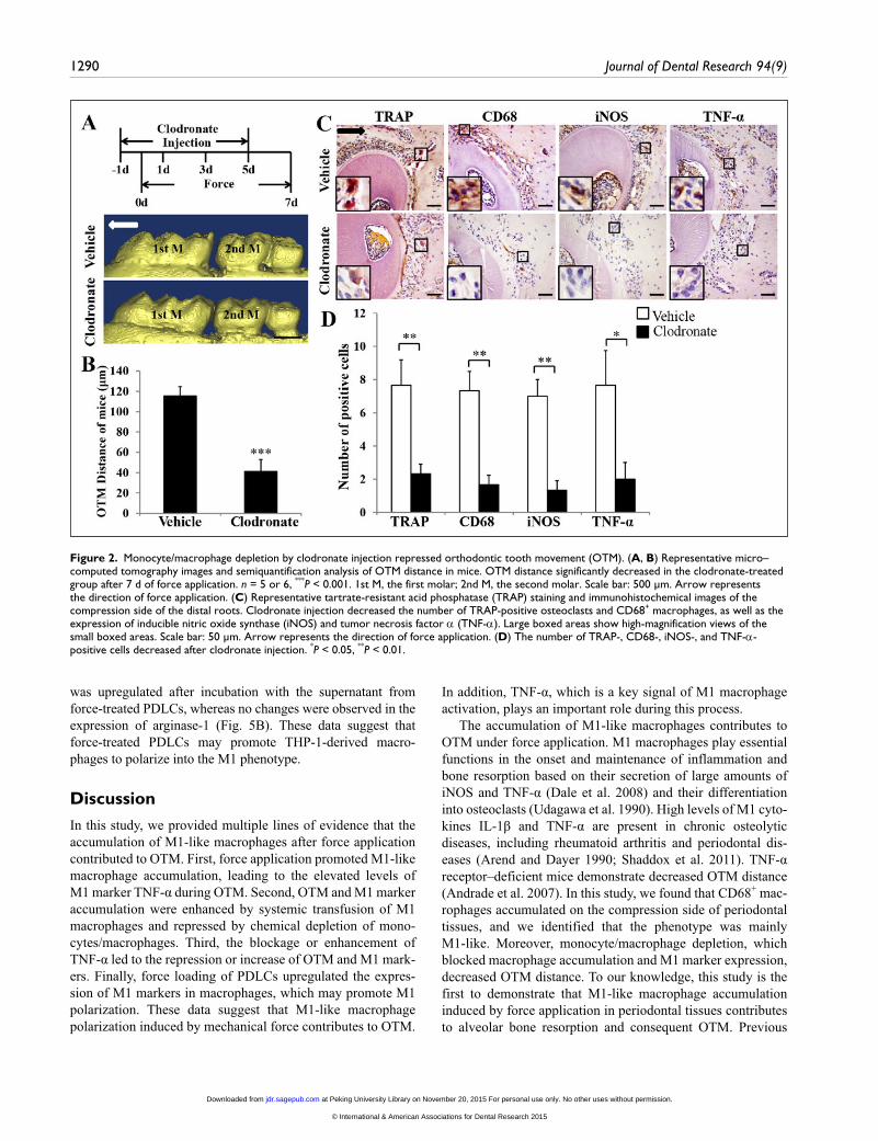
After monocyte/macrophage depletion, OTM distance sig-nificantly decreased to 41 μm, compared with 115 μm of the vehicle group (P < 0.001; Fig. 2A, B). With decreasing OTM distance, the number of tartrate-resistant acid phosphatase (TRAP)–positive osteoclasts and CD68+ macrophages reduced on the compression side in the group treated with clodronate liposomes; the expression of M1 markers TNF-α and iNOS was downregulated as well (Fig. 2C, D).
We further determined the contribution of M1 polarization to OTM. M1 macrophages were confirmed by the high expres-sions of TNF-α and iNOS mRNA (Appendix Fig. 2A). EGFP-labeled cells were found in the compression side on day 7 during OTM, and the green fluorescent signals of EGFP were colocalized with part of CD68+ macrophages (Appendix Fig. 2B). The increase in OTM distance induced by mechanical force in the vehicle group (113 μm) was enhanced by M1 mac-rophage transfusion (145 μm; P < 0.01; Fig. 3A, B). Correspondingly, the number of TRAP-positive osteoclasts and CD68+ macrophages increased on the compression side in the M1 transfusion group; moreover, the expression of iNOS and TNF-α was also upregulated (Fig. 3C, D). These data indi-cate that M1 macrophages play an important role in alveolar bone resorption and consequent OTM. In addition, no signifi-cant change of the OTM distance was detected after M2 mac-rophage transfusion (Appendix Fig. 3), confirming the unique effect of M1 macrophages during OTM.
Mechanical Force–induced OTM Was Repressed by Blockage of TNF-α and Enhanced by Injection of rTNF-αTNF-α could influence M1 macrophage activation and osteo-clast differentiation (Azuma et al. 2000; Mosser and Edwards 2008). In this study, we enhanced or blocked TNF-α by sys-temically injected rTNF-α or TNF-α inhibitor to examine the effect of M1 macrophage activation on OTM.
at Peking University Library on November 20, 2015 For personal use only. No other uses without permission.jdr.sagepub.comDownloaded from
© International & American Associations for Dental Research 2015

M1-like Macrophage Polarization Promotes Orthodontic Tooth Movement 1289
After systemic injection of rTNF-α, the OTM distance increased to 522 μm, compared with 400 μm of the vehicle group (P < 0.001; Fig. 4A, B). By contrast, after TNF-α was blocked through systemic injection of TNF-α inhibitor etaner-cept, the OTM distance decreased to 291 μm, compared with 413 μm of the vehicle group (P < 0.001; Fig. 4E, F). In addi-tion, systemic injection of rTNF-α increased the number of TRAP-positive osteoclasts and CD68+ macrophages on the compression side and upregulated the expression of M1 mark-ers iNOS and TNF-α (Fig. 4C, D). Conversely, blocking TNF-α by etanercept decreased the number of TRAP-positive osteoclasts and CD68+ macrophages and downregulated the expression of iNOS and TNF-α (Fig. 4G, H). These data indi-cate that TNF-α-related M1 macrophage polarization influ-ences bone resorption and consequent OTM.
Force Loading of PDLCs Promoted M1 Polarization of THP-1-derived Macrophages
Since we demonstrated that M1-like macrophages accumulated after force application, we further determined the effect of mechanical force loading of PDLCs on macrophage polariza-tion. THP-1-derived macrophages were cultured with the super-natant from primary cultured PDLCs with or without compressive force loading. After incubation with the superna-tants from force-treated PDLCs, the mRNA expression of M1 markers TNF-α, IL-1β, and IL-6 was upregulated in THP-1-derived macrophages; conversely, the expression of M2 mark-ers DECTIN-1 and arginase-1 did not change, and IL-4R was downregulated (Fig. 5A). Western blot analysis showed that the protein expression of TNF-α in THP-1-derived macrophages
Figure 1. M1-like macrophages accumulated on the compression side of periodontal tissues after orthodontic force application. (A) Representative micro–computed tomography images and semiquantification analysis of the orthodontic tooth movement (OTM) distance in rats. OTM distance gradually increased after force was applied for 5 and 10 d (F5d and F10d). n = 5. ***P < 0.001 vs. control; #P < 0.05 vs. F5d. 1st M, the first molar; 2nd M, the second molar. Arrow represents the direction of force application. Scale bar: 1,000 μm. (B) Representative immunohistochemical images of the compression side of distobuccal roots. The expression levels of CD68 and tumor necrosis factor α (TNF-α) were upregulated in F5d and F10d. Arrow represents the direction of force application. Large boxed areas show high-magnification views of the small boxed areas. Scale bar: 50 μm. (C) The number of CD68- and TNF-α-positive cells increased in F5d and F10d. n = 5; ***P < 0.001 vs. control. (D) Representative immunofluorescence images and hematoxylin and eosin staining on the compression side of distobuccal roots. The number of CD68-positive (red) and inducible nitric oxide synthase (iNOS)–positive (green) M1-like macrophages (merged yellow) increased in F5d and F10d, whereas the number of CD68- (red) and CD163-positive (green) M2-like macrophages (merged yellow) remained unchanged. Corresponding hematoxylin and eosin staining showed the histomorphology of the immunostained sections. Dashed lines mark the outline of the roots. Scale bar: 50 μm. Arrow represents the direction of force application. (E) Semiquantification of double-stained positive cells. M1 proportion increased after force application, whereas M2 proportion did not change. n = 5 or 6; M1-positive ratio: ***P < 0.001 vs. control.
at Peking University Library on November 20, 2015 For personal use only. No other uses without permission.jdr.sagepub.comDownloaded from
© International & American Associations for Dental Research 2015
1290 Journal of Dental Research 94(9)
was upregulated after incubation with the supernatant from force-treated PDLCs, whereas no changes were observed in the expression of arginase-1 (Fig. 5B). These data suggest that force-treated PDLCs may promote THP-1-derived macro-phages to polarize into the M1 phenotype.
DiscussionIn this study, we provided multiple lines of evidence that the accumulation of M1-like macrophages after force application contributed to OTM. First, force application promoted M1-like macrophage accumulation, leading to the elevated levels of M1 marker TNF-α during OTM. Second, OTM and M1 marker accumulation were enhanced by systemic transfusion of M1 macrophages and repressed by chemical depletion of mono-cytes/macrophages. Third, the blockage or enhancement of TNF-α led to the repression or increase of OTM and M1 mark-ers. Finally, force loading of PDLCs upregulated the expres-sion of M1 markers in macrophages, which may promote M1 polarization. These data suggest that M1-like macrophage polarization induced by mechanical force contributes to OTM.
In addition, TNF-α, which is a key signal of M1 macrophage activation, plays an important role during this process.
The accumulation of M1-like macrophages contributes to OTM under force application. M1 macrophages play essential functions in the onset and maintenance of inflammation and bone resorption based on their secretion of large amounts of iNOS and TNF-α (Dale et al. 2008) and their differentiation into osteoclasts (Udagawa et al. 1990). High levels of M1 cyto-kines IL-1β and TNF-α are present in chronic osteolytic diseases, including rheumatoid arthritis and periodontal dis-eases (Arend and Dayer 1990; Shaddox et al. 2011). TNF-α receptor–deficient mice demonstrate decreased OTM distance (Andrade et al. 2007). In this study, we found that CD68+ mac-rophages accumulated on the compression side of periodontal tissues, and we identified that the phenotype was mainly M1-like. Moreover, monocyte/macrophage depletion, which blocked macrophage accumulation and M1 marker expression, decreased OTM distance. To our knowledge, this study is the first to demonstrate that M1-like macrophage accumulation induced by force application in periodontal tissues contributes to alveolar bone resorption and consequent OTM. Previous
Figure 2. Monocyte/macrophage depletion by clodronate injection repressed orthodontic tooth movement (OTM). (A, B) Representative micro–computed tomography images and semiquantification analysis of OTM distance in mice. OTM distance significantly decreased in the clodronate-treated group after 7 d of force application. n = 5 or 6, ***P < 0.001. 1st M, the first molar; 2nd M, the second molar. Scale bar: 500 μm. Arrow represents the direction of force application. (C) Representative tartrate-resistant acid phosphatase (TRAP) staining and immunohistochemical images of the compression side of the distal roots. Clodronate injection decreased the number of TRAP-positive osteoclasts and CD68+ macrophages, as well as the expression of inducible nitric oxide synthase (iNOS) and tumor necrosis factor α (TNF-α). Large boxed areas show high-magnification views of the small boxed areas. Scale bar: 50 μm. Arrow represents the direction of force application. (D) The number of TRAP-, CD68-, iNOS-, and TNF-α-positive cells decreased after clodronate injection. *P < 0.05, **P < 0.01.
at Peking University Library on November 20, 2015 For personal use only. No other uses without permission.jdr.sagepub.comDownloaded from
© International & American Associations for Dental Research 2015

M1-like Macrophage Polarization Promotes Orthodontic Tooth Movement 1291
studies focused on how local elements affect osteoclastogene-sis of osteoclast precursors (Teitelbaum 2000; Tyrovola et al. 2008). However, the source of accumulated macrophages, which are regarded as osteoclast precursors, is largely unknown. The effect of depletion suggested that circulated monocytes could be a source of M1 accumulation during OTM. This concept was consistent with previous studies, which reported that circulated monocytes are sources of infil-trating macrophages in pathologic processes (Ingersoll et al. 2011; Qian et al. 2011).
Systemic transfusion of M1 but not M2 macrophages pro-motes alveolar bone resorption and OTM. Previous studies have demonstrated that the adoptive transfer of M1 aggravated renal diseases compared with M0 (Ikezumi et al. 2003). Our results of M1 macrophage transfusion were in line with these previous findings. Moreover, no significant change of OTM distance was observed after M2 macrophage transfusion, con-firming the unique effect of M1-like macrophages during OTM. Macrophages are plastic to change to different polariza-tions under different environmental conditions (Sindrilaru et al. 2011; Novak and Koh 2013). During OTM, force-induced
inflammatory microenvironment in periodontal tissues may promote or maintain M1 macrophage polarization, which may reduce the effect of M2 transfusion. How the transfused mac-rophages contribute to OTM is unclear. A previous study showed that macrophages transferred in vivo could traffic to the inflammatory sites (Wang et al. 2007). Our data showed that EGFP-labeled cells were found in the periodontal tissues on day 7 during OTM, indicating that portions of the trans-fused macrophages were recruited to the local periodontal tis-sues and may directly contribute to the OTM. However, only part of the CD68+ macrophages colocalized with the signals of EGFP, suggesting that transfused macrophages may also induce a systemic response and promote circulating mono-cytes/macrophages to accumulate in the local periodontal tis-sues and to increase bone resorption and OTM. Nevertheless, the relative contributions of the transfused and circulating monocytes/macrophages need to be further explored.
Systemic injection of rTNF-α enhanced alveolar bone resorption, consequent OTM, and the expression of M1 mark-ers, whereas the injection of TNF-α inhibitor suppressed them. TNF-α could maintain the activation state of M1 macrophages
Figure 3. Adoptive transfusion of ex vivo polarized M1 macrophages enhanced orthodontic tooth movement (OTM). (A, B) Representative micro–computed tomography images and semiquantification analysis of OTM distance in mice. OTM distance significantly increased in the M1 infusion group after 7 d of force application. n = 5 or 6, **P < 0.01. 1st M, the first molar; 2nd M, the second molar. Arrow represents the direction of force application. Scale bar: 500 μm. (C) Representative tartrate-resistant acid phosphatase (TRAP) staining and immunohistochemical images of the compression side of the distal roots. M1 infusion increased the number of TRAP-positive osteoclasts and CD68+ macrophages, as well as the expression of inducible nitric oxide synthase (iNOS) and tumor necrosis factor α (TNF-α). Large boxed areas show high-magnification views of the small boxed areas. Arrow represents the direction of force application. Scale bar: 50 μm. (D) The number of TRAP-, CD68-, iNOS-, and TNF-α-positive cells increased after M1 transfusion. *P < 0.05, **P < 0.01.
at Peking University Library on November 20, 2015 For personal use only. No other uses without permission.jdr.sagepub.comDownloaded from
© International & American Associations for Dental Research 2015

1292 Journal of Dental Research 94(9)
Figure 4. Blocking tumor necrosis factor α (TNF-α) by etanercept injection repressed orthodontic tooth movement (OTM), whereas injection of recombinant TNF-α (rTNF-α) enhanced OTM. (A, B) Representative micro–computed tomography images and semiquantification analysis of the OTM distance in rTNF-α-treated rats. OTM distance increased after 10 d of force application. Arrow represents the direction of force application. Scale bar: 1000 μm. n = 5 or 6, ***P < 0.001. (C) Representative tartrate-resistant acid phosphatase (TRAP) staining and immunohistochemical images of the compression side of distobuccal roots. Injection of rTNF-α increased the number of TRAP-positive osteoclasts and CD68+ macrophages, as well as the expression of inducible nitric oxide synthase (iNOS) and TNF-α. Large boxed areas show high-magnification views of the small boxed areas. Arrow represents the direction of force application. Scale bar: 50 μm. (D) The number of TRAP-, CD68-, iNOS-, and TNF-α-positive cells increased after rTNF-α injection. *P < 0.05, **P < 0.01. (E, F) Representative micro–computed tomography images and semiquantification analysis of the OTM distance in rats treated with TNF-α inhibitor etanercept. The OTM distance decreased after 12 d of force application. n = 5 or 6, ***P < 0.001. 1st M, the first molar; 2nd M, the second molar. Arrow represents the direction of force application. Scale bar: 1,000 μm. (G) Representative TRAP staining and immunohistochemical images of the compression side of distobuccal roots. Injection of the TNF-α inhibitor decreased the number of TRAP-positive osteoclasts and CD68+ macrophages, as well as the expression of iNOS and TNF-α. Arrow represents the direction of force application. Scale bar: 50 μm. (H) The number of TRAP-, CD68-, iNOS-, and TNF-α-positive cells decreased after TNF-α inhibitor injection. **P < 0.01.
at Peking University Library on November 20, 2015 For personal use only. No other uses without permission.jdr.sagepub.comDownloaded from
© International & American Associations for Dental Research 2015

M1-like Macrophage Polarization Promotes Orthodontic Tooth Movement 1293
in an autocrine manner (Wang et al. 2006). The present study showed that TNF-α expression was upregulated dur-ing OTM. Moreover, enhancement of TNF-α increased the number of M1 macrophages and osteoclasts, accompa-nied by high expression of M1 markers; conversely, reverse effects were detected when TNF-α was suppressed. These data indicate that TNF-α may induce M1 macrophage activation and promote osteoclast differentiation under force application. Nevertheless, mouse and rat OTM models were chosen on the basis of their advantages in cell infu-sion and tooth movement distance, respectively. Our results suggest that TNF-α-related M1-like macrophages play an essential role in mechanical force–induced bone resorption and con-sequent OTM, which may provide insights into novel modalities to accel-erate OTM.
Force-treated PDLCs in periodontal tissues may contribute to M1 macro-phage polarization during OTM. Previous studies have shown that fibroblasts are important in the inflammatory process and can modulate macrophage polar-ization (Smith et al. 1997; El Kasmi et al. 2014). Pulmonary adventitial fibroblasts in pulmonary hypertension sustainedly produce proinflammatory cytokines and chemokines (Li et al. 2011). They could also activate a distinct proinflammatory macrophage phenotype (El Kasmi et al. 2014). Periodontal ligament fibroblasts also influence macrophages to express M2 markers under normal and inflammatory conditions (Konermann et al. 2012). Hence, determining the effect of PDLCs on macrophage polarization under mechanical force stimulation is indispensable. PDLCs are one of the most impor-tant cellular components in periodontal tissues. With mechani-cal force stimuli, PDLCs express multiple inflammatory elements (Lee et al. 2012). Previously we found that mechani-cal force upregulated IFN-γ expression in PDLCs in vivo and in vitro, which may contribute to M1 macrophage activation (He et al. 2015). In this study, we found that mechanical force promoted M1-like macrophage accumulation on the compres-sion side of periodontal tissues. Additionally, the supernatant of force-loaded PDLCs promoted M1 polarization. These data suggest that force-loaded PDLCs may promote M1-like mac-rophage polarization during OTM. However, whether mechan-ical force directly influences macrophage polarization is unclear. Further studies are needed to explore the direct effect of mechanical force on macrophage polarization.
It should be mentioned that slight root resorption was also observed during OTM. The process of OTM may be accompa-nied by orthodontic root resorption. They shared similar histo-logic changes of hard tissue resorption by osteoclasts/
odontoclasts and remodeling. However, clinically, significant tooth movement at the price of severe periodontal ligament damage and root resorption should be avoided. Further work should be done to illustrate the optimal force to gain substantial and stable tooth movement with less periodontal damage.
In conclusion, the accumulation of M1-like macrophages after mechanical force application promotes OTM. During this process, TNF-α, which is a key signal of M1 macrophage acti-vation, plays an important role. These results elucidate the mechanisms of OTM and may provide some clues for acceler-ating OTM.
Author Contributions
D. He, contributed to conception, design, data acquisition, analy-sis, and interpretation, drafted and critically revised the manu-script; X. Kou, contributed to conception, design, data analysis, and interpretation, critically revised the manuscript; R. Yang, con-tributed to design and data interpretation, critically revised the manuscript; D. Liu, X. Wang, Q. Luo, Y. Song, contributed to design, critically revised the manuscript; F. Liu, Y. Yan, contrib-uted to data acquisition, critically revised the manuscript; Y. Gan, contributed to conception and design, critically revised the manu-script; Y. Zhou, contributed to conception, design, and data inter-pretation, critically revised the manuscript. All authors gave final approval and agree to be accountable for all aspects of the work.
Acknowledgments
This study was supported by grants from the National Natural Science Foundation of China (81300897, 81470717) and International S&T Cooperation Program of China (2013DFB
Figure 5. Supernatant from force-treated periodontal ligament cells (PDLCs) enhanced M1 markers of THP-1-induced macrophages. (A) Relative mRNA expression of M1/M2-related genes. The mRNA expression of M1 markers tumor necrosis factor α (TNF-α), interleukin 1β (IL-1β), and IL-6 of THP-1-induced macrophages was upregulated after incubation with the supernatant of force-treated PDLCs for 24 h compared with those incubated with the supernatant of control PDLCs; the mRNA expression of M2 markers DECTIN-1 and arginase-1 did not change, and the expression of IL-4R was downregulated. n = 3, *P < 0.05, **P < 0.01, ***P < 0.001. CS, control supernatant; FS, force-treated supernatant. (B) Western blot of TNF-α and arginase-1. TNF-α expression of THP-1-induced macrophages was upregulated after incubation with the supernatant of force-treated PDLCs for 24 h, whereas no changes were observed in arginase-1. Beta-actin served as an internal control for equal loading. Data represent 3 independent experiments.
at Peking University Library on November 20, 2015 For personal use only. No other uses without permission.jdr.sagepub.comDownloaded from
© International & American Associations for Dental Research 2015

1294 Journal of Dental Research 94(9)
30360). The authors declare no potential conflicts of interest with respect to the authorship and/or publication of this article.
ReferencesAlhashimi N, Frithiof L, Brudvik P, Bakhiet M. 2000. Orthodontic movement
induces high numbers of cells expressing IFN-gamma at mRNA and protein levels. J Interferon Cytokine Res. 20(1):7–12.
Andrade I Jr, Silva TA, Silva GA, Teixeira AL, Teixeira MM. 2007. The role of tumor necrosis factor receptor type 1 in orthodontic tooth movement. J Dent Res. 86(11):1089–1094.
Arend WP, Dayer JM. 1990. Cytokines and cytokine inhibitors or antagonists in rheumatoid arthritis. Arthritis Rheum. 33(3):305–315.
Azuma Y, Kaji K, Katogi R, Takeshita S, Kudo A. 2000. Tumor necrosis factor-alpha induces differentiation of and bone resorption by osteoclasts. J Biol Chem. 275(7):4858–4864.
Brudvik P, Rygh P. 1993. The initial phase of orthodontic root resorption incident to local compression of the periodontal ligament. Eur J Orthod. 15(4):249–263.
Cao H, Kou X, Yang R, Liu D, Wang X, Song Y, Feng L, He D, Gan Y, Zhou Y. 2014. Force-induced Adrb2 in periodontal ligament cells promotes tooth movement. J Dent Res. 93(11):1163–1169.
Dale DC, Boxer L, Liles WC. 2008. The phagocytes: neutrophils and mono-cytes. Blood. 112(4):935–945.
Dunn MD, Park CH, Kostenuik PJ, Kapila S, Giannobile WV. 2007. Local delivery of osteoprotegerin inhibits mechanically mediated bone modeling in orthodontic tooth movement. Bone. 41(3):446–455.
El Kasmi KC, Pugliese SC, Riddle SR, Poth JM, Anderson AL, Frid MG, Li M, Pullamsetti SS, Savai R, Nagel MA, et al. 2014. Adventitial fibroblasts induce a distinct proinflammatory/profibrotic macrophage phenotype in pulmonary hypertension. J Immunol. 193(2):597–609.
Garlet TP, Coelho U, Silva JS, Garlet GP. 2007. Cytokine expression pattern in compression and tension sides of the periodontal ligament during orthodon-tic tooth movement in humans. Eur J Oral Sci. 115(5):355–362.
Gordon S. 2003. Alternative activation of macrophages. Nat Rev Immunol. 3(1):23–35.
He D, Kou X, Luo Q, Yang R, Liu D, Wang X, Song Y, Cao H, Zeng M, Gan Y, et al. 2015. Enhanced m1/m2 macrophage ratio promotes orthodontic root resorption. J Dent Res. 94(1):129–139.
Hunter MM, Wang A, Parhar KS, Johnston MJ, Van Rooijen N, Beck PL, McKay DM. 2010. In vitro-derived alternatively activated macrophages reduce colonic inflammation in mice. Gastroenterology. 138(4):1395–1405.
Ikezumi Y, Atkins RC, Nikolic-Paterson DJ. 2003. Interferon-gamma augments acute macrophage-mediated renal injury via a glucocorticoid-sensitive mechanism. J Am Soc Nephrol. 14(4):888–898.
Ingersoll MA, Platt AM, Potteaux S, Randolph GJ. 2011. Monocyte traffick-ing in acute and chronic inflammation. Trends Immunol. 32(10):470–477.
Konermann A, Stabenow D, Knolle PA, Held SA, Deschner J, Jager A. 2012. Regulatory role of periodontal ligament fibroblasts for innate immune cell function and differentiation. Innate Immun. 18(5):745–752.
Kou XX, Li CS, He DQ, Wang XD, Hao T, Meng Z, Zhou YH, Gan YH. 2015. Estradiol promotes M1–like macrophage activation through cadherin-11 to aggravate temporomandibular joint inflammation in rats. J Immunol. 194(6):2810–2818.
Krishnan V, Davidovitch Z. 2006. Cellular, molecular, and tissue-level reac-tions to orthodontic force. Am J Orthod Dentofacial Orthop. 129(4):469.e1–469.e32.
Lee SI, Park KH, Kim SJ, Kang YG, Lee YM, Kim EC. 2012. Mechanical stress-activated immune response genes via Sirtuin 1 expression in human periodontal ligament cells. Clin Exp Immunol. 168(1):113–124.
Li M, Riddle SR, Frid MG, El Kasmi KC, McKinsey TA, Sokol RJ, Strassheim D, Meyrick B, Yeager ME, Flockton AR, et al. 2011. Emergence of fibro-blasts with a proinflammatory epigenetically altered phenotype in severe hypoxic pulmonary hypertension. J Immunol. 187(5):2711–2722.
Meikle MC. 2006. The tissue, cellular, and molecular regulation of orthodontic tooth movement: 100 years after Carl Sandstedt. Eur J Orthod. 28(3):221–240.
MirHashemi AH, Afshari M, Alaeddini M, Etemad-Moghadam S, Dehpour A, Sheikhzade S, Akhoundi MS. 2013. Effect of atorvastatin on orthodontic tooth movement in male wistar rats. J Dent. 10(6):532–539.
Mosser DM, Edwards JP. 2008. Exploring the full spectrum of macrophage activation. Nat Rev Immunol. 8(12):958–969.
Novak ML, Koh TJ. 2013. Phenotypic transitions of macrophages orchestrate tissue repair. Am J Pathol. 183(5):1352–1363.
Qian BZ, Li J, Zhang H, Kitamura T, Zhang J, Campion LR, Kaiser EA, Snyder LA, Pollard JW. 2011. CCL2 recruits inflammatory monocytes to facilitate breast-tumour metastasis. Nature. 475(7355):222–225.
Ren Y, Hazemeijer H, de Haan B, Qu N, de Vos P. 2007. Cytokine profiles in crevicular fluid during orthodontic tooth movement of short and long dura-tions. J Periodontol. 78(3):453–458.
Seo BM, Miura M, Gronthos S, Bartold PM, Batouli S, Brahim J, Young M, Robey PG, Wang CY, Shi S. 2004. Investigation of multipotent postnatal stem cells from human periodontal ligament. Lancet. 364(9429):149–155.
Shaddox LM, Wiedey J, Calderon NL, Magnusson I, Bimstein E, Bidwell JA, Zapert EF, Aukhil I, Wallet SM. 2011. Local inflammatory markers and systemic endotoxin in aggressive periodontitis. J Dent Res. 90(9):1140–1144.
Sica A, Mantovani A. 2012. Macrophage plasticity and polarization: in vivo veritas. J Clin Investig. 122(3):787–795.
Sindrilaru A, Peters T, Wieschalka S, Baican C, Baican A, Peter H, Hainzl A, Schatz S, Qi Y, Schlecht A, et al. 2011. An unrestrained proinflamma-tory M1 macrophage population induced by iron impairs wound healing in humans and mice. J Clin Investig. 121(3):985–997.
Smith RS, Smith TJ, Blieden TM, Phipps RP. 1997. Fibroblasts as senti-nel cells: synthesis of chemokines and regulation of inflammation. Am J Pathol. 151(2):317–322.
Sunderkotter C, Nikolic T, Dillon MJ, Van Rooijen N, Stehling M, Drevets DA, Leenen PJ. 2004. Subpopulations of mouse blood monocytes differ in matu-ration stage and inflammatory response. J Immunol. 172(7):4410–4417.
Taddei SR, Moura AP, Andrade I Jr, Garlet GP, Garlet TP, Teixeira MM, da Silva TA. 2012. Experimental model of tooth movement in mice: a stan-dardized protocol for studying bone remodeling under compression and tensile strains. J Biomech. 45(16):2729–2735.
Teitelbaum SL. 2000. Bone resorption by osteoclasts. Science. 289(5484):1504–1508.
Tyrovola JB, Spyropoulos MN, Makou M, Perrea D. 2008. Root resorp-tion and the OPG/RANKL/RANK system: a mini review. J Oral Sci. 50(4):367–376.
Udagawa N, Takahashi N, Akatsu T, Tanaka H, Sasaki T, Nishihara T, Koga T, Martin TJ, Suda T. 1990. Origin of osteoclasts: mature monocytes and macrophages are capable of differentiating into osteoclasts under a suitable microenvironment prepared by bone marrow-derived stromal cells. Proc Natl Acad Sci U S A. 87(18):7260–7264.
van Amerongen MJ, Harmsen MC, van Rooijen N, Petersen AH, van Luyn MJ. 2007. Macrophage depletion impairs wound healing and increases left ventricular remodeling after myocardial injury in mice. Am J Pathol. 170(3):818–829.
Wang H, Peters T, Kess D, Sindrilaru A, Oreshkova T, Van Rooijen N, Stratis A, Renkl AC, Sunderkotter C, Wlaschek M, et al. 2006. Activated macro-phages are essential in a murine model for T cell-mediated chronic psoriasi-form skin inflammation. J Clin Investig. 116(8):2105–2114.
Wang Y, Wang YP, Zheng G, Lee VW, Ouyang L, Chang DH, Mahajan D, Coombs J, Wang YM, Alexander SI, et al. 2007. Ex vivo programmed macrophages ameliorate experimental chronic inflammatory renal disease. Kidney Int. 72(3):290–299.
Zhang Q, Atsuta I, Liu S, Chen C, Shi S, Shi S, Le AD. 2013. IL-17-mediated M1/M2 macrophage alteration contributes to pathogenesis of bisphospho-nate-related osteonecrosis of the jaws. Clin Cancer Res. 19(12):3176–3188.
at Peking University Library on November 20, 2015 For personal use only. No other uses without permission.jdr.sagepub.comDownloaded from
© International & American Associations for Dental Research 2015