cellular and molecular changes in orthodontic tooth...
TRANSCRIPT
Review ArticleTheScientificWorldJOURNAL (2011) 11, 1788–1803ISSN 1537-744X; doi:10.1100/2011/761768
Cellular and Molecular Changes in OrthodonticTooth Movement
Shahrul Hisham Zainal Ariffin,1 Zulham Yamamoto,1 lntan Zarina Zainol Abidin,1
Rohaya Megat Abdul Wahab,2 and Zaidah Zainal Ariffin3
1School of Biosciences and Biotechnology, Faculty of Science and Technology,Universiti Kebangsaan Malaysia, 43600 Bangi, Selangor, Malaysia
2Department of Orthodontics, Faculty of Dentistry, Universiti Kebangsaan Malaysia,Jalan Raja Muda Abdul Aziz, 50300 Kuala Lumpur, Malaysia
3Department of Microbiology, Faculty of Applied Science, Universiti Teknologi MARA,40450 Shah Alam, Malaysia
Received 13 September 2011; Accepted 10 October 2011
Academic Editor: Salvatore Cuzzocrea
Tooth movement induced by orthodontic treatment can cause sequential reactions involving theperiodontal tissue and alveolar bone, resulting in the release of numerous substances from thedental tissues and surrounding structures. To better understand the biological processes involvedin orthodontic treatment, improve treatment, and reduce adverse side effects, several of thesesubstances have been proposed as biomarkers. Potential biological markers can be collected fromdifferent tissue samples, and suitable sampling is important to accurately reflect biological pro-cesses. This paper covers the tissue changes that are involved during orthodontic tooth movementsuch as at compression region (involving osteoblasts), tension region (involving osteoclasts), den-tal root, and pulp tissues. Besides, the involvement of stem cells and their development towardsosteoblasts and osteoclasts during orthodontic treatment have also been explained. Several pos-sible biomarkers representing these biological changes during specific phenomenon, that is, boneremodelling (formation and resorption), inflammation, and root resorption have also been pro-posed. The knowledge of these biomarkers could be used in accelerating orthodontic treatment.
KEYWORDS: Orthodontic, stem cells, biomarker, bone remodelling, inflammation, root resorption
Correspondence should be addressed to Shahrul Hisham Zainal Ariffin, [email protected] Rohaya Megat Abdul Wahab, [email protected] © 2011 Shahrul Hisham Zainal Ariffin et al. This is an open access article distributed under the Creative CommonsAttribution License, which permits unrestricted use, distribution, and reproduction in any medium, provided the original work is properlycited.Published by TheScientificWorldJOURNAL; http://www.tswj.com/
TheScientificWorldJOURNAL (2011) 11, 1788–1803
1. INTRODUCTION
The success of orthodontic treatment is influenced by a number of factors, including periodontal health,oral hygiene, and orthodontic forces [1]. The development of new methods to accelerate orthodontic toothmovement (OTM) has been sought by clinicians as a way to shorten treatment times, reduce adverse effectssuch as pain, discomfort, dental caries, and periodontal diseases, and minimize iatrogenic damages such asroot resorption and the subsequent development of nonvital teeth.
Tooth movement induced by orthodontic force application is characterised by remodelling changesin the dental and periodontal tissues [2]. Two interrelated processes involved in OTM are (1) deflection,or bending, of the alveolar bone and (2) remodelling of the periodontal tissues, including the dental pulp,periodontal ligament (PDL), alveolar bone, and gingiva. The applied force causes the compression of thealveolar bone and the PDL on one side, while on the opposite side the PDL is stretched [3].
When exposed to varying degrees of magnitude, frequency, and duration of mechanical loading,bone and adjacent periodontal tissues show extensive macroscopic and microscopic changes. Mechanicalloading also alters periodontal tissue vascularity and blood flow, resulting in the local synthesis andrelease of various molecules such as neurotransmitters, cytokines, growth factors, colony-stimulatingfactors (cytokines that involved in maturing of various leucocyte, macrophage, and monocyte line), andarachidonic acid metabolites. The released molecules evoke cellular responses in the various cell types inand around teeth, providing a favourable microenvironment for tissue deposition or resorption [2]. Variouscell-signalling pathways are activated, which ultimately stimulate PDL turnover, as well as localised boneresorption and bone deposition [4].
Orthodontic tooth movement consists of three phases: the initial phase, the lag phase, and the postlagphase. The initial phase is characterised by immediate and rapid movement and occurs 24 hours to 48 hoursafter the first application of force to the tooth. This rate is largely attributed to the displacement of the toothin the PDL space. The lag phase lasts 20 to 30 days and shows relatively little to no tooth displacement.This phase is marked by PDL hyalinisation in the region of compression. No subsequent tooth movementoccurs until the cells complete the removal of all of the necrotic tissues. The postlag phase follows the lagphase, during which the rate of movement increases [2].
The sequence of events following OTM can be characterized using suitable biomarkers. A biomarkeris a substance that is measured and evaluated objectively as an indicator of normal biologic processes,pathogenic processes, or pharmacologic responses to a therapeutic intervention [5]. In investigatingbiomarkers, the rate, amount, and activity of the released substances not only reflect the activity of individualcells but also indicate the metabolic activity in the involved tissues or organs [6].
A good biomarker demonstrates high specificity and sensitivity. Specific markers are prominent onlyin OTM. Sensitive markers should have the ability to inform about the biological condition in terms ofperiodontal tissue changes and their relationships with the particular phase of OTM. Knowledge of theongoing process can lead to proper mechanical loading and thus shorten the period of treatment, which canaid in avoiding adverse effects associated with orthodontic treatment.
This paper will discuss the tissue changes that are involved during OTM such as at compressionregion (involving osteoblasts), tension region (involving osteoclasts), and dental root and pulp tissues. Theinvolvement of stem cells and their development towards osteoblasts and osteoclasts during orthodontictreatment also will be discussed in this paper. Several possible biomarkers representing these biologicalchanges during specific phenomenon, that is, bone remodelling (formation and resorption), inflammation,and root resorption, have been identified. In this paper, we suggest several biomarkers that are involvedduring OTM such as ALP (bone formation) and TRAP 5a (bone resorption) which both involved in boneremodelling, LDH (inflammation), and DSPP (root resorption). The observation of these biomarkers canpotentially be observed using sampling from four different sampling procedures, that is, tissue (biopsy),serum, GCF, and saliva. We propose that GCF and saliva are the best and practical sampling procedureto observe the suggested biomarkers. The knowledge of these biomarkers can be used in determining theprecise force and duration that should be used for each tooth and ultimately produce optimal treatments
1789
TheScientificWorldJOURNAL (2011) 11, 1788–1803
with minimal side effects or accelerate the treatments. We conducted literature search by using key wordssuch as OTM, orthodontic treatment, orthodontic markers, and orthodontic in search engine. In addition,we also manually search related literature at Universiti Kebangsaan Malaysia Library. Suitable papers andimportant result of the paper will be discussed and cited.
2. TISSUE AND CELL CHANGES DURING ORTHODONTIC TOOTH MOVEMENT
The early response of periodontal tissues to mechanical stress involves several metabolic changes that enabletooth movement. A slight change in the thickness of the PDL occurred after 1 hour of orthodontic forceloading, while more significant changes were seen after 6 hours [33]. The periodontal tissues primarilyaffected by orthodontic forces can be divided histologically into two main regions: the compression regionand tension region. Other regions that are affected are the dental root and pulp.
2.1. Compression Region
The compression region is an area that is pressed by the orthodontic appliance in the direction of the force.Compression results in the deformation of blood vessels and disarrangement of tissues surrounding teeth.Subsequently, blood flow and periodontal tissue changes may adapt to the compression force. Metabolicchanges can occur to the cells of the periodontal ligament as a result of hypoxia and decreased nutrientlevels.
In hypoxic conditions, cells will rely on anaerobic glycolysis. Many enzymes involved in ananaerobic metabolism can be potential markers. Lactate dehydrogenase is an example of a molecule thataccumulates during anaerobic metabolism [34]. Cells that adapt via metabolic changes will continue to liveand cells that cannot adapt to the ischaemic condition will die [35]. The dead cell will lyse, releasing all ofits contents to the milieu and subsequently causing the activation of local inflammatory processes.
Mechanical forces often cause hyalinisation leading to necrosis in the PDL and lead to boneresorption. Hyalinisation occurs as cell-free areas of the PDL, in which the normal tissue architectureand staining characteristics of collagen in the processed histologic material have been lost. Distortionsin the normal periodontal fibre arrangement were observed [36]. Numerous cell fragments (debris), areasof degraded matrix interspersed between the intact collagen fibrils [37], and, in some cases, pyknotic nucleiwere also present in hyalinisation areas [36]. In rat models, the onset of hyalinisation of the compressedPDL was found after 24 hours of orthodontic force application. Macrophages are ultimately responsible forremoving the hyalinised tissues.
Alveolar bone resorption occurs at the compression areas during tooth movement. Bone resorptionoccurs through osteoclastic activity by osteoclast thus creating cavity in bone known as lacunae that laterwill be filled in by osteoblast cells to cover the cavity. Two processes involved in bone resorption arethe solubilisation of minerals and the degradation of the organ matrix, which mainly consists of type Icollagen. These processes are driven by proteolytic enzymes and, in particular, matrix metalloproteinasesand lysosomal cysteine proteinases [9].
According to the concept of tissue response after OTM, bone repair at the compression region onlyoccurs when the magnitude of the force decreases [38]. However, electron microscopy observations showedthat tissue repair and the formation of bone occur at the pressure areas even with the maintenance of theorthodontic forces in humans [39, 40]. As soon as the osteoclasts become inactive and move away fromthe bone surface, the compression areas display bone formation [37]. Bone remodelling markers may beexamined by measuring osteoclastic and osteoblastic activities at the compression regions during the earlyphases of OTM.
2.2. Tension Region
In the tension region, new bone is formed as a result of forces applied by braces during orthodontictreatment. Osteoblasts are differentiated from the local precursor cells, that is, mesenchymal stem cells.
1790
TheScientificWorldJOURNAL (2011) 11, 1788–1803
Mature osteoblasts form the osteoids and the mineralisation processes follow [10]. In addition, endothelialnitric oxide synthase (eNOS) was shown to mediate the bone formation in the tension area [11], which inturn suggests that eNOS could be useful markers for osteoblastic activity. Enzyme profiles have also beeninvestigated in relation to alveolar bone formation at tension sites. Another biochemical marker that may beuseful during osteoblastic activity is alkaline phosphatase (ALP) [41, 42].
2.3. Dental Root and Pulp
One adverse effect caused by orthodontic treatment is root resorption, which is a common iatrogenicconsequence in the field of orthodontics [43] and may start during the early stages of orthodontic treatment[44, 45]. Irreversible root resorption is caused by excessive forces or decreased resistance to normal forces.Roots do not shorten naturally with age, unless forces overcompress the PDL [44]. Some odontoclasts residein root resorption site indicating that odontoclasts play central roles in root [9]. Study on markers involvedduring odontoclast activity indicates that they can be potential markers for root resorption activity.
Biomechanical treatment factors such as magnitude, duration, direction, and type of force (e.g.,intermittent, interrupted, and continuous) can have an impact on root resorption [46]. In the PDL of heavilyapplied orthodontic rat molars, foci of lymphocytic infiltration were observed, which reflects inflammatoryresponses to applied orthodontic forces [15]. The discontinuation of or reductions in orthodontic forces canstop root resorption and initiate the healing process in the cementum. Intermittent forces result in shortertooth movement distances, but they are also associated with less severe root resorption than with continuousforces. However, the relationship between the amount of tooth movement and root resorption is less clear[46], and other unknown factors may influence the extent and depth of the root resorption [43].
In rats, matrix metalloproteinase-1 (MMP-1) and cathepsin K are important in root resorptionduring tooth movement because they degrade the collagenous bone matrix in a mode similar to boneresorption [9]. MMP-1 mRNA was detected in fibroblastic cells, cementoblasts, and osteoblasts but not inodontoclasts or osteoclasts. Moreover, MMP-1 mRNA was highly expressed in some cementocytes locatednear odontoclasts and in many osteocytes [9]. On the other hand, cathepsin K mRNA was expressed onlyin odontoclasts and osteoclasts [9, 41, 42]. Other cathepsins such as cathepsin B, L, and S are expressed byosteoclasts in low amounts [9]. These indicated that MMP-1 and cathepsins warrant further observation inhuman as markers for tooth root resorption activity.
However, it is still difficult to find precise biomarkers for root resorption or nonvital teeth becauseosteoclasts are also activated. Therefore, a conventional radiograph is still a cheap, effective, and importantway to monitor root resorption. However, it is not an adequate tool in the diagnosis of apical shortening,lateral or cervical root gaps, enlargement of root canals, and external root radiolucencies in early stages.It also has the limitation of being a 2-dimensional image [43, 47]. In contrast, computed tomography canalso be used to evaluate root resorption using a 3-dimensional approach [43]. However, the accuracy of thisapproach in determining root resorption warrants further evaluation.
3. DEVELOPMENT OF DENTAL STEM CELLS DURING OTM
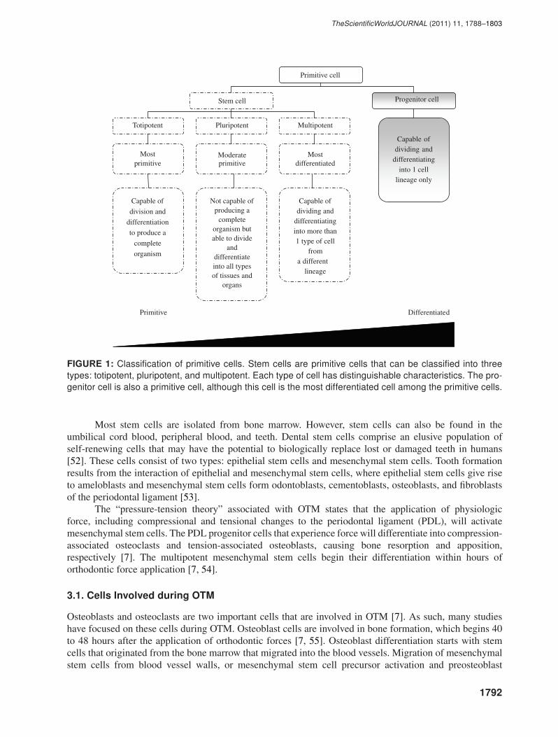
Stem cells are those with the ability to divide for indefinite periods of time and with the ability todifferentiate into a variety of cell types. Stem cells can be divided into three categories: totipotent,pluripotent, and multipotent cells (Figure 1) [48, 49]. Totipotent cells are the most primitive cells, followedby pluripotent cells. The multipotent cell type is the most differentiated type of stem cell (Figure 1) [50].
Totipotent cells have the potential to differentiate into any type of cell in the body and are capableof developing into a complete organism. After several cell division cycles, totipotent cells will develop intopluripotent cells. Pluripotent cells are capable of dividing and differentiating into any type of cell, tissue, ororgan (Figure 1). However, these cells are not capable of developing into a complete organism. Multipotentcells have more limited capacities than do pluripotent cells (Figure 1). Other types of primitive cells includeprogenitor cells, which are able to differentiate into only one type of mature cell (Figure 1) [41, 51].
1791
TheScientificWorldJOURNAL (2011) 11, 1788–1803
Primitive cell
Progenitor cell
Totipotent Pluripotent Multipotent
Mostprimitive
Moderateprimitive
Mostdifferentiated
Capable ofdividing and
differentiatinginto 1 cell
lineage only
Stem cell
Capable of
division and
differentiation
to produce a
complete
organism
Not capable ofproducing a
completeorganism butable to divide
anddifferentiateinto all typesof tissues and
organs
Primitive Differentiate d
Capable ofdividing and
differentiatinginto more than1 type of cell
fromdifferentlineage
a
FIGURE 1: Classification of primitive cells. Stem cells are primitive cells that can be classified into threetypes: totipotent, pluripotent, and multipotent. Each type of cell has distinguishable characteristics. The pro-genitor cell is also a primitive cell, although this cell is the most differentiated cell among the primitive cells.
Most stem cells are isolated from bone marrow. However, stem cells can also be found in theumbilical cord blood, peripheral blood, and teeth. Dental stem cells comprise an elusive population ofself-renewing cells that may have the potential to biologically replace lost or damaged teeth in humans[52]. These cells consist of two types: epithelial stem cells and mesenchymal stem cells. Tooth formationresults from the interaction of epithelial and mesenchymal stem cells, where epithelial stem cells give riseto ameloblasts and mesenchymal stem cells form odontoblasts, cementoblasts, osteoblasts, and fibroblastsof the periodontal ligament [53].
The “pressure-tension theory” associated with OTM states that the application of physiologicforce, including compressional and tensional changes to the periodontal ligament (PDL), will activatemesenchymal stem cells. The PDL progenitor cells that experience force will differentiate into compression-associated osteoclasts and tension-associated osteoblasts, causing bone resorption and apposition,respectively [7]. The multipotent mesenchymal stem cells begin their differentiation within hours oforthodontic force application [7, 54].
3.1. Cells Involved during OTM
Osteoblasts and osteoclasts are two important cells that are involved in OTM [7]. As such, many studieshave focused on these cells during OTM. Osteoblast cells are involved in bone formation, which begins 40to 48 hours after the application of orthodontic forces [7, 55]. Osteoblast differentiation starts with stemcells that originated from the bone marrow that migrated into the blood vessels. Migration of mesenchymalstem cells from blood vessel walls, or mesenchymal stem cell precursor activation and preosteoblast
1792
TheScientificWorldJOURNAL (2011) 11, 1788–1803
formation, occurs about 10 hours after the application of force [7]. This sequence of cellular activities occursduring the development of the stem cell into osteoblast and osteoclast cells may be useful in determiningpotential markers associated with OTM. Osteoclast cells are multinucleated cells which degrade and resorbbone. Osteoclast cells which work together with osteoblast cells in bone remodelling are derived fromhaematopoietic stem cells [42, 51, 56].
4. MARKERS FOR ORTHODONTIC TOOTH MOVEMENT
Applying orthodontic forces to teeth will ultimately result in movement. The main phenomena, both beforeand after tooth movement, are alveolar bone remodelling, tissue inflammation, and root resorption. Each ofthese events can potentially be detected using suitable markers.
4.1. Markers of Alveolar Bone Remodelling
As orthodontic forces are applied to teeth, the compression region shows an elevation in osteoclastic activity.Meanwhile, in the tension region, osteoblasts begin to proliferate and mineralise the extracellular matrix.This orchestra results in alveolar bone remodelling [10].
Chemokines may contribute to differential bone remodelling in response to orthodontic forcesthrough the establishment of distinct microenvironments in the sites of both compression and tension [57].The principal trigger for OTM is most likely the strain experienced by the PDL cells, bone-related cells,and the extracellular matrix. This strain leads to changes in gene expression in the cells via interactionsbetween the cells and the extracellular matrix [4]. One of the examples are matrix metalloproteinases(MMPs). MMPs break down the extracellular matrix and are important in bone remodelling. Compressioninduces an increase in MMP-1 protein levels after 1 hour. However, the increase lasted for 2 hours andsubsequently disappeared. Tension led to significantly increased levels of MMP-1 protein after just 1 hourof force application and also subsequently disappeared [31].
MMP-2 protein was induced by compression and increased significantly in a time-dependent fashion,reaching a peak after 8 hours of force application. On the tension side, MMP-2 was significantly increasedafter 1 hour but gradually returned to basal levels within 8 hours [31]. This result indicates that MMP-2could be used during very early stages of orthodontic treatment as a marker for active tooth movement.
4.1.1. Bone Formation Marker
Bone formation is primarily due to osteoblastic activities. Therefore, bone formation markers are usuallyosteoblastic enzymes or byproducts of bone formation such as type I procollagen. Type I procollagen wassecreted by osteoblast cells.
The cleavages of procollagen will produce two types of procollagen, that is, procollagen type I C-terminal propeptide (PICP) and procollagen type I N-terminal propeptide (PINP) that were proposed to bemeasured as bone formation markers [58]. However, PICP and PINP are markers that can only indicate theformation of type I collagen and not totally bone specific [58]. Therefore, other markers that specificallyactivated during osteoblast differentiation or activity are needed.
Many genes are involved in osteoblast differentiation. The transcription factor (TF) Cbfa1 (or Runx-2) is the earliest expressed and the most specific bone formation marker and helps to control osteoblastdifferentiation. Cbfa1-binding site also present at the regulatory sites of most genes that is involved inregulating bone matrix secretion by mature osteoblast [7]. On the other hand, Osterix, a TF, is involvedin bone formation and induces mature osteoblasts to express osteocalcin. In contrast, the expression ofosteocalcin will inhibit osteoblast differentiation [7, 59]. Indicate Cbfa1 and Osterix as potential markersfor early and late osteoblastic activity while osteocalcin as terminal osteoblast differentiation marker.
In addition, bone morphogenetic proteins (BMPs), transforming growth factor-beta (TGF-β) proteinsand growth-factor- (GFs) associated internal signalling molecules are other bone-forming genes that encodeproteins for GFs [7]. BMPs that bind to surface receptors on progenitor and mature osteoblasts can trigger
1793
TheScientificWorldJOURNAL (2011) 11, 1788–1803
a signalling pathway that promotes osteoprogenitor cell differentiation and the upregulation of osteoblastactivity. The expression of Cbfa1 also can be induced by BMPs.
Bone formation can also be promoted by GFs via their interaction with specific surface receptorson osteoblasts, thereby stimulating insulin-like GF-1. Insulin-like GF-1 is a primary mediator of the effectsof growth hormones that have growth-promoting effects on bone, in addition to regulate cell growth anddevelopment. Other studies have found that Msx1 and Msx2 are potential regulators of bone formation. TheMsx1, protein is known as a critical modulator of bone development and remodelling, and Msx2 is an alter-native regulator protein of Cbfa1 expression in bone formation during OTM. Therefore, Cbfa1 (or Runx-2),osterix, osteocalcin, BMPs, TGF-β, GF-associated internal signalling, insulin-like GF-1, Msx1 and Msx2can be used as potential biomarkers during the development of stem cells into osteoblasts during OTM.
Mechanical forces in orthodontic treatment cause the physical distortion of PDL and alveolar bonecells. They can also trigger a multilevel cascade of signal transduction pathways, such as the prostaglandinE2 (PGE2) pathway, that initiate structural and functional changes in extracellular, cell membrane, andcytoskeletal proteins [7]. Subsequent changes in cytoskeletal protein structure and function lead to thecreation of new cells and bone matrix formation [7, 60, 61].
The relationship between ALP and mineralisation has been the subject of many studies sinceRobinson’s first discussion of the enzyme in 1923 [62]. Although extensive knowledge has been gained,the exact role of ALP in mineral formation remains to be established. Several investigations haveproduced consistent findings with regard to the localisation of the enzyme in mineralising tissues. Enzymecytochemical studies have repeatedly demonstratde that ALP activity is closely associated with cell andmatrix vesicle membranes in cartilage calcification, intramembranous osteogenesis, and newly formingdentine [62]. ALP activity was detected in the osteoid areas of new bone formation but not in the calcifiedbone matrix. The cells that showed high ALP activities were preosteoblasts, osteoblasts [41, 42, 51],lining zones, newly embedded osteocytes, endosteal cells, and subperiosteal cells [63]. In conjunction withbone formation induced by orthodontic forces, increased levels of ALP were detected in human gingivalcrevicular fluid (GCF) collected from orthodontically treated human samples. Therefore, they might havebiological activities in the early stages of tooth movement [17, 18].
4.1.2. Bone Resorption Marker
Osteoclastic cells that are involved in bone resorption are specialised multinucleated giant cells thatoriginate from haematopoietic stem cells [42, 64]. The earliest bone resorption marker is the interleukin-1 beta (IL-1β) [7]. PGE2, interleukin-6 (IL-6), and other inflammatory cytokines can also facilitateosteoclastic bone resorption processes [7, 65]. These proteins regulate osteoclastic activity throughactivation of the nuclear factor kappa B (RANK) and of the nuclear factor kappa B ligand (RANKL).Osteoblastic cells also control osteoclastic processes by synthesizing RANKL to promote more osteoclasticdifferentiation [7, 66].
Signal induction between osteoblasts with surface expression of RANKL and osteoclastic precursorscarrying the receptor RANK induces osteoclastic formation and activation [67, 68]. A study by Oshiro etal. [69] has demonstrated changes in RANK, RANKL, and osteoprotegerin (OPG) in the tooth-supportingtissues during OTM, where RANKL stimulation and OPG inhibition are involved in osteoclastogenesis[67, 70]. Compressive forces upregulate RANKL through the PGE2 pathway and thereby supportosteoclastogenesis [67, 70].
The transfer of the local RANKL gene to the periodontal tissue using a Hemagglutinating Virus ofJapan (HVJ) envelope vector was reported to accelerate OTM in 6-week-old male Wistar rats. The activationof transferred RANKL gene in the periodontal tissue indicates that RANKL is involved during active OTMespecially in periodontal area [71]. On the other hand, the activation of the OPG gene, also through genetransfer to periodontal tissues, managed to neutralize RANKL activity and hence inhibits osteoclastogenesisand eventually OTM. There is an indication that the activation of the OPG gene inhibits the OTM process[67]. Furthermore, several cases have demonstrated that increases in RANKL and decreases in OPG can be
1794
TheScientificWorldJOURNAL (2011) 11, 1788–1803
observed during severe orthodontic root resorption [67, 72]. Therefore, we suggest that root resorptive pro-cesses need specific biomarkers other than RANKL or OPG in order to apply optimum force during OTM.
The enzyme assay of acid phosphatase activity in saliva was also introduced as a method used tomeasure biomarkers of OTM. On the basis of the enzymatic profile activity of lactate dehydrogenase,tartrate resistant acid phosphatase (TRAP) and ALP in mixed saliva, Zainal Ariffin et al. [19] suggestedthe reactivation of orthodontic braces from 30 days to 25 days so that the treatment course will be decreasedby approximately 17%.
Other biomarkers of early OTM were investigated in a rat model using a split-mouth design at 3and 24 hours after appliance insertion. The spatial expression patterns of KI-67, RANKL, and Runx2during OTM were mapped by using immunohistochemical staining. The expression of KI-67, a proliferationmarker, and RANKL, a molecule associated with osteoclastic differentiation, increased in the compressionsites of the periodontal ligament subjected to 3 hours of force. On the other hand, there were increasedexpression of KI-67 and Runx2, both markers of osteoblastic precursors, in the areas of tension after 24hours of force. The early expression of RANKL indicates that cells are involved in osteoclastic precursorsignalling at this early stage. In addition, decreased KI-67 expression found near the midpoint of the toothroot is believed to represent the centre of rotation, providing a molecular means of visualising mechanicalloading patterns and runt-related transcription factor 2 (Runx2) [12].
The potential of TRAP as a biomarker of bone resorption has been long recognised [73]. TRAPactivity was very strong in in vitro osteoclast cultures. Refinement of the activity assay to primarily measureTRAP 5b at a pH level of 6.1 was suggested for investigation of osteoclastic activity and bone resorptionrates [73]. This will differentiate between TRAP 5a that have optimum pH at 5.2 compared to TRAP 5bwith optimum pH at around 6, hence TRAP5b activity can be precisely measured. However, no properinvestigation has yet been conducted involving TRAP 5b assayed at a pH level of 6.1.
Nitric oxide (NO) is an important regulator of bone responses to mechanical stress and is producedthrough the activity of constitutive endothelial nitric oxide synthase (eNOS) or inducible nitric oxidesynthase (iNOS). NO mediates adaptive bone formation, protects osteocytes against apoptosis and mediatesosteoclastic activity. High levels of NO reduce osteoclastic activity, while the inhibition of NO productionincreases osteoclastogenesis and osteoclastic activity [11]. Using a rat model and immunohistochemistrytechniques, Tan et al. [11] showed that the number of endothelial-nitric-oxide-synthase- (eNOS-) positiveosteocytes was significantly higher at both 24 and 48 hours than at baseline during tooth movement. After 48hours, however, the number decreased to baseline levels. The number of inducible-nitric-oxide-synthase-(iNOS-) positive osteocytes was significantly higher than baseline levels between 6 and 48 hours, as thenumber increased 3.0- to 4.1-fold. Subsequently, the number decreased to baseline levels. As such, iNOSmediates inflammation-induced bone resorption in areas of compression [11].
Osteocalcin is the most abundant noncollagenous matrix protein found in bone. It is expressed byhighly differentiated osteoblasts and is incorporated into the bony matrix. Smaller osteocalcin fragmentsare thought to be a degradation products of bone matrix, which suggests its potential as a bone resorptionmarker [58].
4.2. Markers of Inflammatory Processes
The host response to orthodontic forces has been described as aseptic and transitory inflammation. Amongthe substances investigated are lactate dehydrogenase (LDH) and aspartate aminotransferase (AST), whichare inflammatory biomarkers found outside cells during necrosis. Increased levels of lactate dehydrogenase[29, 30] and aspartate aminotransferase [20–22] were detected in human GCF samples obtained duringOTM. However, increased levels of lactate dehydrogenase in whole saliva can be associated with periodontaldisease as well, especially with the presence of calculus and periodontal pockets greater than 5 mm [74].Aspartate aminotransferase is also found in periodontitis [75, 76].
Cathepsin B, an intracellular lysosomal cysteine proteinase, can degrade extracellular componentsincluding collagen and cause protein turnover in the lysosomal system. It is also known to play an important
1795
TheScientificWorldJOURNAL (2011) 11, 1788–1803
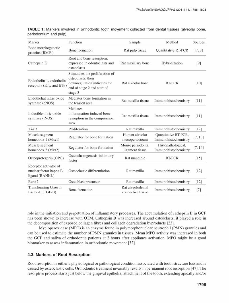
TABLE 1: Markers involved in orthodontic tooth movement collected from dental tissues (alveolar bone,periodontium and pulp).
Marker Function Sample Method Sources
Bone morphogeneticproteins (BMPs)
Bone formation Rat pulp tissue Quantitative RT-PCR [7, 8]
Cathepsin KRoot and bone resorption;expressed in odontoclasts andosteoclasts
Rat maxillary bone Hybridization [9]
Endothelin-1, endothelinreceptors (ETA and ETB)
Stimulates the proliferation ofosteoblasts; theirdownregulation indicates theend of stage 2 and start ofstage 3
Rat alveolar bone RT-PCR [10]
Endothelial nitric oxidesynthase (eNOS)
Mediates bone formation inthe tension area
Rat maxilla tissue Immunohistochemistry [11]
Inducible nitric oxidesynthase (iNOS)
Mediatesinflammation-induced boneresorption in the compressionarea.
Rat maxilla tissue Immunohistochemistry [11]
Ki-67 Proliferation Rat maxilla Immunohistochemistry [12]
Muscle segmenthomeobox 1 (Msx1)
Regulator for bone formationHuman alveolarmucoperiosteum
Quantitative RT-PCR,Immunohistochemistry
[7, 13]
Muscle segmenthomeobox 2 (Msx2)
Regulator for bone formationMouse periodontal
ligament tissueHistopathological,
Immunohistochemistry[7, 14]
Osteoprotegerin (OPG)Osteoclastogenesis-inhibitoryfactor
Rat mandible RT-PCR [15]
Receptor activator ofnuclear factor kappa Bligand (RANKL)
Osteoclastic differentiation Rat maxilla Immunohistochemistry [12]
Runx2 Osteoblast precursor Rat maxilla Immunohistochemistry [12]
Transforming GrowthFactor-B (TGF-B)
Bone formationRat alveolodentalconnective tissue
Immunohistochemistry [7]
role in the initiation and perpetuation of inflammatory processes. The accumulation of cathepsin B in GCFhas been shown to increase with OTM. Cathepsin B was increased around osteoclasts; it played a role inthe decomposition of exposed collagen fibres and collagen degradation byproducts [23].
Myeloperoxidase (MPO) is an enzyme found in polymorphonuclear neutrophil (PMN) granules andcan be used to estimate the number of PMN granules in tissues. Mean MPO activity was increased in boththe GCF and saliva of orthodontic patients at 2 hours after appliance activation. MPO might be a goodbiomarker to assess inflammation in orthodontic movement [32].
4.3. Markers of Root Resorption
Root resorption is either a physiological or pathological condition associated with tooth structure loss and iscaused by osteoclastic cells. Orthodontic treatment invariably results in permanent root resorption [47]. Theresorptive process starts just below the gingival epithelial attachment of the tooth, extending apically and/or
1796
TheScientificWorldJOURNAL (2011) 11, 1788–1803
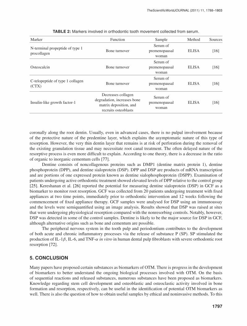
TABLE 2: Markers involved in orthodontic tooth movement collected from serum.
Marker Function Sample Method Sources
N-terminal propeptide of type 1procollagen
Bone turnoverSerum of
premenopausalwoman
ELISA [16]
Osteocalcin Bone turnoverSerum of
premenopausalwoman
ELISA [16]
C-telopeptide of type 1 collagen(CTX)
Bone turnoverSerum of
premenopausalwoman
ELISA [16]
Insulin-like growth factor-1
Decreases collagendegradation, increases bone
matrix deposition, andrecruits osteoblasts
Serum ofpremenopausal
womanELISA [16]
coronally along the root dentin. Usually, even in advanced cases, there is no pulpal involvement becauseof the protective nature of the predentine layer, which explains the asymptomatic nature of this type ofresorption. However, the very thin dentin layer that remains is at risk of perforation during the removal ofthe existing granulation tissue and may necessitate root canal treatment. The often delayed nature of theresorptive process is even more difficult to explain. According to one theory, there is a decrease in the ratioof organic to inorganic cementum cells [77].
Dentine consists of noncollagenous proteins such as DMP1 (dentine matrix protein 1), dentinephosphoprotein (DPP), and dentine sialoprotein (DSP). DPP and DSP are products of mRNA transcriptionand are portions of one expressed protein known as dentine sialophosphoprotein (DSPP). Examination ofpatients undergoing active orthodontic treatment showed elevated levels of DPP relative to the control group[25]. Kereshanan et al. [26] reported the potential for measuring dentine sialoprotein (DSP) in GCF as abiomarker to monitor root resorption. GCF was collected from 20 patients undergoing treatment with fixedappliances at two time points, immediately prior to orthodontic intervention and 12 weeks following thecommencement of fixed appliance therapy. GCF samples were analysed for DSP using an immunoassayand the levels were semiquantified using an image analysis. Results showed that DSP was raised at sitesthat were undergoing physiological resorption compared with the nonresorbing controls. Notably, however,DSP was detected in some of the control samples. Dentine is likely to be the major source for DSP in GCF,although alternative origins such as bone and cementum are possible.
The peripheral nervous system in the tooth pulp and periodontium contributes to the developmentof both acute and chronic inflammatory processes via the release of substance P (SP). SP stimulated theproduction of IL-1β, IL-6, and TNF-α in vitro in human dental pulp fibroblasts with severe orthodontic rootresorption [72].
5. CONCLUSION
Many papers have proposed certain substances as biomarkers of OTM. There is progress in the developmentof biomarkers to better understand the ongoing biological processes involved with OTM. On the basisof sequential reactions and released substances, numerous substances have been proposed as biomarkers.Knowledge regarding stem cell development and osteoblastic and osteoclastic activity involved in boneformation and resorption, respectively, can be useful in the identification of potential OTM biomarkers aswell. There is also the question of how to obtain useful samples by ethical and noninvasive methods. To this
1797
TheScientificWorldJOURNAL (2011) 11, 1788–1803
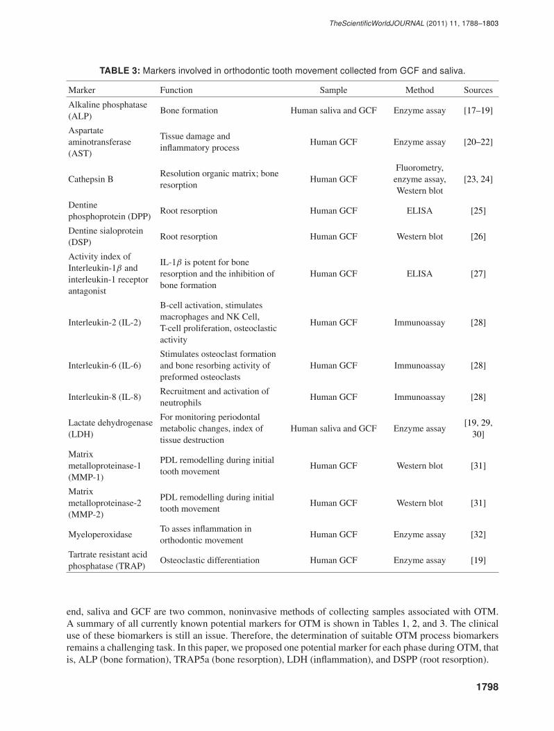
TABLE 3: Markers involved in orthodontic tooth movement collected from GCF and saliva.
Marker Function Sample Method Sources
Alkaline phosphatase(ALP)
Bone formation Human saliva and GCF Enzyme assay [17–19]
Aspartateaminotransferase(AST)
Tissue damage andinflammatory process
Human GCF Enzyme assay [20–22]
Cathepsin BResolution organic matrix; boneresorption
Human GCFFluorometry,
enzyme assay,Western blot
[23, 24]
Dentinephosphoprotein (DPP)
Root resorption Human GCF ELISA [25]
Dentine sialoprotein(DSP)
Root resorption Human GCF Western blot [26]
Activity index ofInterleukin-1β andinterleukin-1 receptorantagonist
IL-1β is potent for boneresorption and the inhibition ofbone formation
Human GCF ELISA [27]
Interleukin-2 (IL-2)
B-cell activation, stimulatesmacrophages and NK Cell,T-cell proliferation, osteoclasticactivity
Human GCF Immunoassay [28]
Interleukin-6 (IL-6)Stimulates osteoclast formationand bone resorbing activity ofpreformed osteoclasts
Human GCF Immunoassay [28]
Interleukin-8 (IL-8)Recruitment and activation ofneutrophils
Human GCF Immunoassay [28]
Lactate dehydrogenase(LDH)
For monitoring periodontalmetabolic changes, index oftissue destruction
Human saliva and GCF Enzyme assay[19, 29,
30]
Matrixmetalloproteinase-1(MMP-1)
PDL remodelling during initialtooth movement
Human GCF Western blot [31]
Matrixmetalloproteinase-2(MMP-2)
PDL remodelling during initialtooth movement
Human GCF Western blot [31]
MyeloperoxidaseTo asses inflammation inorthodontic movement
Human GCF Enzyme assay [32]
Tartrate resistant acidphosphatase (TRAP)
Osteoclastic differentiation Human GCF Enzyme assay [19]
end, saliva and GCF are two common, noninvasive methods of collecting samples associated with OTM.A summary of all currently known potential markers for OTM is shown in Tables 1, 2, and 3. The clinicaluse of these biomarkers is still an issue. Therefore, the determination of suitable OTM process biomarkersremains a challenging task. In this paper, we proposed one potential marker for each phase during OTM, thatis, ALP (bone formation), TRAP5a (bone resorption), LDH (inflammation), and DSPP (root resorption).
1798
TheScientificWorldJOURNAL (2011) 11, 1788–1803
ACKNOWLEDGMENTS
The authors would like to thank the Ministry of Higher Education Malaysia (FRGS/1/2011/SG/UKM/02/13,and UKMDD-03-FRGS0030-2010), Ministry of Science, Technology and Innovation Malaysia (09-05-MGI-GMB002), and Universiti Kebangsaan Malaysia (UKM-OUP-KPB-33-170/2011 and UKM-GUP-2011-093) for the financial grants during this study.
REFERENCES
[1] D. Cardaropoli and L. Gaveglio, “The influence of orthodontic movement on periodontal tissues level,” Seminarsin Orthodontics, vol. 13, no. 4, pp. 234–245, 2007.
[2] V. Krishnan and Z. Davidovitch, “Cellular, molecular, and tissue-level reactions to orthodontic force,” AmericanJournal of Orthodontics and Dentofacial Orthopedics, vol. 129, no. 4, pp. 469–e1, 2006.
[3] C. Dolce, J. Scott Malone, and T. T. Wheeler, “Current concepts in the biology of orthodontic tooth movement,”Seminars in Orthodontics, vol. 8, no. 1, pp. 6–12, 2002.
[4] T. Bartzela, J. C. Turp, E. Motschall, and J. C. Maltha, “Medication effects on the rate of orthodontic toothmovement: a systematic literature review,” American Journal of Orthodontics and Dentofacial Orthopedics, vol.135, no. 1, pp. 16–26, 2009.
[5] M. Taba, J. Kinney, A. S. Kim, and W. V. Giannobile, “Diagnostic biomarkers for oral and periodontal diseases,”Dental Clinics of North America, vol. 49, no. 3, pp. 551–571, 2005.
[6] D. Bernardi, M. Zaninotto, and M. Plebani, “Requirements for improving quality in the measurement of bonemarkers,” Clinica Chimica Acta, vol. 346, no. 1, pp. 79–86, 2004.
[7] R. S. Masella and M. Meister, “Current concepts in the biology of orthodontic tooth movement,” AmericanJournal of Orthodontics and Dentofacial Orthopedics, vol. 129, no. 4, pp. 458–468, 2006.
[8] Y. Enokiya, S. Hashimoto, T. Muramatsu et al., “Effect of stretching stress on gene transcription related to early-phase differentiation in rat periodontal ligament cells,” The Bulletin of Tokyo Dental College, vol. 51, no. 3, pp.129–137, 2010.
[9] S. Domon, H. Shimokawa, Y. Matsumoto, S. Yamaguchi, and K. Soma, “In situ hybridization for matrixmetalloproteinase-1 and cathepsin K in rat root-resorbing tissue induced by tooth movement,” Archives of OralBiology, vol. 44, no. 11, pp. 907–915, 1999.
[10] S. Sprogar, T. Vaupotic, A. Cor, M. Drevensek, and G. Drevensek, “The endothelin system mediates bonemodeling in the late stage of orthodontic tooth movement in rats,” Bone, vol. 43, no. 4, pp. 740–747, 2008.
[11] S. D. Tan, R. Xie, J. Klein-Nulend et al., “Orthodontic force stimulates eNOS and iNOS in rat osteocytes,”Journal of Dental Research, vol. 88, no. 3, pp. 255–260, 2009.
[12] P. J. Brooks, D. Nilforoushan, M. F. Manolson, C. A. Simmons, and S. G. Gong, “Molecular markers of earlyorthodontic tooth movement,” Angle Orthodontist, vol. 79, no. 6, pp. 1108–1113, 2009.
[13] F. Wehrhan, P. Hyckel, J. Ries et al., “Expression of Msx-1 is suppressed in bisphosphonate associatedosteonecrosis related jaw tissue-etiopathology considerations respecting jaw developmental biology-relatedunique features,” Journal of Translational Medicine, vol. 8, article no. 96, 2010.
[14] T. Watanabe, K. Nakano, R. Muraoka et al., “Role of Msx2 as a promoting factor for Runx2 at the periodontaltension sides elicited by mechanical stress,” European Journal of Medical Research, vol. 13, no. 9, pp. 425–431,2008.
[15] E. Low, H. Zoellner, O. P. Kharbanda, and M. A. Darendeliler, “Expression of mRNA for osteoprotegerin andreceptor activator of nuclear factor kappa β ligand (RANKL) during root resorption induced by the applicationof heavy orthodontic forces on rat molars,” American Journal of Orthodontics and Dentofacial Orthopedics, vol.128, no. 4, pp. 497–503, 2005.
[16] S. Adami, A. Zivelonghi, V. Braga et al., “Insulin-like growth factor-1 is associated with bone formation markers,PTH and bone mineral density in healthy premenopausal women,” Bone, vol. 46, no. 1, pp. 244–247, 2010.
[17] G. Perinetti, M. Paolantonio, M. D’Attilio et al., “Alkaline phosphatase activity in gingival crevicular fluid duringhuman orthodontic tooth movement,” American Journal of Orthodontics and Dentofacial Orthopedics, vol. 122,no. 5, pp. 548–556, 2002.
1799

TheScientificWorldJOURNAL (2011) 11, 1788–1803
[18] A. A. A. Asma, R. Megat Abdul Wahab, and S. H. Zainal Ariffin, “Crevicular alkaline phosphatase activity duringorthodontic tooth movement: canine retraction stage,” Journal of Medical Sciences, vol. 8, no. 3, pp. 228–233,2008.
[19] S. H. Zainal Ariffin, M. F. Ellias, R. Megat Abdul Wahab, Y. Bakar, and S. Senafi, “Profiles of lactatedehydrogenase, tartrate resistant acid phosphatase and alkaline phosphatase in saliva during orthodontictreatment,” Sains Malaysiana, vol. 39, no. 3, pp. 405–412, 2010.
[20] G. Perinetti, M. Paolantonio, M. D’Attilio et al., “Aspartate aminotransferase activity in gingival crevicular fluidduring orthodontic treatment. A controlled short-term longitudinal study,” Journal of Periodontology, vol. 74, no.2, pp. 145–152, 2003.
[21] R. Megat Abdul Wahab, S. H. Zainal Ariffin, and K. Khazlina, “The activity of aspartate aminotransferase duringcanine retraction (Bodily Tooth Movement) in orthodontic treatment,” Journal of Medical Sciences, vol. 8, no. 6,pp. 553–558, 2008.
[22] R. Megat Abdul Wahab, S. H. Zainal Ariffin, and K. Khazlina, “Preliminary study of aspartate aminotransferaseactivity in gingival crevicular fluids during orthodontic tooth movement,” Journal of Applied Sciences, vol. 9, no.7, pp. 1393–1396, 2009.
[23] S. H. Rhee, J. Kang, and D. S. Nahm, “Cystatins and cathepsin B during orthodontic tooth movement,” AmericanJournal of Orthodontics and Dentofacial Orthopedics, vol. 135, no. 1, pp. 99–105, 2009.
[24] Y. Sugiyama, M. Yamaguchi, M. Kanekawa et al., “The level of cathepsin B in gingival crevicular fluid duringhuman orthodontic tooth movement,” European Journal of Orthodontics, vol. 25, no. 1, pp. 71–76, 2003.
[25] J. Mah and N. Prasad, “Dentine phosphoproteins in gingival crevicular fluid during root resorption,” EuropeanJournal of Orthodontics, vol. 26, no. 1, pp. 25–30, 2004.
[26] S. Kereshanan, P. Stephenson, and R. Waddington, “Identification of dentine sialoprotein in gingival crevicularfluid during physiological root resorption and orthodontic tooth movement,” European Journal of Orthodontics,vol. 30, no. 3, pp. 307–314, 2008.
[27] L. R. Iwasaki, J. E. Haack, J. C. Nickel, R. A. Reinhardt, and T. M. Petro, “Human interleukin-1β and interleukin-1 receptor antagonist secretion and velocity of tooth movement,” Archives of Oral Biology, vol. 46, no. 2, pp.185–189, 2001.
[28] G. Basaran, T. Ozer, F. A. Kaya, and O. Hamamci, “Interleukins 2, 6, and 8 levels in human gingival sulcusduring orthodontic treatment,” American Journal of Orthodontics and Dentofacial Orthopedics, vol. 130, no. 1,pp. 7.e1–7.e6, 2006.
[29] E. Serra, G. Perinetti, M. D’Attilio et al., “Lactate dehydrogenase activity in gingival crevicular fluid duringorthodontic treatment,” American Journal of Orthodontics and Dentofacial Orthopedics, vol. 124, no. 2, pp.206–211, 2003.
[30] G. Perinetti, E. Serra, M. Paolantonio et al., “Lactate dehydrogenase activity in human gingival crevicular fluidduring orthodontic treatment: a controlled, short-term longitudinal study,” Journal of Periodontology, vol. 76, no.3, pp. 411–417, 2005.
[31] G. Cantarella, R. Cantarella, M. Caltabiano, N. Risuglia, R. Bernardini, and R. Leonardi, “Levels of matrixmetalloproteinases 1 and 2 in human gingival crevicular fluid during initial tooth movement,” American Journalof Orthodontics and Dentofacial Orthopedics, vol. 130, no. 5, pp. 568–e11, 2006.
[32] A. M. Marcaccini, P. A. F. Amato, F. V. Leao, R. F. Gerlach, and J. T. L. Ferreira, “Myeloperoxidase activityis increased in gingival crevicular fluid and whole saliva after fixed orthodontic appliance activation,” AmericanJournal of Orthodontics and Dentofacial Orthopedics, vol. 138, no. 5, pp. 613–616, 2010.
[33] Y. Nakamura, K. Noda, S. Shimoda et al., “Time-lapse observation of rat periodontal ligament during functionand tooth movement, using microcomputed tomography,” European Journal of Orthodontics, vol. 30, no. 3, pp.320–326, 2008.
[34] S. Passarella, L. de Bari, D. Valenti, R. Pizzuto, G. Paventi, and A. Atlante, “Mitochondria and l-lactatemetabolism,” FEBS Letters, vol. 582, no. 25-26, pp. 3569–3576, 2008.
[35] Y. Kitase, M. Yokozeki, S. Fujihara et al., “Analysis of gene expression profiles in human periodontal ligamentcells under hypoxia: the protective effect of CC chemokine ligand 2 to oxygen shortage,” Archives of OralBiology, vol. 54, no. 7, pp. 618–624, 2009.
1800
TheScientificWorldJOURNAL (2011) 11, 1788–1803
[36] M. von Bohl, J. C. Maltha, J. W. von den Hoff, and A. M. Kuijpers-Jagtman, “Focal hyalinization duringexperimental tooth movement in beagle dogs,” American Journal of Orthodontics and Dentofacial Orthopedics,vol. 125, no. 5, pp. 615–623, 2004.
[37] L. Bonafe-Oliveira, R. M. Faltin, and V. E. Arana-Chavez, “Ultrastructural and histochemical examination ofalveolar bone at the pressure areas of rat molars submitted to continuous orthodontic force,” European Journalof Oral Sciences, vol. 111, no. 5, pp. 410–416, 2003.
[38] K. Reitan and P. Rygh, “Tissue reactions in orthodontics,” in Orthodontics Current Principles and Techniques, T.M. Graber, R. L. Vanarsdall, and K. W. L. Vig, Eds., CV Mosby, St Louis, Mo, USA, 4th edition, 2005.
[39] R. M. Faltin, K. Faltin, F. G. Sander, and V. E. Arana-Chavez, “Ultrastructure of cementum and periodontalligament after continuous intrusion in humans: a transmission electron microscopy study,” European Journal ofOrthodontics, vol. 23, no. 1, pp. 35–49, 2001.
[40] M. A. Casa, R. M. Faltin, K. Faltin, and V. E. Arana-Chavez, “Root resorption on torqued human premolars shownby tartrate-resistant acid phosphatase histochemistry and transmission electron microscopy,” Angle Orthodontist,vol. 76, no. 6, pp. 1015–1021, 2006.
[41] M. D. Yazid, S. H. Zainal Ariffin, S. Senafi, M. A. Razak, and R. Megat Abdul Wahab, “Determination ofthe differentiation capacities of murines’ primary mononucleated cells and MC3T3-E1 cells,” Cancer CellInternational, vol. 10, article 42, 2010.
[42] S. H. Zainal Ariffin, I. Z. Zainol Abidin, M. D. Yazid, and R. Megat Abdul Wahab, “Differentiation analyses ofadult suspension mononucleated peripheral blood cells of Mus musculus,” Cell Communication and Signaling,pp. 29–35, 2010.
[43] J. Kurol, P. Owman-Moll, and D. Lundgren, “Time-related root resorption after application of a controlledcontinuous orthodontic force,” American Journal of Orthodontics and Dentofacial Orthopedics, vol. 110, no.3, pp. 303–310, 1996.
[44] E. F. Harris, “Root resorption during orthodontic therapy,” Seminars in Orthodontics, vol. 6, no. 3, pp. 183–194,2000.
[45] I. Smale, J. Artun, F. Behbehani, D. Doppel, M. Van’t Hof, and A. M. Kuijpers-Jagtman, “Apical root resorption6 months after initiation of fixed orthodontic appliance therapy,” American Journal of Orthodontics andDentofacial Orthopedics, vol. 128, no. 1, pp. 57–67, 2005.
[46] T. Kumasako-Haga, T. Konoo, K. Yamaguchi, and H. Hayashi, “Effect of 8-hour intermittent orthodontic forceon osteoclasts and root resorption,” American Journal of Orthodontics and Dentofacial Orthopedics, vol. 135,no. 3, pp. 278–e1, 2009.
[47] C. Estrela, M. R. Bueno, A. H. G. De Alencar et al., “Method to evaluate inflammatory root resorption by usingcone beam computed tomography,” Journal of Endodontics, vol. 35, no. 11, pp. 1491–1497, 2009.
[48] S. H. Zainal Ariffin, R. Megat Abdul Wahab, I. Ismail, N. M. Mahadi, and Z. Zainal Ariffin, “Stem cells, cytokinesand their receptors,” Asia-Pacific Journal of Molecular Biology and Biotechnology, vol. 13, no. 1, pp. 1–13, 2005.
[49] N. Shanthly, M. R. Aruva, K. Zhang, B. Mathew, and M. L. Thakur, “Stem cells: a regenerative pharmaceutical,”Quarterly Journal of Nuclear Medicine and Molecular Imaging, vol. 50, no. 3, pp. 205–216, 2006.
[50] S. H. Zainal Ariffin, R. Megat Abdul Wahab, I. Z. Zainol Abidin, S. Sahidan, M. M. Nor, and Z. Zainal Ariffin,“Stem cell in blood development,” Sains Malaysiana, vol. 34, pp. 21–26, 2005.
[51] I. Z. Zainol Abidin, S. H. Zainal Ariffin, R. Megat Abdul Wahab, S. Sahidan, and Z. Zainal Ariffin, “Osteoclastand osteoblast development of Mus musculus haemopoietic mononucleated cells,” Journal of Biological Sciences,vol. 8, no. 3, pp. 506–516, 2008.
[52] P. C. Yelick and J. P. Vacanti, “Dental stem cells,” in Handbook of Stem Cells, P. Andrews, J. Cibelli, R. Edwardset al., Eds., Academic Press, Amsterdam, The Netherlands, 2004.
[53] G. Bluteau, H. U. Luder, C. De Bari, and T. A. Mitsiadis, “Stem cells for tooth engineering,” European Cells andMaterials, vol. 16, pp. 1–9, 2008.
[54] M. K. Sutherland, J. C. Geoghegan, C. Yu et al., “Sclerostin promotes the apoptosis of human osteoblastic cells:a novel regulation of bone formation,” Bone, vol. 35, no. 4, pp. 828–835, 2004.
[55] W. E. Roberts, S. S. Huja, and J. A. Roberts, “Bone modeling: biomechanics, molecular mechanisms, and clinicalperspectives,” Seminars in Orthodontics, vol. 10, no. 2, pp. 123–161, 2004.
1801

TheScientificWorldJOURNAL (2011) 11, 1788–1803
[56] A. E. Grigoriadis, M. Kennedy, A. Bozec et al., “Directed differentiation of hematopoietic precursors andfunctional osteoclasts from human ES and iPS cells,” Blood, vol. 115, no. 14, pp. 2769–2776, 2010.
[57] T. P. Garlet, U. Coelho, C. E. Repeke, J. S. Silva, F. D. Q. Cunha, and G. P. Garlet, “Differential expressionof osteoblast and osteoclast chemmoatractants in compression and tension sides during orthodontic movement,”Cytokine, vol. 42, no. 3, pp. 330–335, 2008.
[58] R. A. Hannon and R. Eastell, “Bone markers and current laboratory assays,” Cancer Treatment Reviews, vol. 32,no. 1, pp. 7–14, 2006.
[59] K. S. Lee, H. J. Kim, Q. L. Li et al., “Runx2 is a common target of transforming growth factor β1 and bonemorphogenetic protein 2, and cooperation between Runx2 and Smad5 induces osteoblast-specific gene expressionin the pluripotent mesenchymal precursor cell line C2C12,” Molecular and Cellular Biology, vol. 20, no. 23, pp.8783–8792, 2000.
[60] J. Parkin and B. Cohen, “An overview of the immune system,” The Lancet, vol. 357, no. 9270, pp. 1777–1789,2001.
[61] A. J. Zhu and M. P. Scott, “Incredible journey: how do developmental signals travel through tissue?” Genes andDevelopment, vol. 18, no. 24, pp. 2985–2997, 2004.
[62] D. C. Morris, J. C. Randall, and H. C. Anderson, “Light microscopic localization of alkaline phosphatase in fetalbovine bone using immunoperoxidase and immunogold-silver staining procedures,” Journal of Histochemistry &Cytochemistry, vol. 36, no. 3, pp. 323–327, 1988.
[63] D. Miao and A. Scutt, “Histochemical localization of alkaline phosphatase activity in decalcified bone andcartilage,” Journal of Histochemistry & Cytochemistry, vol. 50, no. 3, pp. 333–340, 2002.
[64] T. Miyamoto and T. Suda, “Differentiation and function of osteoclasts,” The Keio Journal of Medicine, vol. 52,no. 1, pp. 1–7, 2003.
[65] N. Alhashimi, L. Frithiof, P. Brudvik, and M. Bakhiet, “Orthodontic tooth movement and de novo synthesis ofproinflammatory cytokines,” American Journal of Orthodontics and Dentofacial Orthopedics, vol. 119, no. 3,pp. 307–312, 2001.
[66] G. Karsenty, “The complexities of skeletal biology,” Nature, vol. 423, no. 6937, pp. 316–318, 2003.
[67] G. E. Wise and G. J. King, “Mechanisms of tooth eruption and orthodontic tooth movement,” Journal of DentalResearch, vol. 87, no. 5, pp. 414–434, 2008.
[68] H. Yasuda, N. Shima, N. Nakagawa et al., “A novel molecular mechanism modulating osteoclast differentiationand function,” Bone, vol. 25, no. 1, pp. 109–113, 1999.
[69] T. Oshiro, A. Shiotani, Y. Shibasaki, and T. Sasaki, “Osteoclast induction in periodontal tissue duringexperimental movement of incisors in osteoprotegerin-deficient mice,” The Anatomical Record, vol. 266, no.4, pp. 218–225, 2002.
[70] H. Kanzaki, M. Chiba, Y. Shimizu, and H. Mitani, “Dual regulation of osteoclast differentiation by periodontalligament cells through RANKL stimulation and OPG inhibition,” Journal of Dental Research, vol. 80, no. 3, pp.887–891, 2001.
[71] H. Kanzaki, M. Chiba, A. Sato et al., “Cyclical tensile force on periodontal ligament cells inhibitsosteoclastogenesis through OPG induction,” Journal of Dental Research, vol. 85, no. 5, pp. 457–462, 2006.
[72] M. Yamaguchi, Y. Ozawa, H. Mishima, N. Aihara, T. Kojima, and K. Kasai, “Substance P increases productionof proinflammatory cytokines and formation of osteoclasts in dental pulp fibroblasts in patients with severeorthodontic root resorption,” American Journal of Orthodontics and Dentofacial Orthopedics, vol. 133, no. 5,pp. 690–698, 2008.
[73] L. T. Yam and A. J. Janckila, “Tartrate-resistant acid phosphatase (TRACP): a personal perspective,” Journal ofBone and Mineral Research, vol. 18, no. 10, pp. 1894–1896, 2003.
[74] V. A. de la Pena, P. Diz Dios, and R. Tojo Sierra, “Relationship between lactate dehydrogenase activity in salivaand oral health status,” Archives of Oral Biology, vol. 52, no. 10, pp. 911–915, 2007.
[75] N. Ozmeric, “Advances in periodontal disease markers,” Clinica Chimica Acta, vol. 343, no. 1-2, pp. 1–16, 2004.
[76] B. Zappacosta, A. Manni, S. Persichilli et al., “Salivary thiols and enzyme markers of cell damage in periodontaldisease,” Clinical Biochemistry, vol. 40, no. 9-10, pp. 661–665, 2007.
1802

TheScientificWorldJOURNAL (2011) 11, 1788–1803
[77] A. Smidt, E. Nuni, and D. Keinan, “Invasive cervical root resorption: treatment rationale with an interdisciplinaryapproach,” Journal of Endodontics, vol. 33, no. 11, pp. 1383–1387, 2007.
This article should be cited as follows:
Shahrul Hisham Zainal Ariffin, Zulham Yamamoto, lntan Zarina Zainol Abidin, Rohaya Megat AbdulWahab, and Zaidah Zainal Ariffin, “Cellular and Molecular Changes in Orthodontic Tooth Movement,”TheScientificWorldJOURNAL, vol. 11, pp. 1788–1803, 2011.
1803

Submit your manuscripts athttp://www.hindawi.com
Hindawi Publishing Corporationhttp://www.hindawi.com Volume 2014
Oral OncologyJournal of
DentistryInternational Journal of
Hindawi Publishing Corporationhttp://www.hindawi.com Volume 2014
Hindawi Publishing Corporationhttp://www.hindawi.com Volume 2014
International Journal of
Biomaterials
Hindawi Publishing Corporationhttp://www.hindawi.com Volume 2014
BioMed Research International
Hindawi Publishing Corporationhttp://www.hindawi.com Volume 2014
Case Reports in Dentistry
Hindawi Publishing Corporationhttp://www.hindawi.com Volume 2014
Oral ImplantsJournal of
Hindawi Publishing Corporationhttp://www.hindawi.com Volume 2014
Anesthesiology Research and Practice
Hindawi Publishing Corporationhttp://www.hindawi.com Volume 2014
Radiology Research and Practice
Environmental and Public Health
Journal of
Hindawi Publishing Corporationhttp://www.hindawi.com Volume 2014
The Scientific World JournalHindawi Publishing Corporation http://www.hindawi.com Volume 2014
Hindawi Publishing Corporationhttp://www.hindawi.com Volume 2014
Dental SurgeryJournal of
Drug DeliveryJournal of
Hindawi Publishing Corporationhttp://www.hindawi.com Volume 2014
Hindawi Publishing Corporationhttp://www.hindawi.com Volume 2014
Oral DiseasesJournal of
Hindawi Publishing Corporationhttp://www.hindawi.com Volume 2014
Computational and Mathematical Methods in Medicine
ScientificaHindawi Publishing Corporationhttp://www.hindawi.com Volume 2014
PainResearch and TreatmentHindawi Publishing Corporationhttp://www.hindawi.com Volume 2014
Preventive MedicineAdvances in
Hindawi Publishing Corporationhttp://www.hindawi.com Volume 2014
EndocrinologyInternational Journal of
Hindawi Publishing Corporationhttp://www.hindawi.com Volume 2014
Hindawi Publishing Corporationhttp://www.hindawi.com Volume 2014
OrthopedicsAdvances in