follow after spontaneous artery dissection: of five...
TRANSCRIPT
Heart 1996;75:206-209
CASE STUDY
Follow up after spontaneous coronary arterydissection: a report of five cases
Patrizio Zampieri, Silvio Aggio, Loris Roncon, Massimo Rinuncini, Carlo Canova,Gabriele Zanazzi, Roberto Fiorencis, Pietro Zonzin
AbstractFive cases of spontaneous coronary arterydissection (SCAD) are reported, three inwomen and two in men (mean age 44years; range 28-65), all of whom suffereda myocardial infarction. Common riskfactors for coronary artery disease werepresent in the two men; in the femalegroup one patient was taking an oral con-traceptive, one was in the postpartumperiod, and the third was a smoker. Onlythe three women received intravenousalteplase and their ejection fraction wasnormal; both men had impaired left ven-tricular function. Two patients had SCADof the left anterior descending coronaryartery and three of the right coronaryartery. Only the two men had angio-graphic features of coronary atheroscle-rotic involvement. No patients requiredsurgical revascularisation or percuta-neous transluminal coronary angioplasty.At a mean follow up of 27 months (range 6to 40) all patients were alive and all butone were asymptomatic.
(Heart 1996;75:206-209)
Keywords: myocardial infarction; coronary artery dis-section; risk factors
Department ofCardiology, OspedaleCivile, Rovigo, ItalyP ZampieriS AggioL RonconM RinunciniC CanovaG ZanazziR FiorencisP Zonzin
Correspondence to:Dr P Zonzin, Divisione deCardiologia, Via tre Martiri,45100 Rovigo, Italy.Accepted for publication16 August 1995
Primary or spontaneous coronary artery dis-section (SCAD) is a rare cause of ischaemicheart disease occurring predominantly inyoung, otherwise healthy women. It is oftenfatal and its incidence may be underestimated.To date the cases of a little more than 100patients with this diagnosis have beenreported. Most of these patients died as adirect result of the dissection. Only a few long
term survivors have been described.' The pat-tern and severity of presentation are variableand relate to the extent of dissection, its rate ofdevelopment, and the vessel involved. Notunexpectedly dissection of the left main coro-nary artery or of the left anterior descendingcoronary artery (LAD) may have the most dis-astrous consequences including sudden death,extensive infarction, severe pump failure, andmalignant arrhythmias. Pre-existing athero-sclerotic disease may play a role in the clinicalpresentation, as may concurrent coronaryspasm. Angiographic diagnosis of SCAD isextremely rare. This is a report of five cases ofSCAD diagnosed by selective coronary arteri-ography from 2225 consecutive patients(0 2%) who underwent angiography at thedepartment of cardiology of Rovigo, Italy,from January 1989 to August 1993.
Case reportClinical data of patient population are sum-marised in the table.
CASE 1A 28 year old woman was admitted to coro-nary care unit two hours after the onset of pre-cordial pain. She had no history of previouscardiovascular disease, no collagen tissue dis-ease, no trauma, and no risk factors for coro-nary artery disease. She had a full termuncomplicated spontaneous vaginal deliverysix weeks before admission to our hospital.Physical examination was normal. The elec-trocardiogram showed ST elevation in leadsV2, V3, and aVF. The echocardiogramshowed an akinesis of the posterior segment ofthe left ventricular wall. The patient wastreated with intravenous alteplase (100 mg).Serial electrocardiograms and serum enzyme
Clinical characteristics ofour cases with spontaneous coronary artery dissection
Case Age Sex MI TT V EF Follow up
1 28 F Inferior + RCA 0-72 A 6 months2 42 F Anterior + LAD 0-62 A 24 months3 38 F Anterior + LAD 0-71 A 38 months4 65 M Inferior - RCA 0-42 A 33 months5 51 M Inferior - LCA+RCA 0-26 24 months
MI, myocardial infarction; 1-1, thrombolytic therapy; V, vessel; EF, ejection fraction; F, female; M, male; A, asymptomatic; RCA,right coronary artery; LAD, left anterior descending coronary artery; LCA, left circumflex coronary artery.
206
on 27 August 2018 by guest. P
rotected by copyright.http://heart.bm
j.com/
Heart: first published as 10.1136/hrt.75.2.206 on 1 F
ebruary 1996. Dow
nloaded from

Follow up after spontaneous coronary artery dissection: a report offive cases
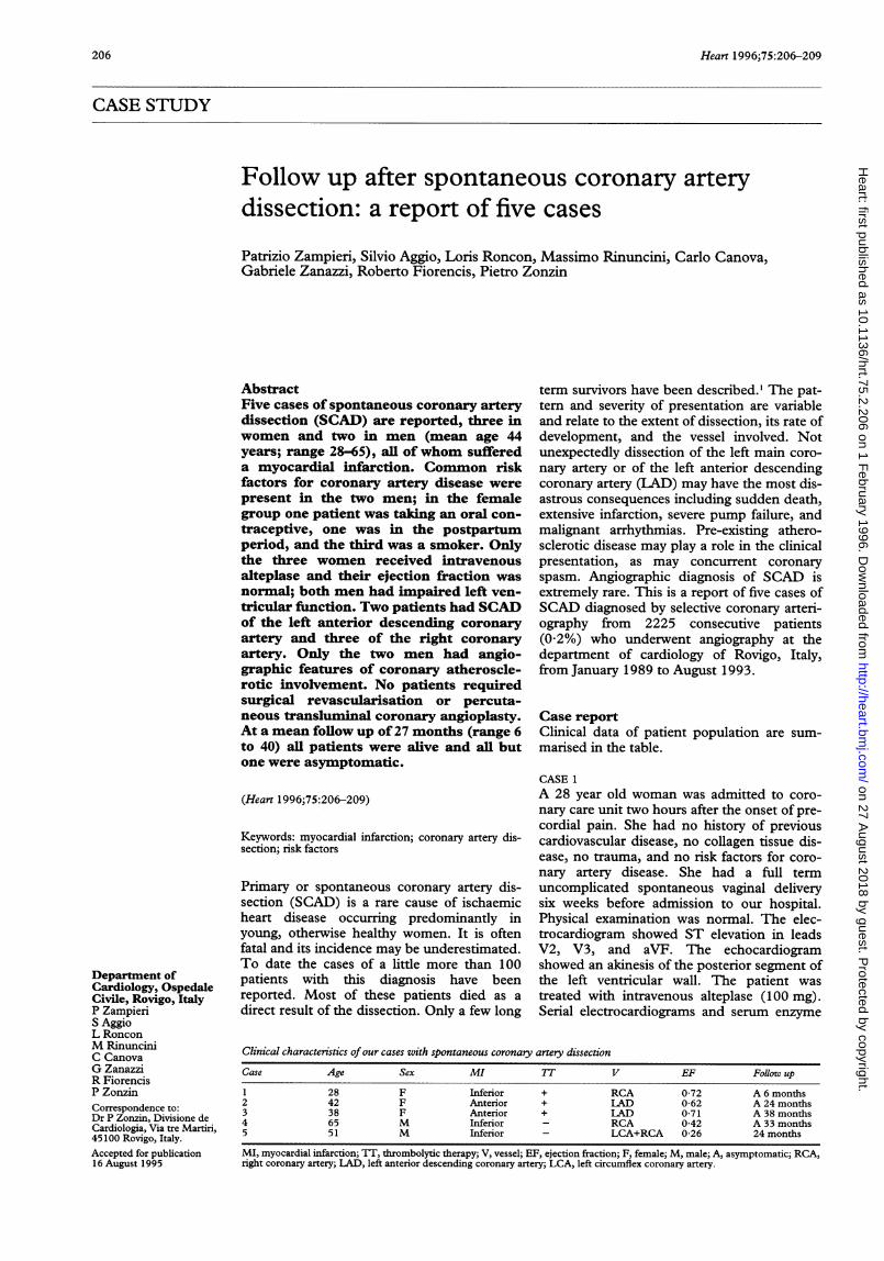
Figure 1 Left anterior oblique view with cranial angulation of the left coronary artery.Arrow points to intimalflap in the mid portion of the left anterior descending coronary
artery.
levels showed acute myocardial infarction.During the following three days she had multi-ple episodes of chest pain with ST elevation inthe inferior leads. She was given ,B blockersand intravenous glyceryl trinitrate and sheimproved symptomatically. Her later clinicalcourse was uneventful. Thirteen days aftermyocardial infarction the patient underwentcoronary and left ventricular angiography. Leftventricular angiography showed mild hypo-kinesis of the posterobasal and diaphragmaticleft ventricular wall, with an ejection fractionof 0-72. Coronary angiography showed dissec-tion of the right coronary artery. The dissec-tion resulted in mild luminal narrowing of the
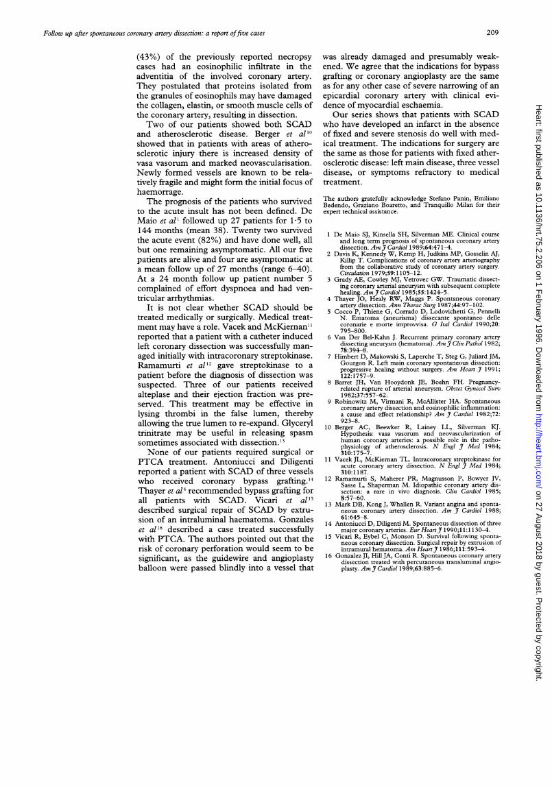
Figure 2 Left coronary angiogram (right anterior oblique) ofpatient number 3. Arrowpoints to the distal left anterior descending coronary artery where a dual lumen is present,separated by a flap-like spiral defect indicative of intimal dissection. The subintimalhaematoma does not totally obstruct the lumen, as distal passage of dye occurs.
mid and distal segments of the artery. Thepatient no longer complained of chest pain,either at rest or during a submaximal exercisetest. She was discharged on the 16th day andshe was still asymptomatic at a six monthsclinical follow up.
CASE 2A 42 year old woman who had previously beenin good health experienced the acute onset ofsevere pain in the chest and left arm shortlyafter preparing breakfast at home. Three hourslater she was admitted to our coronary careunit with an electrocardiographic and echocar-diographic pattern of acute anteroseptalmyocardial infarction. She had experiencedthree full term uncomplicated pregnancies, thelast one three years previously; she was nottaking oral contraceptives and had a normalmenstrual status. She was a heavy smoker (20cigarettes a day). She was given intravenousalteplase with improvement in her clinicalstate. However, serial electrocardiograms andserum enzyme levels showed evolution ofacute myocardial infarction. Her clinicalcourse was uneventful. Ten days after admis-sion the patient underwent coronary angiogra-phy. Left ventriculography showed mildimpairment of contractility in the anteriorwall. The left coronary arteriogram showed aflap-like filling defect in the mid LAD (fig 1).The patient was discharged with medicaltreatment (/3 blockers and aspirin) after a nega-tive submaximal exercise test. At a 24 monthfollow up she remained totally asymptomatic.
CASE 3This 38 year old woman had experiencedanginal pain while swimming. She was admit-ted to a hospital four hours later with evidenceof acute anterior myocardial infarction andreceived intravenous alteplase. A month latershe was referred to our cardiology departmentfor further evaluation of her coronary arterydisease. She had been taking an oral contra-ceptive at the time of her myocardial infarc-tion, and had no further coronary risk factors.Physical examination was normal. A submaxi-mal exercise test was negative. Left heartcatheterisation revealed a normal left ventricu-lar ejection fraction and apical akinesis.Coronary angiography showed a flap-like spi-ral filling defect indicative of an intimal dissec-tion in the distal LAD, comprising the lumen(fig 2). Nevertheless anterograde flow was pre-served. She was given medical treatment andwas asymptomatic at a 38 month clinical fol-low up.
CASE 4A 65 year old man was referred to our hospitalfor angiographic evaluation of his coronaryartery disease. He had experienced a postero-lateral myocardial infarction two years before.He had remained asymptomatic for the follow-ing period. A treadmill thallium test showed aposterolateral scar and an inferior reversibledefect. The patient was a heavy smoker andhad a history of hypertension and diabetes.Left ventriculography showed posterolateral
207
on 27 August 2018 by guest. P
rotected by copyright.http://heart.bm
j.com/
Heart: first published as 10.1136/hrt.75.2.206 on 1 F
ebruary 1996. Dow
nloaded from

Zampieri, Aggio, Roncon, Rinuncini, Canova, Zanazzi, et al
Figure 3 Right anterior oblique angiographic view of the left coronary artery showing(arrow) a serpiginous filling defect in the obtuse marginal branch of the left circumflexartery.
akinesis and mild anterolateral hypokinesiswith an ejection fraction of 0-42. Coronaryangiography revealed diffuse mild three vesselatherosclerotic disease with a long dissectionof the right coronary artery in its mid and distalportions. We did not advise any surgical revas-cularisation and the patient was asymptomaticat a 33 month follow up.
CASE 5This 51 year old man, who had several riskfactors, had an inferior myocardial infarctiontwo years before admission. In the years fol-lowing this he experienced neither angina nor
dyspnoea. After a positive exercise test he wasreferred to us for coronary angiographic evalu-ation. Left ventriculography and coronary
angiography revealed severely impaired leftventricular function (EF 0 26) and dissectionsof the right coronary artery in its mid and distalportions and of the marginal obtuse branch ofthe left circumflex artery (fig 3). The antero-grade flow was preserved in both arteries.There were signs of diffuse atheroscleroticcoronary artery disease with no evidence ofsevere focal stenosis. We decided to treat thepatient medically. At a 24 month follow up hecomplained of effort dyspnoea (New YorkHeart Association grade II) and had ventriculararrhythmias which were treated with amio-darone.
DiscussionSpontaneous coronary artery dissection isextremely uncommon. Dissecting aneurysmsof the coronary vessels are much more likely tobe iatrogenic,' secondary to blunt chesttrauma,3 or associated with an aortic dissectinganeurysm or with Marfan's syndrome.Definite physical features of the Marfan syn-drome were absent in all our patients, andthere was no history of previous trauma.
In most cases of SCAD the diagnosis isestablished on necropsy examination. A reviewof published reports shows that the usual clini-cal presentation of this condition was eithersudden death or acute myocardial infarctionfollowed in a short time by death. Though ourpatients had an acute myocardial infarctionthey survived the acute insult and all but onewere asymptomatic at follow up.The angiographic diagnosis during life of
SCAD is very unusual.'4 We defined dissec-tion as the detection at coronary angiographyof a radiolucent area within the lumen of thevessel, with or without contrast persistencewithin the dissection after wash out of the con-trast from the remaining portion of the vessel.The lumen of the vessel was enlarged at thesite of dissection; this pattern is caused by thepresence of dye inside the coronary wall and isimportant in the differential diagnosis withintracoronary thrombus.SCAD typically occurs in young, healthy
women. In a series reviewed in 1990 by Coccoet al,5 74 out of 97 cases were women, with anaverage of 39 years. It is striking that 24 of 74patients were in the puerperium and one hadbeen taking an oral contraceptive for the lastthree years. Approximately 50% of patientswith a coronary artery dissection die suddenlyand many others (18-20%) will die within afew hours. The clinical presentation is other-wise similar to that of patients with atheroscle-rotic coronary artery disease, although theaverage age is younger and risk factors may beabsent. SCAD may be recurrent;6 it may alsoheal spontaneously.7 Van Der Bel-Kahn6reported the case of a 40 year old man inwhom a dissection of the obtuse marginalartery had apparently healed, but who died at alater date of a second dissection of the LAD.
Himbert et al7 described progressive angio-graphic healing of a spontaneous dissection ofthe left main coronary artery. The LAD is themost commonly involved artery (52-5%),5 fol-lowed by the right coronary artery (24%), theleft main stem (13.5%), and the left coronaryartery (2%).
Various hypothesis have been offered toexplain the aetiology of SCAD. Hypertensiondoes not seem to be a risk factor, since it isusually absent.5 Any unifying theory of coro-nary artery dissection must explain theremarkable predilection for women, especiallyduring the puerperium. Changes in the arterialwall during pregnancy have been well docu-mented and include fragmentation of reticu-lum fibres, hypertrophy of smooth musclecells, and alterations in the protein and acidmucopolysaccharide content of the media.8The current belief is that degeneration of theground substance causes a weakening of thetunica media of the vessels during pregnancy;the effect of straining during labour and deliv-ery may initiate intimal rupture, with subse-quent haemorrage into the media days orweeks later. SCAD has occurred in men, how-ever, thus indicating multifactorial aetiology.Angiitis has been implicated in the pathogene-sis of SCAD. Robinowitz et a19 found in anecropsy study that their eight patients and 31
208
on 27 August 2018 by guest. P
rotected by copyright.http://heart.bm
j.com/
Heart: first published as 10.1136/hrt.75.2.206 on 1 F
ebruary 1996. Dow
nloaded from
Follow up after spontaneous coronary artery dissection: a report offive cases
(43%) of the previously reported necropsy
cases had an eosinophilic infiltrate in theadventitia of the involved coronary artery.They postulated that proteins isolated fromthe granules of eosinophils may have damagedthe collagen, elastin, or smooth muscle cells ofthe coronary artery, resulting in dissection.Two of our patients showed both SCAD
and atherosclerotic disease. Berger et al'0showed that in patients with areas of athero-sclerotic injury there is increased density ofvasa vasorum and marked neovascularisation.Newly formed vessels are known to be rela-tively fragile and might form the initial focus ofhaemorrage.The prognosis of the patients who survived
to the acute insult has not been defined. DeMaio et all followed up 27 patients for 1-5 to144 months (mean 38). Twenty two survivedthe acute event (82%) and have done well, allbut one remaining asymptomatic. All our fivepatients are alive and four are asymptomatic ata mean follow up of 27 months (range 6-40).At a 24 month follow up patient number 5complained of effort dyspnoea and had ven-
tricular arrhythmias.It is not clear whether SCAD should be
treated medically or surgically. Medical treat-ment may have a role. Vacek and McKiernan" I
reported that a patient with a catheter inducedleft coronary dissection was successfully man-
aged initially with intracoronary streptokinase.Ramamurti et al 12 gave streptokinase to a
patient before the diagnosis of dissection was
suspected. Three of our patients receivedalteplase and their ejection fraction was pre-served. This treatment may be effective inlysing thrombi in the false lumen, therebyallowing the true lumen to re-expand. Glyceryltrinitrate may be useful in releasing spasmsometimes associated with dissection. 13None of our patients required surgical or
PTCA treatment. Antoniucci and Diligentireported a patient with SCAD of three vesselswho received coronary bypass grafting. 14
Thayer et al4 recommended bypass grafting forall patients with SCAD. Vicari et all'5described surgical repair of SCAD by extru-sion of an intraluminal haematoma. Gonzaleset al16 described a case treated successfullywith PTCA. The authors pointed out that therisk of coronary perforation would seem to besignificant, as the guidewire and angioplastyballoon were passed blindly into a vessel that
was already damaged and presumably weak-ened. We agree that the indications for bypassgrafting or coronary angioplasty are the sameas for any other case of severe narrowing of anepicardial coronary artery with clinical evi-dence of myocardial eschaemia.Our series shows that patients with SCAD
who have developed an infarct in the absenceof fixed and severe stenosis do well with med-ical treatment. The indications for surgery arethe same as those for patients with fixed ather-osclerotic disease: left main disease, three vesseldisease, or symptoms refractory to medicaltreatment.
The authors gratefully acknowledge Stefano Panin, EmilianoBedendo, Graziano Boaretto, and Tranquillo Milan for theirexpert technical assistance.
1 De Maio SJ, Kinsella SH, Silverman ME. Clinical courseand long term prognosis of spontaneous coronary arterydissection. Am Jf Cardiol 1989;64:471-4.
2 Davis K, Kennedy W, Kemp H, Judkins MP, Gosselin AJ,Killip T. Complications of coronary artery arteriographyfrom the collaborative study of coronary artery surgery.Circulation 1979;59:1105-12.
3 Grady AE, Cowley MJ, Vetrovec GW. Traumatic dissect-ing coronary arterial aneurysm with subsequent completehealing. Am J7 Cardiol 1985;55:1424-5.
4 Thayer JO, Healy RW, Maggs P. Spontaneous coronaryartery dissection. Ann Thorac Surg 1987;44:97-102.
5 Cocco P, Thiene G, Corrado D, Lodovichetti G, PennelliN. Ematoma (aneurisma) dissecante spontaneo dellecoronarie e morte improvvisa. G Ital Cardiol 1990;20:795-800.
6 Van Der Bel-Kahn J. Recurrent primary coronary arterydissecting aneurysm (hematoma). Am 7 Clin Pathol 1982;78:394-8.
7 Himbert D, Makowski S, Laperche T, Steg G, Juliard JM,Gourgon R. Left main coronary spontaneous dissection:progressive healing without surgery. Am Heart J7 1991;122:1757-9.
8 Barret JH, Van Hooydonk JE, Boehn FH. Pregnancy-related rupture of arterial aneurysm. Obstet Gynecol Surv1982;37:557-62.
9 Robinowitz M, Virmani R, McAllister HA. Spontaneouscoronary artery dissection and eosinophilic inflammation:a cause and effect relationship? Am _7 Cardiol 1982;72:923-8.
10 Berger AC, Beewker R, Lainey LL, Silverman KJ.Hypothesis: vasa vasorum and neovascularization ofhuman coronary arteries: a possible role in the patho-physiology of atherosclerosis. N Engl _7 Med 1984;310:175-7.
11 Vacek JL, McKiernan TL. Intracoronary streptokinase foracute coronary artery dissection. N Engl . Med 1984;310:1187.
12 Ramamurti S, Maherer PR, Magnusson P, Bowyer JV,Sasse L, Shaperman M. Idiopathic coronary artery dis-section: a rare in vivo diagnosis. Clin Cardiol 1985;8:57-60.
13 Mark DB, Kong J, Whallen R. Variant angina and sponta-neous coronary artery dissection. Am )I Cardiol 1988;61:645-8.
14 Antoniucci D, Diligenti M. Spontaneous dissection of threemajor coronary arteries. EurHeartJ 1990;11:l 130-4.
15 Vicari R, Eybel C, Monson D. Survival following sponta-neous coronary dissection. Surgical repair by extrusion ofintramural hematoma. Am Heart_7 1986;111:593-4.
16 Gonzalez JI, Hill JA, Conti R. Spontaneous coronary arterydissection treated with percutaneous transluminal angio-plasty. Am j7 Cardiol 1989;63:885-6.
209
on 27 August 2018 by guest. P
rotected by copyright.http://heart.bm
j.com/
Heart: first published as 10.1136/hrt.75.2.206 on 1 F
ebruary 1996. Dow
nloaded from