focus on lyme-borreliosis
TRANSCRIPT

Focus onLyme-Borreliosis
Infectious Disease
FOR OUTSIDE THE US AND CANADA ONLY

II

1
Dr. Thomas W. Talaska, Specialist in Microbiology and Infection Epidemi-ology.Since 1992, his work has been focused on tick-borne diseases, especially on epidemiological and diagnostic projects related to Lyme borreliosis and other infections caused by parasites (i.e. babesiosis, ehrlichiosis and rickettsiosis).Dr. Talaska coordinates the Interdisciplinary Consulting Group for Lyme borreliosis organized by the Brandenburg Medical Association. He is the Director of the Institute for Tick-Borne Diseases, whose activity was addressed to the prevention and on informing the population about the possible risks of such insect bites.
Prof. Andreas Krause, Specialist in Internal Medicine and Rheumatology, has conducted studies about the pathogenesis of Lyme borreliosis and the immune response of cells related to this disease. He has also worked on studies concerning arthritic diseases associated with infections and psychoneuroimmunology. Until 2002, Prof. Krause was Director of the Day Clinic for Rheumatology at the Charité University Hospital. Since then, he has been Head of the department of the Internal Medicine/Rheumatology and Clinical Immunology Department, Rheumatology Institute, Imman-uel-Krankenhaus, Berlin-Wannsee, and since October 2005 he has also been Head of the department of the Rheumatology Clinic, Berlin-Buch.
Prof. Elisabeth Aberer is specialized in dermatology and venereal diseases. She has a Master’s Degree in Dermohistopathology. Since April 1995 she has been Senior Consultant of the Dermatology Clinic, University of Graz. Her studies have mainly focused on clinical signs and symptoms, treatment and diagnostic issues related to Lyme borreliosis. Prof. Aberer has formed a research center specialized in Lyme borreliosis at the Vienna General Hospital. She has directed two projects designed to promote scientific research: “Die pathogenetische Bedeutung von B. burgdorferi in der Haut” (The pathogenic meaning of B. burgdorferi on the skin, 1989-1992) and “Harn - PCR zur Routinediagnose der Lyme-Borreliose” (Urine PCR in rou-tine screening for Lyme borreliosis, 1999-2002). She is the Director of the Research Center specialized in General and Self-Immune Pathology and, Director of the Quality Control Department, University Clinic of Derma-tology and Venereology, Graz. Laboratory diagnostics mainly focus on immunodiagnostics in autoimmune dermatological and primary systemic diseases and on specialized diagnostics for Lyme borreliosis.
l Authors
Elisabeth Aberer
Thomas Talaska
Andreas Krause

2
Since July 2000 Prof. Reinhard Kaiser has been the Director of the Neur-ology Clinic, Pforzheim Hospital. After following experimental training at the Institute of Virology and Immunology, University of Würzburg, Pro-fessor Kaiser has been involved, since 1989 in the diagnostics, treatment and clinical observation of patients suffering from neuroborreliosis and tick-borne meningoencephalitis (FSME). His clinical studies have focused on differential diagnostics, on the treatment of patients with acute and chronic inflammation of the nervous system and of those suffering from neurological disorders who are serologically positive for borreliosis, even in absence of evident symptoms of neurological form.
Dr. Volker Fingerle - Specialist in Microbiology and Infectious Epidemi-ology - has worked at the Max von Pettenkofer Institute of Hygiene and Medical Microbiology, Ludwig Maximilians Universität (LMU), Munich, since 1990. He works at the National Reference Center (NRZ) for Borreliae and oversees the consiliary lab for Ehrlichea. His studies have mainly focused on training and consulting activities targeted at colleagues and dealing with the epidemiology, diagnostics and treatment of Lyme borreliosis and of anaplasmosis (ehrlichiosis). His fields of research are epidemiol ogy, diagnostics and pathogenesis of Lyme borreliosis and granulocytic anaplasmosis in humans.
Prof. Bettina Wilske is Microbiologist at the Max von Pettenkofer-Institute of Hygiene and Medical Microbiology, Ludwig Maximilians Universität (LMU), Munich. She is also the head of the National Reference Center (NRZ) for Borreliae and carries out medical consulting assignments on the prophylaxis, diagnostics and treatment of Lyme borreliosis. She has been studying Lyme borreliosis since the early ‘80s, especially focusing on the microbiology, diagnostics, epidemiology and analysis of the pathogenic agent’s antigen structure. She has authored the MiQ 12 Lyme borreliosis (quality standard for the microbiological diagnosis of Lyme borreliosis) edited by the DGHM (German Association for Hygiene and Microbiology), whose English version can be found on the homepage of the DGHM (http://www.dghm.org/red/index.html?cname=MIQ). Bettina Wilske
Reinhard Kaiser
Volker Fingerle
Authors

3
l Index
Epidemiological, biological, and ecological aspects of Lyme borreliosis ......................................................... 5• Thomas Talaska
Rheumatological and other internal manifestations of Lyme borreliosis ............................................ 25• Andreas Krause
Dermatological manifestations of Lyme borreliosis ................. 37• Elisabeth Aberer
Neuroborreliosis ........................................................................... 51• Reinhard Kaiser
Lyme borreliosis diagnostics ....................................................... 67• Volker Fingerle, Bettina Wilske
Contacts ........................................................................................ 83

4

55
As the science of disease prevention, epidemiology plays a
significant role in the planning and evaluation of public health and health politics. It is used in conjunction with laboratory research to identify risk factors for disease, elucidate the mechanisms by which they develop, and finally to formulate prevention strategies. In Germany, the instrument for epidemiological evaluation of infectious diseases is the Infectious Diseases Control Law (IfSG), which came into force on January 1, 2001; however, this law applies only to infections transmitted directly from person to person or foodborne, and does not, cover Lyme borreliosis, by far the most common vectorborne disease in central Europe. However, all of the former East German states and Berlin have laws regarding registration of both occurrence and positive laboratory results of the disease. An effective, affordable, regionally tailored prevention programme including immunisation schemes cannot be
realised without knowledge of disease prevalence, incidence, and risk factors. Such epidemiological information is also essential to gauging the success of any preventive measures. Although Lyme borreliosis has an extremely low mortality rate, its frequency alone has a significant economic effect. This “economical impact” is defined by the World Health Organisation (WHO) as the total cost of visits to physicians, laboratory diagnostics, drugs, hospital stay, lost work time, additional health care and, in certain cases, invalidity [5]. Personal suffering, reduced quality of life and ability to perform should not be ignored, even though these are difficult to quantify. In occupations with high exposure rates, Lyme borreliosis has considerable significance as a workrelated disease.The WHO European regional office in Copenhagen published a report in 2004 on vectorborne diseases, including tickborne diseases, and their significance in Europe. The incidence of such illness is significantly higher
Epidemiological, biological, and ecological aspects of Lyme borreliosisl Thomas Talaska

6
Epidemiological, biological, and ecological aspects of Lyme borreliosis
than has been assumed by medical practit ioners and public health departments – often diagnoses are not made and treatment is delayed. This requires action! In the absence of the public interest associated with dramatic outbreaks, the need for surveillance and control of not only the disease but also its vectors is easily overlooked, resulting in insufficient support for epidemiological research, even by the public health authorities [4].
Borrelia: ticks – hostsThe epidemiology of Lyme borreliosis in humans is closely associated with the tick’s life cycle, its habitat, and the interaction between tick, host – including humans – and the particular species of Borrelia involved. Thus it is necessary here to make a short excursion into the world of ticks in order to understand this zoonosis.Borrelia are obligate parasites with no freeliving stages, and their propagation relies on complex zoonotic transmission cycles involving rodents as the main reservoir hosts and ticks as vectors.In Europe there are three species confirmed pathogenic to humans: Borrelia afzelii Borrelia garinii Borrelia burgdorferi sensu strictoTwo other species show evidence of pathogenicity: Borrelia valaisana Borrelia lusitaniaeRecently an additional Borrelia
burgdorferi genospecies was isolated from erythema migrans lesions [7]: Borrelia spielmanii (also designated
A14S).For Borrelia, humans are deadend hosts.The main vector of Lyme borreliosis in central Europe is Ixodes ricinus (Fig. 1). This tick species has a very broad spectrum of vertebrate hosts (reptiles, birds, especially migratory species, and mammals), compared with other native tick species, and is thus well suited for transmitting pathogens to a wide variety of species.
How does a tick bite?Once a tick finds a suitable attachment site on a host, it uses its mouthparts, the chelicerae, to cut through the host’s skin. The tick pushes its hypostome, a chitin tube with recurved teeth, into the small wound created. In effect, the tick stabs or pricks its host. The tick hypostome is
Fig. 1. A mouse with Ixodes ricinus larvae and nymphs

7
however not a completely closed tube, as in insects, and to facilitate suction, the hypostome must be cemented into the skin by a salivary secretion at the site of penetration. While feeding, the tick is stuck fast to the host – thus the generic name Ixodes (from Greek ixos, meaning “glue” [13]).
Transmission of Borrelia to ticks is required for human infection and can take place in various ways. Transovarial transmission to offspring in systemically infected ticks is relatively uncommon, affecting less than 5% of Ixodes ricinus larvae. A more common route of transmission occurs when larvae are infected by feeding on blood, usually from infected forest mice. Transstadial transmission is then possible to nymphs and finally adult ticks. This crossstage transmission is highly dependent on the Borrelia species involved, the distribution of Borrelia in the tick (midgut only or systemic, including salivary glands), and on the host species. It also depends on the sensitivity of Borrelia species to the lytic action of the host’s innate immune defences, the complement system.If a tick infected with Borrelia afzelii draws a bird’s blood, the complement system in this liquid “meal” is able to destroy the Borrelia in the tick’s midgut. If the tick is infected only in the midgut, then the Borrelia can neither be transferred to the next tick developmental stage nor to
the host (a nonpermissive system). If, however, the tick is systemically infected, deactivation in the midgut occurs, but the tick remains infected and transstadial transmission occurs, while the bird host is protected by its complement system (a semipermissive system). The bird has no reservoir competence for this strain. Reservoirincompetent host species can have a zooprophylactic effect through which the dissemination of Borrelia can be hindered. Nuncio et al. describe an example of this effect on the Portuguese island of Madeira, where the native
hosts are lizards (Madeira wall lizard, Teira dugesii) in either non or semipermissive systems (Fig. 2) [12, 15].The circulation of Borrelia on Madeira can be maintained by introduced mice and rats [16].
Cofeeding is an additional transmission route between ticks. Borrelia can be
Fig. 2. Teira dugesii, Funchal, Madeira (photo: Robert Klose)
8
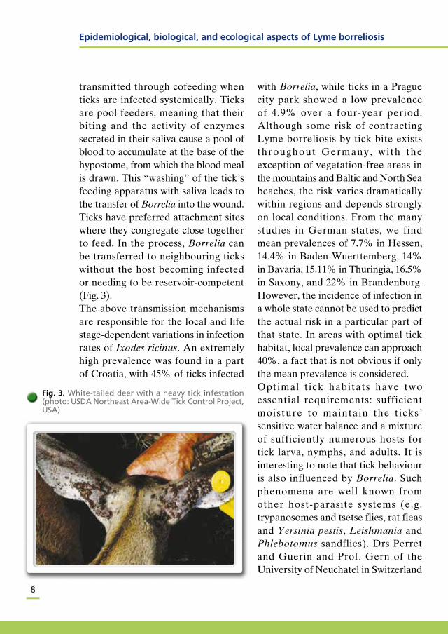
transmitted through cofeeding when ticks are infected systemically. Ticks are pool feeders, meaning that their biting and the activity of enzymes secreted in their saliva cause a pool of blood to accumulate at the base of the hypostome, from which the blood meal is drawn. This “washing” of the tick’s feeding apparatus with saliva leads to the transfer of Borrelia into the wound. Ticks have preferred attachment sites where they congregate close together to feed. In the process, Borrelia can be transferred to neighbouring ticks without the host becoming infected or needing to be reservoircompetent (Fig. 3).The above transmission mechanisms are responsible for the local and life stagedependent variations in infection rates of Ixodes ricinus. An extremely high prevalence was found in a part of Croatia, with 45% of ticks infected
with Borrelia, while ticks in a Prague city park showed a low prevalence of 4.9% over a fouryear period. Although some risk of contracting Lyme borreliosis by tick bite exists throughout Germany, with the exception of vegetationfree areas in the mountains and Baltic and North Sea beaches, the risk varies dramatically within regions and depends strongly on local conditions. From the many studies in German states, we find mean prevalences of 7.7% in Hessen, 14.4% in BadenWuerttemberg, 14% in Bavaria, 15.11% in Thuringia, 16.5% in Saxony, and 22% in Brandenburg. However, the incidence of infection in a whole state cannot be used to predict the actual risk in a particular part of that state. In areas with optimal tick habitat, local prevalence can approach 40%, a fact that is not obvious if only the mean prevalence is considered. Optimal t ick habitats have two essential requirements: sufficient moisture to maintain the ticks’ sensitive water balance and a mixture of sufficiently numerous hosts for tick larva, nymphs, and adults. It is interesting to note that tick behaviour is also influenced by Borrelia. Such phenomena are well known from other hostparasite systems (e.g. trypanosomes and tsetse flies, rat fleas and Yersinia pestis, Leishmania and Phlebotomus sandflies). Drs Perret and Guerin and Prof. Gern of the University of Neuchatel in Switzerland
Fig. 3. White-tailed deer with a heavy tick infestation (photo: USDA Northeast Area-Wide Tick Control Project, USA)
Epidemiological, biological, and ecological aspects of Lyme borreliosis

9
demonstrated that, in dry atmospheric conditions, infected ticks move farther and more often from their original location than uninfected ticks. This increases the likelihood of finding new hosts on which to feed – and thus of new infections with Borrelia [10]. Prevalence rates also vary according to tick life stage; for example in Thuringia a study of ticks found attached to patients showed infection in 0% of larvae, about 15% of nymphs, and about 19% of adult females [6].
The diversity of interactions between Borrelia, ticks, and hosts outlined here demonstrates the need for sophisticated biological and epidemiological analyses of Lyme borreliosis. In addition, one must also include ecological factors such as vegetation, geology, hydrology, temperature, and rainfall. At present, we are only able to give a descriptive account of the epidemiology of Lyme borreliosis in humans.
Factors influencing the epidemiology of Lyme borreliosis in humans Tick species Borrelia species Presence/density of reservoir
competent hosts Presence/density of tick habitats which
humans enter Use of forest and agricultural land Forest structure and kinds of biotopes Seasonal changes in temperature,
sunshine, and rainfall
Ground moisture and geological characteristics
Longterm temperature develop ments Risk and recreational behaviour of
humans Certainty of the diagnosis of Lyme
borreliosis
EpidemiologyWorldwide, epidemiological data on Lyme borreliosis as an “emerging infectious disease” are inadequate, with the exception of those from the USA (Centers of Disease Control, or CDC, and the Division of VectorBorne Diseases). In Europe there are currently very few national notification or registration systems. The epidemiological data available for Germany are based mostly on studies aimed at only a single clinical manifestation or are limited to very small regions within a German state. Other countries have regional registration systems, such as in Austria and France. Russia has the beginnings of a regional notification system based on a highly centralised diagnosis of Borrelia. Slovenia probably has the oldest epidemiological databank in Europe, based on a notification system for Lyme borreliosis originating in 1998 [21].What actually is borreliosis? Borrelia are not new pathogens – typical clinical cases were described as early as the end of the 1800s (acrodermatitis chronica atrophicans, erythema chronicum migrans, Bannworth’s syndrome).
10
In 1996 Ohlenbusch confirmed the presence of Borrelia in ticks more than 100 years old from museum collections in Berlin and Vienna (Borrelia burgdorferi sensu stricto, Borrelia garinii, Borrelia afzelii [17]). The significant increase in borreliosis, considered a rarity with unknown aetiology but welldefined symptoms as recent as 20 years ago, is surprising. The establishment of clinical case definitions of Lyme borreliosis was an important requirement for collecting valid data. Those created by the CDC were very stringent and developed specifically for epidemiological purposes. However, a number of European manifestations of the disease were not included because they are practically unknown in the USA. In 1996 the European Union for Concerted Action on Risk Assessment in Lyme Borreliosis (EUCALB) established valid case definitions for Europe that allow standardisation of registered, defined clinical cases [20]. The existence of yet undefined and hence unregistered clinical manifestations must be considered. The possibility of simultaneous transmission of multiple tickborn diseases necessitates differential diagnosis. In fact, simultaneous infection with Borrelia and the pathogens causing human granulocytic ehrlichiosis (HGE), Anaplasma phagocytophilum , or Babesia spp. are documented. These present a clinical picture that differs from the course typical for Lyme
borreliosis. The significance of infection with Rickettsia slovaca and Rickettsia helvetica is still largely unclear.What explains the increase in Lyme disease? Socioecological factors are implicated in a study of an area with endemic Lyme disease in the northeastern USA where the movement of large numbers of people from populated urban centres to forested rural areas increased the risk of exposure to tick bites [8]. At the same time, this area experienced a decrease in agricultural land use, and an increase in bushes and trees abutting directly to inhabited areas, with a concomitant increase in tick hosts. The WHO Report on Europe also describes massive ecological changes that have taken place since the end of the Second World War. In particular, intensive reforestation has brought (and continues to bring) changes in the flora and fauna. The population densities of wild boar and deer have increased, leading to greater tick density. Similar developments can also be seen in Germany, but specific studies on this problem are lacking.It is probable that continued climatic change will influence the frequency of vectorborne diseases. The distribution and seasonality of diseases transmitted by ectothermic insects and ticks are particularly sensitive to global temperature changes. Warmer winters favour tick survival and movement into new areas where they were previously rare or absent. Global warming also
Epidemiological, biological, and ecological aspects of Lyme borreliosis

11
leads to longer active periods for ticks in spring and autumn and probably increases human recreational activities in these periods, resulting in higher risk of exposure to ticks. Examples include Sweden, where the distribution of tickborne encephalitis (TBE) has expanded northward, and the Czech Republic, where (TBE) expansion has occurred over the last 30 years into higher areas of the Bohemian Forest [25]. How the situation with Lyme borreliosis will develop remains to be determined, although it is likely to follow the pattern of TBE. The first data confirming this came from the Czech Republic [5].A temperaturerelated lengthening of the tick season in the presence of a large number of reservoircompetent hosts can also lead to faster tick developmental cycles and thus greater tick density; transit from egg to adult can be shortened from 54 to 18 months.In November of 2004, the European Union project “Emerging Diseases in a Changing European Environment” was started. Prof. Sarah Randolph of Oxford University is leading the “TickBorne Diseases” section. Its aim is to explain the increase in tickborne diseases on a European scale and to create predictive statistical and biological models.
The development of borreliosis epidemiology in BrandenburgThe first studies on the epidemiology of Lyme borreliosis in Germany began in the state of Brandenburg under the
auspices of the 1994 WHO Consultation on the Development and Application of Geographical Methods in the Epidemiology of Zoonoses [3]. In that year, the “Geographical Epidemiology of Borreliosis in Brandenburg” project was started, in which a voluntary notification schedule was introduced and the Lyme Disease Case Report Form adapted from the American Case Report Questionnaire in conjunction with the CDC. Starting in November 1996 the German Federal Infectious Diseases Law was extended to require that all clinical occurrences of Lyme borreliosis in Brandenburg be reported using the notification schedule and all positive laboratory results to be registered. This was the first opportunity to collect and analyse a data set covering an entire German state. In the same year, the Lyme Borreliosis Interdisciplinary Advisory Group was established by the Brandenburg State Medical Board to advise medical practitioners, patients and selfhelp groups. In 2001, the Brandenburg State Ministry for Work, Social Affairs, Women, and Health founded the Regional Counselling Centre for TickBorne Diseases. In addition to its advisory role, this service coordinates and carries out studies, evaluates tests, and makes additional analyses of registered cases of Lyme borreliosis. In addition, issues involving HGE, Rickettsia helvetica, and babesiosis are considered there in cooperation with, among others, the Robert Koch

12
Institute (RKI) in Berlin, the WHO in Geneva, the CDC Division of VectorBorne Diseases at Fort Collins, the University of Frankfurt/Main, and Tulane University in New Orleans.
Geographic information systems and Lyme disease Computerbased geographic in for mation systems (GIS) represent a modern tool for analysing risk factors and producing risk maps for the epidemiology of infectious diseases, particularly tickborne diseases [3]. This in turn can provide the basis for locally adapted prevention strategies similar to the timetested methods used in veterinary medicine and veterinary disease control. The GIS systems are based on arearelated databases with parameters that include cases of infection, geographic coordinates, adminis t rat ive s t ructures , and population density. They contain digital coordinate reference maps showing, for example, administrative boundaries, biotopes, geology, hydrology, weather, and other relevant factors [14, 18, 19].
The computer programme “EpiInfo”, f r e e w a r e f o r e p i d e m i o l o g i c a l surveillance originally developed by the CDC, includes databases, analytical tools, and EpiMap, a component program that can produce cartographic representations of epidemiological data. The databases are compatible with the GIS system ArcView, which allows coordinates from study areas or new distribution areas to be recorded locally using global positioning systems (GPS) and precisely located on coordinate reference maps. Other geographic parameters such as vegetation and
Fig. 4. Distribution of borreliosis cases in Brandenburg 2003 (database: EpiInfo 3.3; created using ArcView 3.2 [23])
Fig. 5. Example of satellite-supported biotope carto-graphy – alder sections (marked blue) in the Spree forest (photos: Landsat TM; processed with ArcView 3.2; A. Ober 2003)
Epidemiological, biological, and ecological aspects of Lyme borreliosis

13
watercourses can be superimposed on these maps for analysis (Fig. 4).EpiInfo is also useful for evaluating satellite photographs, in which for example different vegetation types produce different patterns of reflection spectra. The ability to precisely superimpose satellite photographs onto digital maps permits onsite biotope searching and analysis using GPS. Computer searches for biotypes with similar characteristics are also facilitated (Fig. 5).Areas with a potentially higher risk of Lyme borreliosis can be located on the computer screen and related to data from regions with a higher incidence of borreliosis – a major prerequisite for the development of risk maps.
Epidemiological data on Lyme borreliosis in BrandenburgThe first incidence data on Lyme borreliosis in Brandenburg were collected on a volunteer basis from 1994 to 1996, i.e. until introduction of the notification requirement. The incidence was ten cases per 100,000 inhabitants but with marked regional differences, which may, however, have been strongly related to notification behaviour. In 1997, 400 clinical cases were registered. Two studies in Maryland and Connecticut, USA shortly after the introduction of a notification requirement showed that only 10–15% of cases fitting the CDC case definition were actually registered. Applying this
factor optimistically to Brandenburg (ignoring undiagnosed cases and those not presenting erythema migrans, as these can only be speculated) would predict 4,000 clinically relevant cases of borreliosis. Developments in the USA show that the number of
1997 1998 1999 2000 2001 2002 2003 2004
350
300
250
200
150
100
50
0
Cases
Fig. 6. Lyme borreliosis in Brandenburg – registered clinical cases per month until 31.12.2004 (graphic: T. Talaska)
* preliminary data
1995
2500
2000
1500
1000
500
0
Cases1,981
1,823
1,4101,3551,240
783819
451
198164
1996 1997 1998 1999 2000 2001 2002 2003 2004*
Fig. 7. Clinical cases of Lyme borreliosis from 1995 to 2004 (graphic: T. Talaska)

14
registered cases increased markedly in the years following introduction of the notification system, especially when the notifying physician was informed on regional epidemiological data. It is assumed that with an established notification system, about a third of
infections are reported (D.T. Dennis, personal communication). If this is the case, Brandenburg’s 1,240 registered borreliosis cases in 2000 represent an actual value of about 4,200. Such high estimates suggest that the increased incidence of borreliosis is probably only a function of the notification changes, and that the actual estimates for the last four years were much lower. Nevertheless, one cannot ignore that subsequent years have shown a steady increase in registered cases of 10–15% annually. This trend has also been found in other German states with notification requirements, as G. Hesse reported at the 2004 Thuringia Workshop on TickBorne Diseases in Erfurt (Figs. 6, 7, 8, 9, 10).
Regional variability in the state of BrandenburgAs already indicated, there are significant differences between districts in the registered incidence of borreliosis. The highest incidence in Brandenburg was reached in 2000 in the OderSpree district (89.3/100,000 inhabitants), the Uckermark (89.0/100,000), and the Barnim (74.6/100,000). In general, there has been a trend toward higher incidence in eastern Brandenburg since 1996. This appears to depend not on risk behaviour but rather on the density of suitable biotopes for the ticks. It is still necessary to determine whether more and smaller habitat areas with higher contact for humans exist in
2000
1800
1600
1400
1200
1000
800
600
400
200
0
Thuringia
1991
Brandenburg SaxonyMecklenburg-
West Pomerania
1992
1993
1994
1995
1996
1997
1998
1999
2000
2001
2002
2003
Saxony-Anhalt Berlin
Fig. 8. Borreliosis in individual German states (graphic: G. Hesse, Erfurt 2004)
1995
80
70
60
50
40
30
20
10
01996 1997 1998 1999 2000 2001 2002 2003
Mor
bidi
ty
1994
Brandenburg
Saxony
Thuringia
Mecklenburg-West Pomerania
Saxony-Anhalt
Fig. 9. Borreliosis morbidity from 1994 to 2003 (graphic: G. Hesse, Erfurt 2004)
Epidemiological, biological, and ecological aspects of Lyme borreliosis
15
the eastern part of Brandenburg than the west. A typical such case is the municipality of Scharmuetzelsee in the OderSpree district, a densely forested recreational centre for people visiting the lake area. Inhabitants of the town of Bad Saarow stated they are regularly bitten by ticks in their gardens, in which mice are very common and visits by wild animals (e.g. roe deer) are not uncommon. Scharmuetzelsee had the highest incidence in Brandenburg in 2000, with 237 cases per 100,000 inhabitants. This figure does not include visitors from Berlin who were affected. This example shows considerable similarities to observations made in the USA [8]. Considering the OderSpree district more closely, one finds immense differences in incidence at the local level. These ranged from 10/100,000 inhabitants in Beeskow to the previously mentioned 237/100,000 in adjacent Scharmuetzelsee. Mapping cases in the OderSpree district on a satellite photograph, most of the registered cases were localised close to forested wetlands. Sections with very low incidence included large agricultural areas, but even there, the few registered cases were associated with small lakes or water courses.
In general Brandenburg is rich in forest and wetlands, but not all forested land is suitable tick habitat. Ticks require a moist microclimate for survival [11] and protection from the cold in winter.
These are best provided by several annual layers of slowly decomposing leaf cover, especially beech and oak. Such deciduous and mixed forest tracts together comprise less than 5% of forest cover in the state. There are large areas
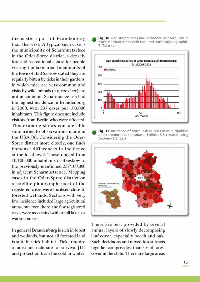
Age-specific incidence of Lyme Borreliosis in BrandenburgTotal 2001-2003
500
400
300
200
100
0
Incidence
50Age (years)
1 100
Fig. 10. Registered cases and incidence of borreliosis in those German states with required notification (graphic: T. Talaska)
Fig. 11. Incidence of borreliosis in 2003 in municipalities and communities (database: EpiInfo 3.3 created using ArcView 3.2 [23])

16
of commercial pine monoculture. Areas forested in this way are poor habitats for ticks because pine needle layers retain less humidity directly over the soil surface than deciduous leaf layers in the warmer part of the year. Recent changes in forestry practice in this region have aimed at increasing the deciduous and mixed forest components to 40% over a 10year period to return the forest to its original natural makeup. It is anticipated that the extent of suitable tick habitat will also increase, and therefore provisions have been made to monitor tick density and thus the potential risk of Lyme borreliosis in these areas (Fig. 11).
Clinical dataErythema migrans, an early clinical manifestation, is the most common registered sign of borreliosis in all states of Germany, and the trend from
1997 to 2004 shows that its proportion increased from 61.7% to 84.5% over that period. It is now higher than in Austria (77.7%) and the USA (76.0%) [24]. It is also evident that the growing proportion of erythema migrans cases accompanies significant reductions in the proportions of reported cases of both the more advanced disseminated borreliosis (19.2% in 1997 vs 5.6% in 2004) and cases which cannot be clearly clinically classified into the EUCALB case definitions (19.1% in 1997 to 8.7% in 2004). Given the notification requirement from 1996 and continuing education organised by the Interdisciplinary Lyme Borreliosis Advisory Group, a possible explanation for this trend is that the early stage of borreliosis is more frequently and accurately diagnosed and treated due to increasing physician and patient awareness. This would reduce the number of patients developing late manifestations such as disseminated borreliosis. However, these cannot be completely avoided, as a significant proportion of infected individuals present with the disseminated form at anamnesis. Only 21% of cases of Lyme arthritis registered in 1999 and 2000 had been previously diagnosed with erythema migrans. The decreasing proportion of “unclear” cases is an indication that Lyme borreliosis is generally being diagnosed with more certainty. However, it is still necessary to analyse these ambiguous clinical
1997 1998 1999 2000 2003 2004
100%
80%
60%
40%
20%
0%
Other K/L ACA NB LA EM
K/L = Karditis/Lymphadenosis cutis benigna;ACA = Acrodermatitis chronica atrophicans;NB = Neuroborreliosis; LA = Lyme arthritis; EM = Erythema migrans
Fig. 12. Different clinical manifestations in percentages
Epidemiological, biological, and ecological aspects of Lyme borreliosis

17
cases and if possible expand the current case definitions. When analysing cases from 2003 and 2004, it is necessary to consider a certain bias in registered manifestations toward erythema migrans due to the RKI case definition of Lyme borreliosis (Fig. 12).Considering the conservatively estimated cost of 10,000 Euro per case of disseminated borreliosis and the relatively certain relationship between erythema migrans and development of disseminated borreliosis in the absence of intervention, the economic importance of information distribution and continuing education is evident.According to this very simplified model, about 1 million Euros could have been saved by the prevention campaign in Brandenburg over the last few years. (Fig. 13).
Model system in the Oder-Spree districtThe OderSpree district was chosen as a study area to analyse risk factors for borreliosis and the prevalence of Borrelia in ticks due to the high incidence of Lyme borreliosis found there. The study was carried out in conjunction with the RKI from June to December 1999. In addition, the geographic distribution of cases was calculated to determine whether it was random or there were indeed highrisk areas within the district (which, as indicated previously, is very heterogeneous).
One significant risk factor, as predicted, was activity in private gardens, particularly those bordering the forest (Fig. 14).This is probably due to a lack of awareness of the risk, which most
1997EM-Cases
2000Disseminated Borreliosis
1000
800
600
400
200
0
EM LA LA/Prev NB NB/Prev
-260,000Euro
-820,000Euro
250
200
150
100
50
0
EM = Erythema migrans; LA = Lyme arthritis; NB = Neuroborreliosis
Fig. 13. Estimates of borreliosis occurrence in Branden-burg with and without preventive measures (highest estimates) for 1997 and 2000
Fig. 14. Typical “tick biotope” in the Oder-Spree district: a garden adjacent to the forest kept close to natural condition
18
individuals seem to associate only with activities in the woods. Light clothing is often worn while gardening and, if nobody search for ticks follows, there is a distinct risk of infection. Unfor tunately it was also determined that, in spite of familiarity with personal pre vention measures, they were actually carried out by only 40% of those que stioned. This requires action by the health authorities and additional prevention strategies directed not only at individual activities. It showed that personal prevention can be very effective, when actually carried out. Our experience from the OderSpreewald district shows that it is
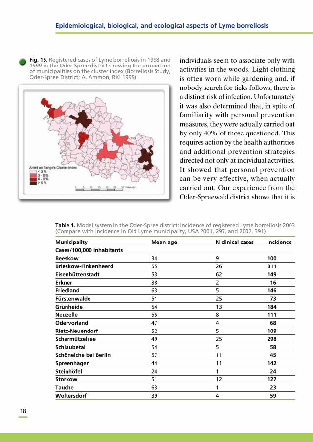
Fig. 15. Registered cases of Lyme borreliosis in 1998 and 1999 in the Oder-Spree district showing the proportion of municipalities on the cluster index (Borreliosis Study, Oder-Spree District; A. Ammon, RKI 1999)
Table 1. Model system in the Oder-Spree district: incidence of registered Lyme borreliosis 2003 (Compare with incidence in Old Lyme municipality, USA 2001, 297, and 2002, 391)
Municipality Mean age N clinical cases Incidence
Cases/100,000 inhabitants
Beeskow 34 9 100
Brieskow-Finkenheerd 55 26 311
Eisenhüttenstadt 53 62 149
Erkner 38 2 16
Friedland 63 5 146
Fürstenwalde 51 25 73
Grünheide 54 13 184
Neuzelle 55 8 111
Odervorland 47 4 68
Rietz-Neuendorf 52 5 109
Scharmützelsee 49 25 298
Schlaubetal 54 5 58
Schöneiche bei Berlin 57 11 45
Spreenhagen 44 11 142
Steinhöfel 24 1 24
Storkow 51 12 127
Tauche 63 1 23
Woltersdorf 39 4 59
Epidemiological, biological, and ecological aspects of Lyme borreliosis

19
frequently ignored [2, 9].In the USA a comprehensive analysis of practical prevention methods requiring minimal effort was done. From the point of view of practicability, acaricideimpregnated feeding stations for wild animals, like those used in the USA, are a realistic alternative. Their application requires minimal administrative effort, and the case reduction achieved in models is very good. Unfortunately, at present this is not possible in Germany due to the difficulty in coordinating the areas of responsibility of the various authorities involved.The geographic distribution shows a clear aggregation of cases in the OderSpree district in particular, and Brandenburg as a whole (Fig. 15).Analyses of notification records show that differences in notification behaviour by physicians can be eliminated as a possible cause. Thus we can assume that the geographic factors previously discussed are responsible for this variation in risk (Table 1) [2].
Forestry workers: a risk groupThe seroprevalence of tickborne diseases in forestry workers was examined in a study carried out in Berlin and Brandenburg [22]. Forestry workers, hunters, and forest managers are more commonly exposed to such diseases than the general population; however, they are also better informed on the risks and their prevention and hence their typical working clothing is
usually wellsuited to preventing tick bites.Our study was carried out on a volunteer basis and included immunoglobulinG (IgG) examinations for seroprevalence against Borrelia burgdorferi sensu lato, as well as the pathogens causing HGE (human granulocytic ehrlichiosis): Anaplasma phagocytophilum, Babesia microtii and Rickettsia helvetica.The risk group as a whole showed 29% Borrelia burgdorferi prevalence, although borreliosis was allegedly diagnosed and treated in only 10.2% of cases. This discrepancy indicates a significant number of asymptomatic and previously undiagnosed cases. In fact, a questionnaire filled out after serodiagnosis showed that an additional 24% of seropositive subjects described symptoms compatible with Lyme borreliosis.
A significant aggregation of seropositive cases occurred in the Barnim region northeast of Berlin. This population showed 6.2% positivity for IgG antibodies to Anaplasma phagocytophilum (FAT, Western blot). In the subsequent case control study, no statistical relationship could be found between HGEpositive cases and potential symptoms. Thus there is evidence of contact with the pathogen, but the course of infection appears to be either asymptomatic or mild.There are only limited data on the frequency of Babesia microtii antibodies
20
in humans in Europe. Our study showed a 1.4% IgGpositive prevalence (FAT, Western blot). Only one indi vidual complained at anamnesis of symptoms consistent with babesiosis after a tick
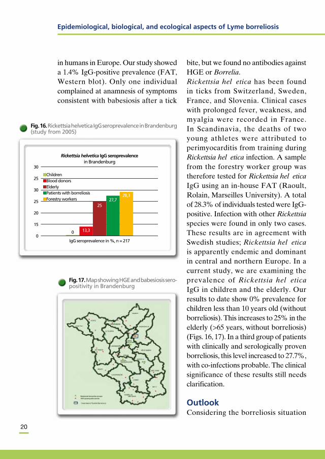
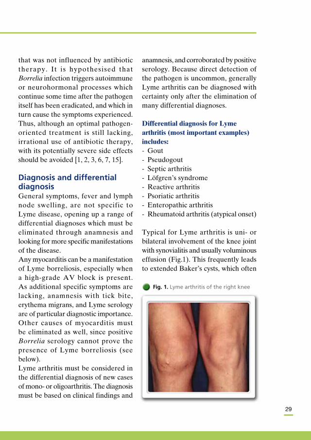
bite, but we found no antibodies against HGE or Borrelia.Rickettsia helvetica has been found in ticks from Switzerland, Sweden, France, and Slovenia. Clinical cases with prolonged fever, weakness, and myalgia were recorded in France. In Scandinavia, the deaths of two young athletes were attributed to perimyocarditis from training during Rickettsia helvetica infection. A sample from the forestry worker group was therefore tested for Rickettsia helvetica IgG using an inhouse FAT (Raoult, Rolain, Marseilles University). A total of 28.3% of individuals tested were IgGpositive. Infection with other Rickettsia species were found in only two cases. These results are in agreement with Swedish studies; Rickettsia helvetica is apparently endemic and dominant in central and northern Europe. In a current study, we are examining the prevalence of Rickettsia helvetica IgG in children and the elderly. Our results to date show 0% prevalence for children less than 10 years old (without borreliosis). This increases to 25% in the elderly (>65 years, without borreliosis) (Figs. 16, 17). In a third group of patients with clinically and serologically proven borreliosis, this level increased to 27.7%, with coinfections probable. The clinical significance of these results still needs clarification.
OutlookConsidering the borreliosis situation
Fig. 17. Map showing HGE and babesiosis sero-positivity in Brandenburg
Rickettsia helvetica IgG seroprevalencein Brandenburg
30
25
30
25
20
15
0
Children Blood donors Elderly Patients with borreliosis Forestry workers
0
IgG seroprevalence in %, n = 217
13,3
2527,7
28,3
Fig. 16. Rickettsia helvetica IgG seroprevalence in Brandenburg (study from 2005)
Epidemiological, biological, and ecological aspects of Lyme borreliosis

21
over the past 8 years, it is apparent that interest in this illness, originally considered a “fashionable disease” in Europe, has increased markedly and that the problem of vectorassociated diseases in general is being taken more seriously. Collection of the epidemiological data from Brandenburg, which according to the CDC are among the best available worldwide for Lyme borreliosis, would not have been possible without the active help of physicians, public health officials, and the Brandenburg state Department of Public Health and Ministry of Health.We hope the German federal government will also become active in the prevention of Lyme borreliosis. A positive signal can be see in the publication in 2002 of the RKI Bulletin of case definitions for this disease conforming to the new IfSG. However, the inclusion of only erythema migrans and what was previously termed neuroborreliosis remains an obstacle to effective epidemiological work. Lyme arthritis, Borreliarelated lymphocytoma, chronic neuro borreliosis, acrodermatitis chronica atrophicans, and Lyme carditis were not included, in contrast with both the CDC and EUCALB definitions. We know from our own epidemiological data that about 15–20% of cases fall outside of the RKI definitions. This biases the actual picture. For example, erythema migrans is found in only 21.5% of Lyme arthritis cases. Cranial nerve paralysis, such as facial nerve
paralysis, by definition requires registration under neuroborreliosis only in cases where intrathecal antibodies are present, even though it is known that these are not found in all cases because central nervous system involvement is not necessary. Accurate analyses of the clinical spectrum of Lyme borreliosis and the efficiency of prevention programs are not possible using the present case definitions. Since these are still under discussion, it is hoped that more inclusive versions will be forthcoming. In 2004, the RKI wrote in a publication on zoonoses in humans: “An improved database on the fre quency of Lyme borreliosis in Germany is to be strived for. A contribu tion to improved epidemiolo gical data collection would be achieved by including Lyme borreliosis in the list of pathogens requiring official registration according to the IfSG” [1]. We hope this will not remain only a dream.Another unsolved problem is the development of a safe, efficacious vaccine against Lyme borreliosis. The monovalent OspA vaccine approved for use in the USA showed good immunogenicity, but was later removed from the market. A polyvalent OspC vaccine failed to gain approval due to problems with side effects. The development of therapeutically useful polyvalent vaccines based on chimeric antigens is currently underway. However, it will be some time before they are available.

22
Legal bases in those German states with notification requirements for Lyme borreliosis Thuringia: Thuringian Decree on the
Adjustment of Notification Requirements for Infectious Diseases (Thüringer Infektionskrankheitenmeldeverordnung, or ThürIfKr MVO) of 15 February 2003
Saxony: Decree of the Saxon State Ministry for Social Affairs on the Extension of the Notification Requirements for Transmissible Diseases and Pathogens According to the Infectious Diseases Control Law (IfSGMeldeVo) of 3 June 2002
SaxonyAnhalt: Decree on the Extension of Notification Requirements for Transmissible Diseases in the State of SaxonyAnhalt of 24 April 1997
MecklenburgWest Pomerania: State Decree on the Extension of the Notification Requirements for Trans missible Diseases According to the Federal Epidemic Law of 5 February 1992 (a new version for protection against infection is currently being produced)
Berlin: Decree on the Extension of Notification Requirements for Transmissible Diseases according to the Federal Epidemic Law of 13 January 1997
Brandenburg : Decree on the Extension of Notification Requi rements for Infectious Diseases (Infektionskran kheitenmeldeverordnung, or InfKrankMV) of 14 December 2001 (succeeding the SeuchMV of 1 November 1996)
There is no notification requirement for Lyme borreliosis or positive laboratory results in the other German states.
Epidemiological, biological, and ecological aspects of Lyme borreliosis

23
l References 1. Alpers K, Stark K, Hel lenbrandt
W, Ammon A (2004) Zoonotische Infektionen beim Menschen – Übersicht über die epidemiologische Situation in Deutschland. BundesgesundheitsblGesundheitsforschGesundheitsschutz 47:611622
2. Ammon A (2001) Risikofaktoren für LymeBorreliose: Ergebnisse einer Studie in einem Brandenburger Landkreis. RKI Epidemiol Bull 21:147149
3. Anonymous (1994) WHO Consultation on development and application of geographical methods in the epidemiology of zoonoses. WHO/CDS/VPH/94.139
4. Anonymous (2004) The vectorborne human infections of Europe – their distribution and burden on public health. WHO Regional Office for Europe, Copenhagen
5. Danielova V (2005) The risk of tickborne diseases rises in higher altitudes in central Europe (Czech Republik). Abstract. VIII International Potsdam symposium on tickborne diseases, Jena, March 2005
6. Dorn W, Flügel C, Grübner I (2002) Data on humanbiting Ixodes ricinus ticks in a region of Thuringia (Germany). Abstract. Int J Med Microbiol 291 [Suppl 33]:219
7. Fingerle V, SchulteSpechtel U, Göttner G, HizoTeufel C, Hofmannn H, Pfister K, Leonhard S, Weber K, Wilske B (2005) Detection of a new Borrelia burgdorferi s.l. genospecies A14S from patient material and ticks. Abstract. VIII International Potsdam symposium on tickborne diseases, Jena, March 2005
8. Fish D (1997) Ecoepidemiology of tickborne pathogens (Borrelia burgdorferi, Ehrlichia) in the northeastern United States: implications for Europe? In: Süss J, Kahl O (eds) Tickborne encephalitis and Lyme borreliosis. Pabst Science Publishers, pp 1520
9. Fitzner J, Ammon A, Baumann I, Talaska T, Schönberg A, Stöbel K, Fingerle V, Wilske B, Petersen L (2001) Risk factors in
Lymeborreliosis: a German casecontrol study. Poster. VII. International Postdam symposium on tickborne diseases
10. Gern L, Humair PF (2002) Ecology of Borrelia burgdorferi sensu lato in Europe. In: Gray JS, Kahl O, Lane RS, Stanek G (eds) Lyme borreliosis – biology, epidemiology and control. CABI International 149174
11. Kahl O (1995) Betrachtungen zur Ökologie der LymeBorreliose in der Region BerlinBrandenburg. In: Talaska T (ed) Borreliosen in Brandenburg. Tagung Berufsverband Med Mikrobiol. Brandenburg, pp 67
12. Kahl O, Gern L, Eisen L, Lane RS (2002) Ecological research on Borrelia burgdorferi sensu lato: terminology and some methodological pitfalls. In: Gray JS, Kahl O, Lane RS, Stanek G (eds) Lyme borreliosis – biology, epidemiology and control. CABI International 2946
13. Kimmig P (2000) Biologie von Zecken. In: Kimmig P, Hassler D, Braun R (eds) Zecken – Kleiner Stich mit bösen Folgen. Ehrenwert, Munich, pp 1121
14. Kistemann T, Schweickart J, Exner M (2003) Geographische Informationssysteme. In: Krämer A, Reintjes R (eds) Infektionsepidemiologie. Springer, Berlin Heidelberg, pp 109116
15. Kurtenbach K, Schäfer SM, Michaelis S de, Etti S, Sewell HS (2002) Borrelia burgdorferi sensu lato in the vertebrate host. In: Gray JS, Kahl O, Lane RS, Stanek G (eds) Lyme borreliosis – biology, epidemiology and control. CABI International pp 117148
16. Nuncio MS, Schouls LM, Pool I van de, Almeida V, Filipe AR (2002) Ecoepidemiology of Borrelia spp. on Madeira Island, Portugal. Abstract. Int J Med Microbiol 291 [suppl 33]:212
17. Ohlenbusch A (1996) Beiträge zur Diagnostik und Pathogenese der LymeBorreliose und zur Transmission des Erregers Borrelia burgdorferi. Cuvillier, Göttingen

24
18. Schöder W (2005) GIS, geostatistics, metadatabanking and t ree based models for data analysis and mapping in environmental and epidemiology. Abstract. VIII International Potsdam symposium on tickborne diseases, Jena, March 2005
19. Schweickart J, Kistemann T, Leisch H (1998) Der Arbeitskreis “Medizinische Geographie”. Eine interdisziplinäre Antwort auf gesundheitsrelevante Fragestellungen. HHGJ 12:245250
20. Stanek G, O’Connell S, Cimmino M, Aberer E, Kristoferitsch W, Granström M, Guy E, Gray J (1996) European Union concerted action on risk assessment in Lyme borreliosis: clinical case definitions for Lyme borreliosis. Wiener Klin Wochenschr 106:741747
21. Strle F (1999) Lyme borreliosis in Slovenia. Zentralbl Bakteriol 289:643652
22. Talaska T, BätzingFeigenbaum J (2001) Waldarbeiterstudie BerlinBrandenburg 2000 zu zeckenübertragenen und anderen Zoonosen. RKI Epidemiol Bull 16:109110
23. Talaska T, Ober A, Hoffmann C, Schweickart J, Pieper J, Dreissig M (2005) Epidemiological analysis of Lyme borreliosis in the Federal Land Brandenburg with GIS. Abstract. VIII International Potsdam symposium on tickborne diseases, Jena, March 2005
24. TylewskaWierzbanowsk S (1995) Country Reports/WHO Workshop on Lyme borreliosis – diagnosis and surveillance. WHO/CDS/VPH/95.1411
25. Zeman P, Benes C (2004) A tickborne encephalitis ceiling in central Europe has moved upwards during the last 30 years: possible impact of global warming? Int J Med Microbiol 293 [suppl 37]:4854
Epidemiological, biological, and ecological aspects of Lyme borreliosis

25
Rheumatological and other internal manifestations of Lyme borreliosisl Andreas Krause
Lyme arthritis is one of the most common and clinically important
manifestations of Lyme borreliosis. It was discovered and first described about 30 years ago due to a local aggregation of cases in children in the municipalities of Lyme and Old Lyme in Connecticut, USA. In addition, systemic infection with Lyme borreliosis can lead to numerous other rheumatological and generalised internal symptoms, knowledge of which is of particular importance for differential diagnosis of the various clinical pictures.
All three species of Borrelia burgdorferi sensu lato known to be human pathogens (Borrelia burgdorferi sensu stricto, Borrelia garinii, Borrelia afzelii) can cause internal symptoms. Whether the other Borrelia species recently isolated from patients with Lyme borreliosis (e.g. Borrelia valaisiana, Borrelia lusitaniae, Borrelia bissettii) are pathogens remains unclear.
The most important cause of Lyme
arthritis is Borrelia burgdorferi sensu stricto. The fact that this is the only species known to cause Lyme borreliosis in the USA explains why Lyme arthritis occurs more frequently there than in Europe or Asia. Although Borrelia garinii and Borrelia afzelii are considerably more common in Europe than Borrelia burgdorferi sensu stricto, various studies of patients with Lyme arthritis have found either comparable frequencies of all three species or predominantly Borrelia burgdorferi sensu stricto. These data confirm the particularly athritogenic significance of Borrelia burgdorferi sensu stricto while indicating that Borrelia garinii and Borrelia afzelii play an important role in causing Lyme arthritis in Europe [1, 3, 4, 9, 10, 15].
Clinical pictureIt is impractical to divide the clinical course of Lyme borreliosis into three stages, as was previously common, as this incorrectly suggests that victims must go through all these stages and

26
that chronic infections become more disseminated with the involvement of more and more organ systems. In truth, Lyme borreliosis follows a variable course with a wide variety of symptoms. It can spontaneously heal at any phase or be arrested through the action of antibiotics. Except for the early cutaneous manifestation, erythema migrans, Lyme borreliosis usually affects a single organ system, presuming antibiotic treatment has followed the diagnosis. Thus for example a patient suffering from acute neuroborreliosis usually does not get Lyme arthritis, and
Lyme arthritis patients with antibiotic treatment need not fear developing chronic neuroborreliosis. A possible explanation for this is the organ specificity of the different Borrelia burgdorferi species, which attack organ systems preferentially.It has proven clinically valuable, however, to divide Lyme borreliosis into an early or acute stage (with the pathogenic stages of local and disseminated infection), and a late or chronic stage (persistent infection) showing different clinical pictures and variable degrees of response to antibiotic treatment (Table 1).
Table 1. Rheumatological and other internal manifestations of Lyme borreliosis (from [3, 8])
Organ system/phase Symptoms Comments
General symptoms
Early phase Feeling of illness, headache, Can be pronounced, no respiratory subfebrile temperatures, or gastrointestinal symptoms lymph node swelling
Chronic phase As in the acute phase Mostly less distinct
Heart
Early phase Perimyocarditis Uncommon, typical AV block with changing grade, usually complete recovery
Chronic phase Dilated cardiomyopathy, Questionable, individual cases ventricular extrasystoles
Musculoskeletal system
Early phase Arthralgia and myalgia Rarely fleeting arthritis
Chronic phase Arthritis, myositis, Intermittent, uncommon chronic bursitis, enthesitis persistent arthritis, mainly in the knee joint, not the axis skeleton, myositis is uncommon
Other
Early phase Hepatomegaly, hepatitis, Practically never clinically relevant splenomegaly
Chronic phase Vasculitis Uncommon, can lead to ischaemia
Rheumatological and other internal manifestations of Lyme borreliosis

27
Particularly in the early phase, many patients have the feeling of being ill. Swollen lymph nodes and subfebrile temperatures are also possible. These symptoms can be especially evident at the beginning of the infection. Respiratory or gastrointestinal sym p toms, however, are not part of the symptomatology of Lyme disease and are therefore helpful in differentiating it from other infectious diseases. During the chronic phase, these unspecific signs of infection are less common and weaker.
Lyme carditis is rare, occurring in less that 5% of patients. As heart involvement is often subclinical or only accompanied by unspecific symptoms, it is possible for transitory rhythm disturbances or partial atrioventricular (AV) block to go unnoticed if they are not consciously sought. Dramatic and potentially lethal complete AV blocks, which can require temporary pace maker treatment, are rare and usually are rapidly cured with antibiotic and steroid therapy. Whether Lyme borreliosis leads to dilated cardio myopathy (DCMP) in the late phase is still controversial. On the one hand, sporadic borrelialike structures have been detected in myocardial biopsies, while on the other, serological studies do not suggest an association between dilated cardiomyopathy and Lyme borreliosis.
The involvement of other internal organs is possible but is usually not
clinical significant. Hepato and splenomegaly, increased hepatic enzyme levels, and pathological urine results in Lyme borreliosis patients have been described, but relevant functional disturbances of the corresponding organs have not been documented.
Rheumatological symptoms can occur relatively early in the course of the disease (within weeks), with arthralgia, myalgia, or mild shortterm arthritides of individual joints. The typical manifestation of Lyme arthritis occurs however in the chronic phase (several weeks to months after infection). Due to its variable latency, there is no seasonal aggregation of new cases. Arthritis usually manifests as mono or oligo arthritis, with 85% of cases involving at least one knee joint. The ankle and elbow joints can also be involved, while involvement of finger joints, especially as polyarthritis, has only rarely been recorded. Individual exceptions are the arthropathies that appear in association with acrodermatitis chronica atrophicans and often involve toe or finger joints. Due to the frequency with which these occur together, they are commonly referred to as arthrodermatitis.The course of Lyme arthritis is usually sporadic, with recurring inflammation interrupted by intervals in which the intensity of symptoms is reduced or they disappear completely. Over time, these intervals may shorten and the arthritis becomes chronic. Synovial analysis shows acute arthritis with dramatically

28
increased white cell counts of up to 50,000/µl, predominantly neutrophils. Histological findings for the synovial membrane in chronic Lyme borreliosis cannot be distinguished from those in rheumatoid arthritis.Accompanying manifestations related to the musculoskeletal system are bursitis and tenosynovialitis. Important for the differentiation from other, clinically similar spondylarthritides is that the axial skeleton, as for example in sacroiliitis, is not involved in Lyme arthritis. Moreover, there are no typical symptoms clearly differentiating Lyme arthritis from other inflammatory joint diseases.In addition to the myalgia frequently found in the early phase, chronic Lyme arthritis can on rare occasions develop into manifest, proximally accentuated myositis leading to muscle weakness and atrophy.With early diagnosis and treatment, Lyme arthritis has good prognosis and usually heals without further consequences. Erosive courses with chronic arthritis have been reported but are rare. For some patients with Lyme arthritis, cure is not achieved even after multiple antibiotic treatments. In the USA this proportion is estimated at 10% of patients; the epidemiological data for Europe are not adequate for such an estimate. Evidence suggests that these antibioticresistant cases involve infectiontriggered immunopathological mechanisms [1, 3, 5, 14].Socalled postLyme syndrome represents
a special rheumatological problem. In spite of regression of inflammatory manifestations with antibiotic therapy, some patients show persistent unspecific symptoms such as arthralgia, myalgia, sleep disturbances, and fatigue, while others report headache as well as memory and concentration disturbances. Some patients show only transient symptoms in the previously inflamed joints. In children, both cognitive and psychiatric changes such as fear and depression have been recorded. Late start of therapy is a risk factor associated with these problems.Many patients with postLyme syndrome are severely handicapped by the continuing symptoms and suffer from significantly reduced quality of life. The course is similar in many ways to that of chronic fatigue syndrome and fibromyalgia. Recent studies have shown that, contrary to the fear nourished by uncertainty and information from less authoritative sources, the risk of contracting this syndrome is very low. A comparison of previous Lyme borreliosis patients and an agematched control group showed good health in both groups and similar frequency of common unspecific symptoms. Only those patients with, for example, neuroborreliosis who had received inadequate or late antibiotic treatment were more likely to suffer from residual neurological symptoms and pain. Patients with Lyme arthritis at anamnesis complained more of knee pain.In two placebocontrolled studies, symptoms showed a variable course
Rheumatological and other internal manifestations of Lyme borreliosis
29
that was not influenced by antibiotic therapy. It is hypothesised that Borrelia infection triggers autoimmune or neurohormonal processes which continue some time after the pathogen itself has been eradicated, and which in turn cause the symptoms experienced. Thus, although an optimal pathogenoriented treatment is still lacking, irrational use of antibiotic therapy, with its potentially severe side effects should be avoided [1, 2, 3, 6, 7, 15].
Diagnosis and differential diagnosisGeneral symptoms, fever and lymph node swelling, are not specific to Lyme disease, opening up a range of differential diagnoses which must be eliminated through anamnesis and looking for more specific manifestations of the disease.Any myocarditis can be a manifestation of Lyme borreliosis, especially when a highgrade AV block is present. As additional specific symptoms are lacking, anamnesis with tick bite, erythema migrans, and Lyme serology are of particular diagnostic importance. Other causes of myocarditis must be eliminated as well, since positive Borrelia serology cannot prove the presence of Lyme borreliosis (see below).Lyme arthritis must be considered in the differential diagnosis of new cases of mono or oligoarthritis. The diagnosis must be based on clinical findings and
anamnesis, and corroborated by positive serology. Because direct detection of the pathogen is uncommon, generally Lyme arthritis can be diagnosed with certainty only after the elimination of many differential diagnoses.
Differential diagnosis for Lyme arthritis (most important examples) includes: Gout Pseudogout Septic arthritis Löfgren’s syndrome Reactive arthritis Psoriatic arthritis Enteropathic arthritis Rheumatoid arthritis (atypical onset)
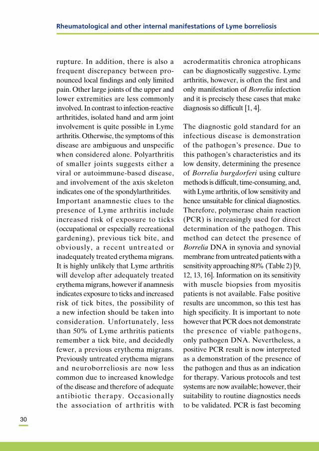
Typical for Lyme arthritis is uni or bilateral involvement of the knee joint with synovialitis and usually voluminous effusion (Fig.1). This frequently leads to extended Baker’s cysts, which often
Fig. 1. Lyme arthritis of the right knee
30
rupture. In addition, there is also a frequent discrepancy between pronounced local findings and only limited pain. Other large joints of the upper and lower extremities are less commonly involved. In contrast to infectionreactive arthritides, isolated hand and arm joint involvement is quite possible in Lyme arthritis. Otherwise, the symptoms of this disease are ambiguous and unspecific when considered alone. Polyarthritis of smaller joints suggests either a viral or autoimmunebased disease, and involvement of the axis skeleton indicates one of the spondylarthritides.Important anamnestic clues to the presence of Lyme arthritis include increased risk of exposure to ticks (occupational or especially recreational gardening), previous tick bite, and obviously, a recent untreated or inadequately treated erythema migrans. It is highly unlikely that Lyme arthritis will develop after adequately treated erythema migrans, however if anamnesis indicates exposure to ticks and increased risk of tick bites, the possibility of a new infection should be taken into consideration. Unfortunately, less than 50% of Lyme arthritis patients remember a tick bite, and decidedly fewer, a previous erythema migrans. Previously untreated erythema migrans and neuroborreliosis are now less common due to increased knowledge of the disease and therefore of adequate antibiotic therapy. Occasionally the association of arthritis with
acrodermatitis chronica atrophicans can be diagnostically suggestive. Lyme arthritis, however, is often the first and only manifestation of Borrelia infection and it is precisely these cases that make diagnosis so difficult [1, 4].
The diagnostic gold standard for an infectious disease is demonstration of the pathogen’s presence. Due to this pathogen’s characteristics and its low density, determining the presence of Borrelia burgdorferi using culture methods is difficult, timeconsuming, and, with Lyme arthritis, of low sensitivity and hence unsuitable for clinical diagnostics. Therefore, polymerase chain reaction (PCR) is increasingly used for direct determination of the pathogen. This method can detect the presence of Borrelia DNA in synovia and synovial membrane from untreated patients with a sensitivity approaching 80% (Table 2) [9, 12, 13, 16]. Information on its sensitivity with muscle biopsies from myositis patients is not available. False positive results are uncommon, so this test has high specificity. It is important to note however that PCR does not demonstrate the presence of viable pathogens, only pathogen DNA. Nevertheless, a positive PCR result is now interpreted as a demonstration of the presence of the pathogen and thus as an indication for therapy. Various protocols and test systems are now available; however, their suitability to routine diagnostics needs to be validated. PCR is fast becoming
Rheumatological and other internal manifestations of Lyme borreliosis

31
an established component of laboratory diagnostics, particularly for Lyme arthritis [9, 11, 12, 16].
The most common laboratory method for routine diagnosis is serology, i.e. determining the presence of specific antibodies against Borrelia burgdorferi (see the relevant chapter in this book).In the early phase of infection, Borrelia serology is often still negative, as measurable concentrations of antibodies develop slowly over weeks. Given continued clinical suspicion, a shortterm serological examination of disease progression demonstrating seroconversion will confirm the diagnosis. In chronic phases of the illness such as Lyme arthritis, serology is less problematic, as in general there are clearly positive antibody values for a whole series of Borrelia antigens. Significantly elevated IgG levels can be found but, in spite of pathogen persistence, specific IgM antibodies are seldom detected. In other cases, IgM antibodies can persist for years after successful treatment and thus cannot be used as an indication for therapy or evidence of persistence of the infection.
Due to the high sensitivity of the test, a false negative serology for Lyme arthritis is very unlikely. However, the lack of IgM antibodies does not exclude a current infection or active illness. The main problem here lies in the interpretation of serological findings: positive IgG values in the chronic phase of Lyme borreliosis are indistin guishable from positive titres after an acute illness or infection (socalled serum scar). This means that the banding pattern on immunoblot does not allow determination of the time of infection or inference of a possible ongoing infection. Immune responses are individual, in part genetically determined and dependent on, for example, extruded pathogen antigens. The antibody response is therefore also variably strong and in some cases nonexistent. The value of a positive Borrelia serology is thus dependent on the clinical symptoms. Only in cases with sufficient clinical evidence does positive serology have high diagnostic value. In those with unspecific symptoms however, positive serology is of limited diagnostic value. Due to the high sensitivity of the test,
Table 2. Sensitivity of methods for direct proof of B. burgdorferi (from [16])
Material Sensitivity
Skin biopsy (erythema migrans, acrodermatitis) 50–70% in culture or PCR
Liquor (acute neuroborreliosis) 10–30% in culture or PCR
Synoviaa 50–70% in PCR
aEven higher sensitivity (up to 80%) by examination of the synovial tissue
32
negative serology practically eliminates the diagnosis of Lyme arthritis.Given the slowness of changes in immune response during the chronic phase, successive examinations over time are seldom helpful, as they produce significant findings only at intervals of several months. In such cases, chronic borreliosis can often be surmised with only a limited probability but seldom proven [3, 5, 13, 16].An additional difficulty stems from the fact that serological tests are not standardised; tests performed in different laboratories often provide different results, which could be mistakenly interpreted as the titre course or an effect of therapy. This commonly leads to false diagnosis, unnecessary fears of continued disease progression, and unnecessary treatment.Thus Lyme arthritis can be diagnosed with sufficient certainty either by standard anamnesis and clinical symptoms together with positive serology or, in case of less typical symptoms, by demonstrating the presence of the pathogen. A careful rheumatological differential diagnosis is also always necessary.
Criteria for the diagnosis of Lyme arthritis Association with pathognomonic
extraarticular manifestations Typical pattern of joint involvement Exclusion of possible differential
diagnoses IgG antibodies against Borrelia
burgdorferi
Positive Borrelia PCR for the synovia or synovial membrane
Depending on the constellation of findings, it is possible to distinguish between certain, probable, and possible Lyme arthritis. This also helps indicate the possible need for critical reexamination of the diagnosis during the course of the disease. If the first four criteria are fulfilled, e.g., when the arthritis is associated with obvious acrodermatitis chronica atrophicans in a patient with highly positive Borrelia serology, then the findings are unequivocal and allow definite diagnosis. The same is true for gonarthritis patients with positive serology, and positive Borrelia PCR result from synovial fluid once the differential diagnoses have been excluded. In practice however, the suspected diagnosis is based only on criteria 2 and 4, leaving diagnostic uncertainty, and even after eliminating other possible diagnoses, one can only speak of a probable diagnosis; or, for example, if there is also psoriasis that cannot be excluded by other cause, only a diagnosis of possible Lyme arthritis remains [2, 4, 8, 10].
TherapyAntibiotic therapy should be started as soon as possible after diagnosis in order to shorten the course of the disease, cure it, and prevent progression or the development of chronic disease. Treatment is stage and symptom
Rheumatological and other internal manifestations of Lyme borreliosis
33
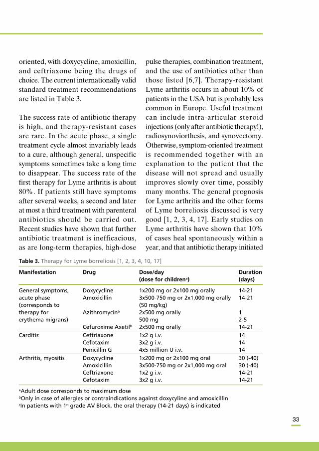
oriented, with doxycycline, amoxicillin, and ceftriaxone being the drugs of choice. The current internationally valid standard treatment recommendations are listed in Table 3.
The success rate of antibiotic therapy is high, and therapyresistant cases are rare. In the acute phase, a single treatment cycle almost invariably leads to a cure, although general, unspecific symptoms sometimes take a long time to disappear. The success rate of the first therapy for Lyme arthritis is about 80%. If patients still have symptoms after several weeks, a second and later at most a third treatment with parenteral antibiotics should be carried out. Recent studies have shown that further antibiotic treatment is inefficacious, as are longterm therapies, highdose
pulse therapies, combination treatment, and the use of antibiotics other than those listed [6,7]. Therapyresistant Lyme arthritis occurs in about 10% of patients in the USA but is probably less common in Europe. Useful treatment can include intraarticular steroid injections (only after antibiotic therapy!), radiosyno viorthesis, and synovectomy. Otherwise, symptomoriented treatment is recommended together with an explanation to the patient that the disease will not spread and usually improves slowly over time, possibly many months. The general prognosis for Lyme arthritis and the other forms of Lyme borreliosis discussed is very good [1, 2, 3, 4, 17]. Early studies on Lyme arthritis have shown that 10% of cases heal spontaneously within a year, and that antibiotic therapy initiated
Table 3. Therapy for Lyme borreliosis [1, 2, 3, 4, 10, 17]
Manifestation Drug Dose/day Duration (dose for childrena) (days)
General symptoms, Doxycycline 1x200 mg or 2x100 mg orally 14-21 acute phase Amoxicillin 3x500-750 mg or 2x1,000 mg orally 14-21 (corresponds to (50 mg/kg) therapy for Azithromycinb 2x500 mg orally 1 erythema migrans) 500 mg 2-5 Cefuroxime Axetilb 2x500 mg orally 14-21
Carditisc Ceftriaxone 1x2 g i.v. 14 Cefotaxim 3x2 g i.v. 14 Penicillin G 4x5 million U i.v. 14
Arthritis, myositis Doxycycline 1x200 mg or 2x100 mg oral 30 (-40) Amoxicillin 3x500-750 mg or 2x1,000 mg oral 30 (-40) Ceftriaxone 1x2 g i.v. 14-21 Cefotaxim 3x2 g i.v. 14-21
aAdult dose corresponds to maximum dosebOnly in case of allergies or contraindications against doxycyline and amoxicillincIn patients with 1st grade AV Block, the oral therapy (14-21 days) is indicated

34
even several years after disease onset is effective [14].
Case studiesFirst case descriptionA 38yearold male patient had complained for 4 months of painful swelling in the right knee. As this problem first occurred after a skiing holiday, trauma was suspected, although the patient could not remember such incident. Magnetic resonance imaging showed minor chondropathy and degenerative changes to the outer meniscus. Symptomatic treatment with diclofenac led to only shortterm improvement. Further anamnesis showed no significant previous illness. About a year previously, the patient had been bitten on the left side of the chest by a tick probably while working in the garden. The tick had been removed with a tweezers. Shortly thereafter, erythema occurred which lasted 1 day and then spontaneously disappeared. Indications for another cause of the arthritis, additional rheumatological symptoms, and extraarticular manifestations were neither reported nor found on clinical examination. The latter was unre markable with the exception of florid, leftsided gonarthritis with substantial synovitis. Arthrosonography confirmed this finding and showed partly edematous, partly proliferative synovialitis, and joint swelling with a poor echo (estimated volume 50 ml) but no popliteal cyst.
Laboratory findings showed limited humoral inflammatory activity with increased erythrocyte sedimentation rate and elevated Creactive protein (CRP). All other routine parameters were normal. The patient was human leukocyte antigen (HLA)B27negative. Borrelia serology was highly positive for IgG antibodies against Borrelia burgdorferi using enzymelinked immunosorbent assay (ELISA). ImmunoglobulinM antibodies were not present. These results were confirmed by immunoblot using antibodies against, among others, the proteins OspC, p39, flagellin, VlsE, and p83/100. Analysis of synovial fluid from the right knee joint showed a highly inflamed knee effusion with 10,000 leukocytes/mm3 (90% neutro phils). Probable Lyme arthritis was diagnosed that likely resulted from the reported tick bite. Whether the erythema following the tick bite was erythema migrans remains unclear. Its regression after only 1 day suggests that this was not the case, although erythema migrans is sometimes very pale and can go unnoticed by patients. Oral treatment with 200 mg/day of doxycycline was initiated. Acemetacin (60 mg) as required was given as an antiinflammatory in addition to cold treatment and physiotherapy. The arthritis healed slowly under this treatment, with symptomatic treatment continuing 4 weeks after the antibiotic therapy was finished. The patient was symptomfree after 3 months.
Rheumatological and other internal manifestations of Lyme borreliosis

35
Second case descriptionA 55yearold, obese female had complained for several months of pain in the right knee. Additionally, she had had lumbar back pain for “as long as she could remember”. Anamnesis indicated psoriasis vulgaris from early adulthood and typical erythema migrans on the left upper thigh 6 years previously that had been treated for 2 weeks with an unknown antibiotic. She suffered tick bites every year. Having changed her general practitioner shortly before, the new physician conjectured that her rheumatological problems were caused by Lyme arthritis. A serological examination was positive.From the clinical rheumatological point of view, both iliosacral joints were markedly swollen and painful when moved, and the right knee joint showed malposition of the valgus with light inflammation. Subsequent anamnesis indicated, at least for earlier years, an inflammatory character to the back pain with a distinct maximum in the early morning and reduction with movement.Radiology showed bilateral sacroiliitis and rightsided gonarthritis.Laboratory findings showed increased values for erythrocyte sedimentation rate and CRP. Human leukocyte antigenB27 was present. Borreliosis serology was positive, with low titres of IgG against OspC, flagellin, p58, and p60. Analysis of synovial fluid from the knee showed less than 1,000 leukocytes/mm3, as seen in arthritic irritation.
A diagnosis of active rightsided valgus gonarthritis was made, with probable psoriatic spondylarthritis associated with HLAB27, and Lyme borreliosis with erythema migrans 6 years previously. There was no evidence of florid Lyme disease.Sacroiliitis does not manifest in Lyme arthritis. Synovial analysis clearly showed that the knee problems, which could also be related to psoriatic arthritis in differential diagnosis, did not result from Lyme arthritis but, taken together with the other clinical and radiological findings, were apparently caused by active arthritis. The serological findings also speak against Lyme arthritis, as the typical clearly positive IgG antibody response against numerous Borrelia proteins, e.g. VlsE and p38/100, was not present. The findings suggest a serum scar after Lyme borreliosis some years previously. The erythema migrans 6 years earlier helps to explain this serological result. An association between Borrelia and the current rheumatological problems could not be made due to the time span involved. In addition, the development of a disease course involving an organ cannot be expected after adequate antibiotic treatment of erythema migrans. This case shows how carefully a serological finding must be interpreted in relation to clinical symptoms and other results.

36
l References1. Burmester GR, Kamradt T, Krause
A (2004) Lyme disease. In: Isenberg DA, Maddison PJ, Woo P, Glass D, Breedveld FC (eds) Oxford textbook of rheumatology. pp 613620
2. European Union concerted action on Lyme borreliosis (EUCALB). http://www.dis. strath.ac.uk/vie/LymeEU/index.htm
3. Franz JK, Krause A: Lyme disease (Lyme borreliosis). Best Practice & Research Clinical Rheumatology 17: 241264, 2003
4. Herzer P, Krause A. LymeBorreliose. In: Classen M et al. (Hrsg.) Rationelle Diagnostik und Therapie in der Inneren Medizin, Urban&Fischer, München, 2006, in press
5. Huppertz HI, Krause A. LymeBorreliose. Internist 44: 175183, 2003
6. Kalish RA, et al. Evaluation of study patients with Lyme disease, 1020year followup. J Infect Dis 183: 453460, 2001
7. Klempner MS, et al. Two controlled trials of antibiotic treatment in patients with persistent symptoms and a history of Lyme disease. N Engl J Med 34: 8592, 2001
8. Krause A, Herzer P. Frühdiagnostik der LymeArthritis. Z f Rheumatol 2005 (in press)
9. Lünemann J, et al. Rapid typing of Borrelia burgdorferi sensu lato species in specimens from patients with different manifestations of Lyme borreliosis. J Clin Micrbiol 39: 11301133, 2001
10. Nationales Referenzzentrum für Borrelien am Max von PettenkoferInstitut München. http://pollux.mpk.med.unimuenchen.de/ alpha1/nrzborrelia/lb/framelb.html
11. Nocton JJ, Dressler F, Rutledge BJ, Rys N, Persing DH, Steere AC. Detection of B. burgdorferi DNA by polymerase chain reaction in synovial fluid in Lyme arthritis. N Engl J Med 330: 229234, 1999
12. Priem S, et al. Detection of Borrelia burgdorferi by polymerase chain reaction in synovial membrane, but not synovial fluid from patients with persisting Lyme arthritis after antibiotic therapy. Ann Rheum Dis 57: 118121, 1998
13. Quality standards for the microbiological d i a g n o s i s o f i n f e c t i o u s d i s e a s e s : LymeBorreliose (MiQ 12 2000 Lyme Borreliosis).
http://pollux.mpk.med.unimuenchen.de/ alpha1/nrzborrelia/miqlyme/framemiqlyme.html
14. Steere AC, Schoen RT, Taylor E. The clinical evolution of Lyme arthritis. Ann Intern Med 107: 725731, 1987
15. Steere AC, Coburn J, Glickstein L. The emergence of Lyme disease. J Clin Invest 113: 10931101, 2004
16. Wilske B. Diagnosis of Lyme borreliosis in Europe. VectorBorne Zoonotic Dis 3: 215227, 2003
17. Wormser GP, et al. Practice guidelines for the treatment of Lyme disease. Clin Infect Dis 31: 114, 2000
Rheumatological and other internal manifestations of Lyme borreliosis

37
As the organ contacting the environment, the skin is exposed
to many external influences. It is also a point of entry for pathogens, which spread via the skin if the barrier function or immune system is no longer intact. In contrast, the transmission of pathogens such as Borrelia spp. by arthropods can occur in fully healthy individuals.Whether Lyme borreliosis (LB) manifests, leading to a specific clinical picture, depends on the genetic disposition of the patient, duration of the tick bite and virulence of the pathogen [20, 22, 26, 28].Cutaneous symptoms of LB were already recognized many decades before discovery of the pathogen, Borrelia burgdorferi, by W. Burgdorfer in 1983. It has also been known since the 1950s that erythema migrans (EM), Borrelia lymphocytoma (BL), and acrodermatitis chronica atrophicans (ACA) can be successfully treated with penicillin [19, 21].
What happens after a tick bite?The transmission of Lyme borreliosis by ticks, in Europe predominantly by the Ixodes ricinus species, is reported in only about 60% of patients [30]. Studies have shown that insects such as mosquitoes, horse flies, sandflies, gnats, and wasps have no organ suitable for survival of the Borrelia pathogen as exists in the tick gut [13].
Unspecific arthropod reactionsAfter a tick has attached to the skin for an extended time, a local immune reaction occurs that, however, may go unnoticed. An itchy swelling or lump may then form at the site of the bite, from which a tick granuloma can develop. An inflamed patch several centimetres in diameter can also develop and then disappears over a number of days.
Classic cutaneous symptoms of Lyme borreliosis (Table 1)Erythema migransErythema migrans can develop from an
Dermatological manifestations ofLyme borreliosisl Elisabeth Aberer

38
unspecific reaction to a tick bite after only 1–2 days. It is characterised by a slowly expanding inflamed patch. In most cases however there is a latent phase of 4–20 days before the formation of this patch. If not treated, it can increase to 50 cm or more in diameter. The erythema can vary in form from round to oval to irregular. Subjectively it can lead to slight itching. Local lymph node swelling is possible [11, 28].
Clinical forms of erythema migrans The color can be bluish in the center
while the edge is bright red. There are often streaky or patchy radial branches at the periphery (Fig. 1).
A bright red, moving ring can form, with healing in the center (Fig. 2).
A small vesiculous variant can appear (Fig. 3).
A homogenous erythema without a marked border can form (Fig. 4).
Children often show a unilateral reddening of a cheek or a stripe that is only visible on some days. Temporary disappearance followed by reappearance of the erythema has been reported. Rubbing a suspect location with a wooden spatula can lead to reappearance of the erythema.
Fig. 1. Fig. 3.
Fig. 2.
Dermatological manifestations of Lyme borreliosis

39
Disseminated early infectionWhile only about two thirds of patients have only cutaneous infection (erythema migrans minor), the remaining third have simultaneous, generalised symptoms as a sign of haematogenic dissemination [34]. Patients with this EM major form report flulike symptoms, light fever, headache, joint and muscle pain, swollen lymph nodes, and fatigue. Occasionally, rapid heartbeat and cardiac rhythm disturbances are observed [23]. Multiple EM lesions can also result from haematogenic dissemination as well as from
Fig. 4.
Table 1. Differential diagnosis of Lyme borreliosis
Cutaneous manifestation Differential diagnosis
Erythema migrans
Bluish center Fixed drug eruption Morphea
Homogenous reddening Fixed drug eruption Erysipeloid Subacute cutaneous lupus erythematosus
Vesicular form Contact dermatitis Herpes simplex
Multiple erythemata Urticaria
Borrelia lymphocytoma
On the ear Othaematoma Erysipelas Relapsing polychondritis
On the mamilla Breast cancer
In other places Malignant lymphoma
Acrodermatitis chronica atrophicans
Light red discoloration of the distal extremities Stasis dermatitis
Diffuse livid discoloration Chronic venous insufficiency Venous thrombosis of the leg
Acral form: fingers/toes Acrocyanosis
Livid reticular discoloration on the extremities Phospholipid antibody syndrome Livedo reticularis/racemosa

40
simultaneous infection from multiple tick bites.
Erythema chronicum migransIf not treated, EM can last several weeks or months while growing continuously. Spontaneous healing is possible, but its frequency is difficult to determine.
Borrelia lymphocytomaBorrelia Lymphocytoma (BL) was first described as lym phadenosis cutis benigna (Bäfverstedt’s disease), a pseudolymphoma with mixed polyclonal B and T cell proli feration [6]. After a latent period of several weeks, BL most commonly manifests in children as a reddish to bluish node in the area of the earlobe (Fig. 5), livid reddish disco loration or swelling in the area of
the helix, and a flat node one or more centimetres in size in the scrotal area. BL may also occur on one mammilla or areola mammae as an enlargement or nodular lesion (Fig. 6). These nodes can start in an expanding EM that the patient may or may not remember. As EM often occurs on the heads of children, the redness frequently goes unnoticed and a lymphocytoma on the ear is the first recognised symptom.
Acrodermatitis chronica atrophicansAcrodermatitis chronica atrophicans begins as an inconspicuous, poorly defined reddening of the skin (Fig. 7), subjectively without symptoms, which becomes slowly darker. It occurs on the distal extremities, elbows, extensor
Fig. 5. Fig. 6.
Dermatological manifestations of Lyme borreliosis

41
sides of the upper arms, back of the hand, and upper and lower legs.
Case reportsThe development of ACA with neuropathy. A 57yearold female observed an EM on her lower leg after a tick bite. Correct diagnosis was not made, and the erythema spread to the upper thigh and buttocks over the next 2 years. After 3 years an enlarged lymph node was removed from the right inguinal area which showed unspecific lymphadenitis. The patient reported intermittent redness on the buttocks. Two years later, leftsided bartholinitis appeared and later disappeared. Twelve years after the
tick bite, the patient noted a burning sensation and weakness in both thighs as well as a bright red discoloration with undefined borders and light swelling in the left ankle region. This led her to visit a dermatologist, where finally ACA was diagnosed clinically and verified with a biopsy and serology (Fig. 7). The dermatological changes healed under antibiotic treatment with 2 g of ceftriaxone daily for 15 days. The accompanying neuropathy improved slowly.
CommentsThe clinical course of ACA can rarely be studied so clearly. This patient distinctly shows the clinical persistence of the disease as described in the literature [14, 29]. Reactivation of Borrelia burgdorferi and the development of ACA were also found in two other patients, one injured on a rose thorn while gardening and the other on a metal spike at home. This indicates that latent infections occur in the extremities where temperature is lower, the optimum Borrelia growth temperature being 33–34°C.Bluish, cushionlike swellings also develop in ACA. Sometimes individual fingers are swollen and discolored blue (Fig. 8). Swelling of the feet can lead to the patient’s requiring a larger shoe size. More reticulate discoloration can occur on the extensor sides of the upper arms and thighs. The skin gradually becomes thin and wrinkled, and the bluish
Fig. 7.

42
discoloration decreases. A thin, almost transparent skin remains, with blood vessels visible underneath (Fig. 9).The neuropathy often reported by patients does not respond to antibiotics as well as the dermatological changes do [12]. Solid, extraarticular (fibrous) nodes form over the elbows, knees, and finger joints. These can easily be lifted in a fold of the skin and, unlike rheumatic nodes, are not painful.Skin sclerosis is possible in shin and underarm extensor regions (tibial and ulnar bands) where ACA is present. This can be differentiated from linear scleroderma by its undefined edges. In addition to classic ACA, aneto dermic lesions can occur also.
Genospecies-specific courses of Lyme borreliosisThe species Borrelia burgdorferi sensu lato is subdivided into three genospecies: Borrelia burgdorferi sensu stricto, Borrelia afzelii, and Borrelia garinii; all three of these can cause EM. Various genospeciesspecific courses of EM have been reported [7]. Of these, only Borrelia burgdorferi sensu stricto is found in the USA. This species causes the least number of Borrelia infections in Europe. In our study in the Vienna area, Borrelia burgdorferi sensu lato was isolated from the skin of 28 patients. Of these, 23 had Borrelia afzelii (17 with EM, six with ACA), three had Borrelia garinii (all with EM), and two had Borrelia burgdorferi sensu stricto
Fig. 8.
Fig. 9.
Dermatological manifestations of Lyme borreliosis

43
infection (one with EM, one positive blood culture in disseminated LB). In and around the city of Graz, Austria 11 strains were isolated, of which five were Borrelia garinii from EM and six Borrelia afzelii (four EM, one ACA, one morphea). A similar pattern has been found in other studies [25]. For the other cutaneous symptoms, BL is most commonly caused by Borrelia afzelii (eight of nine cases) [18] and ACA almost exclusively by Borrelia afzelii [24], with mixed infections also possible. This explains why no secondary cutaneous clinical courses (e.g. BL or ACA) of LB have been found in the USA, with the exception of patients who were infected in Europe.Differences between EM caused by Borrelia burgdorferi sensu stricto and EM caused by Borrelia afzelii are: shorter incubation time (4 days vs 14), more frequent localisation in the trunk area (50% vs 27%), central lightening of colour in only a third of patients compared with two thirds of Borrelia afzelii infections, and more frequent systemic signs and se ropositivity (89% vs 30%) [30]. In addition, EM caused by Borrelia afzelii differs from that caused by Borrelia garinii–infections with the former occurring more often in June and with Borrelia garinii in September, while Borrelia afzelii EM appears more often on the extremities and less often on the trunk. Growth of the reddened area by Borrelia afzelii reaches 3 cm/day and by Borrelia garinii 12.5 cm/day.
Generalised and local symptoms occur in three quarters of Borrelia garinii patients but only a third of Borrelia afzelii patients [5, 17].
Symptoms after erythema migransA case of EM is clinically cured with 2–3 weeks of antibiotic treatment, sometimes within a few days and sometimes towards the end of the therapy period. Occasionally there is still a livid erythema after 3 weeks that fades several weeks later. If severe symptoms exist at the start of therapy, they may continue after the end of medication and completely disappear several weeks later. Severe symptoms can also arise during therapy (Herxheimerlike reaction) and should not be interpreted as treatment failure. The prescribed antibiotic must not be stopped. Duration of therapy should be increased to 3 weeks if originally set at 2 weeks based on apparently localised EM. About 7% of patients suffer relapse in the following weeks (for up to 6 months) with repeated wandering joint and muscle pain and headaches that can last for days. These symptoms also disappear after several weeks. In severe cases, an additional course of antibiotics is usually prescribed. The occurrence of arthritis or neuroborreliosis has not been observed in LB patients with correctly treated EM over an observation period of 20 years. In two patients, erythema nodosum (EN) accompanied the EM.

44
Case report of erythema migrans with erythema nodosum (EN)A 34yearold male had a 7 x 5 cm annular erythema on the right leg for 2 months and painful, bright red nodes on both shins for a week. He could not remember being bitten by a tick. He also complained of joint pain that had lasted for a year. Xray of the thorax showed bilateral lymphadenopathy (Löfgren’s syndrome). Pathological findings included Creactive protein of 89, erythrocyte sedimentation rate of 44, and angiotensinconverting enzyme of 57.4 U (normal range 18.0–55.0 Units).Immunoblot for Borrelia antibodies was negative before and after therapy. Polymerase chain reaction (PCR) for Borrelia in the urine was positive on five occasions from the 5th to 150th day of treatment but negative on days 210 and 360 [2]. Anti IgM and IgG antibodies specific for Ehrlichia were positive (1:64) over 5 months without a change in titre, but PCR for Ehrlichia in the blood was negative. A pulmonary specialist diagnosed pulmonary sarcoidosis I. Bronchoscopy and bronchoalveolar lavage showed no evidence of pulmonary involvement. A double therapy cycle of 200 mg of doxycyline for 3 weeks and glucocorticosteroid treatment led to cure of the EM, EN, and lymph node swelling.
CommentsDue to the steady titre values and negative PCR, ehrlichiosis could be
eliminated as a cause of the EN and bilateral lymphadenopathy. There were also no fever or unusual laboratory values such as leukopenia. The conclusion is that the EN is associated with the EM. Arthralgia, myalgia, memory disturbances, fatigue syndrome, conjunctivitis, febrile periods, headache, tinnitus, and acute loss of hearing are referred in the literature as symptoms of postLyme syndrome. Seltzer et al. [27] examined the symptom profiles of 212 patients 1–11 years after LB and compared them to a group with equivalent age and place of residence. No differences were found between the groups. It is still unclear how to classify this clinical picture. Perhaps the symptoms are overrated and were influenced by fear of further organ involvement from the LB.
Culture and polymerase chain reaction confirmed atypical formsClinical observations have shown that the potency of Borrelia burgdorferi infection lies in immune stimulation of B cells and the induction of cutaneous sclerosis.
Cutaneous lymphomaThe role of Borrelia burgdorferi in atypical cutaneous proliferation, pseudolymphoma, and malignant lymphoma has been confirmed in many publications. As a cause of malignant lymphoma, it acts similarly
Dermatological manifestations of Lyme borreliosis

45
to Helicobacter pylori or EpsteinBarr virus. A targeted antibiotic (virostatic) therapy can lead to cure in some cases. Comparative studies with lymphoma patients in the USA or Asia showed no association with Borrelia burgdorferi [16, 32], while those in Austria, France, Scotland, Denmark, and Germany clearly did [9, 15].
Circumscribed scleroderma (morphea) and lichen sclerosis Starting in 1985, an association between Borrelia infection and the development of morphea was described in Switzerland, Germany, and Austria by various authors from Basel, Munich, Berlin, and Vienna. According to PCR results and positive Borrelia cultures from skin biopsies and their typing, aggregation of Borreliainduced morphea cases was geographic in nature [4, 10]. Only Borrelia afzelii could be isolated from both morphea and lichen sclerosis.
Diagnostic and therapeutic management of these patients In principle, circumscribed scleroderma has a variety of causes. It can occur after insect bites, immunisations, and trauma. Independently of this, a childhood and early adolescent form exists which is localised to the lines of Blaschko (e.g. linear forms, morphea en coup de sabre) and therefore could be induced during embryonal development. Congenital atrophic morphea has been cured by treatment with ceftriaxone [1]. The
significance of Borrelia to the emergence of this disease is not yet clear. Borrelia serology using immunoblot and Borrelia PCR of a skin biopsy are recommended for diagnosis. The IgG ELISA titres are often negative, but specific bands appear on immunoblot. In Borrelia associated skin sclerosis it is likely that cellular immune reactions predominate, as shown by lymphocyte proliferation tests. Stimulation of lymphocytes by Borrelia burgdorferi was also observed in seronegative patients with morphea and ACA patients with fibrous nodules [3].Patients with morphea react better to systemic therapy with ceftriaxone (for 20 days) than to oral treatment; the same has been observed in seronegative patients. This is the treatment of choice for all patients with progressive morphea and distinct lilac rings, particularly in children.
Dermatoses in which Borrelia burgdorferi infection must be consideredAll pat ients wi th unexpla ined sclerosis [31], myositis, and especially dermatomyositis must be examined for the presence of Borrelia burgdorferi infection. In the literature this infection has also been associated with cases of livid erythema with myopathy, relapsing febrile panniculitis, annular erythema, triggerfinger, SchönleinHenoch and thrombocytopenic purpura, granulomatous thrombophlebitis, temporal arteritis, sarcoidosis, pityriasis rosea, pityriasis lichenoides, anetoderma,

46
Buschke’s scleroderma, eosinophilic fasciitis (Shulman’s syndrome), facial hemiatrophy, granuloma annulare, and interstitial granulomatous dermatitis.
Diagnosis of Lyme borreliosisConsult the European Union for Concerted Action on Risk Assessment in Lyme Borreliosis guidelines at http://www.oeghmp.at/eucalb/.
Erythema migransErythema migrans is usually diagnosed on purely clinical criteria! Anamnesis of a growing red patch is the most important feature; patients frequently do not remember a bite. This patch should be >5 cm. The patch may have to be observed over the course of a week, with a control examination sometimes required. Histological examination is unspecific, showing perivascular lymphohistiocytic infiltrate with sparse eosinophils. Biopsy is needed only when the clinical picture is unclear. Serological examination can show positive IgM antibodies, which should however be confirmed by immunoblot. Depending on the duration of the erythema, IgG antibodies may also be present. These could also result from an earlier infection of known or unknown origin (seroprevalence in healthy individuals 10–30%). Since only 50% of patients have antibodies, serological examination is not required for diagnosis.
Borrelia lymphocytomaThe clinical diagnosis is also decisive
here. Biopsy shows polyclonal B and T cell proliferation. In children, biopsy can be avoided if there is a classic manifestation. In cases of lymphocytoma of the mammilla or other localised areas, a skin biopsy should be carried out. In about 70% of patients, antibodies can be demon strated. Healing after antibiotic therapy can be slow, taking several weeks.
Acrodermatitis chronica atrophicansPositive IgG titre, typical clinical picture, and histological examination are crucial to diagnosing this manifestation of LB. Histology shows atrophy of the epidermis and dermis, telangiectasia, and occasionally bandlike lymphohistiocytic infiltrates with plasma cells. The cure by therapy is very slow, requiring months. Followup after a year is recommended, possibly also a repeat biopsy to determine whether inflammatory infiltrates are present.
SerologyBorrelia serology will be dealt with in detail in the chapter on diagnosis. Here only a possible serological picture for EM is presented (Table 2).
TherapyTreatments of LB with oral penicillin, tetracycline, cefuroxime axetil, and azithromycin are equally effective (Table 3). With antibiotic treatment, EM with or without accompanying symptoms heals without complications in >90% of
Dermatological manifestations of Lyme borreliosis

47
cases. If treatment fails, headaches, joint and muscle pain, and fatigue can occur for a period of 6 months but usually disappear within a few weeks. Secondary manifestations are extremely rare after appropriate treatment. The most recent publications on duration of therapy show that postLyme syndrome occurs in 510% of patients independently of whether treatment lasts 2 or 3 weeks [33].Threeweek treatment is recommended if the erythema lasts >2 months and in
case of severe symptoms. The choice of antibiotic depends on patient compa tibility and possible allergies. Tetracyclines should be avoided if exposure to sun is likely, and are also contraindicated during pregnancy, while breastfeeding, and for children <8 years old. In cases accompanied by fever, severe general symptoms, or when coinfection with ehrlichiosis is suspected, tetracycline is the treatment of choice (Table 3).
Table 2. Serological picture in erythema migrans (Borrelia antibodies)
ELISA IgM ELISA IgG IB IgM IB IgG Interpretation
- - - - EM/no borreliosis?+ - - - EM/unspecific+ - + - EM Confirmation/antibody persistence from older infections+ + + +/- EM Confirmation/reinfection- + - + Older infections+/- - + + EM Confirmation/older infections
Table 3. Antibiotic therapy for Lyme borreliosis
Erythema migrans
Penicillin V 3x1 1,5 Mio 14-20 days
Amoxicillin 3x1 500-750 mg 14-20 days
Doxycycline 1x1 200 mg 14-20 days
Azithromycin 2x1 500 mg 1st day 1x1 500 mg 7 days
Borrelia lymphocytoma
Oral therapy 20 days
Acrodermatitis chronica atrophicans
Oral therapy 30 days
Or ceftriaxone 1x1 2g 20 days
A second treatment with an alternative antibiotic is recommended for the following findings:
Positive serology Severe suffering and unspecific symptoms
EM Continuing arthralgia, headaches for >3 months
BL No cure within 3 months
ACA Histologically verified infiltrate after 1 year

48
PrognosisPathogen persistence was found in three of 48 patients with late symptoms [29]. Reinfection is possible. It is noteworthy that chronic Borrelia infections stimulate lymphocytes, with the possibility of monoclonal lymphocytic proliferation. Persistent, severe disease courses could be due to infection with exceptionally pathogenic Borrelia burgdorferi clones (RST1 isolate [8]). In addition to interspecies variation, additional genetic factors can lead to organspecific manifestations. In general, there is no danger of
developing LB when EM is properly treated. Therapy can fail however, when less efficacious antibiotics such as firstgeneration cephalosporins, roxithromycin, or erythromycin are used in an irregular manner; for example if therapy is interrupted on weekends, as sometimes happens with ceftriaxone therapy. Uncertainty concerning serology remains, as the variable prevalence in the population and persistence of antibodies after therapy complicate the assessment of serology results, making diagnosis difficult.
Dermatological manifestations of Lyme borreliosis

49
l References1. Aberer E, Weissenbacher G (1997) Congenital
anetoderma induced by intrauterine infection? Arch Dermatol 133(4):526527
2. Bergmann AR, Schmidt BL, Derler AM, Aberer EJ (2002) Importance of sample preparation for molecular diagnosis of lyme borreliosis from urine. J Clin Microbiol 40(12):4581–4584
3. Breier P, Klade H, Stanek G, Poitschek C, Kirnbauer R, Dorda W, Aberer E (1996) Lymphoproliferative responses to Borrelia burgdorferi in circumscribed scleroderma. Br J Dermatol 134(2):285–291
4. Breier FH, Aberer E, Stanek G, Khanakaha G, Schlick A, Tappeiner G (1999) Isolation of Borrelia afzelii from circumscribed scleroderma. Br J Dermatol 140(5):925–930
5. Carlsson SA, Granlund H, Jansson C, Nyman D, Wahlberg P (2003) Characteristics of erythema migrans in Borrelia afzelii and Borrelia garinii infections. Scand J Infect Dis 35(1):31–33
6. Colli C, Leinweber B, Müllegger R, Chott A, Kerl H, Cerroni L (2004) Borrelia burgdorferiassociated lymphocytoma cutis: clinicopathologic, immunophenotypic, and molecular study of 106 cases. J Cutan Pathol 31(3):232–240
7. Dam AP van, Kuiper H, Vos K, Widjojokusumo A, Jongh BM de, Spanjaard L, Ramselaar AC, Kramer MD, Dankert J (1993) Different genospecies of Borrelia burgdorferi are associated with distinct clinical manifestations of Lyme borreliosis. Clin Infect Dis 17(4):708–717
8. Dolan MC, Piesman J, Schneider BS, Schriefer M, Brandt K, Zeidner NS (2004) Comparison of disseminated and nondisseminated strains of Borrelia burgdorferi sensu stricto in mice naturally infected by tick bite. Infect Immun 72(9):5262–5266
9. Fouchardiere A de la, Vandenesch F, Berger F (2003) Borreliaassociated primary cutaneous MALT lymphoma in a nonendemic region. Am J Surg Pathol 27(5):702–703
10. Fujiwara H, Fujiwara K, Hashimoto K, Mehregan AH, SchaumburgLever G, Lange R, Schempp C, Gollnick H (1997) Detection
of Borrelia burgdorferi DNA (B garinii or B afzelii) in morphea and lichen sclerosus et atrophicus tissues of German and Japanese but not of US patients. Arch Dermatol 133(1):41–44
11. Hengge UR, Tannapfel A, Tyring SK, Erbel R, Arendt G, Ruzicka T (2003) Lyme borreliosis. Lancet Infect Dis 3(8):489500; Erratum in: Lancet Infect Dis 3(12):815
12. Kindstrand E, Nilsson BY, Hovmark A, Pirskanen R, Asbrink E. Peripheral neuropathy in acrodermatitis chronica atrophicans effect of treatment. Acta Neurol Scand. 2002 Nov;106(5):253
13. KosikBogacka D, KuznaGrygiel W, Bukowska K (2004) The prevalence of spirochete Borrelia burgdorferi sensu lato in ticks Ixodes ricinus and mosquitoes Aedes spp. within a selected recreational area in the city of Szczecin. Ann Agric Environ Med 11(1):105–108
14. Kuiper H, Dam AP van, Spanjaard L, Jongh BM de, Widjojokusumo A, Ramselaar TC, Cairo I, Vos K, Dankert J (1994) Isolation of Borrelia burgdorferi from biopsy specimens taken from healthylooking skin of patients with Lyme borreliosis. J Clin Microbiol 32(3):715–720
15. Leinweber B, Colli C, Chott A, Kerl H, Cerroni (2004) Differential diagnosis of cutaneous infiltrates of B lymphocytes with follicular growth pattern. Am J Dermatopathol 26(1):4–13
16. Li C, Inagaki H, Kuo TT, Hu S, Okabe M, Eimoto T (2003) Primary cutaneous marginal zone Bcell lymphoma: a molecular and clinicopathologic study of 24 asian cases. Am J Surg Pathol 27(8):1061–1069
17. Logar M, RuzicSabljic E, Maraspin V, LotricFurlan S, Cimperman J, Jurca T, Strle F (2004) Comparison of erythema migrans caused by Borrelia afzelii and Borrelia garinii. Infection 32(1):15–19
18. Maraspin V, Cimperman J, LotricFurlan S, RuzicSabljic E, Jurca T, Picken RN, Strle F (2002) Solitary borrelial lymphocytoma in adult patients. Wien Klin Wochenschr 31;114(1314):515–523

50
19. Miescher G, Storck H (1951) Successful penicillin therapy of acrodermatitis atrophicans. Dermatologica 102(46):370–371
20. Nahimana I, Gern L, Blanc DS, Praz G, Francioli P, Peter O (2004) Risk of Borrelia burgdorferi infection in western Switzerland following a tick bite. Eur J Clin Microbiol Infect Dis 23(8):603–608
21. Paschoud JM (1958) Lymphadenosis benigna cutis as a communicable disease. IV. Discussion of study results. Part B: penicillin therapy; lymphadenosis benigna cutis & erythema chronicum migrans, connections & etiology; summary. Der Hautarzt 9(7):311–315
22. Piesman J (1993) Dynamics of Borrelia burgdorferi transmission by nymphal Ixodes dammini ticks. J Infect Dis 167(5):1082–1085
23. PikeljPecnik A, LotricFurlan S, Maraspin V, Cimperman J, Logar M, Jurca T, Strle F (2002) Electrocardiographic findings in patients with erythema migrans.Wien Klin Wochenschr 31;114(1314):510–514
24. Rijpkema SG, Tazelaar DJ, Molkenboer MJ, Noordhoek GT, Plantinga G, Schouls LM, Schellekens JF (1997) Detection of Borrelia afzelii, Borrelia burgdorferi sensu stricto, Borrelia garinii and group VS116 by PCR in skin biopsies of patients with erythema migrans and acrodermatitis chronica atrophicans. Clin Microbiol Infect 3(1):109–116
25. Schaarschmidt D, Oehme R, Kimmig P, Hesch RD, Englisch S (2001) Detection and molecular typing of Borrelia burgdorferi sensu lato in Ixodes ricinus ticks and in different patient samples from southwest Germany. Eur J Epidemiol 17(12):1067–1074
26. Seinost G, Dykhuizen DE, Dattwyler RJ, Golde WT, Dunn JJ, Wang IN, Wormser GP, Schriefer ME, Luft BJ (1999) Four clones of Borrelia burgdorferi sensu stricto cause invasive infection in humans. Infect Immun 67(7):3518–3524
27. Seltzer EG, Gerber MA, Cartter ML, Freudigman K, Shapiro ED (2000) Longterm
outcomes of persons with Lyme disease. JAMA 2;283(5):609–616
28. Stanek G, Strle F (2003) Lyme borreliosis. Lancet 15;362(9396):1639–1647
29. Strle F, Cheng Y, Cimperman J, Maraspin V, LotricFurlan S, Nelson JA, Picken MM, RuzicSabljic E, Picken RN (1995) Persistence of Borrelia burgdorferi sensu lato in resolved erythema migrans lesions. Clin Infect Dis 21(2):380–389
30. Strle F, Nadelman RB, Cimperman J, Nowakowski J, Picken RN, Schwartz I, Maraspin V, AgueroRosenfeld ME, Varde S, LotricFurlan S, Wormser GP (1999) Comparison of cultureconfirmed erythema migrans caused by Borrelia burgdorferi sensu stricto in New York State and by Borrelia afzelii in Slovenia. Ann Intern Med 5;130(1):32–36
31. Wackernagel A, Bergmann AR, Aberer E (2005) Acute exacerbation of systemic scleroderma in Borrelia burgdorferi infection. J Eur Acad Dermatol Venereol 19(1):93–96
32. Wood GS, Kamath NV, Guitart J, Heald P, Kohler S, Smoller BR, Cerroni L (2001) Absence of Borrelia burgdorferi DNA in cutaneous Bcell lymphomas from the United States. J Cutan Pathol 28(10):502–507
33. Wormser GP, Ramanathan R, Nowakowski J, McKenna D, Holmgren D, Visintainer P, Dornbush R, Singh B, Nadelman RB (2003) Duration of antibiotic therapy for early Lyme disease. A randomized, doubleblind, placebocontrolled trial. Ann Intern Med 6;138(9):697–704
34. Wormser GP, McKenna D, Carlin J, Nadelman RB, Cavaliere LF, Holmgren D, Byrne DW, Nowakowski J (2005) Brief communication: hematogenous dissemi nation in early Lyme disease. Ann Intern Med 3;142(9):751–755
Dermatological manifestations of Lyme borreliosis

51
For laymen the terms Borrelia infection, borreliosis, and neuro
borreliosis are commonly used as synonyms. The reason for this confusion is the incorrect assumption that unspecific, generalised symptoms are usually caused by a disease of the nervous system, and that a borrelial infection discovered by chance during examination is causing these symptoms. Here the uncritical request for Borrelia serology analysis in response to unspecific symptoms has a high a priori chance of returning positive results due to the high seroprevalence in the population. Even using an optimal test method, such as ELISA using recombinant proteins from three Borrelia spp., only a negative result provides useful information. The following is a critical discussion of the typical neurological symptoms of neuroborreliosis, the problem of unspecific symptoms, and postLyme syndrome.
PathogenesisLaboratory investigations have shown that Borrelia burgdorferi sensu lato has
a natural affinity for cells of the central and peripheral nervous systems, where it concentrates in the region of the meninges and nerve roots. This because these cells have surface proteoglycans and galactocerebrosides, which provide binding sites for the outer surface protein Osp A of Borrelia burgdorferi sensu lato [2]. The OspA lipoprotein is also of particular importance in the pathogenesis of neuroborreliosis. Initially it induces proliferation of astrocytes and lym phocytes via the synthesis of proinflammatory cytokines (e.g. IL1, IL6, and IL8). Later however, it causes their apoptosis from increased production of tumour necrosis factor alpha [30]. The general feeling of illness and, in cases of acute neuroborreliosis, frequently pain can be explained by the increased synthesis of immunotransmitters and their attachment to neuronal receptors, but the mechanisms leading to the disturbance of neurological function in chronic forms are not yet known. Antibodies and autoreactive T lymphocytes specific for neuronal
Neuroborreliosisl Reinhard Kaiser

52
and glial proteins are commonly found, but their significance is also unclear. A correlation between these la boratory findings and the clinical course and prognosis of the disease has not been established [14, 18].
Clinical pictureBorrelia infection of the nervous system is usually acute, and seldom chronic. Symptoms lasting for ≥6 months are criteria for the chronic form.
Diseases affecting the peripheral nervous systemMeningopolyradiculoneuritis (Bannwarth’s syndrome)After erythema migrans, meningopolyrad icu loneur i t i s (Bannwarth ’ s syndrome) is the second most common mani festation of acute Borrelia infection in Europe [19, 34]. Diagnosis is characterised by the effects of Borreliainduced inflammation in the meninges (meningitis), spinal cord root (radiculitis), and cranial nerves, which may occur concurrently or consecutively but also alone.
MeningitisMeningitis is relatively uncommon in adult Europeans but can be a major problem in children [4, 7]. Headaches often are only moderately pronounced, however their intensity can fluctuate substantially within a few days or weeks. Meningitis, fever, nausea, vomiting, and vertigo are uncommon.Suspected diagnosis is confirmed
by demonstrating pleocytosis in the CSF and an elevated Borreliaspecific antibody index.
RadiculitisThe symptoms of radiculitis develop on average within 4–6 weeks (range 1–12 weeks) after the tick bite. The first symptoms are predominantly night time radicular pain in the extremities and circling pain in the trunk that do not respond to simple analgesics. Maximum pain is often reached within a few hours or days. It reaches an intensity that patients report not having previously experienced, is constantly present, and often prevents sleep. As there is usually no demonstrable neurological deficit during this phase, the possibility of borreliosis is often not considered. Most patients however show sensory neurological irritation and neurological deficits within a few weeks: symptoms involving sensitivity to stimulus are commonly reported although corresponding deficits are rare. Occasionally the segment with the most intense pain later becomes numb. Paresis is more common than sensitivity disturbances and is typically distributed asymmetrically, more commonly affecting the limb bitten by the tick or where the erythema migrans occurred. In cases of radiculitis, the pain usually decreases within a few days after beginning antibiotic treatment. Differential diagnoses for such extreme pain and pleocytosis are Varicella zoster
Neuroborreliosis

53
virus reactivation and carcinomatous meningitis. Pathological findings on magnetic resonance imaging are uncommon for radiculitis.
Cranial nerve paresisA b o u t 6 0 % o f p a t i e n t s w i t h Bannwarth’s syndrome have cranial nerve paresis. With the exception of the olfactory nerve, reports indicate that any brain region can be affected. In most cases, the paresis occurs within a few weeks after the erythema migrans. The facial nerve paresis is by far the most common, affecting about 80% of cases, and is bilateral in 40% of these (acute > chronic course). Facial paresis in neuroborreliosis is occasionally isolated, in which case it cannot be differentiated from idiopathic cases. Clarification is best made by analysis of the CSF. If pleocytosis is absent–even with the presence of Borreliaspecific IgG antibodies in the serum–an idiopathic case is most likely. If Borreliaspecific IgM antibodies are found in the serum, it is not possible to exclude borreliosis as a cause of the paresis, even in the absence of pleocytosis and antibodies in the CSF. In such cases (particularly if there are also Borreliaspecific IgG antibodies in the serum), a 14day oral course of antibiotics (e.g. 1x200 mg doxycycline or 3x750 mg amoxicillin per day) is recommended. As with idiopathic facial paresis, about 5–10% of patients suffer from subsequent disturbances [5, 17].Second in frequency after facial paresis
is abducens paresis, which is bilateral in about 10% of cases. All the other cranial nerves are affected much less often. The significance of Borrelia infection in vestibulocochlear nerve or vestibular organ diseases is still unconfirmed. A number of large epidemiological studies were unable to show a definite causal relationship between acute vertigo and serologically determined borreliosis infection [27]. Only in a single study of patients with acute hearing loss did analysis of CSF by lumbar puncture reveal inflammatory changes. It is noteworthy that patients with such inflammatory CSF signs and positive Borrelia serology responded better to treatment and/or had better prognoses than those with acute idiopathic hearing loss [13]. These findings suggest a recommendation not only for Borrelia serology but also for lumbar puncture in cases of acute hearing loss. Chronic neuroborreliosis can occasionally also be associated with a significant loss in hearing.
Polyneuritis/polyneuropathyThe term polyneuritis is used to describe cases in which there is evidence of an inflammatory cause of a polyneuropathy (PNP). In PNP, disturbances in neurological function or dysaesthesia are usually distal, affecting the extremities. In European patients distal polyneuritis resulting from Borrelia infection is almost always found in association with acrodermatitis chronica

54
atrophicans (ACA) [16]. All other cases of Borreliainduced polyneuritis should be considered with scepticism, as most publications on the subject lack sufficient proof and an adequate differential diagnosis is not defined. The major hindrances to collecting evidence are the large number of possible causes of PNP combined with the very high seroprevalence of Borrelia antibodies in the general population (between 10% and 30% depending on region). Additionally, only a small proportion of Borrelia infections (<5%) manifest clinically. Thus the association of PNP and serum Borrelia antibodies is more coincidental than causal in most patients. Only with additional evidence of pathological CSF changes can causality be accepted. This however, would suggest a diagnosis of radiculitis, because CSF involvement is not generally associated with the predominantly distal changes seen in a typical PNP. Using such diagnostic and therapeutic methods, we have been unable to find a proven Borreliainduced PNP in the last 15 years.
Diseases of the central nervous systemEncephalitisClinical symptoms of borreliosis involving the central nervous system have seldom been observed. When present, they occur more frequently in the chronic than the acute course. Cases of encephalitis involving Borrelia
show no characteristics pointing to such aetiology. Reported disturbances have included quantitatively and qual i tat ively reduced states of consciousness, focal and generalised seizures, mono and hemiparesis, hemianopsia, aphasia, dysarthria and coordination disturbances. Individual cases have presented with choreiform and dystonic motor disturbances, transitory Parkinson’s diseaselike symptoms, pseudotumour cerebri in association with autoimmune thyroiditis, obstructive hydrocephalus, temporary organic brain syndrome with memory and concentration disturbances, opsoclonus, and cerebral “pseudolymphoma” (lymphocytic infiltrate as a consequence of local collection of Borrelia in the cerebrum).
MyelitisThe clinical symptoms of the rare, acute Borrelia myelitis include transverse sensory and motor symptoms as well as bladder disturbances, usually accompa nied by fever. These cannot be differentiated from myelitis of viral aetiology. Chronic myelitis, which was diagnosed more frequently in the past, is now relatively rare and usually develops slowly over months to years. At first, the patients notice increased fatigue while walking, more frequent stumbling, progressive reduction in walking distance and finally the development of a spasticataxic gait disorder and urinary dysfunction. Two thirds of such patients
Neuroborreliosis

55
with para or tetraparesis described in the literature showed severe clinical symptoms [1, 9]. In about 60% of patients with myelitis, there were also signs of encephalitis, and of these, 40% had additional cranial nerve paresis. Magnetic resonance imaging seldom showed signal changes in the spinal cord.Borreliainduced inflammation of the spinal cord and nerve roots (myeloradiculitis) can sometimes be painless or lack changes in sensitivity such as those in a motor neuron illness. The causal relationship between Borrelia infection and the development of amyotrophic lateral sclerosis suggested by some authors is based only on seroprevalence, and no case has been confirmed using appropriate diagnostic tests.
Cerebral vasculitisIn rare cases, cerebral symptoms are caused by Borreliainduced cerebral vasculitis. More than a third of the reported cases occur in patients younger than 30 [24, 32]. Its course is usually acute, involving an infarct in the posterior basin (thalamus, brainstem) (Fig. 1), and can be fatal. Autopsies have revealed obliterative vasculitis with substantial intima swelling and only discrete fibrosis of the media but marked thickening of the adventitia with numerous lymphocytic infiltrates. Additionally, chronic lymphocyte infiltration of the meninges was present. Diagnosis is based on positive serology, pathological CSF findings, MRI, and
magnetic resonance angiography. Due to its rare aetiology, regular checks for antibodies in cases of cerebral insult are not necessary.
MyositisMyositis is considered a very rare manifestation of borreliosis affecting the musculoskeletal system. The pain and paresis are usually focal, and muscle enzyme levels are seldom elevated. Histopathology shows nodular or interstitial myositis with little degeneration of the muscle fibres and few macrophage but many CD4positive Tlymphocytes [31]. This finding differentiates Borrelia myositis from other inflammatory myopathies in which Blymphocytes usually predominate. Diagnosis is supported by electromyography, clinical symptoms conforming to the serology, and immunological and molecular demonstration of Borrelia burgdorferi sensu lato in the biopsy.
Fig. 1. Brainstem infarct as a result of cerebral vasculitis due to Borrelia

56
Problem cases in clinical practiceBorrelia encephalopathy
This diagnosis comes predominantly from the American literature and shows various unspecific symptoms such as reduced performance, increased fatigue, irritability, emotional instability, as well as disturbances in sleep, concentration, and memory [22]. A causal relationship b e t w e e n t h e s e s y m p t o m s a n d serologically determined Borrelia infection is hardly conceivable without evidence of inflammatory signs in the CSF and the persistence of symptoms after antibiotic therapy. The positive Borrelia serology in patients lacking inflammatory changes in the CSF, should be interpreted instead as resulting from a clinically unapparent past infection (seroprevalence), and possible causes for these symptoms other that Borrelia must be sought in the differential diagnostic. When, a positive Borrelia serology is associated with inflammatory changes in the CSF and other neurological deficits, all of these indications, singly or in combination, can be interpreted as symptoms of neuroborreliosis.
Post-Lyme syndromePostLyme syndrome (PLS) was defined in 1996 as a complex of symptoms that persist more than 6 months after appropriately treated Lyme disease [6]. Two groups of 23 patients of comparable age and gender who had received antibiotic treatment were compared,
one group without persistent symptoms and the other without. Fibromyalgia was diagnosed in seven patients with PLS, three had chronic fatigue syndrome, and ten had “other” mild symptoms. Patients with PLS showed conspicuous deficits in concentration and memory tests and complained more frequently of sleep disturbances and mood changes than the control population. It was concluded from these results that Lyme borreliosis could lead to persistent complaints in a proportion of patients despite antibiotic treatment. It was not possible to show what factors, other than the Borrelia infection, were responsible for development of these symptoms.Elkins et al. examined 30 patients with PLS using psychological and neuropsychological tests [12]. Unlike other authors, they could not find relevant deficits compared with a normal group. The only feature observed was a reduced proportion of positive emotions. However, such conspicuous emotional features are also found in chronic fatigue syndrome patients with no history of Lyme borreliosis, calling into question the credibility of PLS as a cause.Klempner et al. examined 78 Borrelia antibodypositive and 51 antibodynegative patients with a history of borreliosis and PLS in a doubleblind, placebocontrolled study to test the value of longterm antibiotic therapy (2 g ceftriaxone for 30 days plus 200 mg doxycycline for 60 days or placebo). The study had to be stopped
Neuroborreliosis

57
prematurely, as an interim analysis showed that symptoms such as muscle pain, fatigue, and dysaesthesia could not be eliminated by such treatment [25]. Similar results were found in two other largescale antibiotic studies, of which neither showed a positive effect on the unspecific symptoms [23, 26].A critical review of this literature shows no plausible evidence for a chronic Lyme disease or PLS without proof of presence of the pathogen. In particular, immunological and microbiological findings that could explain the persistence of these unspecific symptoms are lacking.
Fibromyalgia syndromeThe diagnostic criteria of fibromyalgia are: Generalised pain by anamnesis (i.e.
left and rightsided, upper and lower body, axis skeleton)
Pain on at least 11 of 18 tender points with finger pressure (nine on each body side)
Insertion point of the cisternal muscles Transverse processes of the spine C5–7 Central upper edge of the M. trapezius
and M. supraspinatus Cartilage/bone boundary of the
second rib Radial epicondyle Regio glutaea lateralis Trochanter major Medial fat pad of the knee proximal to
the line of the joint
In 1992 Dinerman and Steere described the appearance of fibromyalgic symptoms during acute Lyme arthritis or up to 5 months after acute Lyme disease [10]. Additional antibiotic therapy led to temporary improvement that lasted only a few months. The authors attributed this temporary success to a suggestive or placebo effect of the antibiotic, since sleep deficit, lack of training, and changed muscle metabolism can act as trigger functions, as can infections with various pathogens, head trauma, and emotional factors. Steere discussed the clinical differentiation between Lyme arthritis and fibromyalgia in his 1995 review article. Local joint inflammation predominates in Lyme arthritis; whereas patients with fibromyalgia show generalised symptoms such as increased fatigue, head, joint, and muscle pain, diffuse paraesthesia, and concentration disturbances [35]. The appearance of fibromyalgia weeks or months after Lyme disease will be discussed in a subsequent article by a panel of experts who are however quite critical of any causal relationship to previous Borrelia infection.A retrospective study conducted by Shadick et al. compared the longterm results of 186 patients with clinically probable Lyme disease with those of 167 control individuals with no evidence of earlier Borrelia infection [33]. Six years after the acute illness, the frequency of neurological and cognitive deficits in the Lyme disease
58
patients was not higher than that of the controls. During the initial illness, patients in the borreliosis group complained mostly of fever, headache, photophobia, and neck stiffness (symptoms of meningitis). The authors deduce from their results that the longterm prognosis for borreliosis is usually good and that fibromyalgia is not a typical residual effect.As neither the pathogenesis nor aetiology of fibromyalgia is known, the cause of the syndrome by some earlier event, e.g. a Borrelia infection, cannot be determined. This needs to be taken into consideration when expert opinion is given. Most authors agree that additional antibiotic treatment does not lead to a cure of the symptoms. A proportion of patients do however benefit from antidepressive therapy with serotonin reuptake inhibitors.
Chronic fatigue syndromeDiagnostic criteria for chronic fatigue syndrome (CFS).The main criterion of CFS is persistent fatigue or rapid tiring for at least 6 months which: Cannot be explained by another illness
and is recent Does not result from a chronic stress
situation and can not be significantly improved by bed rest
Is so pronounced that average performance is distinctly reduced
At least four secondary criteria of CFS must be present at or after the onset of
fatigue and for diagnosis must persist for at least 6 months: Neck pain Painful cervical or axillary lymph nodes Muscle pain Wandering, noninflammatory arthralgia Newly developed headaches Concentrat ion problems and
disturbances in shortterm memory No recovery after sleep Increased, generalised fatigue
persisting >24 h after a previously tolerated activity
Coyle et al. (1994) questioned the importance of Borrelia infection to the development of CFS. They examined 13 patients who had had borreliosis and 12 controls without previous clinical evidence of such disease [8]. The only remarkable result was notable variations in the cerebrospinal fluid of one third of subjects in both groups; however, a specific influence of Borrelia infection on the cerebrospinal fluid could not be found. A similar result was found by Pollark et al., who examined 138 CFS patients for antibodies to Borrelia burgdorferi sensu lato in a risk area for this disease [29]. Only eight serum samples were borderline positive, but using immunoblot they were negative. In various case control studies a large number of pathogens, including Borrelia burgdorferi sensu lato, and various other factors (gender, number of childbirths, silicon implants,
Neuroborreliosis

59
tendency to depression) have been investigated as possible causes of CFS, however no aetiology has been identified.To summarise, the aetiology and pathogenesis of CFS as well as fibromyalgia are still unknown, and a causal relationship with previous Borrelia infection has not been demonstrated.
Pain syndromePain of unknown origin. Such pain, with no cause found by differential diagnosis, could indicate the beginning of Borrelia radiculitis if Borrelia antibodies are present in the serum. The next step is lumbar puncture in order to test this possible diagnosis. If the CSF findings are normal, it is highly unlikely that the pain is related to a Borrelia infection, and the search for other causes must be intensified.Borrelia infections can cause, in rare cases, a complex regional pain syndrome (previously called Sudeck’s dystrophy). Treatment involves eliminating the infection (e.g. by 1x2 g ceftriaxone/day for 14 to 21 days) and symptomatic therapy of the sympathetic nervous systemsupported pain syndrome.
Health disturbancesA whole spectrum of health problems are attributed to chronic borreliosis by patients with positive Borrelia serology. This selfdiagnosis is supported by
appropriate questionnaires patterned after that of Burrascano. Even when there is no doubt about the presence of chronic Borrelia infection, as with ACA or chronic neuroborreliosis (with appropriate inflammatory indications in the CSF), persistent health problems cannot be assumed to stem from chronic borreliosis in the absence of organspecific pathology consistent with this diagnosis, despite positive Borrelia serology. Costbenefit analyses speak against speculative treatment of unspecific symptoms in cases of positive Borrelia serology.
Differential diagnosisIn cases of neuroborreliosis it is necessary, in principle, to consider other pathogens (bacterial and viral), autoimmunity (multiple sclerosis, lupus erythematosus), and granulomatous diseases of the nervous system (neurosarcoidosis) in the differential diagnosis. In the USA it is necessary, in rare cases of polyneuropathy after a tick bite, to consider a toxic or allergic neuropathy caused by tick saliva.
DiagnosticsThe diagnosis of neuroborreliosis is initially determined clinically, then confirmed by serology and CSF analysis. The probability of correct diagnosis can be divided into difference grades depending on clinical and laboratory findings [19].

60
Possible neuroborreliosis: Typical clinical picture (cranial
n e r v e d e f i c i t s , m e n i n g i t i s /meningoradiculitis, focal neurological deficits)
Borreliaspecific IgG and/or IgM antibodies in serum
CSF findings not available
Probable neuroborreliosis is as “possible borreliosis” but also with: Positive CSF findings with lympho
cytic pleocytosis, blood/CSF barrier disturbances, and/or intrathecal immunoglobin synthesis and
Elimination of other possible causes of the symptoms present
Certain neuroborreliosis is as “probable borreliosis” but also includes: Intrathecal synthesis of Borrelia
specific antibodies (IgG and/or IgM) in the CSF or
Positive PCR for the CSF
T y p i c a l C S F f i n d i n g s f o r neuroborreliosis show lymphocytic pleocytosis with numerous plasma cells and a definite barrier disruption (increased albumin and total protein levels). In addition, the acute form also displays pronounced intrathecal IgM that progresses to include synthesis of both IgG and IgA in the chronic form. The diagnosis can be confirmed by demonstration of Borreliaspecific intrathecal antibody synthesis. This can be done most easily by comparing
specific antibody con centrations in the CSF and serum with respect to the total immunoglobulin concentrations in both compartments [21]. An antibody concentration at least 1.5 times higher per unit of immu noglobin in the CSF indicates intrathecal synthesis. Pathogenspecific intrathecal antibody synthesis can also be demonstrated by different banding patterns using immunoblot when the immunoglobulin concentrations in the CSF and serum are equivalent. Intrathecal Borrelia b u r g d o r f e r i s p e c i f i c a n t i b o d y production can persist for many years or decades.
TherapyCeftriaxone and cefotaxim, given for sufficient duration, are equally effective for treating neuroborreliosis [28]. The former shows very good in vitro results for minimum inhibitory concentration and availability in brain tissue. Both antibiotics reach CSF concentrations up to ten times higher than required to inhibit bacterial growth, even under the physiological conditions of the blood/CSF barrier. The clinical value of ceftriaxone for the treatment of erythema migrans, Lyme arthritis, and neuroborreliosis has been described in numerous studies and case reports. As a general rule, a dosage of 1x2 g ceftriaxone/day given over 30 min is adequate. If the infusion is too rapid and/or the dose higher, then precipitation of a calcium salt of ceftriaxone in the
Neuroborreliosis
61
gallbladder may occur (accumulation of biliary sludge). Such precipitates usually do not cause symptoms and disappear at the conclusion of therapy. If the therapy lasts more than 2 weeks, sonography of the gallbladder should be done to check for biliary sludge. The effect of treatment duration on outcome has been clearly shown in several studies. In one case, Borrelia burgdorferi sensu lato could be isolated from the CSF of a patient 7 months after a 10day ceftriaxone treatment regimen [28, 36]; while another study showed a substantial number of patients complaining of unspecific problems weeks after short duration treatment. Our own study with 101 patients diagnosed with certain neuro borreliosis showed no such findings or problems after 14day–21day for chronic neuroborreliosis–treatment with ceftria xone [20]. There are no criteria suppor ting a longer duration. Thus a minimum 14day treatment should be given.
Although ceftriaxone showed clinical efficacy equivalent to that of penicillin G in comparative studies, some authors find significant advantage in the onceaday drug application and the higher bactericidal CSF concentrations attained by the former. Pharmacological data on ceftriaxone can be found at www.rxlist. com/cgi/generic3/ceftriax_cp.htm.The advantage of cefotaxim over ceftriaxone lies in its lower biliary excretion (no accumulation of biliary
sludge); its disadvantage is a shorter halflife with the resulting need of more frequent application. Study results demonstrate cefotaxime’s efficiency in both acute and chronic forms of borreliosis.Penicillin has a similar efficiency to doxycycline for certain forms of acute borreliosis. In a Danish prospective study, 170 neuroborreliosis patients were successfully treated with 2x10 mega penicillin G over 10 days [15].Doxycycline is probably the most commonly used antibiotic for borreliosis. Studies testing a new antibiotic normally use doxycycline for comparison. In most of these studies, it was as effective as the alternative test substances. It has been tested using various doses and durations of treatment, and it is generally agreed that the minimum dose is 200 mg/day, with a minimum duration of 14 days. In cases of uncomplicated meningopolyneuritis, 300 mg/day of doxycycline over 21 days is a possible alternative to 2 g/day ceftriaxone over 14 days [11]. Controlled tests determining the optimal dose of doxycycline are not available.
Ophthalmological diseasesClinical pictureIn certain cases, Borrelia infection can also lead to ophthalmological manifestations: follicular conjunctivitis, interstitial keratitis, vitritis, iridocyclitis, episcleritis, anterior uveitis, choroiditis, and retinal vasculitis [3]. Visual
62
deterioration in cases of optic neuritis usually develops subacutely over several weeks and only with early treatment can a good prognosis be given.
DiagnosticsAnamnesis, the clinical picture, and evidence of specific IgM and IgG antibodies support the diagnosis. However, they seldom demonstrate causality between Borrelia infection and the ophthalmological illness. Only in the case of papillitis can CSF findings confirm the diagnosis. Culture of the pathogen and PCR do not play a role in confirming diagnosis.
TherapyProspective studies are lacking due to the rarity of these diseases. For treatment, primarily thirdgeneration cephalosporins are recommended (e.g. 2 g/day of ceftriaxone or 3x2 g/day of cefotaxime) due to their favourable penetration.
Case reportCase 1: acute neuroborreliosisA 45year old male programmer first experienced mild back pain about 3 weeks after a long hike in the Black Forest. Within a few days the pain had disseminated to include the right leg, and its intensity increased to such an extent that he visited his local physician. The patient showed a positive nerve stretch reflex, but neurological findings were otherwise normal. The physician
suspected a slipped disk and ordered MRI of the lumbar spine. A small lateral protrusion was found at L4/5 but there was no nerve compression. Primary therapy therefore included analgesics, physiotherapy, and application of warmth. Despite these measures, the pain increased and the patient was unable to sleep at night. Ten days after the pain began, he developed facial paralysis. The physician prescribed cortisone (100 mg prednisolone), which did not affect the paralysis, but the other symptoms improved. Four days after beginning this treatment, the pain increased again, this time to the extent that the patient was admitted to hospital. Neurological examination showed peripheral, bilateral facial paralysis and mild impairment of foot lifting. Borrelia serology was positive for IgM and IgG antibodies, and CSF analysis showed lymphocytic pleocytosis with 200 cells/µl, an increased albumin quotient of 10x103, and intrathecal synthesis of IgM. At 1.3, the Borreliaspecific antibody index was still within normal limits. In spite of this, acute neuroborreliosis was suspected based on anamnesis and symptoms. Treatment with 2 g/day of ceftriaxone i.v. was started. After 3 days the patient reported marked reduction in pain and at the end of the 14day treatment it had disappeared completely. In addition, the leg paralysis was no longer apparent on discharge from the hospital. The bilateral facial paralysis disappeared completely about
Neuroborreliosis

63
6 weeks after the end of treatment. Fol lowup CSF examination at 3 months showed normal cell count and an intact blood/CSF barrier, decreasing IgM synthesis, and low IgG synthesis with evidence of oligoclonal bands. A year later, the Borreliaspecific antibody index was significantly higher at 2.5, indicating previous Borrelia infection of the nervous system, but CSF findings remain normal. The patient was symptomfree 6 weeks after the end of treatment.
Case 2: chronic neuroborreliosisA 40yearold female resumed jogging in March 2000 after a 3month winter pause and noticed a considerable reduction in her normal running distance. Despite training three times a week, she remained unable to reach her previous year’s distance, 5 km at a stretch. She also noted an increased tendency to stumble not only in the forest but also on any uneven or rough surface. In June 2000 she noticed light urinary incontinence and increased micturition, which could not be explained through urinalysis. The following August she stumbled over a curb in the darkness, fell to the ground, and broke her lower arm after an evening of dancing, when she had also noticed a certain “stiffness”. On admittance to hospital, she mentioned instability while walking, so a neurologist was consulted. Further examination showed pronounced ataxia while standing and walking as
well as light spastic paraparesis in the legs. Magnetic resonance imaging of the brain and spinal cord for suspected multiple sclerosis was pathologically unremarkable. Subsequent lumbar puncture showed pleocytosis at 100 cells/µl, total protein of 2500 mg/l, and intrathecal IgG synthesis of 70%. Test results for sarcoidosis, vasculit is , neurolysis , and HIV infection were negative. However, the high Borreliaspecific antibody index of 5.8 was occasion for initiating a 3week treatment regimen with 2 g of ceftriaxone per day. Control examination of the patient’s CSF 1 year later was unremarkable except for the clear presence of oligoclonal bands, and both general and Borreliaspecific IgG synthesis.

64
l References 1. Ackermann R, Gollmer E, Rehse
Kupper B (1985) [Progressive Borrelia encephalomyelitis. Chronic manifestation of erythema chronicum migrans disease of the nervous system] Progressive BorrelienEnzephalomyelitis. Chronische Manifestation der ErythemachronicummigransKrankheit am Nervensystem. Dtsch Med Wochenschr 110:1039–1042
2. Bay I, Narayan K, Dail D, Sondey M, Hodzic E, Barthold SW, Pachner AR, Cadavid D (2004) Spinal cord involvement in the nonhuman primate model of Lyme disease. Lab Invest 84:160–172
3. Balcer LJ, Winterkorn JM, Galetta SL (1997) Neuroophthalmic manifestations of Lyme disease. J Neuroophthalmol 17: 108–121
4. Belman AL, Iyer M, Coyle PK, Dattwyler R (1993) Neurologic manifestations in children with North American Lyme disease. Neurology 43:2609–2614
5. Berglund J, Stjernber L, Ornstein K, TykessonJoelsson K, Walter H (2002) 5y Followup study of patients with neuroborreliosis. Scand J Infect Dis 34:421–425
6. Bujak DI, Weinstein A, Dornbush, RL (1996) Clinical and neurocognitive features of the post Lyme syndrome. J Rheumatol 23:1392–1397
7. Christen HJ (1996) Lyme neuroborreliosis in children. Ann Med 28:235–240
8. Coyle PK, Krupp LB, Doscher C, Amin K (1994) Borrelia burgdorferi reactivity in patients with severe persistent fatigue who are from a region in which Lyme disease is endemic. Clin Infect Dis 18 [Suppl 1]:S24–S27
9. Depre A, Sindic CJ, Bukasa K, Bigaignon G, Laterre C (1988) [Borrelia burgdorferi encephalomyelitis] Formes encephalomyelitiques de l’infection a Borrelia burgdorferi. Rev Neurol (Paris ) 144:416–420
10. Dinerman H, Steere AC (1992) Lyme disease associated with fibromyalgia. Ann Intern Med 117:281–285
11. Dotevall L, Hagberg L (1999) Successful oral doxycycline treatment of Lyme diseaseassociated facial palsy and meningitis. Clin Infect Dis 28:569–574
12. Elkins LE, Pollina DA, Scheffer SR, Krupp LB (1999) Psychological states and neuropsychological performances in chronic Lyme disease. Appl Neuropsychol 6:19–26
13. Finizia C, Jonsson R, Hanner P (2001) Serum and cerebrospinal fluid pathology in patients with sudden sensorineural hearing loss. Acta Otolaryngol 121:823–830
14. GarciaMonco JC, Benach JL (1997) Mechanisms of injury in Lyme neuroborreliosis. Semin Neurol 17:57–62
15. Hansen K, Lebech AM (1992) The clinical and epidemiological profile of Lyme neuroborreliosis in Denmark 1985–1990. A prospective study of 187 patients with Borrelia burgdorferi specific intrathecal antibody production. Brain 115 (Pt 2):399–423
16. Hopf H (1975) Peripheral neuropathy in acrodermatitis chronica atrophicans (Herxheimer). J Neurol Neurosurg 38:458
17. James DG (1997) Differential diagnosis of facial nerve palsy. Sarcoidosis Vasc Diffuse Lung Dis 14:115–120
18. Kaiser R (1995) Intrathecal immune response in patients with neuroborreliosis: specificity of antibodies for neuronal proteins. J Neurol 242:319–325
19. Kaiser R (1998) Neuroborreliosis. J Neurol 245:247–255
20. Kaiser R (2004) [Clinical courses of acute and chronic neuroborreliosis following treatment with ceftriaxone]. Nervenarzt 75:553–557
21. Kaiser R, Lucking CH (1993) Intrathecal synthesis of specif ic antibodies in neuroborreliosis. Comparison of different ELISA techniques and calculation methods. J Neurol Sci 118:64–72
22. Kaplan RF, JonesWoodward L (1997) Lyme encephalopathy: a neuropsycho logical perspective. Semin Neurol 17:31–37
23. Kaplan RF, Trevino RP, Johnson GM, Levy L, Dornbush R, Hu LT, Evans J, Weinstein A, Schmid CH, Klempner MS (2003) Cognitive function in posttreatment Lyme disease: do additional antibiotics help? Neurology 60:1916–1922 these are out of alphabetical order
24. Keil R, Baron R, Kaiser R, Deuschl G (1997)
Neuroborreliosis

65
[Vasculitis course of neuroborreliosis with thalamic infarct] Vaskulitische Verlaufsform einer Neuroborreliose mit Thalamusinfarkt. Nervenarzt 68:339–341
25. Klempner MS, Hu LT, Evans J, Schmid CH, Johnson GM, Trevino RP, Norton D, Levy L, Wall D, McCall J, Kosinski M, Weinstein A (2001) Two controlled trials of antibiotic treatment in patients with persistent symptoms and a history of Lyme disease. N Engl J Med 345:85–92
26. Krupp LB, Hyman LG, Grimson R, Coyle PK, Melville P, Ahnn S, Dattwyler R, Chandler B (2003) Study and treatment of post Lyme disease (STOPLD): a randomized double masked clinical trial. Neurology 60:1923–1930
27. Peltomaa M, Pyykko I, Seppala I, Viljanen M (1998) Lyme borreliosis – an unusual cause of vertigo. Auris Nasus Larynx 25:233–242
28. Pfister HW, PreacMursic V, Wilske B, Schielke E, Sorgel F, Einhaupl KM (1991) Randomized comparison of ceftriaxone and cefotaxime in Lyme neuroborreliosis. J Infect Dis 163:311–318
29. Pollark RJ, Komaroff AL, Telford SR, Gleit, Fagioli L, Brunet LR, Spielman A (1995) Borrelia burgdorferi infection is rarely found in patients with chronic fatigue syndrome. Clin Infect Dis 20:467–468
30. Ramesh G, Alvarez AL, Roberts ED, Dennis VA, Lasater BL, Alvarez X, Philipp MT (2003)
Pathogenesis of Lyme neuroborreliosis: Borrelia burgdorferi lipoproteins induce both proliferation and apoptosis in rhesus monkey astrocytes. Eur J Immunol 33:2539–2550
31. Reimers CD, Koning J de, Neubert U, PreacMursic V, Koster JG, MullerFelber W, Pongratz DE, Duray PH (1993) Borrelia burgdorferi myositis: report of eight patients. J Neurol 240:278–283
32. Schmiedel J, Gahn G, Kummer R von, Reichmann H (2004) Cerebral vasculitis with multiple infarcts caused by lyme disease. Cerebrovasc Dis 17:79–81
33. Shadick NA, Phillips CB, Sangha O, Logigian EL, Kaplan RF, Wright EA, Fossel AH, Fossel K, Berardi V, Lew RA, Liang MH (1999) Musculoskeletal and neurologic outcomes in patients with previously treated Lyme disease. Ann Intern Med 131:919–926
34. Stanek G, Flamm H, Groh V, Hirschl A, Kristoferitsch W, Neumann R, Schmutzhard E, Wewalka G (1987) Epidemiology of borrelia infections in Austria. Zentralbl Bakteriol Mikrobiol Hyg [A] 263:442–449
35. Steere AC (1995) Musculoskeletal manifestations of Lyme disease. Am J Med 98:44S–48S
36. Treib J, Fernandez A, Haass A, Grauer MT, Holzer G, Woessner R (1998) Clinical and serologic followup in patients with neuroborreliosis. Neurology 51:1489–1491

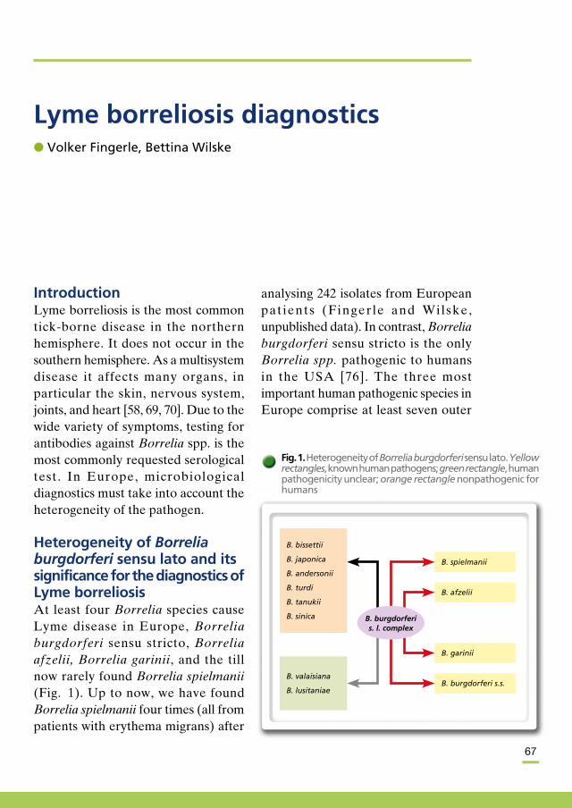
67
IntroductionLyme borreliosis is the most common tickborne disease in the northern hemisphere. It does not occur in the southern hemisphere. As a multisystem disease it affects many organs, in particular the skin, nervous system, joints, and heart [58, 69, 70]. Due to the wide variety of symptoms, testing for antibodies against Borrelia spp. is the most commonly requested serological test. In Europe, microbiological diagnostics must take into account the heterogeneity of the pathogen.
Heterogeneity of Borrelia burgdorferi sensu lato and its significance for the diagnostics of Lyme borreliosisAt least four Borrelia species cause Lyme disease in Europe, Borrelia burgdorferi sensu stricto, Borrelia afzelii, Borrelia garinii, and the till now rarely found Borrelia spielmanii (Fig. 1). Up to now, we have found Borrelia spielmanii four times (all from patients with erythema migrans) after
analysing 242 isolates from European pat ients (Fingerle and Wilske, unpublished data). In contrast, Borrelia burgdorferi sensu stricto is the only Borrelia spp. pathogenic to humans in the USA [76]. The three most important human pathogenic species in Europe comprise at least seven outer
Lyme borreliosis diagnosticsl Volker Fingerle, Bettina Wilske
Fig. 1. Heterogeneity of Borrelia burgdorferi sensu lato. Yellow rectangles, known human pathogens; green rectangle, human pathogenicity unclear; orange rectangle nonpathogenic for humans
B. spielmanii
B. bissettii
B. japonica
B. andersonii
B. turdi
B. tanukii
B. sinica
B. valaisiana
B. lusitaniae
B. afzelii
B. garinii
B. burgdorferi s.s.
B. burgdorferi s. l. complex

68
Lyme borreliosis diagnostics
surface protein A (OspA) serotypes [83]. Skin isolates are predominantly Borrelia afzeli i (OspA type 2), particularly those from patients with acrodermatitis chronica atrophicans, which incidentally is also not found in the USA [10, 53, 83]. Isolates from CSF and ticks are variable but show a predominance of Borrelia garinii (OspA types 37) [18, 72, 79, 83]. Sequence analysis of OspA amplicons using polymerase chain reaction (PCR) on arthrocentesis from patients with Lyme arthritis are distinctly heterogeneous [17, 73]. Other PCR studies showed mostly Borrelia burgdorferi sensu stricto (5S/23S rRNA intergenic spacer PCR [48] or flagellin PCR [35, 36]).Borrelia afzelii and Borrelia garinii are the most common European species. The third most common, Borrelia burgdorferi sensu stricto, is particularly rare in eastern Europe (see review by Hubalek and Halouzka 1997 [32]). A great deal of heterogeneity can occur even in very small areas [18, 21, 49, 61, 64] however, distinct regional accumulations of certain species or subspecies have also been observed [45, 49, 55]. Multispecies infections in ticks have also been reported [32], although such infections are less commonly found in humans [14, 73, 79]. The heterogeneity of the pathogen (Fig. 1) makes the diagnostics of Lyme borreliosis in Europe substantially more difficult than in the USA and must be taken into consideration in
the development of diagnostic reagents such as PCR primers and antigens. Thus when considering the frequently used OspA PCR, it is important that not only representatives of all human pathogenic species be detected but also the various OspA types of the heterogeneous Borrelia garinii group [18]. In addition, PCR should also distinguish Borrelia valaisiana and Borrelia lusitaniae because both are potential human pathogens, as indicated by a number of PCR and culture results [63, 75, 78]. We recently developed a powerful OspA PCR assay to determine and differentiate the various European species and OspA subtypes [49]. In serodiagnostic procedures, the strains’ hetergeneity must also be taken into account, as certain important diagnostic proteins are decidedly heterogeneous and thus differ in their reactivity with patient sera. For example, among representative strains of Borrelia burgdorferi sensu stricto, Borrelia afzelii, and Borrelia garinii (B31, PKo, and PBi), amino acid sequence identities for DbpA (Osp17) are 40–44%, and those for OspC are 54–68%. DbpA in particular has much greater amino acid sequence heterogeneity than DNA heterogeneity, which indicates immunoselection. Interestingly, highly heterogeneous proteins can still show highly conserved immunogenic epitopes (e.g. the C6 peptide from VlsE) [46, 47].

69
Microbiological diagnosticsQuality standards for the microbiological diagnosis of Lyme borreliosisThe German Society for Hygiene and Microbiology (DGHM) has published quality standards for the diagnosis of Lyme borreliosis formulated by a committee of experts (MiQ 12 LymeBorreliose) [86]. The free English version is available at http://www.dghm.org/ red/index. html?cname=MIQ.Except for the pathognomic clinical manifestation of erythema migrans, the diagnosis of Lyme borreliosis normally requires microbiological confirmation. For this purpose, determination of the presence of antibodies is usually used, while demonstration of the pathogen using culture or PCR is limited to special cases, for example with ambiguous serological test results or difficult clinical cases. Demonstration of the pathogen’s presence should be made only in specialised laboratories.
Detection of the pathogenDetection using cultureCulture using modified Kelly medium
[59, 85], which expends considerable time and material, can help in special cases when the clinical picture strongly suggests Lyme borreliosis despite negative serology (seronegative Lyme borreliosis) [86]. Such cases may involve atypical erythema migrans, especially acute neuroborreliosis with short disease duration and no proof of antibodies in the CSF, or patients with immunodeficiency. The sensitivity of pathogen detection in culture is generally poor (Table 1).
Detection using PCRCurrently no standard procedures are available for either sample preparation or the PCR itself. Borrelia PCR should enable diagnosis of the Borrelia species, i.e. the laboratory report should supply information on which human pathogenic species is present. The diagnostic sensitivity of PCR is approximately the same as for culture, whereas it is distinctly easier to detect Borrelia in tissue than in body fluids [2, 6, 35, 39, 44, 71, 87]. Only in the case of synovial fluid is PCR appreciably better than culture (Table 1; [52]).
Table 1. Sensitivity of culture and PCR methods for detecting Borrelia burgdorferi
Sample Sensitivity
Skin (erythema migrans, acrodermatitis) 50–70% with culture or PCR
Cerebrospinal fluid (acute neuroborreliosis) 10–30% with culture or PCR
Synovial fluid* (Lyme arthritis) 50–70% with PCR (culture is extremely seldom positive)
*higher sensitivity with synovial biopsy compared to synovial fluid

70
Lyme borreliosis diagnostics
Detection of antibodies to Borrelia burgdorferiIt is generally accepted that serological examination should follow a twostep approach [12, 37, 85, 86]: (1) serological screening which, only if reactive, should be followed by (2) a confirmatory examination. A sensitive screening procedure is recommended,
e.g. enzymelinked immunosorbent assay (ELISA) or a similar procedure such as chemiluminescence immunoassay (CLIA), which can be confirmed by immunoblot if reactivity is present.
Screening procedure. At least an improved secondgeneration test (using for example recombinant material
Table 2. Reactivity of the various homologues of VlsE and DbpA using line immunoblot for early manifestations. Data from Goettner et al. 2005
A. IgG reactivity
DbpA from DbpA VlsE from VlsE
Quantity PBie PBr PKo B31e reactive PBie PKoe PKa2 reactive
(n) n (%) n (%) n (%) n (%) n (%) n (%) n (%) n (%) n (%)
EMa 15 0 2 4 1 5 12 9 6 12 (0,0) (13,3) (26,7) (6,7) (33,3) (80,0) (60,0) (40,0) (80)
NBb 50 19 20 17 6 39 44 41 41 46 (38,0) (40,0) (34,0) (12,0) (78,0) (88,0) (82,0) (82,0) (92,0)
Controlsc 110 2 2 0 0 4 1 3 0 4 (1,8) (1,8) (0,0) (0,0) (3,6) (0,9) (2,7) (0,0) (3,6)
B. IgM reactivity
DbpA from DbpA VlsE from VlsE Quantity PBie PBre PKoe B31e reactived PBie PKoe PKa2e reactived
(n) n (%) n (%) n (%) n (%) n (%) n (%) n (%) n (%) n (%)
EMa 15 0 0 1 0 1 8 4 3 8 (0,0) (0,0) (6,7) (0,0) (6,7) (53,3) (26,7) (20,0) (53,3)
NBb 50 9 13 2 0 22 26 7 6 28 (18,0) (26,0) (4,0) (0,0) (44,0) (52,0) (14,0) (12,0) (56,0)
Controlsc 110 0 1 0 0 1 6 0 0 6 (0,0) (0,9) (0,0) (0,0) (0,9) (5,4) (0,0) (0,0) (5,4)
aErythema migransbNeuroborreliosiscSera from blood donors were used as controls n=60, patients with syphilis n=10, patients with fever of unknown origin n=30, rheumatic factor-positive sera n=10d At least one homologous protein reactiveeThe Borrelia strains belong to the following species: PBi to Borrelia garinii OspA type 4, PBR to B. garinii OspA type 3, PKo to Borrelia afzelii, and B31 and PKa2 to Borrelia burgdorferi
71
as antigen) should be used for the screening procedure [86] due to possible crossreactions with other bacteria. The strains used as a source of antigens should express OspC, the dominant antigen of the IgM immune response, and DbpA, an immunodominant antigen of the IgG response [86]. Recently, specific recombinant antigens (e.g VlsE) or synthetic peptides (e.g. the C6 peptide derived from VlsE) were used successfully on samples from American [4, 43, 47] and European patients [20, 22, 46, 54]. Moreover, DbpA which, like VlsE, is often hard to extract from Borrelia in culture, has also proved to be a sensitive antigen [22, 54] (Table 2).
Confirmatory test (immunoblot). As a confirmatory test, immunoblot requires a high specificity of at least 95%. Using recombinant antigens, the identification of diagnostic bands is substantially easier than with lysate blots.The American immunoblot criteria recommended by the centers for disease control cannot be applied in Europe [28, 29, 65]. Dressler et al. [16] have shown that the immune response of European patients is confined to a markedly narrower spectrum of Borrelia proteins than that of American patients. In a study using sera from Germany and another using sera from various European countries, Hauser et al. have shown that strainspecific criteria for interpretation must be defined [28,
29]. Different rules for interpretation must therefore be established in order to achieve equivalent sensitivity and specificity using different Borrelia strains for immunoblot antigens. The criteria for interpreting immunoblots recommended by the DGHM were published in “MiQ 12 LymeBorreliose” [86].
Patients with early manifestations have an immune response based on only a few proteins, while those with late manifestations (acrodermatitis or arthritis) have IgG antibodies against a broad spectrum of antigens (Fig. 2).Using recombinant antigens has a number of advantages over the use of whole cell lysate antigen: (a) specific antigens can be selected (e.g. p83/100, BmpA), (b) homologous antigens from various strains can be combined (e.g. DbpA [Osp17], OspC, BmpA), (c) truncated antigens with a higher specificity can be produced (internal flagellin fragment), and (d) antigens expressed primarily in vivo but not in culture are available (e.g. DbpA and in particular VlsE [30, 67, 81]). Commercial recombinant blots can be better standardised than whole cell lysate blots.The sensitivity of immunoblots based on recombinant antigens has been improved considerably in the last few years by additional antigens and the establishment of line immunoblots [22, 67, 81]. For the first time, this has

72
allowed significantly higher sensitivity with recombinant immunoblots than with whole cell lysate blots Recombinant immunoblot technology is an essential step towards standardising and increasing sensitivity, as homologous antigens from various strains (especially those with low sequence identity such as DbpA) and antigens that are only expressed in vivo (e.g. VlsE) can be used.
Serological results at various stages of the diseaseSerological test results must always be interpreted in relation to the
cl inical f indings. For this , case definitions are helpful [60, 68, 86]. In stage I (erythema migrans), only 20–50% of patients are seropositive for IgM and/or IgG antibodies [3, 26, 77] (Table 3), and IgM antibodies are prevalent. A possible exception is the immune response to VlsE. In American patients with erythema migrans, VlsE IgG antibodies were detectable before the Borreliaspecific IgM response (44% vs 19% of patients in acute erythema migrans and 59% vs 43% in convale scent erythema migrans) [4]. In European patients with erythema
Fig. 2. Line immunoblot: IgG immune response in patients with various manifestations of Lyme borreliosis. Late Lyme borreliosis: sera from patients with acrodermatitis chronica atrophicans or Lyme arthritis. The Borrelia strains belong to the species: PBi to Borrelia garinii OspA type 4, PBR to Borrelia garinii OspA type 3, 20047 to Borrelia garinii unknown OspA type, PKo to Borrelia afzelii, and B31 and PKa2 to Borrelia burgdorferi sensu stricto
Early LB
Antigen (Strain) Erythema migrans Neuroborreliosis
Late LB Controls
Lyme borreliosis diagnostics

73
migrans, earlier IgG response against VlsE was observed in 20 of 23 cases confirmed by culture (87%). The IgM response was not studied [46]. In stage II (acute neuroborreliosis), the rate of seropositivity increases (IgM and/or IgG antibodies) to 70–90% [24, 25, 80, 82]. In principle, patients with early manifestations can be seronegative, particularly those with shortterm illness. A check of the serological course should be done in such cases. At 6 weeks or more after onset of the disease, 100% of stage II neuroborreliosis patients are seropositive [24]. In cases with late manifestations (stage III, acrodermatis and arthritis), IgG antibodies could be demonstrated in all patients examined [26, 37, 80, 82]. A negative IgG test therefore speaks against diagnosis of latestage Lyme borreliosis. A positive IgM test with negative IgG is likewise not diagnostically relevant for latestage Lyme borreliosis [86].For the diagnosis of neuroborreliosis, evidence of intrathecal antibody production (i.e. in the CSF/serum index) is particularly important [84]. Combined with additional changes in the CSF such as lymphocytic pleo
cytosis and barrier disturbance, this provides decisive diagnostic evidence. For diagnosing chronic borreliosis of the central nervous system, a positive IgG CSF/serum index is essential (see EUCALB case definitions [68, 86]), whereas chronic peripheral neuropathy usually displays no intrathecal antibody production [42].As serological results can vary substantially and antibodies can persist for a long time even in successfully treated patients, serological course controls are not suitable for determining therapy failure. Until recently, a fall in VlsE specific antibodies was considered an indicator for the success of therapy [57]. This could not however be confirmed in a European study [56].The presence of specific antibodies does not indicate the presence of a clinical manifestation. Antibody tests can also be positive due to previous clinical or subclinical infection. The less specific the patients’ symptoms are, the less is the positive predictive value. In normal, healthy individuals, seropositivity varies with age and outdoor activity (e.g. in a study from Bavaria between 5% and 20% [62]).
Table 3. Sensitivity for the presence of antibodies in cases of Lyme borreliosisStage Sensitivity Antibody class
I 20-50% Predominantly IgM
II 70-90% With short-term illness predominantly IgM, with longer-term illness predominantly IgG
III Near 100% Usually only IgGa
aThe presence of IgM antibodies without IgG is not diagnostic for late manifestation

74
Methods not recommended for the diagnosis of Lyme borreliosisEvidence of borreliosis from a tick removed from a patientExamination of ticks for the presence of Borrelia burgdorferi is valuable only if (1) with a negative test result there is sufficient certainty that no infection with Borrelia burgdorferi has occurred or (2) with a positive result there is sufficient likelihood of a clinical manifestation of the infection.The fo l lowing po int s must be considered for evaluating this method of examination:1. Not every tick bite is discovered.
Ixodes ricinus larvae and nymphs can hardly be recognised by normal vision and are often unconsciously removed with a fingernail. Thus, negative results can lead to a false sense of security, and infection can be contracted through the unrecognised bite.
2. Infected ticks usually do not transmit the pathogen through biting, and, should an infection result, only some cases become clinically relevant (e.g. Nahimana et al. [51] in which only three of 17 individuals who seroconverted became clinically ill).
3. The diagnostic sensitivity and specificity of the various methods for analysing ticks is largely unclear. Influencing factors include transport time, DNA extraction, presence of inhibitory substances (e.g. haemo
globin or scutum of the tick), laboratory contamination and the genetic heterogeneity of Borrelia burgdorferi sensu lato.
4. The great majority of the resulting illnesses are simple to diagnose and efficiently to treat [34].
5. No European study has been able to demonstrate that antibiotic prophylaxis or therapy following a bite by an infected tick has more advantages than disadvantages.
To summarise, examining t icks removed from humans for Borrelia burgdorferi sensu lato cannot be recommended. The reasons are the low infection and manifestation rates, insufficient negative and positive predictive value of the method, good disease prognosis, and no evidence of a reasonable prophylactic treatment. Action useful after tick bites has been proposed by a group of experts [38].
Significance of the lymphocyte transformation testI n p r i n c i p l e , t h e l y m p h o c y t e transformation test (LTT) measures proliferation of lymphocytes in patient blood after stimulation with an antigen (e.g. sonicated Borrelia, recombinant Borrelia antigens). The test is therefore an attempt to determine cellular immunity against specific antigens. In 1988, Dattwyler et al. showed that LTT could be positive even in cases of seronegative chronic borreliosis that
Lyme borreliosis diagnostics
75
developed after prompt treatment for an early manifestation [13]. However, a series of studies published since then have shown no advantage of LTT over serology in routine Lyme borreliosis diagnostics [5, 9, 15, 19, 31, 33, 40, 66, 74].Krause et al. used LTT to examine 24 patients with various manifestations of Lyme disease and 30 patients with arthritis of unrelated aetiology as a control group, including also 20 healthy individuals [40]. For proof of antibodies, the sensitivity was 83% and specificity 92%. For LTT with a specificity of 92%, the sensitivity was 71%, while for a sensitivity of 83% the specificity was 72%. Krause recently questioned whether LTT could contribute to routine clinical diagnostics. In addition, due to the test’s low specificity, he reemphasised the high likelihood of false positive results and concluded that LTT should not be used for the diagnosis of Lyme borreliosis [41].Dressler et al. examined 42 patients with chronic Lyme borreliosis and 77 control individuals [15]. A sonicated Borrelia burgdorferi sensu stricto strain was used as antigen. The sensitivity of the test was given at 45% (serology 93%) with specificity of 95%. (No data were provided for serology.) Four of nine laboratory personnel examined also were positive using LTT.In 1996 Huppertz et al. examined 55 children with Lyme arthritis and 48 control patients [33]. Two unsonicated Borrelia burgdorferi sensu stricto strains were used as the antigen source.
The sensitivity of the test was 77% with a specificity of 78%. For eight of the children with Lyme arthritis, the stimulation index was negative before therapy but positive after therapy. In the authors’ opinion, their study demonstrates that the test is valuable for neither prognosis nor control of the disease course. They concluded: “Lymphocyte proliferation assay will rarely aid in finding the correct diagnosis when clinical presentation and antiborrelial serology do not match.”The studies of LTT to date show that it is not well standardised and, compared with serology, is less sensitive and particularly less specific. Therefore, LTT is not suited for routine diagnosis of Lyme borreliosis.Meanwhile, the ELISPOT test (a commercially available LTT modification) has been recommended for human granulocytic ehrlichiosis, including an indication that coinfection has considerable significance for the prognosis and course of infection with Borrelia. Here we would point out that to date no case of autochthonous human granulocytic ehrlichiosis in Germany has been published and therefore use of this test is already questionable on epidemiological grounds.
The significance of cystic forms of Borrelia burgdorferiSocalled cystic forms, also known as spheroblasts, forms without cell walls,
76
or L forms, can be induced in vitro by various stress factors such as culture in CFS, extreme changes in pH value, and increased temperature [1, 7, 8, 50]. Purely L forms proved infectious to mice [23]. Whether these forms are significant for pathogenesis of the disease or to diagnostics is however still unclear. In the meantime, assays allegedly specific for the Borrelia burgdorferi sensu lato form without cell walls are available. However, they have not been adequately evaluated and thus cannot be recommended for Lyme borreliosis diagnostics [11].
The visual contrast sensitivity testThe visual contrast sensitivity test (VCS) has been recommended by Hartmann and MüllerMarienburg for chronically ill patients with unspecific
symptoms such as myalgia, muscle twitching, joint pain, low energy, fatigue, diarrhea, constipation, and similar symptoms [27]. This test is based on the hypothesis that Borrelia burgdorferi produces a lipophilic neurotoxin which, among other activities, binds to the optic nerves, making the disease detectable using a VCS test due to reduced function, i.e. a deficit in recognising grey tones [27]. We do not know of any scientific publication that supports the suppositions on which this test is based. As the neurotoxin is subject to the enterohepatic circulation, it is also recommended that chole styramine be used to break the toxic cycle.From our point of view, we urgently advise against therapy with cholestyramine and the use of VCS tests for the diagnosis of Lyme borreliosis.
Lyme borreliosis diagnostics

77
l References 1. Alban, PS, Johnson, PW, Nelson, DR.
Serumstarvationinduced changes in protein synthesis and morphology of Borrelia burgdorferi . Microbiol 146(2000)119127
2. Arnez M, RuzicSabljic E, Ahcan J, RadselMedvescek A, PleterskiRigler D, Strle F. Isolation of Borrelia burgdorferi sensu lato from blood of children with solitary erythema migrans. Pediatr Infect Dis J 2001; 3:251255
3. Åsbrink, E.; Hovmark, A.; Hederstedt, B. Serologic studies of erythema chronicum migrans Afzelius and acrodermatitis chronica atrophicans with indirect immunofluorescence and enzymelinked immunosorbent assays. Acta Derm Venereol 1985; 65:509514
4. Bacon, RM, Biggerstaff, BJ, Schriefer, M, Gilmore, RD, Philipp, MT, Steere, A, Wormser, GP, Marques, AR, and Johnson, BJB. Improved serodiagnosis of Lyme disease by kinetic ELISAs using recombinant VlsE1 or peptide antigens of Borrelia burgdorferi compared with twotiered testing. J Infect Dis; 2003; 187:11871199
5. Bauer, Y.; Hofmann, H.; Jahraus, O.; Mytilineos, J.; Simon, M. M.; Wallich, R. Prominent T cell response to a selectively in vivo expressed Borrelia burgdorferi outer surface protein (pG) in patients with Lyme disease. Eur. J. Immunol.: 31(2001)767776
6. Brettschneider, S.; Bruckbauer, H.; Klugbauer, N.; Hofmann, H. Diagnostic value of PCR for detection of Borrelia burgdorferi in skin biopsy and urine samples from patients with skin borreliosis. J. Clin. Microbiol.: 36(1998)26582665
7. Brorson, O.; Brorson, S. H. In vitro conversion of Borrelia burgdorferi to cystic forms in spinal fluid, and transformation to mobile spirochetes by incubation in BSKH medium. Infection: 26(1998)144150
8. Brorson, O.; Brorson, S. H. A rapid method
for generating cystic forms of Borrelia burgdorferi and their reversal to mobile spirochetes. APMIS 106 (1998)11311141
9. Buechner, S. A.; Lautenschlager, S.; Itin, P.; Bircher, A.; Erb, P. Lymphoproliferative responses to Borrel ia burgdorfer i in patients with erythema migrans, acrodermatitis chronica atrophicans, lymphadenosis benigna cut is , and morphea. Arch. Dermatol.: 131(1995)673677
10. Canica, MM, Nato, F, Du Merle, L, Mazie, JC, Baranton, G, Postic, D. Monoclonal antibodies for identification of Borrelia afzelii sp. nov. associated with late cutaneous manifestations of Lyme borreliosis. Scand J Infect Dis 1993; 25:441448.
11. Centers for Disease Control and Prevention. Caution regarding testing for Lyme disease. Morbid Mortal Wkly Rprt 2005; 54:125
12. Centers for Disease Control and Prevention. Recommendations for Test Performance and Interpretation from the Second National Conference on Serologic Diagnosis of Lyme Disease. Morbid Mortal Wkly Rprt 1995;44:590
13. Dattwyler, R. J.; Volkman, D. J.; Luft, B. J.; Halperin, J. J.; Thomas, J.; Golightly, M. G. Seronegative Lyme disease: Dissociation of specific T and Blymphocyte responses to Borrelia burgdorferi. N. Engl. J. Med.: 319(1988)14411446
14. Demaerschalck, I, Messaoud, AB, De Kesel, M, Hoyois, B, Lobet, Y, Hoet, P, Bigaignon, G, Bollen, A, Godfroid, E. Simultaneous presence of different Borrelia burgdorferi genospecies in biological fluids of Lyme disease patients. J Clin Microbiol: 1995; 33:602608
15. Dressler, F, Natalino, H, Yoshinari, Steere, A C. The Tcell proliverative assay in the diagnosis of lyme borreliosis. Ann Int Med 115(1991)533539
16. Dressler, F, Ackermann, R, Steere, AC. Antibody responses to the three genomic groups of Borrelia burgdorferi in European Lyme borreliosis. J Infect Dis

78
1994; 169:31331817. Eiffert, H, Karsten, A, Thomssen, R,
Christen HJ. Characterization of Borrelia burgdorferi strains in Lyme arthritis. Scand J Infect Dis 1998; 30:265268
18. Eiffert, H, Ohlenbusch, A, Christen, HJ, Thomssen, R, Spielman, A, Matuschka, FR. Nondifferentiation between Lyme disease spirochetes from vector ticks and human cerebrospinal fluids. J Infect Dis 1995; 171:476479
19. Ekerfelt, C.; Forsberg, P.; Svenvik, M.; Roberg, M.; Bergström, S.; Ernerudh, J. Asymptomatic Borreliaseropositive individuals display the same incidence of Borreliaspecific interferongamma (IFNy)secreting cells in blood as patients with clinical Borrelia infection. Clin. Exp. Immunol.: 115(1999)498502
20. Fingerle, V, SchulteSpechtel, U, Wilske, B. Evaluation of an ELISA based on the C6peptide of VlsE for diagnosis of early neuroborreliosis. Int J Med Microbiol 2002; 292 (Suppl. 34): 217
21. G e r n , L , H u C M , K o c i a n o v a E , Vyrestekova V, Rehacek J. Genetic diversity of Borrelia burgdorferi sensu lato isolates obtained from Ixodes ricinus ticks collected in Slovakia. Europ J Epidemiol (1999)15, 665669
22. Goettner, G., U. SchulteSpechtel, R. Hillermann, G. Liegl, B. Wilske, and V. Fingerle. 2005. Improvement of Lyme borreliosis serodiagnosis by a newly developed recombinant IgG and IgM line immunoblot and addition of VlsE and DbpA homologues. J.Clin.Microbiol. in press
23. Gruntar, I, Malovrh, T, Murgia, R, Cinco, M. Conversion of Borrelia garinii cystic forms to motile spirochetes in vivo. APMIS 105(2001)383388
24. Hansen, K, Hindersson, P, Pedersen, NS. Measurement of antibodies to the Borrelia burgdorferi flagellum improves serodiagnosis in Lyme disease. J Clin Microbiol 1988; 26:338346
25. H a n s e n , K , L e b e c h , A M . L y m e neuroborreliosis: a new sensitive diagnostic
assay for intrathecal sythesis of Borrelia burgdorferi specific immunoglobulin G, A, and M. Ann. Neurol.: 30(1991)197205
26. Hansen, K.; Åsbrink, E. Serodiagnosis of erythema migrans and acrodermatitis chronica atrophicans by the Borrelia burgdorferi flagellum enzymelinked immunosorbent assay. J Clin Microbiol 1989; 27:545551
27. Hartmann und MüllerMarienburg. Indirekter Neurotoxinnachweis durch den “Visual Contrast Sensitivity” Test bei Patienten mit chronischer Borreliose. Neurologie 2003. 248251
28. Hauser, U, Lehnert, G, Lobentanzer, R, Wilske, B. Interpretation criteria for standardized western blots for three european species of Borrelia burgdorferi sensu lato. J Clin Microbiol 1997. 35:14331444
29. Hauser, U, Lehnert, G, Wilske, B. Diagnostic value of proteins of three Borrelia species (Borrelia burgdorferi sensu lato) and implications for development and use of recombinant antigens for serodiagnosis of Lyme borreliosis in Europe. Clin Diagn Lab Immunol 1998; 5:456462
30. Heikkilä, T, Seppälä, I , Saxen, H, Panelius, J, Yrjänäinen, H, Lahdenne, P. Speciesspecific serodiagnosis of Lyme arthritis and neuroborreliosis due to Borrelia burgdorferi sensu stricto, B. afzelii, and B. garinii by using decorin binding protein A. J Clin Microbiol 2002; 40:453460
31. Horowitz, HW, Pavia, CS, Bittker, S, Forseter, G, Cooper, D, Nadelman, RB, Byrne, D, Johnson, RC, Wormser, GP. Sustained cellular immune response to Borrelia burgdorferi: Lack of correlation with clinical presentation and serology. Clin Diagn Lab Immunol 1(1994)4:373378
32. Hubalek, Z. Halouzka, J. Distribution of Borrelia burgdorferi sensu lato genomic groups in Europe, a review. Eur J Epidemiol 1997; 13: 951957
33. Huppertz, H.I.; Mösbauer, S.; Busch,
Lyme borreliosis diagnostics

79
D. H.; Karch, H. Lymphoproliferative responses to Borrelia burgdorferi in the diagnosis of Lyme arthritis in children and adolescents. Eur. J. Pediatr.: 155(1996)297302
34. Huppertz, H. I.; Böhme, M.; Standaert, S. M.; Karch, H.; Plotkin, S. A. Incidence of Lyme borreliosis in the Würzburg region of Germany. Eur. J. Clin. Microbiol. Infect. Dis.: 18(1999)697703
35. Jaulhac, B, CharyValckenaere, I, Sibilia, J, Javier, RM, Piemont, Y, Kuntz, JL, Monteil, H, Pourel, J Detection of Borrelia burgdorferi by DNA amplification in synovial tissue samples from patients with Lyme arthritis. Arthritis Rheum 1996; 39:736745
36. Jaulhac, B.; Heller, R.; Limbach, F. X.; Hansmann, Y.; Lipsker, D.; Monteil, H.; Sibilia, J.; Piemont, Y. Direct molecular typing of Borrelia burgdorferi sensu lato species in synovial samples from patients with Lyme arthritis. J. Clin. Microbiol.: 38(2000)18951900
37. Johnson, BJB, Robbins, KE, Balley, RE, Cao, BL, Sviat, SL. Craven R B, Mayer L W, Dennis D T: Serodiagnosis of Lyme disease: accuracy of a twostep approach using a flagellabased ELISA and immunoblotting. J Infect Dis 1996; 174:346353
38. Kaiser, R. Teilnehmer der Expertenkonferenz: Frühsommermeningoenzephalitis und LymeBorreliosePrävention vor und nach Zeckenstich. Dtsch med Wochenschr 1998; 123:847853
39. Kar lsson, M, HovindHougen, K, Svenungsson, B, Stiernstedt, G. Cultivation and characterization of spirochetes from cerebrospinal fluid of patients with Lyme borreliosis J Clin Microbiol 1990; 28:473479
40. Krause, A, Brade, V, Schoerner, C, Solbach, W, Kalden, J R, Burmester, G. T cell proliferation induced by Borrelia burgdorferi in patients with lyme borreliosis. Arthr Rheum 34(1991)393402
41. Krause, A. Verraten Lymphos die Borreliose? Medical Tribune 38(2003)14:26
42. Kristoferitsch, W. Chronical peripheral
neuropathy. in: Weber, K.; Burgdorfer, W. (eds.) Aspects of Lyme borreliosis. Berlin, Heidelberg, New York, Springer (1993) 219227
43. Lawrenz, MB, Hardham, JM, Owens, RT, Nowakowski, J, Steere, AC, Wormser, GP, Norris, SJ. Human antibody responses to VlsE antigenic variation protein of Borrelia burgdorferi. J Clin Microbiol 1999; 37:39974004
44. Lebech, A. M., K. Hansen, F. Brandrup, O. Clemmensen, and L. HalkierSorensen. 2000. Diagnostic value of PCR for detection of Borrelia burgdorferi DNA in clinical specimens from patients with erythema migrans and Lyme neuroborreliosis. Mol.Diagn. 5:139150
45. Lencakova D, HizoTeufel C, Petko B, SchulteSpechtel U, Stanko M, Wilske B, Fingerle V. Prevalence of Borrelia burgdorferi s.l. OspA types in Ixodes ricinus ticks from selected localities in Slovakia and Poland.. Int J Med Microbiol. 2006; Suppl 1:10818.
46. Liang, FT, Aberer, E, Cinco, M, Gern, L, Hu, CM, Lobet, YN, Ruscio, M, Voet, PE, Weynants, VE, Philipp, MT. Antigenic conservation of an immunodominant invariable region of the VlsE lipoprotein among european pathogenic genospecies of Borrelia burgdorferi sl. J Infect Dis 2000; 182:14551462.
47. Liang, FT, Alvarez, AL, Gu, Y, Nowling, JM, Ramamoorthy, R, Philipp, MT. An immunodominant conserved region within the variable domain of VlsE, the variable surface antigen of Borrelia burgdorferi. J Immunol 1999; 163:55665573.
48. Lünemann, JD, Zarmas, S, Priem, S, Franz, J, Zschenderlein, R, Aberer, E, Klein, R, Schouls, L, Burmester, GR, Krause, A. Rapid typing of Borrelia burgdorferi sensu lato species in specimens from patients with different manifestations of Lyme borreliosis. J Clin Microbiol 2001; 39:11301133
49. Michel, H.; Wilske, B.; Hettche, G.; Goettner, G.; Heimerl, C.; Reischl, U.; SchulteSpechtel, U.; Fingerle, V.

80
An ospApolymerase chain reaction/restriction fragment length polymorphismbased method for sensitive detection and reliable differentiation of all European Borrelia burgdorferi sensu lato species and OspAtypes. Med. Microbiol. Immunol.: 193(2004)219226
50. Murgia, R.; Cinco, M. Induction of cystic forms by different stress conditions in Borrelia burgdorferi. APMIS: 112(2004)5762
51. Nahimana, I.; Gern, L.; Blanc, D. S.; Praz, G.; Francioli, P.; Peter, O. Risk of Borrelia burgdorferi infection in western Switzerland following a tick bite. Eur. J. Clin. Microbiol. Infect. Dis.: 23(2004)603608
52. Nocton, J J, Dressler, F, Rutledge, B J, Rys P N, Persing, D H, Steere, A C. Detection of Borrelia burgdorferi DNA by polymerase chain reaction in synovial fluid from patients with Lyme arthritis. N Engl J Med 1994; 330:229234
53. Ohlenbusch , A, Matuschka , FR, Richter, D, Christen, HJ, Thomssen, R, Spielman, A, Eiffert, H. Etiology of the acrodermatitis chronica atrophicans lesion in Lyme disease. J Infect Dis 1996; 174:421423
54. Panelius, J., P. Lahdenne, H. Saxen, S. A. Carlsson, T. Heikkila, M. Peltomaa, A. Lauhio, and I. Seppala. 2003. Diagnosis of Lyme neuroborreliosis with antibodies to recombinant proteins DbpA, BBK32, and OspC, and VlsE IR6 peptide. J. Neurol. 250:13181327
55. Péter, O, Bretz, A.G., Bee, D. Occurence of different genospecies of Borrelia burgdorferi sensu lato in ixodid ticks of Valais, Switzerland. Eur J Epidemiol 1995; 11:463467
56. Peltomaa,M., McHugh,G., and Steere,A.C. (2003). Persistence of the antibody response to the VlsE sixth invariant region (IR6) peptide of Borrelia burgdorferi after successful antibiotic treatment of Lyme disease. J. Infect. Dis. 187, 11781186
57. Philipp,M.T., Marques,A.R., Fawcett,P.T.,
Dally,L.G., and Martin,D.S. (2003). C6 test as an indicator of therapy outcome for patients with localized or disseminated lyme borreliosis. J. Clin. Microbiol. 41, 49554960
58. Pfister, HW, Wilske, B, Weber, K. Lyme borreliosis: basic science and clinical aspects. Lancet 1994; 343:10131016
59. PreacMursic, V, Wilske, B, Reinhardt, S. Culture of Borrelia burgdorferi on six solid media. Eur J Microbiol Infect Dis. 1991; 10:10761079
60. Rauer, S., R. Kaiser, H. W. Kölmel, H. W. Pfister, and B. Wilske. Leitlinien für Diagnostik und Therapie in der Neurologie. Herausgegeben von der Kommission “Leitlinien” der Deutschen Gesellschaft für Neurologie, Stuttgart:Georg Thieme Verlag, 2005, 121
61. Rauter, C, Oehme, R, Diterich, I, Engele, M, Hartung, T. Distribution of clinically relevant Borrelia genospecies in ticks assessed by a novel, singlerun, realtime PCR. J Clin Microbiol 2002;40:3643
62. Reimer, B.; Marschang, A.; Fingerle, V.; Wilske, B.; v. Sonnenburg, F.; v. Hoecke, C. Epedimiology of Lyme borreliosis in SouthEastern Bavaria (Germany). Zent. bl. Bakteriol. 1999; 289:653654
63. Rijpkema S G T, Tazelaar D J, Molkenboer M J C H, Noordhoek G T, Plantinga G, Schouls L M, Schellekens J F P. Detection of Borrelia afzelii, Borrelia burgdorferi sensu stricto, Borrelia garinii and group VS116 by PCR in skin biopsies of patients with erythema migrans and acrodermatitis chronica atrophicans. Clin Microbiol Infect. 1997;3:109116
64. Rijpkema, S, Golubic, D, Molkenboer, M, VerbeckDe Kruif, N, Schellekens, J. Idenfication of four genomic groups of Borrelia burgdorferi sensu lato in Ixodes ricinus ticks collected in a Lyme borreliosis endemic region of northern Croatia. Exp Appl. Acarol 1996; 20: 2330
65. Robertson, J, Guy, E, Andrews, N, Wilske, B, Anda, P, Granström, M, Hauser, U, Moosmann, Y, Sambri, V, Schellekens, J, Stanek, G, Gray, J. A European multicenter
Lyme borreliosis diagnostics

81
study of immunoblotting in serodiagnosis of Lyme borreliosis. J Clin Microbiol (2000)38:20972102
66. Schempp, C.; Owsianowski, M.; Lange, R.; Gollnick, H. Comparison of Borrelia burgdorferi ultrasonicate and whole B. burgdorferi cells as a stimulus for tcell proliferation and GMCSF secretion in vitro. Zbl. Bakt.: 279(1993)417425
67. SchulteSpechtel, U. Lehnert, G.; Liegl, G, Fingerle, V, Heimerl, C, Johnsson, BJB, Wilske,B. Significant improvement of the recombinant Borrelia IgG immunoblot by addition of VlsE and a DbpA homologue derived from B. garinii for the diagnosis of early Neuroborreliosis. J Clin Microbiol. 2003; 41:12991303
68. Stanek, G, O’Connell, S, Cimmino, M, Aberer, E, Kristoferitsch, W, Granström, M, Guy, E, Gray, J. European Union concerted action on risk assessment in Lyme borreliosis: clinical case definitions for Lyme borreliosis. Wien Klin Wochenschr 1996; 108/23:741747
69. Stanek, G, Strle, F. Lyme borreliosis. Lancet 2003; 362:1639–1647
70. Steere, AC. Medical progress Lyme disease. N Engl J Med 1989; 321:586596.
71. Strle, F.; Nadelman, R. B.; Cimperman, J.; Nowakowski, J.; Picken, R. N.; Schwartz, I.; Maraspin, V.; AgueroRosenfeld, M. E.; Varde, S.; LotricFurlan, S.; Wormser, G. P. Comparison of cultureconfirmed erythema migrans caused by Borrelia burgdorferi sensu stricto in New York State and by Borrelia afzelii in Slovenia. Ann. Intern. Med.: 130(1999)3236
72. Van Dam, AP, Kuiper, H, Vos, K, Widjojokusumo, A, De Jongh, BM, Spanjaard, L, Ramselaar, ACP, Kramer, MD, Dankert, J. Different genospecies of Borrelia burgdorferi are associated with distinct clinical manifestations of Lyme borreliosis. Clin Infect Dis 1993 ; 17:708717
73. Vasiliu, V, Herzer, P, Rössler, D, Lehnert, G, Wilske, B. Heterogeneity of Borrelia burgdorferi sensu lato demonstrated by an ospAtypespecific PCR in synovial fluid from patients with Lyme arthritis. Med
Microbiol Immunol 1998; 187:9710274. Vaz, A.; Glickstein, L.; Field, J. A.;
McHugh, G.; Sikand, V. K.; Damle, N.; Steere, A. C. Cellular and humoral immune responses to Borrelia burgdorferi antigens in patients with culturepositive early Lyme disease. Infect. Immun.: 69(2001)74377444
75. Wang, G, van Dam, AP, Dankert, J. Phenotypic and genetic characterization of a novel Borrelia burgdorferi sensu lato isolate from a patient with Lyme borreliosis. J Clin Microbiol 1999a; 37:30253028
76. Wang, G, van Dam, AP, Schwartz, I, Dankert, J. Molecular typing of Borrelia burgdorferi sensu lato taxonomic, epidemiological, and clinical implications. Clin Microbiol Rev 1999b; 12:633653
77. Weber, K, PreacMursic, V, Wilske, B, Thurmayr, R, Neubert, U, Scherwitz, C. A randomized trial of ceftriaxone versus oral penicillin for treatment of early Lyme borreliosis. Infection 1990; 18:9196
78. Wilske, B. Microbiological diagnosis in Lyme borreliosis. Int. J. Med. Microbiol. 2002, 291,(Suppl.)33:114119
79. Wilske, B, Busch, U, Eiffert, H, Fingerle, V, Pfister, HW, Rössler, D, PreacMursic, V. Diversity of OspA and OspC among cerebrospinal fluid isolates of Borrelia burgdorferi sensu lato from patients with neuroborreliosis in Germany. Med Microbiol Immunol 1996; 184:195201
80. Wilske, B, Fingerle, V, Herzer, P, Hofmann, A, Lehnert, G, Peters, H, Pfister, HW, PreacMursic, V, Soutschek, E, Weber, K. Recombinant immunoblot in the serodiagnosis of Lyme borreliosis, comparison with indirect immunofluoerescence and enzymelinked immunosorbent assay. Med Microbiol Immunol 1993a; 182:255270
81. Wilske, B, Habermann, C, Fingerle, V, Hillenbrand, B, JaurisHeipke, S, Lehnert, G, Pradel, I, Rössler, D, SchulteSpechtel, U. An improved recombinant IgG immuno blot for serodiagnosis of Lyme borreliosis. Med

82
Microbiol Immunol 1999; 188:13914482. Wilske, B.; PreacMursic, V. Microbiological
diagnosis of Lyme borreliosis. in: Weber, K.; Burgdorfer, W. (eds.) Aspects of Lyme borreliosis. Berlin, Heidelberg, New York, Springer(1993b)267300
83. Wilske, B, PreacMursic, V, Göbel, U B, Graf, B, JaurisHeipke, S, Soutschek, E, Schwab, E, Zumstein, G. An OspA serotyping system for Borrelia burgdorferi based on reactivity with monoclonal antibodies and OspA sequence analysis. J Clin Microbiol 1993c; 31:340350
84. Wilske, B, Schierz, G, PreacMursic, V, von Busch, K, Kühbeck, R, Pfister, HW, Einhäupl, K. Intrathecal production of specific antibodies against Borrelia burgdorferi in patients with lymphocytic men ingorad icu l i t i s (Bannwar th ’ s syndrome). J Infect Dis 1986; 153:304314
85. Wilske, B, Schriefer, M. Borrelia. In Manual of
Clinical Microbiology (8th edition). Edited by Murray, P. R.; Baron, E. J.; Jorgensen, J. H.; Pfaller, M. A.; Yolken, R. H. (eds.) Manual of Clinical Microbiology (8th edition) Volume 1. Washington, D. C., ASM Press(2003)937954
86. Wilske, B, Zöller, L, Brade, V, Eiffert, M, Göbel, UB, Stanek, G unter Mitarbeit von HW Pfister. MIQ 12 LymeBorreliose. In Qualitätsstandards in der mikrobiologisch infektiologischen Diagnostik. Edited by Mauch H and Lütticken R; im Auftrag der Deutschen Gesellschaft für Hygiene und Mikrobiologie (DGHM). Urban & Fischer Verlag, München Jena; 2000 (english version: http://www.dghm.org/red/index.html? cname=MIQ)
87. Zore, A.; RuzicSabljic, E.; Maraspin, V.; Cimperman, J.; LotricFurlan, S.; Pikelj, A.; Jurca, T.; Logar, M.; Strle, F. Sensitivity of culture and polymerase chain reaction for the etiologic diagnosis of erythema migrans. Wien. Klin. Wochenschr.: 114(2002)606609
Lyme borreliosis diagnostics

83
Dr. med. Thomas TalaskaPraxis für Mikrobiologie und Infektionsepidemiologie RudolfBreitscheidStr.1015295 Groß Lindowthomas.talaska@tonline.de
Prof. Dr. med. Andreas KrauseImmanuel Krankenhaus GmbHRheumakliniken BerlinWannsee und BerlinBuchKönigstr.63 Karower Str.1114109 Berlin 13125 [email protected]
Prof. Dr. med. Elisabeth AbererUniversitätsklinik für DermatologieMedizinische Universität GrazA8036 Grazelisabeth.aberer@medunigraz.at
Prof. Dr. med. Reinhard KaiserNeurologische KlinikKlinikum Pforzheim GmbHKanzlerstraße 2675175 [email protected]
Dr. med. Volker FingerleMax von PettenkoferInstitut, LMU MünchenNationales Referenzzentrum für Borrelien PettenkoferStrasse 9a80336 [email protected]muenchen.de
Prof. Dr. med. Bettina WilskeMax von PettenkoferInstitut, LMU MünchenNationales Referenzzentrum für Borrelien PettenkoferStrasse 9a80336 MunichBettina.Wilske@mvpbak.med.unimuenchen.de
l Contacts


M08
7000
4173
/A 0
3/14
DiaSorin S.p.A.Via Crescentino, snc
13040 Saluggia (VC) ItalyTel. +39 0161 487 526/947
Fax +39 0161 487 670www.diasorin.com