diagnosis of lyme borreliosis in europe - lymenet.de · diagnosis of lyme borreliosis in europe ......
TRANSCRIPT
VECTOR-BORNE AND ZOONOTIC DISEASESVolume 3, Number 4, 2003© Mary Ann Liebert, Inc.
Review
Diagnosis of Lyme Borreliosis in Europe
BETTINA WILSKE
ABSTRACT
In Europe, Lyme borreliosis is caused by at least three species, B. burgdorferi sensu stricto, B. afzelii and B. garinii.Thus microbiological diagnosis in European patients must consider the heterogeneity of Lyme disease borreliaefor development of diagnostic tools such as PCR primers and diagnostic antigens. According to guidelines of theGerman Society of Hygiene and Microbiology, the serological diagnosis should follow the principle of a two-stepprocedure. A sensitive ELISA differentiating IgM and IgG is recommended as the first step. In case the ELISA isreactive, it is followed by immunoblots (IgM and IgG) as the second step. The reactive diagnostic bands shouldbe clearly identified, which is easy if recombinant antigens are used. The sensitivity and standardization of im-munoblots has been considerably enhanced by use of recombinant antigens instead of whole cell lysates. Im-proved sensitivity resulted from use of recombinant proteins that are expressed primarily in vivo (e.g., VlsE) andcombination of homologous proteins from different strains of borrelia (e.g., DbpA). It also appears promising touse recombinant proteins (DbpA, VlsE, others) or synthetic peptides (the conserved C6 peptide derived from VlsE)as ELISA antigens. At present, detection rates for serum antibodies are 20–50% in stage I, 70–90% in stage II, andnearly 100% in stage III Lyme disease. The main goals for the future are to improve specificity in general and sen-sitivity for diagnosis of early manifestations (stage I and II). Detection of the etiological agent by culture or PCRshould be confined to specific indications and specialised laboratories. Recommended specimens are skin biopsyspecimens, CSF and synovial fluid. The best results are obtained from skin biopsies with culture or PCR (50–70%)and synovial tissue or fluid (50–70% with PCR). CSF yields positive results in only 10–30% of patients. Methodsthat are not recommended for diagnostic purposes are antigen tests in body fluids, PCR of urine, and lymphocytetransformation tests. Key Words: Lyme borreliosis—Borrelia burgdorferi—Diagnosis. Vector-Borne Zoonotic Dis.3, 215–227.
215
INTRODUCTION
LYME BORRELIOSIS is a multisystem disease in-volving many organs such as the skin, the
nervous system, the joints, and the heart (Steereet al. 1989, Pfister et al. 1994). This condition isthe most frequent tick-borne disease in thenorthern hemisphere. Due to the diversity ofclinical symptoms, Lyme disease is often con-sidered in a differential diagnosis. Examina-tions for antibodies against Borrelia burgdorferiare thus in high demand, and are among themost frequently requested serological tests
in microbiological laboratories. Microbiologicaldiagnosis in European patients must considerthe heterogeneity of Lyme disease borreliae inEurope.
HETEROGENEITY OF LYME DISEASE BORRELIAE IN EUROPE
AND ITS IMPACT FOR MICROBIOLOGICAL DIAGNOSIS
In Europe, Lyme borreliosis is caused by atleast three species: B. burgdorferi sensu stricto,
Max von Pettenkofer Institute, University of Munich, National Reference Center for Borreliae, Munich, Germany.
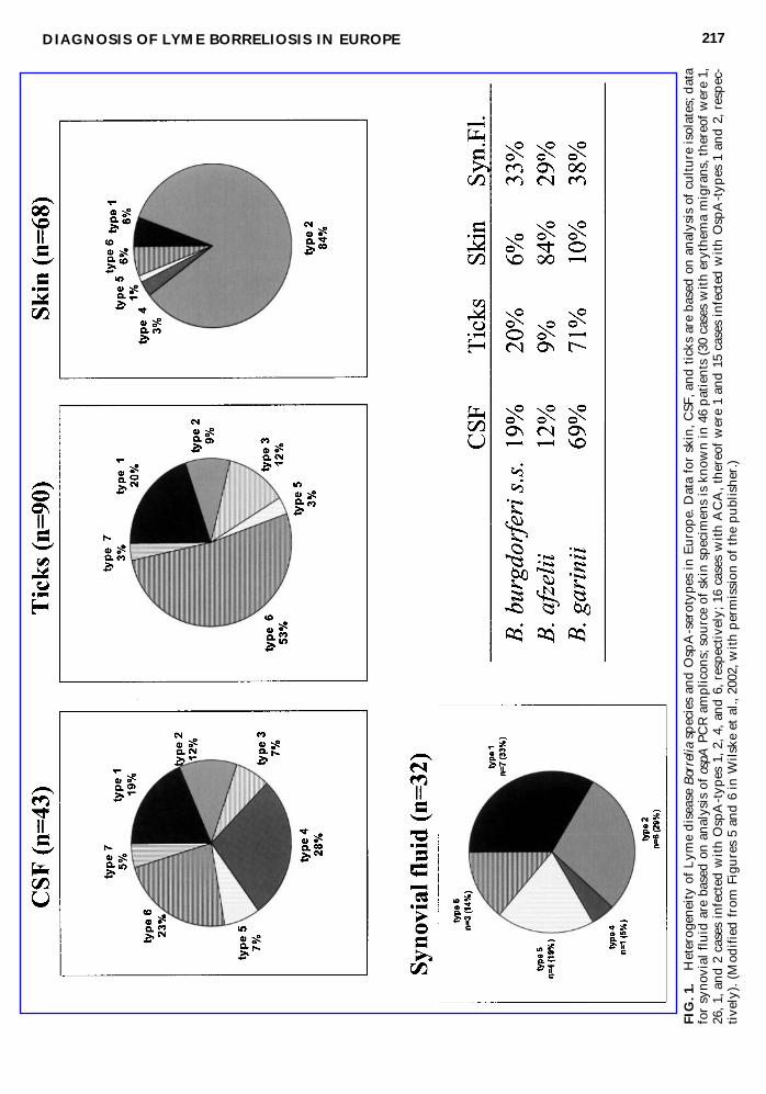
B. afzelii, and B. garinii. In contrast, B. burgdor-feri sensu stricto is the only human-pathogenicspecies in the United States (Wang et al. al1999b). The three human-pathogenic speciescomprise at least seven OspA-serotypes in Eu-rope (Fig. 1) (Wilske et al. 1993c). Skin isolatesprimarily belong to B. afzelii (OspA-type 2), es-pecially those from patients with acrodermati-tis chronica athrophicans, a chronic skin dis-ease not present in America (Canica et al. 1993,Ohlenbusch et al. 1996, Wilske et al. 1993c) (seealso legend of Fig. 1). Isolates from CSF andticks are heterogeneous with a predominanceof B. garinii (Eiffert et al. 1995, van Dam et al.1993, Wilske et al. 1996, Wilske et al. 1993c).Sequence analysis of polymerase chain reac-tion (PCR) ospA amplicons from synovial fluidof Lyme arthritis patients revealed hetero-geneity (Eiffert et al. 1998, Vasiliu et al. 1998),whereas other studies found mainly B.burgdorferi s.s. using PCR based on the 5S/23SrRNA intergenic spacer region (Lünemann etal. 2001) or the flagellin gene (Jaulhac et al.1996 and 2000). The most frequent genomicgroups in Europe B. afzelii and B. garinii occuracross the continent and the islands, whereasthe third frequent group B. burgdorferi s.s. hasonly rarely been isolated in Eastern Europe (fora survey, see Hubalek et al. 1997). Strains maybe very heterogeneous even within small ar-eas (Eiffert et al. 1995, Gern et al. 1999, Michelet al. 2003, Rauter et al. 2002, Rijpkema et al.1996). On the other side a focal prevalence ofcertain species or subtypes was also observed(Michel et al. 2003, Peter et al. 1995). Mixed in-fections have been repeatedly observed in ixo-did ticks (for a survey, see Hubalek et al. 1997)and sometimes also in specimens from patients(Demaerschalck et al. 1995, Vasiliu et al. 1998,Wilske et al. 1996). The heterogeneity of thecausative strains (Fig. 1) is a challenge for themicrobiological diagnosis of Lyme borreliosisin Europe and must be kept in mind for de-velopment of diagnostic tools such as PCRprimers and diagnostic antigens. For example,ospA PCR has been widely used. Here, it is im-portant to be sure that not only representativesof the three species are detected, but also thedifferent ospA-types of the heterogeneous B.garinii group (Eiffert et al. 1995). In addition,PCR should detect B. valaisiana and the re-
cently detected new genotype A14S since B.valaisiana and genotype A14S might also bepathogenic for humans, as suggested by posi-tive PCR results or cultures obtained from skinbiopsy specimens in a few studies (Rijpkemaet al. 1997, Wang et al. 1999a, Wilske et al.2002). An ospA PCR for detection and differ-entiation of the various European species andOspA-types has been described by Michel(2003).
Most of the proteins relevant for serodiag-nosis are heterogeneous. Interspecies aminoacid sequence identities are for example only40–44% for DbpA (Osp17) and 54-68% forOspC for representative strains of B. burg-dorferi sensu stricto, B. afzelii, and B. garinii(strains B31, PKo, and PBi, respectively)(Table 1). Especially DbpA has a much higheramino acid sequence heterogeneity com-pared to the DNA sequence heterogeneity in-dicating immune selection. However, highlyheterogeneous proteins sometimes have con-served immunogenic epitopes (e.g., the C6peptide of VlsE) (Liang et al. 1999, Liang etal. 2000).
GUIDELINES FOR THEMICROBIOLOGICAL DIAGNOSIS
OF LYME BORRELIOSIS
The German Society of Hygiene and Micro-biology (DGHM) has recently published guide-lines for the microbiological diagnosis of Lymeborreliosis written by an expert committee(MiQ 12 Lyme-Borreliose) (Wilske et al. 2000).The English version is accessible via internet(www.dghm.org/red/index.html?cname5MIQ). Except in cases with the pathognomic clin-ical manifestation erythema migrans, the diag-nosis of Lyme borreliosis usually requires confirmation by means of a microbiological di-agnostic assay. Antibody detection methodsmainly are used for this purpose, whereas de-tection of the causative agent by culture isola-tion and nucleic acid techniques is confined tospecial situations, such as to clarify clinicallyand serologically ambiguous findings. Appli-cation of these methods should be reserved tolaboratories specialized in this type of exami-nation.
WILSKE216
DIAGNOSIS OF LYME BORRELIOSIS IN EUROPE 217
FIG. 1
.Heterog
eneity of Lym
e disease Borrelia
species an
d O
spA-serotyp
es in
Europe. D
ata for skin, C
SF, a
nd ticks are based
on an
alysis of cu
lture is
olates; d
ata
for sy
novial fluid are based
on an
alysis of ospA
PCR amplicon
s; sou
rce of skin sp
ecim
ens is kno
wn in 46 patients (30 cases with erythe
ma migrans
, the
reof w
ere 1,
26, 1
, and
2 cases infected w
ith Osp
A-typ
es 1, 2
, 4, a
nd 6, respe
ctively; 16 cases with ACA, the
reof w
ere 1 an
d 15 cases infected
with Osp
A-typ
es 1 and
2, respec-
tive
ly). (M
odified from Figures 5 an
d 6 in
Wils
ke et al., 20
02, w
ith permission
of the pu
blishe
r.)
SPECIMENS FOR THEMICROBIOLOGICAL DIAGNOSIS
For culture and PCR, skin biopsy samples arethe most promising specimens. In general poorresults are obtained from body fluids with theexception of PCR of synovial fluid. Examina-tion of urine (PCR, antigen detection) is not rec-ommended nor the examination (PCR or IFA)of ticks removed from patients in order to de-cide antibiotic prophylaxis (Brettschneider etal. 1998, Kaiser et al. 1998, Klempner et al. 2001,Wilske et al. 2000). Examination of ticks shouldbe performed only for epidemiological or otherscientific studies. For antibody determination,serum or CSF can be investigated. CSF exami-nation should always be done together withserum antibody analysis (determination of theCSF/serum antibody index).
DIRECT DETECTION METHODS
Culture
B. burgdorferi can be cultivated in modifiedKelly’s medium (Preac-Mursic et al. 1991,
Wilske and Schriefer 2002). This, however, is avery time-consuming method (generation timeof B. burgdorferi is about 7–20 h) characterised bylow sensitivity, especially in body fluids (Arnezet al. 2001, Åsbrink et al. 1985, Karlsson et al.1990, Strle et al. 1999, Zore et al. 2002) (Table 2).Culturing may be of help in individual cases ifthe clinical picture suggests Lyme borreliosis de-spite a negative antibody assay (seronegativeLyme borreliosis), for example, in atypical ery-thema migrans, suspected acute neuroborrelio-sis without detection of intrathecal antibodies orin the case of suspected Lyme borreliosis in pa-tients with immune deficiencies.
PCR
There is no standardized method for thepreparation of specimens nor for performingthe PCR itself. For DNA amplification underexperimental conditions various target se-quences have been used by specialised labora-tories, for example, from plasmid-borne genessuch as ospA and ospB, or chromosomal genessuch as the genes for the flagellar protein orp66 (clone 2H1), or from gene segments of the 16S rRNA or the 5S/23S rRNA intergenic
WILSKE218
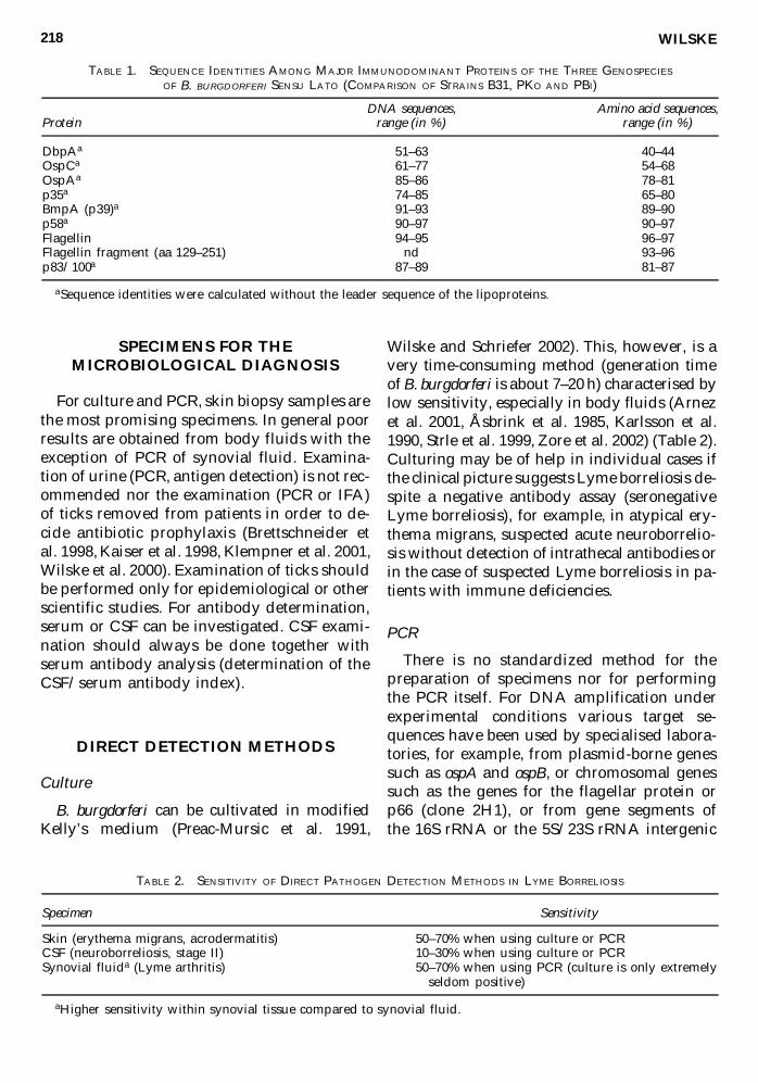
TABLE 1. SEQUENCE IDENTITIES AMONG MAJOR IMMUNODOMINANT PROTEINS OF THE THREE GENOSPECIES
OF B. BURGDORFERI SENSU LATO (COMPARISON OF STRAINS B31, PKO AND PBI)
DNA sequences, Amino acid sequences,Protein range (in %) range (in %)
DbpAa 51–63 40–44OspCa 61–77 54–68OspAa 85–86 78–81p35a 74–85 65–80BmpA (p39)a 91–93 89–90p58a 90–97 90–97Flagellin 94–95 96–97Flagellin fragment (aa 129–251) nd 93–96p83/100a 87–89 81–87
aSequence identities were calculated without the leader sequence of the lipoproteins.
TABLE 2. SENSITIVITY OF DIRECT PATHOGEN DETECTION METHODS IN LYME BORRELIOSIS
Specimen Sensitivity
Skin (erythema migrans, acrodermatitis) 50–70% when using culture or PCRCSF (neuroborreliosis, stage II) 10–30% when using culture or PCRSynovial fluida (Lyme arthritis) 50–70% when using PCR (culture is only extremely
seldom positive)
aHigher sensitivity within synovial tissue compared to synovial fluid.
spacer region (for a survey, see Schmidt et al.1997). Borrelia PCR should allow diagnosis ofthe Borrelia species, that is, the medical reportshould contain information as to which of thethree species pathogenic for humans has beenfound. The diagnostic sensitivity of PCR isabout the same as the sensitivity of culture.Borreliae are detected with much more diffi-culty from body fluids than from tissue spec-imens by either PCR or culture (Arnez et al.2001, Jaulhac et al. 1996, Karlsson et al. 1990).Solely PCR of synovial fluid seems to surpassculture significantly in sensitivity (Nocton etal. 1994).
Sensitivity of culture and PCR
Table 2 gives a survey about sensitivity of di-rect detection methods in clinical specimens
from patients with Lyme borreliosis. Culture andPCR have the highest detection rates (50–70%) inskin biopsies from patients with erythema mi-grans or acrodermatitis chronica atrophicans(Åsbrink et al. 1985a, van Dam et al. 1993, vonStedingk et al. 1995, Weber et al. 1990, Zore et al.2002). In contrast borreliae are detected by PCRor culture in the CSF of only 10–30% of patientswith neuroborreliosis (Eiffert et al. 1995, Karls-son et al. 1990, Wilske and Preac-Mursic 1993b).CSF isolates are more frequently obtained frompatients with short duration of disease than frompatients with disease of long duration (Karlssonet al. 1990). It is surprising that borreliae are de-tected by PCR in 50–70% in the synovial fluidsof Lyme arthritis patients, but culture is rarelysuccessful (Eiffert et al. 1998, Vassiliu et al. 1998).The best PCR results are obtained from synovialtissue, not fluid (Jaulhac et al. 1996).
DIAGNOSIS OF LYME BORRELIOSIS IN EUROPE 219
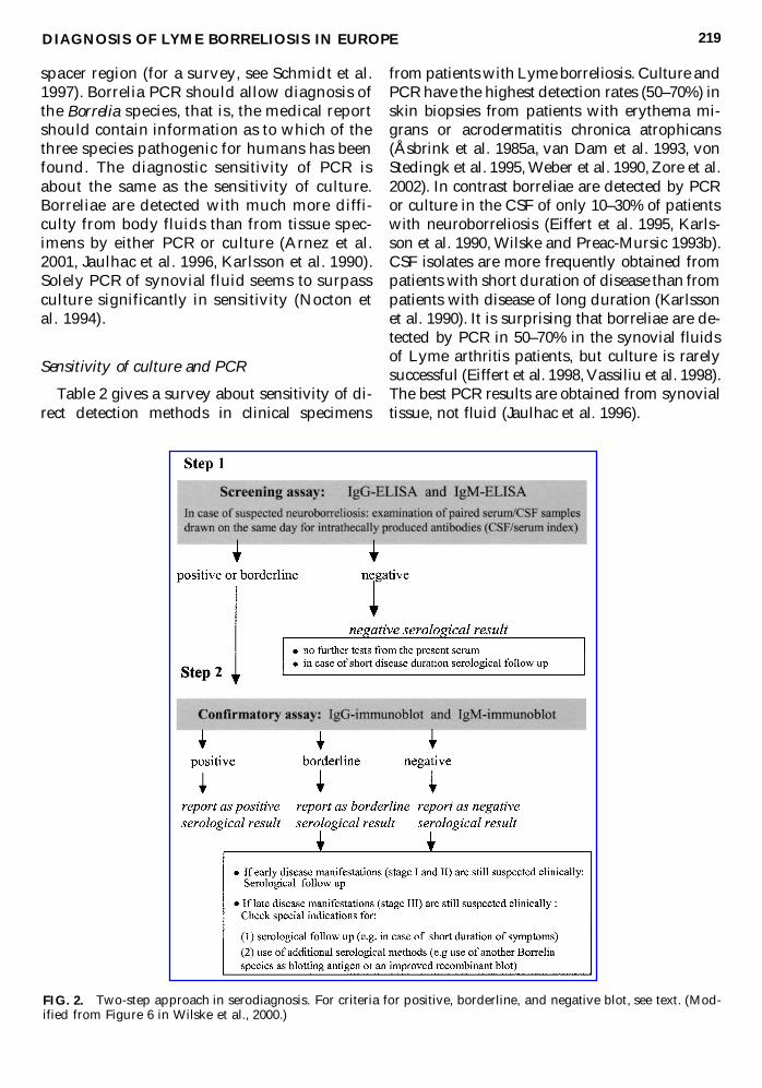
FIG. 2. Two-step approach in serodiagnosis. For criteria for positive, borderline, and negative blot, see text. (Mod-ified from Figure 6 in Wilske et al., 2000.)
ANTIBODY DETECTION
It is generally accepted that serological ex-amination should follow the principles of a twostep approach (Centers for Disease Control andPrevention 1995, Johnson et al. 1996, Wilske etal. 2000, Wilske and Schriefer 2002): (1) A sero-logical screening assay and (2) in the event ofa positive or equivocal result a confirmatory assay. A sensitive ELISA is recommended,which—in case it is reactive—should be con-firmed by the immunoblot (Fig. 2).
ELISA
The ELISA tests used for screening should beat least second generation tests (Wilske et al.2000), which have been improved with respectto cross reactivity with other bacteria (e.g., ex-tract antigen with previous Reiter treponemeadsorption) (Wilske et al. 1993a) or purified in-tact flagella as antigen (Hansen et al. 1988).Strains used as antigen source should expressOspC the immunodominant antigen of the IgMresponse and DbpA an immunodominant anti-gen of the IgG response (Wilske et al. 2000). Re-cently specific recombinant antigens (i.e., VlsE)or synthetic peptides (i.e., the C6 peptide de-rived from VlsE) have been successfully usedin the United States (Bacon et al. 2003, Lawrenzet al. 1999, Liang et al. 1999) and in a study withEuropean sera from patients with erythema mi-grans, acrodermatitis, and arthritis (C6 pep-tide) (Liang et al. 2000). Very recently also pa-tients with neuroborreliosis stage II have beeninvestigated with the C6 ELISA (IgG test) andcompared to the recombinant immunoblot(Fingerle et al. 2002). Of 36 sera 31 were posi-tive by immunoblot and 34 by the C6-ELISA.Two of the 31 immunoblot positive sera wereonly borderline in the C6-ELISA, these sera hadantibodies against recombinant DbpA and p58and DbpA and VlsE respectively. The C6-ELISA appears to be sufficiently sensitive as ascreening test for IgG antibodies in patientswith neuroborreliosis if also borderline resultsare included. However, VlsE has other im-munodominant epitopes besides the C6 regionthat could improve diagnostic sensitivity; het-erogeneity of those immunodominant epitopesespecially must be considered in Europe (Göt-
tner et al. 2002). The IgM and IgG immune re-sponses of Lyme borreliosis patients in recom-binant immunoblots should suggest the bestcombination of antigens for the developmentof recombinant ELISAs.
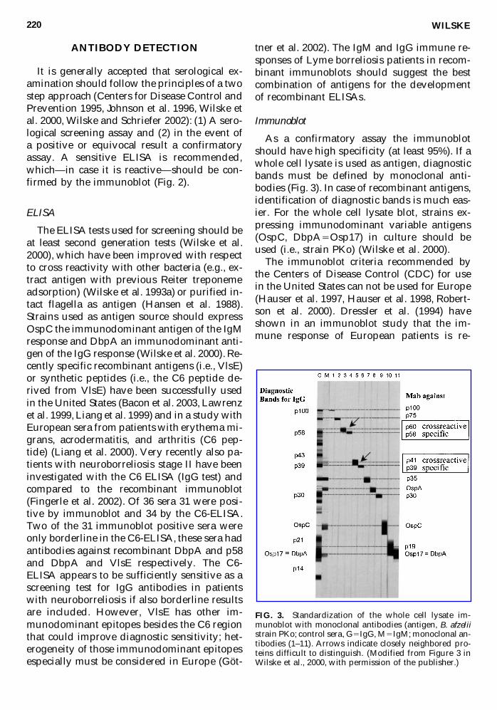
Immunoblot
As a confirmatory assay the immunoblotshould have high specificity (at least 95%). If awhole cell lysate is used as antigen, diagnosticbands must be defined by monoclonal anti-bodies (Fig. 3). In case of recombinant antigens,identification of diagnostic bands is much eas-ier. For the whole cell lysate blot, strains ex-pressing immunodominant variable antigens(OspC, DbpA5Osp17) in culture should beused (i.e., strain PKo) (Wilske et al. 2000).The immunoblot criteria recommended by
the Centers of Disease Control (CDC) for usein the United States can not be used for Europe(Hauser et al. 1997, Hauser et al. 1998, Robert-son et al. 2000). Dressler et al. (1994) haveshown in an immunoblot study that the im-mune response of European patients is re-
WILSKE220
FIG. 3. Standardization of the whole cell lysate im-munoblot with monoclonal antibodies (antigen, B. afzeliistrain PKo; control sera, G5IgG, M5IgM; monoclonal an-tibodies (1–11). Arrows indicate closely neighbored pro-teins difficult to distinguish. (Modified from Figure 3 inWilske et al., 2000, with permission of the publisher.)
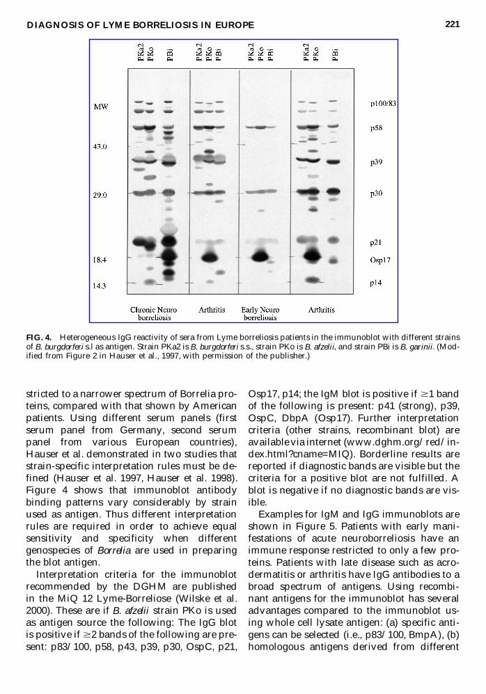
stricted to a narrower spectrum of Borrelia pro-teins, compared with that shown by Americanpatients. Using different serum panels (firstserum panel from Germany, second serumpanel from various European countries),Hauser et al. demonstrated in two studies thatstrain-specific interpretation rules must be de-fined (Hauser et al. 1997, Hauser et al. 1998).Figure 4 shows that immunoblot antibodybinding patterns vary considerably by strainused as antigen. Thus different interpretationrules are required in order to achieve equal sensitivity and specificity when differentgenospecies of Borrelia are used in preparingthe blot antigen.
Interpretation criteria for the immunoblotrecommended by the DGHM are published in the MiQ 12 Lyme-Borreliose (Wilske et al.2000). These are if B. afzelii strain PKo is usedas antigen source the following: The IgG blotis positive if $2 bands of the following are pre-sent: p83/100, p58, p43, p39, p30, OspC, p21,
Osp17, p14; the IgM blot is positive if $1 bandof the following is present: p41 (strong), p39,OspC, DbpA (Osp17). Further interpretationcriteria (other strains, recombinant blot) areavailable via internet (www.dghm.org/red/in-dex.html?cname=MIQ). Borderline results arereported if diagnostic bands are visible but thecriteria for a positive blot are not fulfilled. Ablot is negative if no diagnostic bands are vis-ible.
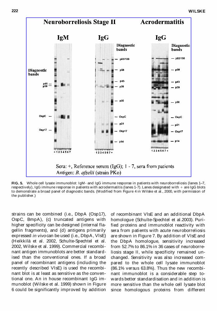
Examples for IgM and IgG immunoblots areshown in Figure 5. Patients with early mani-festations of acute neuroborreliosis have an immune response restricted to only a few pro-teins. Patients with late disease such as acro-dermatitis or arthritis have IgG antibodies to abroad spectrum of antigens. Using recombi-nant antigens for the immunoblot has severaladvantages compared to the immunoblot us-ing whole cell lysate antigen: (a) specific anti-gens can be selected (i.e., p83/100, BmpA), (b)homologous antigens derived from different
DIAGNOSIS OF LYME BORRELIOSIS IN EUROPE 221
FIG. 4. Heterogeneous IgG reactivity of sera from Lyme borreliosis patients in the immunoblot with different strainsof B. burgdorferi s.l as antigen. Strain PKa2 is B. burgdorferi s.s., strain PKo is B. afzelii, and strain PBi is B. garinii. (Mod-ified from Figure 2 in Hauser et al., 1997, with permission of the publisher.)
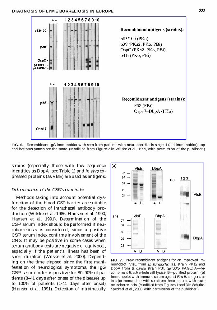
strains can be combined (i.e., DbpA (Osp17),OspC, BmpA), (c) truncated antigens withhigher specificity can be designed (internal fla-gellin fragments), and (d) antigens primarilyexpressed in vivo can be used (i.e., DbpA, VlsE)(Heikkilä et al. 2002, Schulte-Spechtel et al.2002, Wilske et al. 1999). Commercial recombi-nant antigen immunoblots are better standard-ised than the conventional ones. If a broadpanel of recombinant antigens (including therecently described VlsE) is used the recombi-nant blot is at least as sensitive as the conven-tional one. An in house recombinant IgG im-munoblot (Wilske et al. 1999) shown in Figure6 could be significantly improved by addition
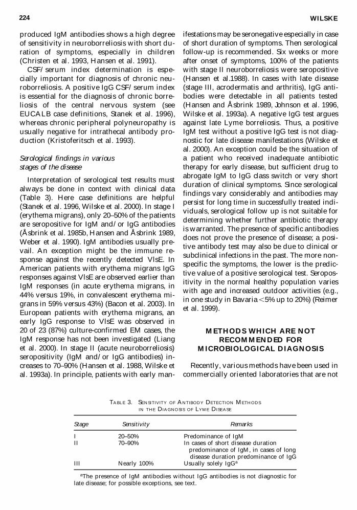
of recombinant VlsE and an additional DbpAhomologue (Schulte-Spechtel et al.2003). Puri-fied proteins and immunoblot reactivity withsera from patients with acute neuroborreliosisare shown in Figure 7. By addition of VlsE andthe DbpA homologue, sensitivity increasedfrom 52.7% to 86.1% in 36 cases of neuroborre-liosis stage II, while specificity remained un-changed. Sensitivity was also increased com-pared to the whole cell lysate immunoblot(86.1% versus 63.8%). Thus the new recombi-nant immunoblot is a considerable step to-wards better standardisation and in addition ismore sensitive than the whole cell lysate blotsince homologous proteins from different
WILSKE222
FIG. 5. Whole cell lysate immunoblot: IgM- and IgG immune response in patients with neuroborreliosis (lanes 1–7,respectively), IgG immune response in patients with acrodermatitis (lanes 1-7). Lanes designated with 1 are IgG blotsto demonstrate a broad panel of diagnostic bands. (Modified from Figure 4 in Wilske et al., 2000, with permission ofthe publisher.)
strains (especially those with low sequenceidentities as DbpA, see Table 1) and in vivo ex-pressed proteins (as VlsE) are used as antigens.
Determination of the CSF/serum index
Methods taking into account potential dys-function of the blood-CSF barrier are suitablefor the detection of intrathecal antibody pro-duction (Wilske et al. 1986, Hansen et al. 1990,Hansen et al. 1991). Determination of theCSF/serum index should be performed if neu-roborreliosis is considered, since a positiveCSF/serum index confirms involvement of theCNS. It may be positive in some cases whenserum antibody tests are negative or equivocal,especially if the patient’s illness has been ofshort duration (Wilske et al. 2000). Depend-ing on the time elapsed since the first mani-festation of neurological symptoms, the IgGCSF/serum index is positive for 80–90% of pa-tients (8–41 days after onset of the disease) upto 100% of patients (.41 days after onset)(Hansen et al. 1991). Detection of intrathecally
DIAGNOSIS OF LYME BORRELIOSIS IN EUROPE 223
FIG. 6. Recombinant IgG immunoblot with sera from patients with neuroborreliosis stage II (old immunoblot); topand bottoms panels are the same. (Modified from Figure 2 in Wilske et al., 1999, with permission of the publisher.)
FIG. 7. New recombinant antigens for an improved im-munoblot: VlsE from B. burgdorferi s.s. strain PKa2 andDbpA from B. garinii strain PBr. (a) SDS- PAGE: A—re-combinant E. coliwhole cell lysate; B—purified protein. (b)Immunoblot with immune serum against E. coli, antigens asin a. (c) Immunoblot with sera from three patients with acuteneuroborreliosis. (Modified from Figures 1 and 3 in Schulte-Spechtel et al., 2003, with permission of the publisher.)
produced IgM antibodies shows a high degreeof sensitivity in neuroborreliosis with short du-ration of symptoms, especially in children(Christen et al. 1993, Hansen et al. 1991).
CSF/serum index determination is espe-cially important for diagnosis of chronic neu-roborreliosis. A positive IgG CSF/serum indexis essential for the diagnosis of chronic borre-liosis of the central nervous system (see EUCALB case definitions, Stanek et al. 1996),whereas chronic peripheral polyneuropathy isusually negative for intrathecal antibody pro-duction (Kristoferitsch et al. 1993).
Serological findings in various stages of the disease
Interpretation of serological test results mustalways be done in context with clinical data(Table 3). Here case definitions are helpful(Stanek et al. 1996, Wilske et al. 2000). In stage I(erythema migrans), only 20–50% of the patientsare seropositive for IgM and/or IgG antibodies(Åsbrink et al. 1985b, Hansen and Åsbrink 1989,Weber et al. 1990). IgM antibodies usually pre-vail. An exception might be the immune re-sponse against the recently detected VlsE. InAmerican patients with erythema migrans IgGresponses against VlsE are observed earlier thanIgM responses (in acute erythema migrans, in44% versus 19%, in convalescent erythema mi-grans in 59% versus 43%) (Bacon et al. 2003). InEuropean patients with erythema migrans, anearly IgG response to VlsE was observed in 20 of 23 (87%) culture-confirmed EM cases, the IgM response has not been investigated (Lianget al. 2000). In stage II (acute neuroborreliosis)seropositivity (IgM and/or IgG antibodies) in-creases to 70–90% (Hansen et al. 1988, Wilske etal. 1993a). In principle, patients with early man-
ifestations may be seronegative especially in caseof short duration of symptoms. Then serologicalfollow-up is recommended. Six weeks or moreafter onset of symptoms, 100% of the patientswith stage II neuroborreliosis were seropositive(Hansen et al.1988). In cases with late disease(stage III, acrodermatis and arthritis), IgG anti-bodies were detectable in all patients tested(Hansen and Åsbrink 1989, Johnson et al. 1996,Wilske et al. 1993a). A negative IgG test arguesagainst late Lyme borreliosis. Thus, a positiveIgM test without a positive IgG test is not diag-nostic for late disease manifestations (Wilske etal. 2000). An exception could be the situation ofa patient who received inadequate antibiotictherapy for early disease, but sufficient drug toabrogate IgM to IgG class switch or very shortduration of clinical symptoms. Since serologicalfindings vary considerably and antibodies maypersist for long time in successfully treated indi-viduals, serological follow up is not suitable fordetermining whether further antibiotic therapyis warranted. The presence of specific antibodiesdoes not prove the presence of disease; a posi-tive antibody test may also be due to clinical orsubclinical infections in the past. The more non-specific the symptoms, the lower is the predic-tive value of a positive serological test. Seropos-itivity in the normal healthy population varieswith age and increased outdoor activities (e.g.,in one study in Bavaria ,5% up to 20%) (Reimeret al. 1999).
METHODS WHICH ARE NOTRECOMMENDED FOR
MICROBIOLOGICAL DIAGNOSIS
Recently, various methods have been used incommercially oriented laboratories that are not
WILSKE224
TABLE 3. SENSITIVITY OF ANTIBODY DETECTION METHODS
IN THE DIAGNOSIS OF LYME DISEASE
Stage Sensitivity Remarks
I 20–50% Predominance of IgMII 70–90% In cases of short disease duration
predominance of IgM, in cases of longdisease duration predominance of IgG
III Nearly 100% Usually solely IgGa
aThe presence of IgM antibodies without IgG antibodies is not diagnostic forlate disease; for possible exceptions, see text.
sufficiently evaluated for diagnostic purposes.Among them are the antigen tests in body flu-ids, PCR of urine, and lymphocyte transfor-mation tests. These tests are not recommendedfor microbiological diagnosis. They are unreli-able and some of them are in addition very ex-pensive, especially if used for therapy control(Brettschneider et al. 1998, Kalish et al. 2003,Klempner et al. 2001).
REFERENCES
Arnez, M, Ruzic-Sabljic, E, Ahcan, J, et al. Isolation of Bor-relia burgdorferi sensu lato from blood of children withsolitary erythema migrans. Pediatr Infect Dis J 2001;3:251–255.
Åsbrink, E, Hovmark, A. Successful cultivation of spiro-chetes from skin lesions of patients with erythemachronicum migrans Afzelius and acrodermatitis chron-ica atrophicans. Acta Pathol Microbiol Immunol ScandB 1985; 93:161–163.
Åsbrink, E, Hovmark, A, Hederstedt, B. Serologic studiesof erythema chronicum migrans Afzelius and acroder-matitis chronica atrophicans with indirect immunoflu-orescence and enzyme-linked immunosorbent assays.Acta Derm Venereol 1985; 65:509–514.
Bacon, RM, Biggerstaff, BJ, Schriefer, M, et al. Improvedserodiagnosis of Lyme disease by kinetic ELISAs usingrecombinant VlsE1 or peptide antigens of Borreliaburgdorferi compared with two-tiered testing. J InfectDis 2003; 187:1187–1199.
Brettschneider, S, Bruckbauer, H, Klugbauer, N, et al. Di-agnostic value of PCR for detection of Borrelia burgdor-feri in skin biopsy and urine samples from patients withskin borreliosis. J Clin Microbiol 1998; 36:2658–2665.
Canica, MM, Nato, F, Du Merle, L, et al. Monoclonal an-tibodies for identification of Borrelia afzelii sp. nov. as-sociated with late cutaneous manifestations of Lymeborreliosis. Scand J Infect Dis 1993; 25:441–448.
Centers for Disease Control and Prevention. Recommen-dations for test performance and interpretation from thesecond National Conference on Serologic Diagnosis ofLyme Disease. Morbid Mortal Wkly Rprt 1995; 44:590.
Christen, H-J, Hanefeld, F, Eiffert, H, et al. Epidemiologyand clinical manifestations of Lyme borreliosis in child-hood. A prospective multicentre study with special re-gard to neuroborreliosis. Acta Paediatr 1993; 386:1–76.
Demaerschalck, I, Messaoud, AB, De Kesel, M, et al. Si-multaneous presence of different Borrelia burgdorferigenospecies in biological fluids of Lyme disease pa-tients. J Clin Microbiol 1995; 33:602–608.
Dressler, F, Ackermann, R, Steere, AC. Antibody re-sponses to the three genomic groups of Borrelia burgdor-feri in European Lyme borreliosis. J Infect Dis 1994;169:313–318.
Eiffert, H, Karsten, A, Thomssen, R, et al. Characteriza-tion of Borrelia burgdorferi strains in Lyme arthritis.Scand J Infect Dis 1998; 30:265–268.
Eiffert, H, Ohlenbusch, A, Christen, H-J, et al. Nondiffer-entiation between Lyme disease spirochetes from vec-tor ticks and human cerebrospinal fluids. J Infect Dis1995; 171:476–479.
Gern, L, Hu, CM, Kocianova, E, et al. Genetic diversity ofBorrelia burgdorferi sensu lato isolates obtained fromIxodes ricinus ticks collected in Slovakia. Eur J Epidemiol1999; 15:665–669.
Göttner, G, Schulte-Spechtel, U, Wilske, B. Antigenic andgenetic heterogeneity of the immunodominant surfaceprotein VlsE among European B. burgdorferi s.l. strains.Int J Med Microbiol 2002; 292:105.
Goossens, HAT, van den Bogaard, AE, Nohlmans, MKE.Evaluation of fifteen commercially available serologi-cal tests for diagnosis of Lyme borreliosis. Eur J ClinMicrobiol. Infect Dis 1999; 18:551–560
Fingerle, V, Schulte-Spechtel, U, Wilske, B. Evaluation ofan ELISA based on the C6-peptide of VlsE for diagno-sis of early neuroborreliosis. Int J Med Microbiol 2002;292 (Suppl. 34): 217.
Hansen, K, Åsbrink, E. Serodiagnosis of erythema mi-grans and acrodermatitis chronica atrophicans by theBorrelia burgdorferi flagellum enzyme-linked im-munosorbent assay. J Clin Microbiol 1989; 27:545–551.
Hansen, K, Cruz, M, Link, H. Oligoclonal Borrelia burgdor-feri-specific IgG antibodies in cerebrospinal fluid inLyme neuroborreliosis. J Infect Dis 1990; 161:1194–1202.
Hansen, K, Hindersson, P, Pedersen, NS. Measurement ofantibodies to the Borrelia burgdorferi flagellum improvesserodiagnosis in Lyme disease. J Clin Microbiol 1988;26:338–346.
Hansen, K, Lebech, A-M. Lyme neuroborreliosis: a newsensitive diagnostic assay for intrathecal sythesis of Bor-relia burgdorferi–specific immunoglobulin G, A, and M.Ann Neurol 1991; 30:197–205.
Hauser, U, Lehnert, G, Lobentanzer, R, et al. Interpreta-tion criteria for standardized western blots for three eu-ropean species of Borrelia burgdorferi sensu lato. J ClinMicrobiol 1997; 35:1433–1444.
Hauser, U, Lehnert, G, Wilske, B. Diagnostic value of pro-teins of three Borrelia species (Borrelia burgdorferi sensulato) and implications for development and use of re-combinant antigens for serodiagnosis of Lyme borrelio-sis in Europe. Clin Diagn Lab Immunol 1998; 5:456–462.
Heikkilä, T, Seppälä, I, Saxen, H, et al. Species-specificserodiagnosis of Lyme arthritis and neuroborreliosisdue to Borrelia burgdorferi sensu stricto, B. afzelii, and B.garinii by using decorin binding protein A. J Clin Mi-crobiol 2002; 40:453–460.
Hubalek, Z, Halouzka, J. Distribution of Borrelia burgdor-feri sensu lato genomic groups in Europe, a review. EurJ Epidemiol 1997; 13: 951–957.
Johnson, BJB, Robbins, KE, Balley, RE, et al. Serodiagno-sis of Lyme disease: accuracy of a two-step approachusing a flagella-based ELISA and immunoblotting. J In-fect Dis 1996; 174:346–353.
Jaulhac, B, Chary-Valckenaere, I, Sibilia, J, et al. Detectionof Borrelia burgdorferi by DNA amplification in synovialtissue samples from patients with Lyme arthritis.Arthritis Rheum 1996; 39:736–745.
Jaulhac, B, Heller, R, Limbach, FX, et al. Direct molecular
DIAGNOSIS OF LYME BORRELIOSIS IN EUROPE 225
typing of Borrelia burgdorferi sensu lato species in syn-ovial samples from patients with Lyme arthritis. J ClinMicrobiol 2000; 38:1895–1900.
Kaiser, R. Teilnehmer der Expertenkonferenz: Frühsom-mermeningoenzephalitis und Lyme-Borreliose-Práven-tion vor und nach Zeckenstich. Dtsch med Wochenschr1998; 123:847–853.
Karlsson, M, Hovind-Hougen, K, Svenungsson, B, et al.Cultivation and characterization of spirochetes fromcerebrospinal fluid of patients with Lyme borreliosis JClin Microbiol 1990; 28:473–479.
Kalish, RS, Wood, J A, Golde, W, et al. Human T lym-phocyte response to Borrelia burgdorferi infection: nocorrelation between human leukocyte function antigentype 1 peptide response and clinical status. J Infect Dis2003; 187:102–108.
Klempner, MS, Schmid, CH, Hu, L, et al. Intralaboratoryreliability of serologic and urine testing for Lyme dis-ease. Am J Med 2001; 110:217–219.
Kristoferitsch, W. Chronical peripheral neuropathy. In:Weber, K, Burgdorfer, W, eds. Aspects of Lyme Borrelio-sis. Berlin: Springer, 1993:219–227.
Lawrenz, MB, Hardham, JM, Owens, RT, et al. Humanantibody responses to VlsE antigenic variation proteinof Borrelia burgdorferi. J Clin Microbiol 1999; 37:3997–4004.
Liang, FT, Alvarez, AL, Gu, Y, et al. An immunodomi-nant conserved region within the variable domain ofVlsE, the variable surface antigen of Borrelia burgdorferi.J Immunol 1999; 163:5566–5573.
Liang, FT, Aberer, E, Cinco, M, et al. Antigenic conserva-tion of an immunodominant invariable region of theVlsE lipoprotein among european pathogenic geno-species of Borrelia burgdorferi sl. J Infect Dis 2000;182:1455–1462.
Lünemann, JD, Zarmas, S, Priem, S, et al. Rapid typing ofBorrelia burgdorferi sensu lato species in specimens frompatients with different manifestations of Lyme borre-liosis. J Clin Microbiol 2001; 39:1130–1133.
Michel, H, Wilske, B, Hettche, G, et al. An ospA-poly-merase chain reaction/restriction fragment lengthpolymorphism-based method for sensitive detectionand reliable differentiation of all European Borreliaburgdorferi sensu lato species and OspA types. Med Mi-crobiol Immunol 2003 (in press).
Nocton, JJ, Dressler, F, Rutledge, BJ, et al. Detection ofBorrelia burgdorferi DNA by polymerase chain reac-tion in synovial fluid from patients with Lyme arthri-tis. N Engl J Med 1994; 330:229–234
Ohlenbusch, A, Matuschka, F-R, Richter, D, et al. Etiol-ogy of the acrodermatitis chronica atrophicans lesionin Lyme disease. J Infect Dis 1996; 174:421–423.
Péter, O, Bretz, AG, Bee, D. Occurence of differentgenospecies of Borrelia burgdorferi sensu lato in ixo-did ticks of Valais, Switzerland. Eur J Epidemiol 1995;11:463–467.
Pfister, H-W, Wilske, B, Weber, K. Lyme borreliosis: basicscience and clinical aspects. Lancet 1994; 343:1013–1016.
Preac-Mursic, V, Wilske, B, Reinhardt, S. Culture of Bor-relia burgdorferi on six solid media. Eur J Microbiol In-fect Dis 1991; 10:1076–1079.
Rauter, C, Oehme, R, Diterich, I, et al. Distribution of clin-ically relevant Borrelia genospecies in ticks assessed bya novel, single-run, real-time PCR. J Clin Microbiol2002; 40:36–43.
Reimer, B, Marschang, A, Fingerle, V, et al. Epedimiologyof Lyme borreliosis in South-Eastern Bavaria (Ger-many). Zent Bl Bakteriol 1999; 289:653–654.
Rijpkema SGT, Tazelaar, DJ, Molkenboer, MJCH, et al.Detection of Borrelia afzelii, Borrelia burgdorferi sensustricto, Borrelia garinii and group VS116 by PCR in skinbiopsies of patients with erythema migrans and acro-dermatitis chronica atrophicans. Clin Microbiol Infect1997; 3:109–116.
Rijpkema, S, Golubic, D, Molkenboer, M, et al. Idenfica-tion of four genomic groups of Borrelia burgdorferi sensulato in Ixodes ricinus ticks collected in a Lyme borrelio-sis endemic region of northern Croatia. Exp Appl Ac-arol 1996; 20:23–30.
Robertson, J, Guy, E, Andrews, N, et al. European multi-center study of immunoblotting in serodiagnosis ofLyme borreliosis. J Clin Microbiol 2000; 38:2097–2102.
Schmidt, BL. PCR in laboratory diagnosis of human Bor-relia burgdorferi infections. Clin Microbiol Rev 1997;10:185–201.
Schulte-Spechtel, U. Lehnert, G, Liegl, G, et al. Significantimprovement of the recombinant Borrelia IgG im-munoblot by addition of VlsE and a DbpA homologuederived from B. garinii for the diagnosis of early neu-roborreliosis. J Clin Microbiol 2003; 41:1299–1303.
Stanek, G, O’Connell, S, Cimmino, M, et al. EuropeanUnion concerted action on risk assessment in Lyme bor-reliosis: clinical case definitions for Lyme borreliosis.Wien Klin Wochenschr 1996; 108/23:741–747.
Steere, AC. Medical progress—Lyme disease. N Engl JMed 1989; 321:586–596.
Strle, F, Nadelman, RB, Cimperman, J, et al. Comparisonof culture-confirmed erythema migrans caused by Bor-relia burgdorferi sensu stricto in New York State and by Borrelia afzelii in Slovenia. Ann Intern Med 1999;130:32–36.
Van Dam, AP, Kuiper, H, Vos, K, et al. Differentgenospecies of Borrelia burgdorferi are associated withdistinct clinical manifestations of Lyme borreliosis. ClinInfect Dis 1993; 17:708–717.
Vasiliu, V, Herzer, P, Rössler, D, et al. Heterogeneity ofBorrelia burgdorferi sensu lato demonstrated by an ospA-type-specific PCR in synovial fluid from patients withLyme arthritis. Med Microbiol Immunol 1998;187:97–102.
Von Stedingk, LV, Olsson, I, Hanson, HS, et al. Poly-merase chain reaction for detection of Borrelia burgdor-feri DNA in skin lesions of early and late Lyme borre-liosis. Eur J Clin Microbiol Infect Dis 1995; 14:1–5.
Wang, G, van Dam, AP, Dankert, J. Phenotypic and ge-netic characterization of a novel Borrelia burgdorferisensu lato isolate from a patient with Lyme borreliosis.J Clin Microbiol 1999; 37:3025–3028.
Wang, G, van Dam, AP, Schwartz, I, et al. Molecular typ-ing of Borrelia burgdorferi sensu lato taxonomic, epi-
WILSKE226
demiological, and clinical implications. Clin MicrobiolRev 1999; 12:633–653.
Weber, K, Preac-Mursic, V, Wilske, B, et al. A random-ized trial of ceftriaxone versus oral penicillin for treat-ment of early Lyme borreliosis. Infection 1990; 18:91–96
Wilske, B. Microbiological diagnosis in Lyme borreliosis.Int J Med Microbiol 2002; 291:33:114–119.
Wilske, B, Busch, U, Eiffert, H, et al. Diversity of OspAand OspC among cerebrospinal fluid isolates of Borre-lia burgdorferi sensu lato from patients with neurobor-reliosis in Germany. Med Microbiol Immunol 1996;184:195–201.
Wilske, B, Fingerle, V, Herzer, P, et al. Recombinant im-munoblot in the serodiagnosis of Lyme borreliosis,comparison with indirect immunofluoerescence andenzyme-linked immnosorbent assay. Med MicrobiolImmunol 1993; 182:255–270.
Wilske, B, Habermann, C, Fingerle, V, et al. An improvedrecombinant IgG immunoblot for serodiagnosis ofLyme borreliosis. Med Microbiol Immunol 1999;188:139–144.
Wilske, B, Preac-Mursic, V. Microbiological diagnosis ofLyme borreliosis. In: Weber, K, Burgdorfer, W, eds. As-pects of Lyme Borreliosis. Berlin: Springer; 1993:267–300.
Wilske, B, Preac-Mursic, V, Göbel, UB, et al. An OspAserotyping system for Borrelia burgdorferi based on re-activity with monoclonal antibodies and OspA se-quence analysis. J Clin Microbiol 1993; 31:340–350.
Wilske, B, Schierz, G, Preac-Mursic, V, et al. Intrathecalproduction of specific antibodies against Borreliaburgdorferi in patients with lymphocytic meningoradi-culitis (Bannwarth’s syndrome). J Infect Dis 1986;153:304–314.
Wilske, B, Schriefer, M. Borrelia. Murray, PR, Baron, EJ,Jorgensen, JH, eds. In: Manual of Clinical Microbiology,8th ed. Washington, DC: ASM Press; 2003:937–954.
Wilske, B, Zöller, L, Brade, V, et al. MIQ 12 Lyme-borre-liose. In: Qualitätsstandards in der mikrobiologisch infekti-ologischen Diagnostik. Mauch, H, Lütticken, R, eds. Mu-nich: Urban & Fischer Verlag, 2000.
Zore, A, Ruzic-Sabljic, E, Maraspin, V, et al. Sensitiviy ofculture and polymerase chain reaction for the etiologicdiagnosis of erythema migrans. Wien Klin Wochenschr2002; 114:606–609.
Address reprint requests to:Bettina Wilske, M.D., Ph.D.Max von Pettenkofer Institute
University of MunichNational Reference Center for Borreliae
Pettenkofer-Strasse 9aD 80336 Munich, Germany
E-mail: [email protected]
227DIAGNOSIS OF LYME BORRELIOSIS IN EUROPE
This article has been cited by:
1. Albina Poljak, Pär Comstedt, Markus Hanner, Wolfgang Schüler, Andreas Meinke, Benjamin Wizel,Urban Lundberg. 2012. Identification and characterization of Borrelia antigens as potential vaccinecandidates against Lyme borreliosis. Vaccine 30:29, 4398-4406. [CrossRef]
2. Pär Comstedt, Tobias Jakobsson, Sven Bergström. 2011. Global ecology and epidemiology of Borreliagarinii spirochetes. Infection Ecology & Epidemiology 1:0. . [CrossRef]
3. Magdalena Ligor, Paweł Olszowy, Bogusław Buszewski. 2011. Application of medical and analyticalmethods in Lyme borreliosis monitoring. Analytical and Bioanalytical Chemistry . [CrossRef]
4. Gary P. Wormser, Guiqing WangThe Role of Culture and Nucleic Acid Amplification in Diagnosis ofLyme Borreliosis 159-183. [CrossRef]
5. Viera Svihrova, Henrieta Hudeckova, Milos Jesenak, Katarina Schwarzova, Zina Kostanova, Ivan Ciznar.2011. Lyme borreliosis—analysis of the trends in Slovakia, 1999–2008. Folia Microbiologica 56:3, 270-275.[CrossRef]
6. Iwona Wojciechowska-Koszko, Iwona Mączyńska, Zbigniew Szych, Stefania Giedrys-Kalemba. 2011.Serodiagnosis of Borreliosis: Indirect Immunofluorescence Assay, Enzyme-Linked Immunosorbent Assayand Immunoblotting. Archivum Immunologiae et Therapiae Experimentalis 59:1, 69-77. [CrossRef]
7. José J. Pereyra-Rodríguez, José Bernabeu-Wittel, Elías Cañas, Julián Conejo-Mir. 2011. Máculaeritematosa lentamente progresiva. Enfermedades Infecciosas y Microbiología Clínica 29:1, 68-69. [CrossRef]
8. G. Stanek, V. Fingerle, K.-P. Hunfeld, B. Jaulhac, R. Kaiser, A. Krause, W. Kristoferitsch, S. O’Connell, K.Ornstein, F. Strle, J. Gray. 2011. Lyme borreliosis: Clinical case definitions for diagnosis and managementin Europe. Clinical Microbiology and Infection 17:1, 69-79. [CrossRef]
9. Iván Bárcena-Uribarri, Marcus Thein, Anna Sacher, Ignas Bunikis, Mari Bonde, Sven Bergström, RolandBenz. 2010. P66 porins are present in both Lyme disease and relapsing fever spirochetes: A comparisonof the biophysical properties of P66 porins from six Borrelia species. Biochimica et Biophysica Acta (BBA)- Biomembranes 1798:6, 1197-1203. [CrossRef]
10. Monica L. Vieira, Zenaide M. de Morais, Amane P. Gonçales, Eliete C. Romero, Silvio A. Vasconcellos,Ana L.T.O. Nascimento. 2010. Lsa63, a newly identified surface protein of Leptospira interrogans bindslaminin and collagen IV. Journal of Infection 60:1, 52-64. [CrossRef]
11. D. Hulínská, J. Votýpka, D. Vaňousová, J. Hercogová, V. Hulínský, H. Dřevová, Z. Kurzová, L. Uherková.2009. Identification of Anaplasma phagocytophilum and Borrelia burgdorferi sensu lato in patients witherythema migrans. Folia Microbiologica 54:3, 246-256. [CrossRef]
12. A. Krause, V. Fingerle. 2009. Lyme-Borreliose. Zeitschrift für Rheumatologie 68:3, 239-254. [CrossRef]13. Barbro Hedin Skogman, Stefan Croner, Maria Nordwall, Mattias Eknefelt, Jan Ernerudh, Pia Forsberg.
2008. Lyme Neuroborreliosis in Children. The Pediatric Infectious Disease Journal 27:12, 1089-1094.[CrossRef]
14. Gaia Codolo, Amedeo Amedei, Allen C. Steere, Elena Papinutto, Andrea Cappon, Alessandra Polenghi,Marisa Benagiano, Silvia Rossi Paccani, Vittorio Sambri, Gianfranco Del Prete, Cosima Tatiana Baldari,Giuseppe Zanotti, Cesare Montecucco, Mario Milco D'Elios, Marina de Bernard. 2008. Borrelia burgdorferiNapA-driven Th17 cell inflammation in lyme arthritis. Arthritis & Rheumatism 58:11, 3609-3617.[CrossRef]
15. N FOMENKO, N LIVANOVA, N CHERNOUSOVA. 2008. Diversity of Borrelia burgdorferi sensu lato innatural foci of Novosibirsk region. International Journal of Medical Microbiology 298, 139-148. [CrossRef]
16. J ZAJKOWSKA, M KONDRUSIK, S GRYGORCZUK, S PANCEWICZ, A IZYCKA. 2008. Theusefulness of ‘in vivo’ antigens in the diagnosis of human Lyme borreliosis. International Journal of MedicalMicrobiology 298, 361-364. [CrossRef]
17. Barbro H. Skogman, Stefan Croner, Pia Forsberg, Jan Ernerudh, Pekka Lahdenne, Heidi Sillanpää, IlkkaSeppälä. 2008. Improved Laboratory Diagnostics of Lyme Neuroborreliosis in Children by Detection of
Antibodies to New Antigens in Cerebrospinal Fluid. The Pediatric Infectious Disease Journal 27:7, 605-612.[CrossRef]
18. T. Cerar, E. Ružić-Sabljić, U. Glinšek, A. Zore, F. Strle. 2008. Comparison of PCR methods and culturefor the detection of Borrelia spp. in patients with erythema migrans. Clinical Microbiology and Infection14:7, 653-658. [CrossRef]
19. Elizabeth Valentine-Thon, Karsten Ilsemann, Martin Sandkamp. 2008. Neuartiger Lymphozyten-Transformations-Test (LTT-MELISA ® ) zum Nachweis einer Lyme-Borreliose / A novel lymphocytetransformation test (LTT-MELISA ® ) for Lyme borreliosis. LaboratoriumsMedizin 32:1, 26-34.[CrossRef]
20. Vincent Staszewski, Karen D. McCoy, Torkild Tveraa, Thierry Boulinier. 2007. INTERANNUALDYNAMICS OF ANTIBODY LEVELS IN NATURALLY INFECTED LONG-LIVED COLONIALBIRDS. Ecology 88:12, 3183-3191. [CrossRef]
21. T. Skarpaas, U. Ljøstad, M. Søbye, Å. Mygland. 2007. Sensitivity and specificity of a commercial C6peptide enzyme immuno assay in diagnosis of acute Lyme neuroborreliosis. European Journal of ClinicalMicrobiology & Infectious Diseases 26:9, 675-677. [CrossRef]
22. U. Ljøstad, T. Skarpaas, Å. Mygland. 2007. Clinical usefulness of intrathecal antibody testing in acuteLyme neuroborreliosis. European Journal of Neurology 14:8, 873-876. [CrossRef]
23. Gary P Wormser, Raymond J Dattwyler, Eugene D Shapiro, J Stephen Dumler, Susan O'Connell, JustinD Radolf, Robert B Nadelman. 2007. Single-dose prophylaxis against Lyme disease. The Lancet InfectiousDiseases 7:6, 371-373. [CrossRef]
24. Andrew R Pachner, Israel Steiner. 2007. Lyme neuroborreliosis: infection, immunity, and inflammation.The Lancet Neurology 6:6, 544-552. [CrossRef]
25. Elisabeth Aberer. 2007. Lyme borreliosis ? an update. JDDG 5:5, 406-414. [CrossRef]26. KAI E. KISAND, TIINA PRÜKK, KALLE V. KISAND, SIIRI-MERIKE LÜÜS, IRJA KALBE, RAIVO
UIBO. 2007. Propensity to excessive proinflammatory response in chronic Lyme borreliosis. APMIS 115:2,134-141. [CrossRef]
27. Elizabeth Valentine-Thon, Karsten Ilsemann, Martin Sandkamp. 2007. A novel lymphocyte transformationtest (LTT-MELISA®) for Lyme borreliosis. Diagnostic Microbiology and Infectious Disease 57:1, 27-34.[CrossRef]
28. Stephen J. Traub, Gregory A. CumminsTick-Borne Diseases 982-1008. [CrossRef]29. Tjaša Cerar, Eva Ružic-Sabljic, Jože Cimperman, Franc Strle. 2006. Comparison of immunofluorescence
assay (IFA) and LIAISON® in patients with different clinical manifestations of Lyme borreliosis. Wienerklinische Wochenschrift 118:21-22, 686-690. [CrossRef]
30. A. Smismans, V. J. Goossens, E. Nulens, C. A. Bruggeman. 2006. Comparison of five differentimmunoassays for the detection of Borrelia burgdorferi IgM and IgG antibodies. Clinical Microbiology andInfection 12:7, 648-655. [CrossRef]
31. H PFISTER, T RUPPRECHT. 2006. Clinical aspects of neuroborreliosis and post-Lyme diseasesyndrome in adult patients. International Journal of Medical Microbiology 296, 11-16. [CrossRef]
32. D LENCAKOVA, C HIZOTEUFEL, B PETKO, U SCHULTESPECHTEL, M STANKO, B WILSKE,V FINGERLE. 2006. Prevalence of Borrelia burgdorferi s.l. OspA types in Ixodes ricinus ticks from selectedlocalities in Slovakia and Poland. International Journal of Medical Microbiology 296, 108-118. [CrossRef]
33. S Miertusova Tothova, S Bonin, G Trevisan, G Stanta. 2006. Mycosis fungoides: is it a Borrelia burgdorferi-associated disease?. British Journal of Cancer 94:6, 879-883. [CrossRef]
34. G. Piccolin, G. Benedetti, C. Doglioni, C. Lorenzato, S. Mancuso, N. Papa, L. Pitton, M.C. Ramon, C.Zasio, Dr. , G. Bertiato. 2006. A Study of the Presence of B. burgdorferi, Anaplasma (Previously Ehrlichia)phagocytophilum, Rickettsia, and Babesia in Ixodes ricinus Collected within the Territory of Belluno, Italy.Vector-Borne and Zoonotic Diseases 6:1, 24-31. [Abstract] [Full Text PDF] [Full Text PDF with Links]

35. Beatriz Fernández-Jorge, Manuel Almagro-Sánchez, Raquel Escudero-Nieto, Eduardo Fonseca-Capdevila.2006. Eritema migratorio por Borrelia afzelii. Medicina Clínica 126:6, 237-238. [CrossRef]
36. A. Krause, P. Herzer. 2005. Frühdiagnostik der Lyme-Arthritis. Zeitschrift für Rheumatologie 64:8, 531-537.[CrossRef]
37. R ESCUDERONIETO. 2005. Enfermedades producidas por Borrelia. Enfermedades Infecciosas yMicrobiología Clínica 23:4, 232-240. [CrossRef]
38. Bettina Wilske. 2005. Epidemiology and diagnosis of Lyme borreliosis. Annals of Medicine 37:8, 568.[CrossRef]
39. References 251-324. [CrossRef]