focal adhesion formation by f9 embryonal carcinoma cells...
TRANSCRIPT

2253Journal of Cell Science 108, 2253-2260 (1995)Printed in Great Britain © The Company of Biologists Limited 1995
Focal adhesion formation by F9 embryonal carcinoma cells after vinculin
gene disruption
Tova Volberg1, Benjamin Geiger1, Zvi Kam1, Roumen Pankov2,*, Inbal Simcha2, Helena Sabanay1, Jean-Luc Coll3,†, Eileen Adamson3 and Avri Ben-Ze’ev2,‡
Departments of 1Chemical Immunology and 2Molecular Genetics and Virology, The Weizmann Institute of Science, Rehovot,76100, Israel3La Jolla Cancer Research Foundation, 10901, N. Torrey Pines Rd, La Jolla, CA 92037, USA
*Present address: University of Sofia, Department of Cytology, Faculty of Biology, 8 Dragan Tzankov str. 1421, Sofia, Bulgaria†Present address: Centre Regional Leon Berard, Lab. Biol. Cell., 28 rue Laennec, 69373 Lyon Cedex 08, France‡Author for correspondence
The assembly of focal adhesions was investigated in F9embryonal carcinoma cells in which the expression ofvinculin was eliminated by a targeted disruption of thevinculin gene. Vinculin-deficient F9 cells were capable ofadhering to fibronectin-coated surfaces, though theydisplayed a reduced spreading compared to the parentalcells. Transmission electron microscopy as well as inter-ference reflection microscopy of live cells showed thatvinculin-null F9 cells formed focal adhesions that wereindistinguishable from those of the control cells. Fluores-cent labeling for actin, talin, α-actinin, paxillin and phos-
photyrosinated components indicated that the organizationof all these focal contact-associated components was essen-tially identical in the vinculin-containing and vinculin-nullcells. However, quantitative, digitized microscopyindicated that the intensity of fluorescence labeling in focaladhesions for α-actinin, talin and paxillin was significantlyhigher in cells lacking vinculin. The results suggest thatthere are multiple molecular mechanisms for the formationof focal adhesions in the absence of vinculin.
Key words: adhesion plaques, vinculin knock out, focal contacts
SUMMARY
INTRODUCTION
Adhesion of cultured cells to the extracellular matrix (ECM)is mediated by specialized regions of the plasma membraneknown as focal contacts, or focal adhesion plaques (Burridgeet al., 1988; Geiger and Ginsberg, 1991). At these sites, actinfilaments are bound to transmembrane receptors of the integrinfamily (Hynes, 1987), through a complex of structural ‘plaque’proteins including vinculin (Geiger, 1979), talin (Burridge andConnell, 1983) and α-actinin (Lazarides and Burridge, 1975).In addition to these and other structural components (Luna andHitt, 1992), molecules involved in signal transduction, mainlytyrosine kinases (Volberg et al., 1992), are also localized atfocal adhesion sites, suggesting a role in the transduction ofcontact-triggered signals (Burridge et al., 1992; Ben-Ze’ev,1992; Juliano and Haskill, 1993). Such regulatory moleculesinclude the pp60src tyrosine kinase (Rohrshneider, 1980), thefocal adhesion kinase p125FAK (Schaller et al., 1992), the LIMdomain-containing molecule zyxin (Sadler et al., 1992), theSH2 (Src homology 2)-containing actin binding moleculetensin (Lo et al., 1994), and a major phosphotyrosinatedmolecule paxillin (Turner et al., 1990).
The mechanisms underlying the association of actinfilaments with integrin receptors in these plaques is largelyunknown. Several in vitro binding studies suggested that F-actin
may associate with integrin via talin, vinculin and α-actinin(Horwitz et al., 1986), or alternatively, through α-actinin, whichmay directly bind to the cytoplasmic tail of β1 integrin (Otey etal., 1990, 1993). Other possible molecular interactions betweenactin and focal adhesion components such as talin, tensin orvinculin were also suggested (Muguruma et al., 1990; Lo et al.,1994; Menkel et al., 1994; Goldmann et al., 1994).
Vinculin is a major constitutive component of adhesionplaques and also of many adherens-type cell-cell junctionswhere it is involved, together with catenins and plakoglobin,in linking the microfilaments to the cadherin receptors of cell-cell contacts (Nagafuchi and Takeichi, 1989; Ozawa et al.,1989; Geiger and Ginsberg, 1991; Geiger et al., 1992; Kemler,1993). In recent studies, the modulation of vinculin levels byoverexpression or suppression by antisense cDNA transfectiondemonstrated dramatic effects on cell motility and spreading(Rodríguez et al., 1992a, 1993), stability of filopodia andlamellipodia (Varnum-Finney and Reichart, 1994), as well asanchorage-dependent growth and tumorigenicity (Rodríguez etal., 1992b, 1993).
To directly study the function of vinculin in focal adhesionassembly, we employed an F9 embryonal carcinoma cell linein which both alleles of the vinculin gene were inactivated byhomologous recombination (Coll et al., unpublished data) andcharacterized the consequent effect on cell adhesion, mor-
2254 T. Volberg and others
phology and locomotion. In the present study we show thatvinculin-deficient F9 cells, which have a reduced ability tospread on the substratum, are still capable of forming focaladhesions upon spreading on the ECM. These adhesionplaques were indistinguishable from those formed by theparental F9 cells, when observed by interference reflection ortransmission electron microscopy. The focal adhesions ofvinculin-deficient cells contained actin, α-actinin, paxillin andtalin, as well as phosphotyrosinated proteins. Moreover, quan-titative analysis indicated that the adhesion plaques in vinculin-deficient cells stain more intensely by anti-α-actinin, talin andpaxillin antibodies, suggesting alternative linkage mechanismsbetween actin filaments and the membranes in adhesionplaques which may compensate for the lack of vinculin.
MATERIALS AND METHODS
Cell culture and differentiation F9 embryonal carcinoma cells and the γ229 F9 mutant clone in whichboth alleles of the vinculin gene were inactivated by homologousrecombination (Coll et al., unpublished data) were grown on gelatin-
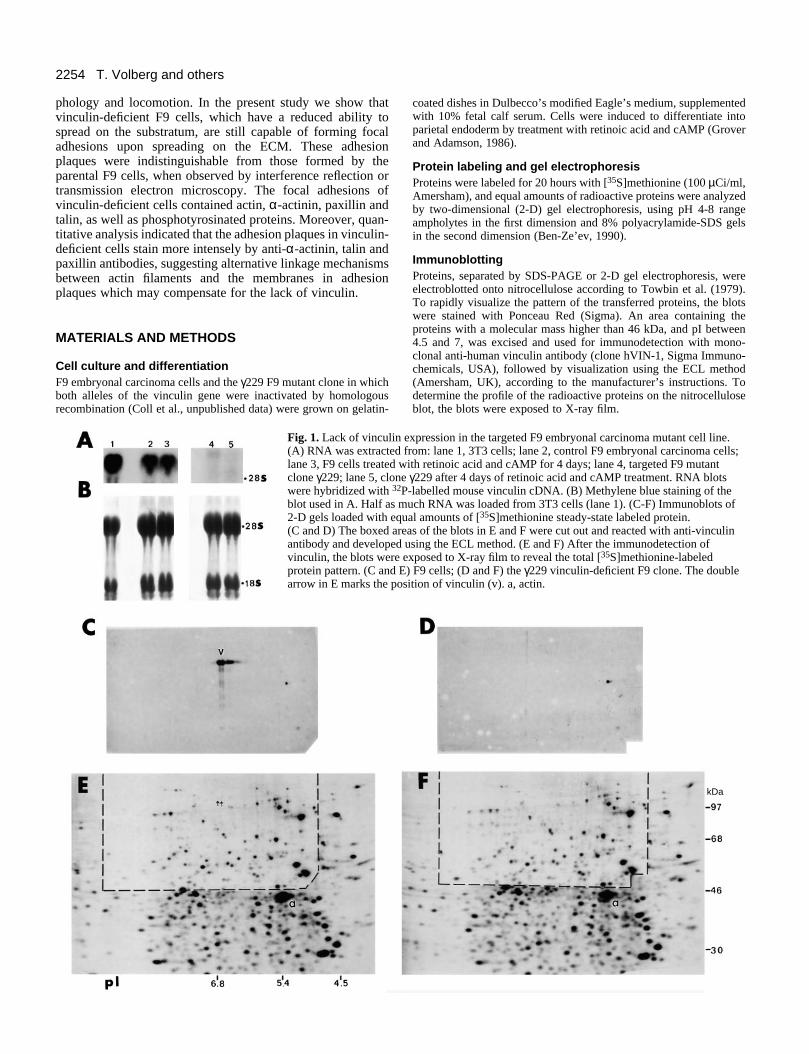
Fig. 1. Lack of vinculin(A) RNA was extractedlane 3, F9 cells treated clone γ229; lane 5, clonwere hybridized with 32
blot used in A. Half as 2-D gels loaded with eq(C and D) The boxed arantibody and developedvinculin, the blots wereprotein pattern. (C and arrow in E marks the po
coated dishes in Dulbecco’s modified Eagle’s medium, supplementedwith 10% fetal calf serum. Cells were induced to differentiate intoparietal endoderm by treatment with retinoic acid and cAMP (Groverand Adamson, 1986).
Protein labeling and gel electrophoresisProteins were labeled for 20 hours with [35S]methionine (100 µCi/ml,Amersham), and equal amounts of radioactive proteins were analyzedby two-dimensional (2-D) gel electrophoresis, using pH 4-8 rangeampholytes in the first dimension and 8% polyacrylamide-SDS gelsin the second dimension (Ben-Ze’ev, 1990).
ImmunoblottingProteins, separated by SDS-PAGE or 2-D gel electrophoresis, wereelectroblotted onto nitrocellulose according to Towbin et al. (1979).To rapidly visualize the pattern of the transferred proteins, the blotswere stained with Ponceau Red (Sigma). An area containing theproteins with a molecular mass higher than 46 kDa, and pI between4.5 and 7, was excised and used for immunodetection with mono-clonal anti-human vinculin antibody (clone hVIN-1, Sigma Immuno-chemicals, USA), followed by visualization using the ECL method(Amersham, UK), according to the manufacturer’s instructions. Todetermine the profile of the radioactive proteins on the nitrocelluloseblot, the blots were exposed to X-ray film.
expression in the targeted F9 embryonal carcinoma mutant cell line. from: lane 1, 3T3 cells; lane 2, control F9 embryonal carcinoma cells;with retinoic acid and cAMP for 4 days; lane 4, targeted F9 mutante γ229 after 4 days of retinoic acid and cAMP treatment. RNA blotsP-labelled mouse vinculin cDNA. (B) Methylene blue staining of themuch RNA was loaded from 3T3 cells (lane 1). (C-F) Immunoblots ofual amounts of [35S]methionine steady-state labeled protein. eas of the blots in E and F were cut out and reacted with anti-vinculin using the ECL method. (E and F) After the immunodetection of exposed to X-ray film to reveal the total [35S]methionine-labeledE) F9 cells; (D and F) the γ229 vinculin-deficient F9 clone. The doublesition of vinculin (v). a, actin.
kDa

2255Adhesion plaques in vinculin null F9 cells
Fig. 2. Morphology and focal adhesion formation by F9 and vinculin-deficient γ229 cells. The morphology of F9 (A) and of the γ229 cells (B) on fibronectin was analyzed by scanning electron microscopy (SEM). The presence of focal adhesions in F9 (C) and γ229 (D) wasexamined by transmission electron microscopy (TEM) of sections cut perpendicular to the substratum. The formation of focal adhesions in livecells on fibronectin-coated substrata was visualized in F9 (E) and γ229 (F) cells by interference reflection microscopy (IRM). The bars in A, B,E and F are 10 µm; in C and D, 1 µm.
2256 T. Volberg and others
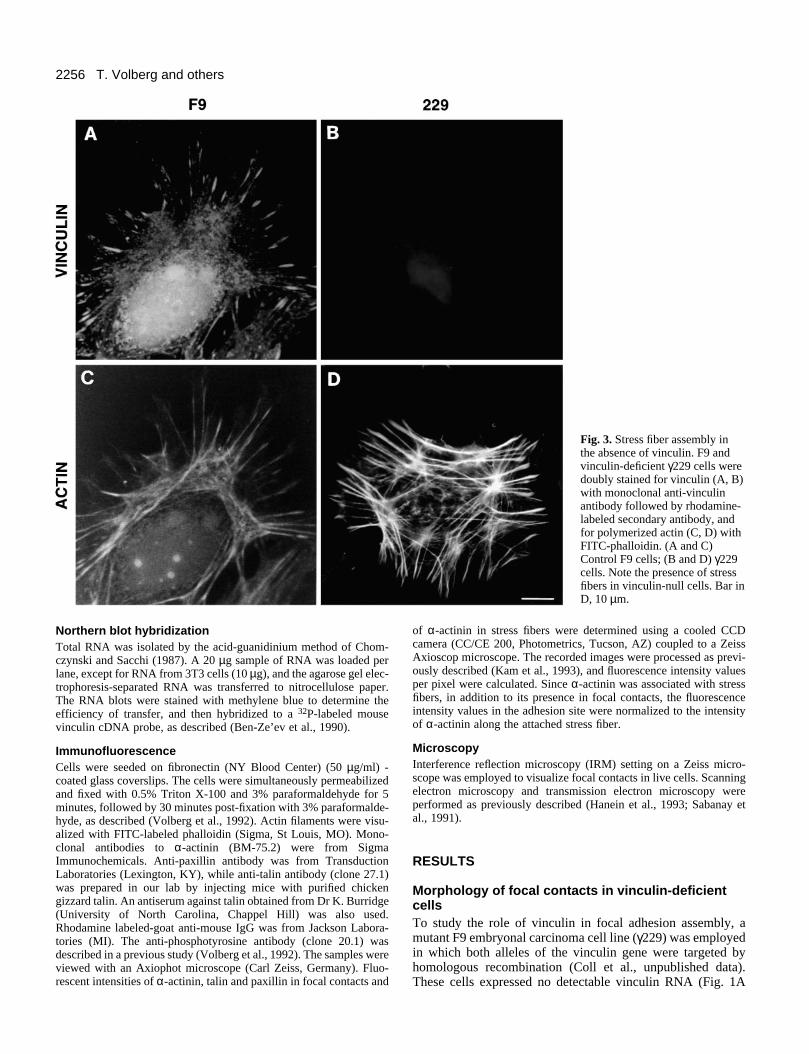
Fig. 3. Stress fiber assembly inthe absence of vinculin. F9 andvinculin-deficient γ229 cells weredoubly stained for vinculin (A, B)with monoclonal anti-vinculinantibody followed by rhodamine-labeled secondary antibody, andfor polymerized actin (C, D) withFITC-phalloidin. (A and C)Control F9 cells; (B and D) γ229cells. Note the presence of stressfibers in vinculin-null cells. Bar inD, 10 µm.
Northern blot hybridizationTotal RNA was isolated by the acid-guanidinium method of Chom-czynski and Sacchi (1987). A 20 µg sample of RNA was loaded perlane, except for RNA from 3T3 cells (10 µg), and the agarose gel elec-trophoresis-separated RNA was transferred to nitrocellulose paper.The RNA blots were stained with methylene blue to determine theefficiency of transfer, and then hybridized to a 32P-labeled mousevinculin cDNA probe, as described (Ben-Ze’ev et al., 1990).
ImmunofluorescenceCells were seeded on fibronectin (NY Blood Center) (50 µg/ml) -coated glass coverslips. The cells were simultaneously permeabilizedand fixed with 0.5% Triton X-100 and 3% paraformaldehyde for 5minutes, followed by 30 minutes post-fixation with 3% paraformalde-hyde, as described (Volberg et al., 1992). Actin filaments were visu-alized with FITC-labeled phalloidin (Sigma, St Louis, MO). Mono-clonal antibodies to α-actinin (BM-75.2) were from SigmaImmunochemicals. Anti-paxillin antibody was from TransductionLaboratories (Lexington, KY), while anti-talin antibody (clone 27.1)was prepared in our lab by injecting mice with purified chickengizzard talin. An antiserum against talin obtained from Dr K. Burridge(University of North Carolina, Chappel Hill) was also used.Rhodamine labeled-goat anti-mouse IgG was from Jackson Labora-tories (MI). The anti-phosphotyrosine antibody (clone 20.1) wasdescribed in a previous study (Volberg et al., 1992). The samples wereviewed with an Axiophot microscope (Carl Zeiss, Germany). Fluo-rescent intensities of α-actinin, talin and paxillin in focal contacts and
of α-actinin in stress fibers were determined using a cooled CCDcamera (CC/CE 200, Photometrics, Tucson, AZ) coupled to a ZeissAxioscop microscope. The recorded images were processed as previ-ously described (Kam et al., 1993), and fluorescence intensity valuesper pixel were calculated. Since α-actinin was associated with stressfibers, in addition to its presence in focal contacts, the fluorescenceintensity values in the adhesion site were normalized to the intensityof α-actinin along the attached stress fiber.
MicroscopyInterference reflection microscopy (IRM) setting on a Zeiss micro-scope was employed to visualize focal contacts in live cells. Scanningelectron microscopy and transmission electron microscopy wereperformed as previously described (Hanein et al., 1993; Sabanay etal., 1991).
RESULTS
Morphology of focal contacts in vinculin-deficientcellsTo study the role of vinculin in focal adhesion assembly, amutant F9 embryonal carcinoma cell line (γ229) was employedin which both alleles of the vinculin gene were targeted byhomologous recombination (Coll et al., unpublished data).These cells expressed no detectable vinculin RNA (Fig. 1A

2257Adhesion plaques in vinculin null F9 cells
Fig. 4. Localization ofphosphotyrosinated proteins in F9and γ229 cells. F9 (A) and γ229(B) cells were stained with mousemonoclonal antibody specific forphosphotyrosine followed by anti-mouse IgG labeled withrhodamine. Bar in B, 10 µm.
lanes 4 and 5). Immunoblot analysis of proteins separated by2-D gel electrophoresis did not detect vinculin even in over-exposed Western blots with anti-vinculin antibody (Fig. 1D).Except for the lack of vinculin, the pattern of major cellularproteins was very similar in the parental (Fig. 1E) and themutant (Fig. 1F) F9 cell lines.
The vinculin-deficient γ229 cells were capable of adheringto fibronectin- and gelatin-coated surfaces, but their spreadingwas apparently impaired (Fig. 2B) compared to the parental F9cells (Fig. 2A), despite the fact that the expression of the majorfibronectin receptor (integrin α5β1) was similar in both celltypes (results not shown). Transmission electron microscopicexamination of sections cut perpendicular to the substratumrevealed that the mutant cells contained electron-dense focaladhesions closely associated with the fibronectin substratum(Fig. 2D), which were indistinguishable from those formed bythe control F9 cells (Fig. 2C). Interference reflectionmicroscopy of the cells demonstrated that both were capableof forming dark regions characteristic of sites of close andfocal cell-substratum contacts (Fig. 2E and F). These observa-tions suggest that the vinculin negative cells did not loose thecapacity to assemble focal adhesions with morphological char-acteristics similar to those of the parental cells.
Organization of focal adhesion components invinculin-deficient cellsImmunofluorescence and fluorescent phalloidin stainingrevealed that despite the absence of vinculin (Fig. 3B), themutant γ229 cells assembled stress fibers (Fig. 3D) that weresimilar to those of control F9 cells (Fig. 3C). Moreover, thefocal adhesions of vinculin-deficient γ229 cells stained brightlywith anti-phosphotyrosine antibody (Fig. 4B), similar tocontrol F9 cells (Fig. 4A), suggesting that stress fibers andphosphotyrosine-rich adhesion plaques can assemble into focaladhesions in the absence of vinculin.
Recent in vitro studies suggested that actin filaments mayassociate with adhesion plaques via several alternative mech-anisms including an interaction between the cytoplasmic tailof β1 integrin with talin and vinculin or directly with α-actinin,an adhesion plaque component that is also a potent actinfilament crosslinker (Otey et al., 1990, 1993). In addition, itwas shown that talin can bind directly to actin in vitro
(Muguruma et al., 1990; Goldmann et al., 1994), and thatpaxillin is a vinculin-binding molecule (Turner et al., 1990).We have therefore analyzed qualitatively and quantitatively thelocal intensities of immunolabeling for these proteins in controlF9 compared to the vinculin-null γ229 cells, by digital fluo-rescence microscopy. As shown in Fig. 5 all these proteinswere localized in characteristic focal adhesions, both in thevinculin-null γ229 cells and in the F9 parental line. Quantita-tive analysis of the local immunofluorescence intensities,however, pointed to significant differences between thevinculin-deficient and the control cells. The results, based onthe analysis of a large number of focal contacts (300-400, in 3independent experiments, for each cell type), indicated that thelabeling intensities for α-actinin (Fig. 5A and B), talin (Fig.5C and D) and paxillin (Fig. 5E and F) were higher by 40%,60% and 80%, respectively, in the vinculin-null cells (Fig. 6).The differences were found to be highly significant (P<0.001-0.005). Western blot analysis with antibodies to paxillin, α-actinin and talin did not detect significant differences in thelevels of these proteins between vinculin-deficient and theparental F9 cells (results not shown). These results imply thatmultiple modes of linking actin filaments to adhesion plaquesexist, and that they are enhanced in the vinculin-deficient F9cells.
DISCUSSION
The primary objective of this study was to investigate the roleof vinculin in the assembly of focal contacts by structural andimmunochemical analysis of the molecular structure of matrixadhesions after specific disruption of the vinculin gene. Theresults presented here, and those described (Coll et al., unpub-lished data), indicate that the elimination of vinculinexpression leads to impaired adhesion to different substrata(Coll et al., unpublished data) and reduced ability to spread onfibronectin (Fig. 2B). Nevertheless, it does not inhibit theformation of focal adhesions, as judged by interference reflec-tion and electron microscopic analyses. Moreover, theassembly of major components of focal adhesions such asactin, α-actinin, talin and paxillin, into adhesion plaques, wasnot impaired; it was even enhanced. This observation suggests

2258 T. Volberg and othersα–
AC
TIN
INTA
LIN
PAX
ILL
IN
fluor
esce
nce
(A.U
.)
F9 229
1500
1000
500
Fig. 5. Localization and quantitation of α-actinin, talin and paxillin in F9 and in vinculin-deficient γ229 cells. F9 (A, C, E) and γ229 cells (B, D, F) were immunofluorescently labeled with monoclonal antibodies to α-actinin (A, B), talin (C, D), and paxillin (E, F), followed byrhodamine-labeled anti-mouse IgG. The fluorescence was recorded using a CCD camera as described in Materials and Methods. and netfluorescence intensity per pixel was calculated and presented using a pseudo color scale. A.U., arbitrary units. Bar in F, 10 µm.
that the linkage of actin to the membrane in these sites includesa ‘network’ of multiple interactions between the various plaqueproteins studied here, and that each of these proteins can bindto focal contacts, also in the absence of vinculin. Such inter-actions may include direct binding of α-actinin to the cyto-plasmic tail of β1 integrin (Otey et al., 1990, 1993). Similarly,
it was shown that the association of talin with focal contactsmight be driven not only by binding to vinculin (Horwitz etal., 1986), but also by interaction with other focal contactmolecules such as α5β1 integrin, which is expressed at normallevels in the vinculin-deficient F9 cells, as well as actin(Muguruma et al., 1990; Goldmann et al., 1994). On the other

2259Adhesion plaques in vinculin null F9 cells
F9 229
talin
F9 229
paxillin
F9 2290
500
1000
1500
2000 α-actinin
fluor
esce
nce
(A.U
.)
Fig. 6. Quantitative analysis of fluorescence intensity of adhesionplaque components in F9 and vinculin-deficient γ229 cells. Thefluorescence intensity of α-actinin, paxillin and talin in individualadhesion plaques from 3 different coverslips for each type of proteinwas determined by digitized quantitative analysis of pseudo colors in300-400 focal adhesions for each protein, as shown in Fig. 5. For α-actinin, the values were normalized against the intensity of stainingof α-actinin in the corresponding stress fiber for each adhesionplaque. The average plus standard deviations are shown.
hand, the presence of paxillin in focal contacts in the vinculin-deficient cells was unexpected, since it was previously demon-strated that the major adhesion plaque component which bindspaxillin is vinculin (Turner et al., 1990). Recent studies haveshown, however, that paxillin can associate with SH3-bindingdomains of tyrosine kinases (Weng et al., 1993), and with theSH2-binding domain of v-Crk (Birge et al., 1993). Theassembly of paxillin in focal adhesions may thus be attributedto such interactions in the vinculin-deficient γ229 cells (Fig.4B).
The availability of the vinculin-null cell line provides furtherinsight into the alternative linkage mechanisms of actinfilaments to adhesion plaques. The F9 cell system and thevinculin-deficient mutant cells enabled us to investigate theinvolvement of vinculin not only in focal contacts, but also incell-cell adherens-type junctions. It has been shown that uponretinoic acid stimulation both F9 and γ229 cells differentiateinto parietal endoderm (Coll et al., unpublished data). Stainingof the differentiated cells for actin, using fluorescent phal-loidin, revealed an abundance of actin along the sites of cell-cell adhesion in both cell types (data not shown). Vinculin wasapparently absent from these cell-cell contacts, both in F9 andin the vinculin-null cells. While the absence of vinculin fromcell-cell adhesions in F9 may explain why the vinculin-deficient cells display normal intercellular adhesions, it raisesan intriguing question concerning the role of vinculin in cell-cell adherens junctions in general: why is vinculin associatedwith some adhesions, for example, in cardiac muscle, variousepithelial and endothelial cells (Geiger, 1979; Geiger et al.,1992), and not with others (such as those in F9 cells, as shownhere)?
It should be pointed out that the absence of vinculin did notsignificantly affect the retinoic acid-mediated biochemicaldifferentiation of F9-derived cells (Coll et al., unpublisheddata). Similar results were also reported for F9 cells in whichthe β1 integrin genes were inactivated by targeted homologousrecombination (Stephens et al., 1993). It is noteworthy that inboth vinculin- and β1 integrin-deficient F9 mutants, cellspreading on the ECM was partially inhibited, suggesting that
while parallel structural linkages between the microfilamentnetwork and the ECM may exist, elimination of even a singlemolecule in this chain can affect the strength of cell adhesionand the ability of cells to spread on the substratum.
The vinculin-null F9 cells will be useful in structure-function analyses employing transfection studies with full-length and mutant forms of vinculin to define the role ofvarious vinculin domains in its association with adhesionplaques.
We thank Dr K. Burridge for the anti-talin antiserum. This studywas supported by grants from the Council For Tobacco Research-USA, from the Minerva Fund, from the Israel Cancer Research Fund(ICRF), from the USA-Israel Binational Fund, and by a grant fromthe Leo and Julia Forchheimer Center for Molecular Genetics at TheWeizmann Institute of Science to A.B.-Z., and United States PublicHealth Service CA 54233 and P30 CA 30199 to E.D.A. B.G. is theE. Neter Professor for Cell and Tumor Biology. A.B.-Z. is theLunenfeld-Kunin Professor for Genetics and Cell Biology.
REFERENCES
Ben-Ze’ev, A. (1990). Application of two-dimensional gel electrophoresis inthe study of cytoskeletal protein regulation during growth activation anddifferentiation. Electrophoresis 11, 191-200.
Ben-Ze’ev, A., Reiss, R., Bendori, R. and Gorodecki., B. (1990). Transientinduction of vinculin gene expression in 3T3 fibroblasts stimulated byserum-growth factors. Cell Regul. 1, 621-636.
Ben-Ze’ev, A. (1992). Cytoarchitecture and signal transduction. Crit. Rev.Eukaryotic Gene Expression 2, 265-281.
Birge, R.B., Fajardo, J.E., Reichman, C., Shoelson, S.E., Songyang, Z.,Cantley, L.C. and Hanafusa, H. (1993). Identification and characterizationof a high-affinity interaction between v-Crk and tyrosine-phosphorylatedpaxillin in CT10-transformed fibroblasts. Mol. Cell. Biol. 13, 4648-4656.
Burridge, K. and Connell, L. (1983). A new protein of adhesion plaques andruffling membranes. J. Cell Biol. 97, 359-367.
Burridge, K., Fath, K., Kelly, T., Nuckolls, G. and Turner, C. (1988). Focaladhesions: Transmembrane junctions between the extracellular matrix andthe cytoskeleton. Annu. Rev. Cell Biol. 4, 487-525.
Burridge, K., Turner, C.E. and Romer, L.H. (1992). Tyrosinephosphorylation of paxillin and pp125FAK accompanies cell adhesion toextracellular matrix: a role in cytoskeletal assembly. J. Cell Biol. 119, 893-903.
Chomczynski, P. and Sacchi, N. (1987). Single step method of RNA isolationby acid guanidinium thiocyanate-phenol-chloroform extraction. Anal.Biochem. 162, 156-159.
Geiger, B. (1979). A 130K protein from chicken gizzard: its localization at thetermini of microfilament bundles in cultured chicken cells. Cell 18, 193-205.
Geiger, B. and Ginsberg, D. (1991). The cytoplasmic domain of adherens-type junctions. Cell Motil. Cytoskel. 20, 1-6.
Geiger, B., Ayalon, O., Ginsberg, D., Volberg, T., Rodríguez Fernández,J.L., Yarden, Y. and Ben-Ze’ev, A. (1992). Cytoplasmic control of cell-adhesion. Cold Spring Harb. Symp. Quant. Biol. 57, 631-642.
Goldmann, W.H., Bremer, A., Haner, M., Aebi, U. and Isenberg, G. (1994).Native talin is a dumbell-shaped homodimer when it interacts with actin. J.Struct. Biol. 112, 3-10.
Grover, A., and Adamson, E.D. (1986). Conditions affecting thedifferentiation of F9 teratocarcinoma cells: Potentiation of response by cyclicAMP. In vitro Cell. Dev. Biol. 22, 280-284.
Hanein, D., Sabanay, H., Addadi, L. and Geiger, B. (1993). Selectiveinteraction of the cells with crystal surfaces. J. Cell Sci. 104, 257-288.
Horwitz, A., Duggan, K., Buck, C.A., Beckerle, M.C. and Burridge, K.(1986). Interactions of plasma membrane fibronectin receptor with talin – atrans-membrane linkage. Nature 320, 531-533.
Hynes, R.O. (1987). Integrins: a family of cell surface receptors. Cell 48, 549-554.
Juliano, R.L. and Haskill, S. (1993). Signal transduction from theextracellular matrix. J. Cell Biol. 120, 577-585.
Kam, Z., Jones, M.O., Chen, H., Agard, D.A. and Sedat, J.W. (1993).

2260 T. Volberg and others
Design and construction of an optimal illumination system for quantitativewide-field multi-dimensional microscopy. Bioimaging 1, 71-81.
Kemler, R. (1993). From cadherins to catenins: cytoplasmic proteininteractions and regulation of cell adhesion. Trends Genet. 9, 317-321.
Lazarides, E. and Burridge, K. (1975). α-Actinin: immunofluorescentlocalization of a muscle structural protein in nonmuscle cells. Cell 6, 289-298.
Lo, S.H., Janmey, P.A., Hartwig, J.H. and Chen, L.B. (1994). Interactions oftensin with actin and identification of its three distinct actin-binding domains.J. Cell Biol. 125, 1067-1075.
Luna, E.J. and Hitt, A.L. (1992). Cytoskeleton-plasma membraneinteractions. Science 258, 955-964.
Muguruma, M., Matsumura, S. and Fukazawa, T. (1990). Directinteractions between talin and actin. Biochem. Biophys. Res. Commun. 171,1217-1223.
Menkel, A.R, Kroemker, M., Bubeck, P., Ronsiek, M., Nikolai, G. andJockush, B.M. (1994). Characterization of an F-actin-binding domain in thecytoskeletal protein vinculin. J. Cell Biol. 126, 1231-1240.
Nagafuchi, A. and Takeichi, M. (1989). Transmembrane control of cadherin-mediated cell adhesion: a 94 kDa protein functionally associated with aspecific region of the cytoplasmic domain of E-cadherin. Cell Regul. 1, 37-44.
Otey, C.A., Pavalko, F.M. and Burridge, K. (1990). An interaction betweenα-actinin and the β1 integrin subunit in vitro. J. Cell Biol. 111, 721-729.
Otey, C.A., Vasquez, G.B., Burridge, K. and Erickson, B.W. (1993).Mapping of the α-actinin binding site within the β1-integrin cytoplasmicdomain. J. Biol. Chem. 268, 21193-21197.
Ozawa, M., Baribault, H. and Kemler, R. (1989). The cytoplasmic domain ofthe cell adhesion molecule uvomorulin associates with three independentproteins structurally related in different species. EMBO J. 8, 1111-1117.
Rodríguez Fernández, J.L., Geiger, B., Salomon, D. and Ben-Ze’ev, A.(1992a). Overexpression of vinculin suppresses cell motility in Balb/c 3T3cells. Cell Motil. Cytoskel. 22, 127-134.
Rodríguez Fernández, J.L., Geiger, B., Salomon, D., Sabanay, I., Zöller, M.and Ben-Ze’ev, A. (1992b). Suppression of tumorigenicity in transformedcells after transfection with vinculin cDNA. J. Cell Biol. 119, 427-438.
Rodríguez Fernández, J.L., Geiger, B., Salomon, D. and Ben-Ze’ev, A.(1993). Suppression of vinculin expression by antisense transfection confers
changes in cell morphology, motility and anchorage-dependent growth of3T3 cells. J. Cell Biol. 122, 1285-1294.
Rohrschneider, L.R. (1980). Adhesion plaques of Rous Sarcoma virus-transformed cells contain the src gene product. Proc. Nat. Acad. Sci. USA 77,3514-3518.
Sabanay, I., Arad, T., Weiner, S. and Geiger, B. (1991). Study of vitrified,unstained frozen tissue sections by cryoimmunoelectron microscopy. J. CellSci. 100, 227-236.
Sadler, I., Crawford, A.W., Michelsen, J.W. and Beckerle, M.C. (1992).Zyxin and cCRP: two interactive LIM domain proteins associated with thecytoskeleton. J. Cell Biol. 119, 1573-1587.
Schaller, M.D., Borgman, C.A., Cobb, B.S., Vines, R.R., Reynolds, A.B.and Parsons, J.T. (1992). pp125FAK, a structurally distinctive proteintyrosine kinase associated with focal adhesions. Proc. Nat. Acad. Sci. USA89, 5192-5196.
Stephens, L.E., Sonne, J.E., Fitzgerald, M.L. and Damsky, C.H. (1993).Targeted deletion of β1-integrins in F9 embryonal carcinoma cells affectsmorphological differentiation but not tissue-specific gene expression. J. CellBiol. 113, 1607-1620.
Towbin, H., Staehelin, T. and Gordon, J. (1979). Electrophoretic transfer ofproteins from acrylamide gels to nitrocellulose sheets: Procedure and someapplications. Proc. Nat. Acad. Sci. USA 76, 4350-4354.
Turner, C.E., Glenney, J.R. and Burridge, K. (1990). Paxillin: a newvinculin-binding protein present in focal adhesions. J. Cell Biol. 111, 1059-1068.
Varnum-Finney, B. and Reichardt, L.F. (1994). Vinculin-deficient PC12 celllines extend unstable lamellipodia and filopodia and have a reduced neuriteoutgrowth. J. Cell Biol. 127, 1071-1084.
Volberg, T., Zick, Y., Dror, R., Sabanay, I., Gilon, C., Levitzki, A. andGeiger, B. (1992). The effect of tyrosine-specific protein phosphorylation onthe assembly of adherens type junctions. EMBO J. 11, 1733-1742.
Weng, Z., Taylor, J.A., Turner, C.E., Brugge, J.S. and Seidel-Dugan, C.(1993). Detection of Src homology 3-binding proteins, including paxillin, innormal and v-Src-transformed Balb-c 3T3 cells. J. Biol. Chem. 268, 14956-14963.
(Received 2 February 1995 - Accepted 13 March 1995)