microtubule distribution during meiosis i in flea-beetle ... · microtubule distributions...
TRANSCRIPT

IntroductionModels of cell division are generally based on studies ofcultured vertebrate somatic cells, in which chromosomesbehave uniformly and the direction of anaphase movement isdetermined by the orientation of sister kinetochores to oppositepoles. However, many other interesting and unusual divisionsexist, and several examples are found in male meiotic cells(White, 1973). Non-uniform chromosome behaviour occurs ingrasshopper spermatocytes, where the single univalent (X)chromosome remains unpaired and moves to one of the twopoles before the start of autosomal anaphase (Nicklas, 1961).In the spermatocytes of other species, single univalents mightact differently, such as moving polewards after the autosomesreach the poles or dividing equationally in meiosis I (Stevens,1909). Non-random chromosome segregation has also beendescribed. In mole-cricket spermatocytes, for example, thelarger partner of a heteromorphic bivalent invariably segregatesto the same spindle pole as the univalent sex chromosome (e.g.Payne, 1916; Camenzind and Nicklas, 1968; Kubai and Wise,1981). In Sciaraspermatocytes (e.g. Gerbi, 1986; Goday andEsteban, 2001), mealy bug spermatocytes (e.g. Brown and Nur,1964; Brown and Weigmann, 1969) and Poeciliopsisoocytes
(e.g. Schultz, 1966; Schultz, 1973; Cimino, 1972), the entirecomplement of male-derived chromosomes segregates to onepole during anaphase, while the female-derived chromosomesmove to the other pole. These examples are just a few of many.
Mechanisms that give rise to non-uniform behaviours of thekinds described above are largely unknown. One possibleexception is the coordinated movements of univalent sexchromosomes in crane-fly spermatocytes. In these cells, thetwo sex chromosomes remain univalent (unpaired) throughoutmeiosis I. They congress to the metaphase plate with thebivalent autosomes and then remain at the equator until theautosomes complete anaphase, at which time they begin theirown anaphase motion to opposite poles (e.g. Forer, 1980).Experiments suggest that this independent behaviour involveslocal signalling between the two univalents. For example, inmicromanipulation experiments, when one of the segregatingunivalents is pushed opposite to its direction of anaphasemotion until it overtakes the other univalent, both univalentsreverse their directions of motion (Forer and Koch, 1973),suggesting that, as the two move to opposite poles, there iscontinuous signalling between them. Signalling also seems tooccur between univalents and autosomes or between their
1235
The meiosis-I spindle in flea-beetle spermatocytes isunusual in that the autosomes and univalent sexchromosomes are separated by a mitochondrial sheath andmove polewards at different times. To help understand thebasis for this interesting chromosome behaviour, and togather more detailed information about it, we studiedmicrotubule distributions throughout meiosis I usingimmunofluorescence and confocal microscopy, and tookcareful measurements of pole and kinetochore positions atall stages of division. Our results show that, by lateprophase, there is a spindle-shaped cytoplasmic array ofmicrotubules in the central part of the cell, with the nucleusat the periphery. Following nuclear envelope breakdown,both autosomes and sex chromosomes become associatedwith cytoplasmic microtubules, although only the
autosomes move centrally to the ‘cytoplasmic spindle’. Thetwo unpaired sex chromosomes remain at the cell peripheryand appear to be connected to each other by a microtubulebundle extending between their kinetochores. Thesebundles often persist into anaphase. Analysis ofmeasurements taken from fixed/stained cells supportsprevious observations that sex chromosomes move partway to the pole in early prometaphase and then stop.The measurements also suggest that during autosomalanaphase, spindle elongation precedes autosome movementto the poles and polewards movement of sex chromosomesis limited or absent when autosomes are moving polewards.
Key words: Microtubules, Meiosis, Chromosome orientation,Acetylated tubulin
Summary
Microtubule distribution during meiosis I in flea-beetle[Alagoasa (Oedionychus )] spermatocytes: evidencefor direct connections between unpaired sexchromosomesPaula J. Wilson 1, Arthur Forer 1,* and Dwayne Wise 2
1Biology Department, York University, Toronto, Ontario M3J 1P3, Canada2Department of Biological Sciences, Mississippi State University, MS 39762, USA*Author for correspondence (e-mail: [email protected])
Accepted 28 November 2002Journal of Cell Science 116, 1235-1247 © 2003 The Company of Biologists Ltddoi:10.1242/jcs.00296
Research Article

1236
spindle fibres (cf. Dietz, 1969; Sillers and Forer, 1981), andlocal signals seem to coordinate the movements of partner half-bivalents to opposite poles during anaphase (Yin and Forer,1996).
The structural and chemical bases for these local‘coordination’ mechanisms are unknown, but they serve toremind us that individual chromosomes respond to local aswell as to global signals, and that understanding theseunconventional divisions might provide clues aboutmechanisms underlying more conventional divisions.Unfortunately, they have been studied less than conventionaltissue culture cells and, aside from Sciara spermatocytes(Gerbi, 1986), no one has studied them genetically. In thisarticle, we describe observations of one of these uniquedivisions: meiosis-I in spermatocytes of flea beetles.
Spermatocyte divisions in flea-beetle species includingAlagoasa(Oedionychus) bicolor and Omophoita cyanipennishave several unique features described in some detail by Virkki(Virkki, 1970; Virkki, 1971; Virkki, 1972; Virkki, 1973;Virkki, 1985; Virkki, 1990). He described meiotic cells withten small autosomal bivalents and two large univalent sexchromosomes, X and Y. The most distinguishing feature ofthese cells was that early in prometaphase I the autosomesbecame separated from the unpaired sex chromosomes, withthe sex chromosomes remaining at the cell periphery and theautosomes moving centrally. The two groups of chromosomesthen seemed to form separate spindles and the autosomesproceeded through the normal stages of meiosis I. Before theautosomes entered anaphase, the two sex chromosomesoriented to opposite poles, moved polewards and then stoppedpart way to the pole and remain stopped until the autosomesentered anaphase.
Recent data (Forer and Wilson, 2000; Green-Marroquinet al., 2001) have shown that, in fact, the sex-chromosomespindle shares poles with the autosomal spindle. Nevertheless,the behaviour of the chromosomes in flea-beetle primaryspermatocytes gives rise to several unanswered questions. Forexample, what are the early events that give rise to the centralautosomal spindle? How are the unpaired sex chromosomesexcluded from the central spindle? How do they orient toopposite poles rather than to the same pole? To what extent arethe movements of the autosomes and sex chromosomesindependent? And, finally, if the sex chromosomes indeed donot move polewards when the autosomes do, how do they avoidthe metaphase/anaphase triggers that presumably govern theautosomes?
Virkki and others have attempted to answer some of thesequestions. Their work indicates that prior to prophase thenucleus is in the middle of the cell; by late prophase I thenucleus is to one side of the cell (Virkki, 1970; Virkki, 1971;Virkki, 1972). A clear zone that can be seen around the nucleusin prophase (Virkki, 1972) seems to disappear beforebreakdown of the nuclear membrane, and by late prophasethere is evidence of asters and of mitochondrial alignment inthe central part of the cell (Virkki, 1972), where the autosomalspindle will eventually be established. Virkki referred to thisearly cytoplasmic alignment as a ‘cytoplasmic spindle’, buthe based his interpretation on the alignment of themitochondria, without information regarding microtubules. Inlater publications (e.g. Virkki, 1985), he suggests that asterspush the nucleus to the cell periphery.
Virkki (Virkki, 1971; Virkki, 1972) showed that theautosomes move centrally following nuclear membranebreakdown, establishing a central spindle, while the unpairedsex chromosomes remain behind. There is little information toexplain why the sex chromosomes do not move with theautosomes, although Virkki argued that the autosomes are‘activated’ for movement before the sex chromosomes (Virkki,1971), or that sex chromosomes make connections with thepoles later than the autosomes (Virkki, 1972). Althoughremaining at the cell periphery, the sex chromosomes do makeconnections to opposite poles and move towards oppositepoles, but the details of this process are unknown. For example,it remains unclear how the unpaired sex chromosomes manageto orient to opposite poles; several studies reject the notion thatthere are connections or physical interactions between the sexchromosomes (Virkki, 1972; Virkki, 1985; Kupfer and Wise,2000a; Green-Marroquin et al., 2001). With respect to sex-chromosome spindle fibres, Virkki (Virkki, 1971; Virkki,1972; Virkki, 1985) described them as syntelic (i.e. each sexchromosome has fibres extending to only one spindle pole);more recently, Green-Marroquin et al. argued that the sex-chromosome spindle connections are initially amphitelic (i.e.each sex chromosome has fibres extending to both poles)and later become syntelic (Green-Marroquin et al., 2001).Unfortunately, none of the studies provide sufficient evidenceto be convincing and none include a comprehensive study ofmicrotubule organization within the two spindles.
Finally, there is the issue of the independent behaviour ofautosomes and sex chromosomes. Virkki (Virkki, 1967; Virkki,1970; Virkki, 1972) argued that as the autosomes proceedthrough prometaphase, metaphase and anaphase the univalentsex chromosomes orient and move part way to opposite polesto form a ‘distance bivalent’ by autosomal metaphase. [Theonly exception mentioned is a case of malorientation, in whichboth sex chromosomes temporarily faced the same pole andseemed to move in a coordinated fashion (Virkki, 1972).]Furthermore, although no direct measurements were available,it was Virkki’s impression that the sex chromosomes movedpolewards roughly around the time that the autosomes did inanaphase (Virkki, 1970). However, Forer and Wilson presenteddata on kinetochore fibre lengths that seemed to argue againstthe interpretation that sex chromosomes move polewards withthe autosomes (Forer and Wilson, 2000). Kupfer and Wise alsodispute this claim, arguing that there is little or no polewardsmovement of sex chromosomes during anaphase (Kupfer andWise, 2000a). To date, no one has addressed the mechanismfor coordinating sex chromosome movements to oppositepoles, nor have there been measurements of autosomal and sex-chromosome movement during meiosis.
Related to the issue of anaphase chromosome motion is therelative contribution of spindle elongation. Virkki did not makesufficient measurements to address the relative contributions ofmovement to the pole (‘anaphase A’) and pole elongation(‘anaphase B’), but Kupfer and Wise suggest frommeasurements on fixed cells that, whereas spindle length doesincrease between prometaphase and anaphase, spindleelongation in anaphase contributes little to autosome separation(Kupfer and Wise, 2000a).
In this article we address some of these unansweredissues and, in particular, present more detailed informationabout microtubule distribution and kinetochore-microtubule
Journal of Cell Science 116 (7)

1237Microtubule distribution during flea-beetle meiosis
interactions throughout the first division, as well asmeasurements of chromosome motion at different stages ofdivision. Unfortunately the animals are difficult to rear inlarge numbers, so extended experimentation is difficult.Nevertheless, as a first step towards extending ourunderstanding of this remarkable division, we have examinedmicrotubule organization in these cells. We studiedmicrotubules via indirect immunofluorescence and confocalmicroscopy, looking at both total tubulin and acetylatedtubulin, the latter being an indicator of more ‘stable’microtubules (Wilson et al., 1994; Wilson and Forer, 1997;Rosenbaum, 2000). We supplemented our observations withstudy of live cells when possible and made extensivemeasurements of chromosome and pole positions at all stagesof division.
The results, reported below, confirm the presence of acytoplasmic spindle prior to prometaphase, show that sexchromosomes have bidirectional kinetochore fibres that oftenpersist into later stages of division, show that sex chromosomesare linked by microtubules and suggest that, during anaphase,spindle elongation precedes autosome-to-pole movement.
Materials and MethodsStocks of Alagoasa bicolorwere initially provided by Niilo Virkki,then maintained in cages in the greenhouse at York University, rearedon Aegiphila and Chlorodendron, using methods essentially asdescribed earlier (Virkki and Zambrana, 1983).
Spermatocytes in division were prepared for live observation or forimmunofluorescence microscopy using methods described previously(Forer and Wilson, 2000). In brief, an animal was put underhalocarbon oil and its single testis removed and placed under oil. Forsome preparations of living cells, portions of the testis were smearedunder oil and cells observed by phase-contrast microscopy using anoil immersion objective (NA=1.3, 100×). For immunofluorescencepreparations and other observations of living cells, portions of testeswere placed in fibrinogen in modified Belar’s Ringer’s solution,spread, mixed with thrombin and then perfused with Belar’s solution,using procedures described previously (Forer and Pickett-Heaps,1998). Living cells affixed to the coverslip with the fibrin clot werestudied live and then prepared for immunofluorescence, or they weredirectly processed for immunofluorescence studies. To process forimmunofluorescence labelling, the coverslip was placed in lysis bufferand subsequently fixed and labelled with antibodies using proceduresdescribed elsewhere (Wilson et al., 1994). Cells generally werestained for both total tubulin (using YL1/2 antibody) and acetylated
tubulin (using 6-11B-1 antibody), though some preparations wereonly single stained (with YL1/2 antibody). Some preparations weretreated additionally with acridine orange (0.001-0.010 mg ml–1 inphosphate-buffered saline), in order to label the chromosomes. In thecase of triple-labelled cells, the acetylated tubulin and chromosomeswere visualized in the same channel. Confocal microscopeobservations were made with a BioRad 600 confocal system attachedto a Nikon Optiphot microscope, using a 60× lens (NA=1.4), asdescribed previously (Wilson et al., 1994). Distances were measuredfrom confocal microscope images using either BioRad (Comos)software or custom software described previously (Wilson et al.,1994).
As described previously (Forer and Wilson, 2000), we usedcomposite images of the entire cell to locate spindle poles. We markedthe spindle-pole positions with a cursor, marked the correspondingsex-chromosome and autosome kinetochore positions with a cursor,and used either BioRad (Comos) software or custom software (Wilsonet al., 1994) to measure distances between the poles and between thekinetochores and the poles. In reporting sex-chromosome distances,we report the value for each chromosome in that cell; in reportingautosomal distances, we measured distances for five or six autosomepairs in each cell and report the average value for each cell. Also asdescribed previously (Forer and Wilson, 2000; Virkki, 1972), in somecells, one or a few bivalents disjoin prematurely; we classified thesecells as ‘metaphase’ and considered as ‘anaphase’ only those cells inwhich all autosomal bivalents were completely (and clearly)disjoined.
ResultsLiving flea-beetle metaphase-I spermatocytes contain twospindles. Ten pairs of autosomes are arranged on a centralspindle, and the large sex chromosomes are on a spindle at theperiphery of the cell, separated from the autosomes by amitochondrial sheath (Fig. 1). Cells to be stained withantibodies were first lysed in a microtubule-stabilizing bufferand then fixed. The relative positions and orientations of thechromosomes were unaltered by lysis (Fig. 1A-D); subsequentimmunofluorescence images revealed an abundance ofmicrotubules in cells at all stages, as described below. In otherspermatocytes, the same stabilizing buffer preserves spindlestructures, including irradiated areas of reduced birefringence(e.g. Wilson et al., 1994; Czaban and Forer, 1994), indicatingthat the basic spindle cytoskeletal architecture is preserved bythese procedures.
Because sex-chromosome behaviour in flea-beetle
Fig. 1.Lysis of a living cell in metaphase I. Cells to be processed for immunofluorescence staining were first lysed in a microtubule stabilizingbuffer, then fixed in 0.2% glutaraldehyde in phosphate-buffered saline. General cell morphology was preserved by this procedure. (A) Cell 5minutes prior to lysis. (B) 50 seconds after perfusion of lysis buffer: the lysis buffer is beginning to reach the cell. (C) 1 minute after perfusion:lysis starting. (D) 6 minutes after perfusion: lysis is completed. Bar, 10 µm.

1238
spermatocytes is ‘out of phase’ with autosomal behaviour, weassessed the stage of lysed cells according to the positions ofthe autosomes. Thus, in describing ‘metaphase’, for example,we refer to the stage in which the autosomes are at themetaphase plate. Observations in this report cover the periodfrom late prophase I to late anaphase I, and focus on thecreation of the central autosomal spindle, the behaviour of thesex chromosomes and the relationship between autosomalmovement and sex-chromosome movement.
Establishing the central autosomal spindleThe spindle is established prior to nuclear envelopebreakdown (NEB)
Late prophase cells have nuclei that are at the cell periphery(Fig. 2A-C), as described previously (Virkki, 1971; Virkki,1972). The peripheral nucleus creates a bulge or out-pocketingof the cell membrane (e.g. Fig. 2B), which usually persiststhroughout later stages of division (e.g. Fig. 1A).
Phase-contrast observations of living prophase cells confirmthe existence of linear cytoplasmic elements in the central areaof prophase cells, arranged in the shape of a mitotic spindle(Fig. 2A-C), presumably the structure that Virkki referred to asthe ‘cytoplasmic spindle’ (Virkki, 1972). Anti-tubulin labellingof fixed cells reveal that these spindle-shaped structures containmicrotubule arrays, with poles at either end (Fig. 2D,E). Inmost cases the microtubules appear to be unacetylated, and insome cells one pole is associated with (or abuts) the nucleus(Fig. 2C,E). Thus, our results confirm the existence ofcytoplasmic microtubules arrayed in the shape of a spindle,organized before the chromosomes are released by thedissolution of the nuclear membrane.
That a central spindle is present prior to NEB means that theautosomes must migrate to the forming spindle followingNEB, rather than the spindle organizing around thechromosomes, as is common in animal cells. Ourimmunofluorescence preparations contained many cells inwhich the sex chromosomes were at the cell periphery andautosomes were scattered between them and the central spindlearea (e.g. Fig. 3A,B), presumably in transit to the centralspindle. Prometaphase therefore includes the time duringwhich autosomes move to the central spindle, enter the centralspindle and congress to the metaphase plate. For purposesof discussion, we have subdivided prometaphase cells intothree stages: (1) early prometaphase, the period when mostautosomes are moving centrally, as deduced from fixed cells
whose autosomes are located between the cell periphery andcentral spindle; (2) mid-prometaphase, the period when mostautosomes have reached the central spindle but have not yetcongressed to the equator; and (3) late prometaphase, theperiod when most but not all of the autosomes have reachedthe metaphase plate. Cells in which all autosomes are at theequator, with well-developed connections to the poles, areconsidered metaphase.
Microtubule interactions with migrating autosomesduring early prometaphase
Immediately following NEB the autosomes and sexchromosomes are intermingled. Throughout prometaphasemicrotubules are present between the poles, with extensivemicrotubule arrays throughout the cell, surrounding both theautosomes and the sex chromosomes (Fig. 3A). Bivalents intransit from the cell periphery to the central part of the cellhave various orientations and microtubule associations.Different microtubule associations include fibres extendingfrom only one half-bivalent to a pole (Fig. 3B,D, closedarrowheads), fibres extending from only one half-bivalent butextending to both poles (Fig. 3B, open arrowheads) and fibresextending from both half-bivalents to both poles (Fig. 3C,open arrowheads). Interestingly, there are also examples ofkinetochore fibres extending away from the central spindle(Fig. 3B,C, arrows). Bivalent orientation varies fromperpendicular to parallel to the spindle axis, and bivalents ofall orientations have microtubule attachments. Becauseexamples of all types of attachments described above are seenin bivalents located between the cell periphery and the centralspindle area, we assume that all were moving or had beenmoving. Most bivalents that are in the central spindlehave normal bipolar microtubule arrangements (Fig. 3E,arrowheads). Thus, following NEB, autosomes appear tointeract with microtubules extending from the poles and beginto move into the central spindle. Association of only one half-bivalent with microtubules from either one or both polesappears to be sufficient to permit movement.
The kinetochore fibres of autosomes in early prometaphase,as they move centrally, are weakly acetylated or unacetylated(Fig. 3C-E), and the attachments can appear quite delicate. Bymid prometaphase, the central autosomal spindle is wellestablished and separate from the sex chromosomes (Fig. 4A-C) and by late prometaphase and metaphase the kinetochorefibres are acetylated (Fig. 4D), indicating that the fibres
Journal of Cell Science 116 (7)
Fig. 2.The cytoplasmic spindle. (A-C) Phase-contrast images of living cells in prophase, revealing spindle-shaped cytoplasmic alignmentsoutside the nucleus. (A) Bar, 10 µm. (B,C) Bar, 5 µm. (D) Labelling of a prophase cell with antibodies to tubulin reveals a spindle-shaped arrayof microtubules in the cytoplasm. The nucleus is at the bottom of the frame. Bar, 10 µm. (E) Prophase cell triple labelled to reveal acetylatedtubulin (green), tyrosinated tubulin (red) and chromosomes (acridine orange labelling, which appears in the green channel). A spindle-shapedstructure containing unacetylated microtubules is evident. The nucleus is at the bottom of the frame. Bar, 10 µm.

1239Microtubule distribution during flea-beetle meiosis
become more stable once the bivalents develop typical bipolarattachments within the central spindle.
Metaphase and anaphase microtubule arraysBy metaphase, the autosomes have congressed to themetaphase plate and the kinetochore fibres are highlyacetylated (Fig. 5C). The kinetochore fibre structure at thisstage is bimorphic, as described previously (Forer and Wilson,2000), in that the kinetochore fibre is thick and highlyacetylated from the kinetochore to roughly halfway to the polebut polewards from this point the bundle splays into severalsmaller microtubule bundles that extend towards the pole.Astral microtubules either are focused at the poles [Fig. 4D;see also Figs 3, 4 in Forer and Wilson (Forer and Wilson,2000)] or form a broad ‘fringe’ of microtubules extending fromthe membrane across the polar ends of the cell [e.g. Fig. 5C;see also Fig. 2 in Forer and Wilson (Forer and Wilson, 2000)].Astral microtubules are acetylated by late prometaphase andunacetylated by late anaphase (Fig. 4E).
In summary, we have confirmed the existence of a spindle-like structure in the prophase cytoplasm and shown that itcontains microtubules organized between two poles. Ourobservations are consistent with the autosomes moving into the
pre-existing cytoplasmic spindle from the periphery of the cellby virtue of interactions with microtubules oriented toward thespindle. Microtubule associations with a single half-bivalentappear to be sufficient to give rise to movement. Once theautosomes reach the central spindle and develop bipolarattachments, the kinetochore fibres become thicker andacetylated, and the astral microtubules become acetylated.
Sex chromosomesOur study of the univalent sex chromosomes focused on threeissues: (1) their failure to move with the autosomes to thecentral spindle; (2) their independent orientation to oppositepoles; and (3) their polewards motion, especially with respectto autosomal motion and pole-to-pole elongation. We paidspecial attention to the timing of the orientation events andto interactions of microtubules with the sex-chromosomekinetochores.
Failure to move with the autosomesDuring prometaphase, the autosomes move into the centralspindle but the sex chromosomes remain at the periphery. Apossible explanation for this behaviour would be the absence
Fig. 3.Confocal serial section reconstructionsof cells in early prometaphase I, showingkinetochore-microtubule associations ofautosomes. (A) Prometaphase cell labelledwith antibodies against tubulin. Bar, 10 µm.(B) Higher magnification image of cell in (A).The closed arrowhead points to a bivalent withassociations to only one pole. Openarrowheads point to a bipolar connectionmade by a single half-bivalent. The sister half-bivalent has microtubule associations only tothe top pole. The arrow points to a half-
bivalent with bidirectionalmicrotubule associations, only one ofwhich points towards a pole. Bar,5 µm. (C-E) Triple-labelledprometaphase cells, stained foracetylated tubulin (green), tyrosinatedtubulin (red) and chromosomes(green). Co-localization of acetylatedand tyrosinated tubulin would resultin a yellow or orange image.(C) Arrowheads point to microtubuleassociations with a bivalent that hasconnections to both poles. The arrowpoints to a kinetochore fibre pointedaway from the poles. Bar, 5 µm.
(D) Different series of sections from the samecell shown in (C). The closed arrowhead pointsto a bivalent with attachments only to one pole:notice the two fibres coming from one half-bivalent, one from each sister chromatid. Bar,5 µm. (E) Arrowheads point to bivalents thathave reached the central spindle andestablished bipolar connections. Bar, 5 µm.(F,G) Stereo images of a prometaphase cellshowing the arrangements of microtubules andchromosomes. Bar, 10 µm.

1240
of kinetochore interactions with microtubules at this stage.Confocal images of cells labelled with anti-tubulin antibodies,however, reveal that the sex-chromosome kinetochores areassociated with microtubules during the period when theautosomes are moving (e.g. Fig. 5A). In fact, we saw noexamples where autosomes had kinetochore fibres and sexchromosomes did not. Therefore, a lack of microtubuleassociations cannot be the reason for the exclusion of the sexchromosomes from the central spindle.
Independent orientation to oppositepoles
We studied the interactions ofmicrotubules with sex-chromosomekinetochores during prometaphase, whenthe sex chromosomes become orientedto opposite poles. There is substantialheterogeneity in the types of interactionsbut, in general, they fall into three broadgroups. In the first group, termed‘bidirectional’, thin microtubule bundlesextend from the kinetochore region in twodirections, usually but not exclusively inthe direction of the two poles (Fig. 5A,B,arrowheads). In some cases the fibreextending to the closest pole is slightlythicker than the fibre extending to thefurther pole but, overall, the fibres arerelatively thin and either unacetylatedor very weakly acetylated. In thesecond group of kinetochore-microtubuleinteractions, termed ‘syntelic’, a singlefibre extends from the kinetochore to asingle pole. The fibre typically isrelatively thick and acetylated (Fig. 5C,arrowheads).
In the third group, termed ‘semi-syntelic’, kinetochores are associatedwith a well-developed, acetylated fibrerunning from the kinetochore to thenearest pole, but there is a second, smallerfibre extending from the kinetochorein the opposite direction (Fig. 5D,arrowheads). The distinction between‘semi-syntelic’ and ‘bidirectional’ isbased on our evaluation of the relativedevelopment of the primary kinetochorefibre and its acetylation relative to the‘secondary’ (poorly acetylated) fibre: inbidirectional fibres, the two microtubulebundles are more similar in size, thinnerand poorly acetylated; in semi-syntelicfibres, there is a clearly dominantacetylated fibre in one direction. Not allfibres fell easily into one of the twocategories and therefore the datapresented include intermediates.
The frequencies of the differentkinetochore-microtubule associations atvarious stages of division are shown inFig. 6, the stages being assigned byscoring autosomes, as described earlier.
The graph shows that bidirectional orientation is characteristicof the earlier part of prometaphase, whereas semi-syntelic andsyntelic fibres are more common in later stages. Thus,orientation to opposite poles is accompanied by a shift frombidirectional kinetochore-microtubule associations to a mainlyunidirectional kinetochore fibre.
It is worth noticing that in many examples of bidirectionalfibres the microtubules appear to interact with the kinetochoresat various angles. In some cases the sides of the microtubules
Journal of Cell Science 116 (7)
Fig. 4.Confocal Z-series showing microtubule organization at different stages of meiosis I.(A-C) Mid-prometaphase cell labelled with antibodies against tubulin and with acridineorange to reveal the chromosomes. (B) and (C) are stereo images. (A) Bar, 5 µm. (B,C). Bar,10 µm. (D) Late prometaphase cell stained for acetylated tubulin (left) and tyrosinatedtubulin (right). The arrowheads indicate the kinetochore fibres of the two sex chromosomes.Bar, 10 µm. (E) Anaphase cell stained for acetylated tubulin (left) and tyrosinated tubulin(right). Bar, 10 µm.

1241Microtubule distribution during flea-beetle meiosis
appear to interact with the face of the kinetochore,consistent with microtubules sliding along thekinetochore rather than inserting into it.
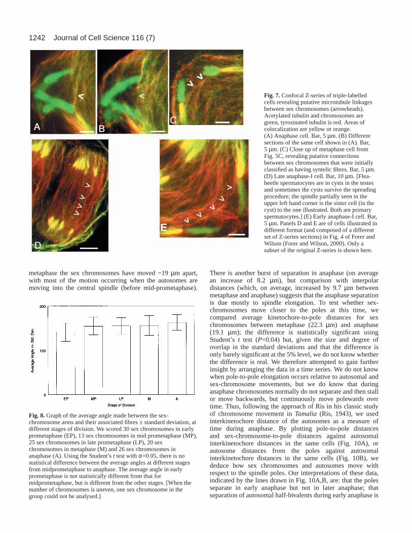
Sex chromosomes are linked by microtubulesWhile analysing microtubule associations with sexchromosomes, we unexpectedly found someprometaphase cells with clear microtubule bundleslinking the kinetochores of the two sex chromosomes(e.g. Fig. 5A,B). This observation prompted us toreanalyse all of our confocal images, lookingspecifically for evidence of microtubules extendingbetween sex-chromosome kinetochores. Weidentified unambiguous interkinetochore connectionsin 6/15 cells in early prometaphase, 1/13 cells in lateprometaphase and 1/10 cells in metaphase.Furthermore, in most of the other cells, we couldidentify putative connections between the sexchromosomes (thin fibres or single microtubulesextending from one sex-chromosome kinetochorein the direction of the other sex-chromosomekinetochore). Putative connections were seen even inlater stages of division, when the sex-chromosomespindle fibres were obvious, well-developed,‘syntelic’ and acetylated. Such putativeinterkinetochore connections were observed in cellsin prometaphase, metaphase and anaphase (Fig. 7).The high frequency of both unambiguous andputative connections suggests that microtubulesmight connect the sex chromosome univalentsthrough all stages of division.
Sex-chromosome arm orientation is extremelyvariable in dividing cells, ranging from perpendicularto parallel to the long axis of the spindle. We tested
whether the sex-chromosome arms change during divisionfrom perpendicular to parallel to the spindle axis, as discussedby Kupfer and Wise (Kupfer and Wise, 2000a), by examiningthe relationship between the angle made between thechromosome arm and its associated spindle fibre at differentstages of division. There is very little change in average angle,regardless of stage (Fig. 8), so we conclude that the orientationof the sex-chromosome arms is not indicative of stage as wemeasured it, and we have not found such analysis useful inanalysing sex chromosome behaviour at different stages.
Independent movement of sex chromosomes andautosomesIn 1970 Virkki suggested that the sex chromosomes movepolewards in anaphase at about the same time that theautosomes do (Virkki, 1970). More recent data (Forer andWilson, 2000) dispute this claim. To help resolve the issue, wemeasured interkinetochore and kinetochore-to-pole distancesof the sex chromosomes and autosomes at different stages ofdivision, classifying stage as described above – by thedisposition of the autosomes. For autosomes, interkinetochoredistance between autosomal half-bivalents remains essentiallyunchanged throughout prometaphase and metaphase, thenincreases during anaphase (Fig. 9).
A different pattern is seen for sex chromosomes (Fig. 9). By
Fig. 5.Triple-labelled cells revealing microtubule-kinetochore associations ofsex chromosomes. Acetylated tubulin and chromosomes are green, tyrosinatedtubulin is red: colocalization results in yellow or orange images.(A,B) Prometaphase cells showing bidirectional associations (arrowheads). Bar,5 µm. (C) Metaphase cell showing syntelic associations (arrowheads). Bar,10µm. (D) Metaphase cell showing semi-syntelic associations (arrowheads).Bar, 10 µm.
Fig. 6.Graph showing the distribution of different types of sex-chromosome/microtubule associations at different stages of division.We scored 29 sex chromosomes in early prometaphase (EP), 14 sexchromosomes in mid prometaphase (MP), 25 sex chromosomes inlate prometaphase (LP), 20 sex chromosomes in metaphase (M) and26 sex chromosomes in anaphase (A). (When the number ofchromosomes is uneven, one sex chromosome in the group could notbe analysed.)
1242
metaphase the sex chromosomes have moved ~19 µm apart,with most of the motion occurring when the autosomes aremoving into the central spindle (before mid-prometaphase).
There is another burst of separation in anaphase (on averagean increase of 8.2 µm), but comparison with interpolardistances (which, on average, increased by 9.7 µm betweenmetaphase and anaphase) suggests that the anaphase separationis due mostly to spindle elongation. To test whether sex-chromosomes move closer to the poles at this time, wecompared average kinetochore-to-pole distances for sexchromosomes between metaphase (22.3 µm) and anaphase(19.1 µm); the difference is statistically significant usingStudent’s t test (P=0.04) but, given the size and degree ofoverlap in the standard deviations and that the difference isonly barely significant at the 5% level, we do not know whetherthe difference is real. We therefore attempted to gain furtherinsight by arranging the data in a time series. We do not knowwhen pole-to-pole elongation occurs relative to autosomal andsex-chromosome movements, but we do know that duringanaphase chromosomes normally do not separate and then stallor move backwards, but continuously move polewards overtime. Thus, following the approach of Ris in his classic studyof chromosome movement in Tamalia (Ris, 1943), we usedinterkinetochore distance of the autosomes as a measure oftime during anaphase. By plotting pole-to-pole distancesand sex-chromosome-to-pole distances against autosomalinterkinetochore distances in the same cells (Fig. 10A), orautosome distances from the poles against autosomalinterkinetochore distances in the same cells (Fig. 10B), wededuce how sex chromosomes and autosomes move withrespect to the spindle poles. Our interpretations of these data,indicated by the lines drawn in Fig. 10A,B, are: that the polesseparate in early anaphase but not in later anaphase; thatseparation of autosomal half-bivalents during early anaphase is
Journal of Cell Science 116 (7)
Fig. 7.Confocal Z-series of triple-labelledcells revealing putative microtubule linkagesbetween sex chromosomes (arrowheads).Acetylated tubulin and chromosomes aregreen, tyrosinated tubulin is red. Areas ofcolocalization are yellow or orange.(A) Anaphase cell. Bar, 5 µm. (B) Differentsections of the same cell shown in (A). Bar,5 µm. (C) Close up of metaphase cell fromFig. 5C, revealing putative connectionsbetween sex chromosomes that were initiallyclassified as having syntelic fibres. Bar, 5 µm.(D) Late anaphase-I cell. Bar, 10 µm. [Flea-beetle spermatocytes are in cysts in the testesand sometimes the cysts survive the spreadingprocedure; the spindle partially seen in theupper left hand corner is the sister cell (in thecyst) to the one illustrated. Both are primaryspermatocytes.] (E) Early anaphase-I cell. Bar,5 µm. Panels D and E are of cells illustrated indifferent format (and composed of a differentset of Z-series sections) in Fig. 4 of Forer andWilson (Forer and Wilson, 2000). Only asubset of the original Z-series is shown here.
Fig. 8.Graph of the average angle made between the sex-chromosome arms and their associated fibres ± standard deviation, atdifferent stages of division. We scored 30 sex chromosomes in earlyprometaphase (EP), 13 sex chromosomes in mid prometaphase (MP),25 sex chromosomes in late prometaphase (LP), 20 sexchromosomes in metaphase (M) and 26 sex chromosomes inanaphase (A). Using the Student’s t test with α=0.05, there is nostatistical difference between the average angles at different stagesfrom midprometaphase to anaphase. The average angle in earlyprometaphase is not statistically different from that formidprometaphase, but is different from the other stages. [When thenumber of chromosomes is uneven, one sex chromosome in thegroup could not be analysed.]

1243Microtubule distribution during flea-beetle meiosis
due primarily to spindle elongation, with little chromosome-to-pole motion; that after the initial spindle elongation theautosomes move to the poles as spindle length remains more-or-less constant; and that, while autosomes are movingpolewards, the sex chromosomes appear not to movepolewards. The data in Fig. 10A suggest that, if there is anypolewards movement of sex chromosomes, it appears to occurearly in anaphase, when the autosomes are not movingpolewards, or to occur at stages in anaphase later than those inour sample.
In conclusion, early in prometaphase, most sexchromosomes are associated with microtubules that extend intwo directions and, in many cases, microtubules connect thetwo chromosomes. As prometaphase proceeds, the fibreextending toward the farther pole is lost or diminished and thefibre oriented to the closer pole becomes larger, moresubstantial and highly acetylated, resulting in a syntelic, orsyntelic-like orientation. Because smaller microtubule bundlesoften extend towards the other sex chromosome at all stages ofdivision, it is possible that, at all stages of division, the sexchromosomes continue to maintain attachments with eachother via small microtubule bundles. With respect to anaphasemotion, sex-chromosome kinetochore-to-pole motion occursprimarily during the latter part of prometaphase. The sexchromosomes separate further during autosomal anaphaseprimarily as a result of spindle elongation. Autosomal half-bivalents, by contrast, seem to separate first through spindleelongation and then through chromosome-to-pole motion asspindle length remains constant.
DiscussionOur findings from combined immunofluorescence and study ofliving cells add significantly to our understanding of meiosis-I events in Alagoasa bicolor. The picture that emerges fromour data confirms that offered by Virkki (Virkki, 1970; Virkki,1971; Virkki, 1972; Virkki, 1985) in many regards and extendshis observations by providing detailed information aboutmicrotubule organization and offering new insights aboutchromosome motion.
Autosomes move into a pre-existing spindleWe confirm the presence of a spindle body in the prophasecytoplasm and, for the first time, provide evidence that it iscomposed of microtubules with focused poles. FollowingNEB, the autosomes interact with microtubules from the
Fig. 9.Plot of average interkinetochore distances for autosomes andsex chromosomes, kinetochore-to-pole distances and pole-to-poledistances at different stages of division, with standard deviations. Togenerate the autosomal data, five bivalent pairs per cell weremeasured and an average distance calculated for each cell. Theaverage distance for data from all cells was then calculated from theindividual cell averages. Sex-chromosome data are simply theaverages of all data taken. We scored 15 cells in early prometaphase(EP), 7 cells in mid prometaphase (MP), 13 cells in lateprometaphase (LP), 10 cells in metaphase (M) and 13 cells inanaphase (A).
Fig. 10.(A) Plot of pole-to-pole distances (open circles) and of distances from the poles of sex-chromosome kinetochores (open and closedtriangles, representing partner kinetochores) as a function of autosomal interkinetochore distance (abscissa), for which we used averagedistances as described in Fig. 9. The lines represent our interpretation: that the poles elongate early in anaphase but not later, and that the sexchromosomes do not move polewards (they remain a constant distance from the poles). (B) The same set of cells, with open circles representingpole-to-pole distances and crosses representing the average distances of the autosomal kinetochores from the spindle poles in these same cells.The lines indicate our interpretation: that autosomal separation during early anaphase is due to spindle elongation and separation later inanaphase is due to movement towards the pole.

1244
central spindle and move into it. The ‘mature’ bipolararrangement of the bivalents, in which a given autosomeis syntelically oriented to the pole opposite to its sisterhomologue, is generally established later in prometaphase,once the bivalents are near or in the central spindle. Given thetypes of microtubule-kinetochore attachments observed forbivalents that are found between the periphery and the centralspindle (and therefore presumably moving centrally), itappears that bivalent movement away from the peripherycan be supported by any type of kinetochore-microtubuleassociation and does not require bipolar attachments. Once inthe central spindle, the autosomes assume bipolar orientationwith well-developed acetylated kinetochore fibres, congress tothe metaphase plate and then enter anaphase.
A cytoplasmic spindle in prophase that the autosomes moveinto during prometaphase seems unique with respect to animalcells, although something similar occurs in bryophytes (e.g.Brown and Lemmon, 1993; Brown and Lemmon, 1997). Inthese cells, a cytoplasmic spindle forms in prophase betweenthe tips of two plastids (which act as microtubule-orientingcentres); the single ‘axial microtubule system’ that they formis reminiscent of Virkki’s ‘cytoplasmic spindle’. The nucleusis to the side of the axial microtubule system and, after NEB,the released chromosomes and axial microtubule systemcombine to form a centrally located spindle [e.g. see Fig. 12 inBrown and Lemmon (Brown and Lemmon, 1993)].
Acetylated vs unacetylated microtubulesOur study for the first time provides information aboutthe distribution of acetylated microtubules in flea-beetlespermatocytes at all stages of the first meiotic division,extending the observations of Wilson and Forer (Wilson andForer, 2000). In general, microtubules that contain acetylatedtubulin are more stable than others and are ‘older’, becausethere is a time lag between polymerization and acetylation[e.g. discussions in Wilson and Forer (Wilson and Forer, 1989;Wilson and Forer, 1997)]. Autosomal and sex-chromosomalkinetochore microtubules are initially not acetylated butbecome acetylated (and hence presumably more stable) bylate prometaphase. Microtubules extending between sex-chromosome kinetochores are poorly acetylated orunacetylated. In metaphase, both the astral microtubules andthe kinetochore microtubules are acetylated (Fig. 5C),although the astral microtubules lose their acetylation in lateranaphase. Whereas acetylation of kinetochore microtubulesis similar to other spermatocytes (e.g. Wilson and Forer,1997), flea-beetle spermatocytes are unique in that fromprometaphase onwards the astral microtubules are acetylated.It is likely that the loss of acetylation later in division is relatedto the need for cytoskeletal reorganization at the end ofdivision.
Why are sex chromosomes excluded from the centralspindle and how do they orient to opposite poles fromthe periphery?It has been suggested that sex chromosomes might fail to movewith the autosomes because they fail to make attachments tothe spindle when the autosomes do (Virkki, 1971; Virkki,1972). However, we see no evidence for this possibility – sex
chromosomes are associated with microtubules early inprometaphase, even though they do not move with theautosomes. It is also possible that sex chromosomes do notmove with the autosomes because they are larger than theautosomes and would be blocked by the presence ofmitochondria surrounding the central spindle, as suggested byone referee. However, evidence from mantid spermatocytessuggests otherwise. In primary spermatocytes of the mantidHumbertiellathe autosomes are separated from the single sexchromosome (Fig. 11) in a spindle that is reminiscent of thatof a flea-beetle spermatocyte. However, in the mantid cells theX chromosome seems to be expelled from the spindle (Hughes-Schrader, 1948), through the mitochondrial sheath, showingthat mitochondria do not block the movement of largechromosomes.
Yet another possible explanation comes from ourobservations of sex-chromosome associations withmicrotubules. Our results show that the X and Y chromosomesare not always syntelic, as suggested by Virkki (Virkki, 1971;Virkki, 1972; Virkki, 1985), nor initially amphitelic, assuggested by others (Kupfer and Wise, 2000b; Green-Marroquin et al., 2001). Although there are bidirectionalassociations between the sex-chromosome kinetochores andmicrotubules in prometaphase, they appear in many cases to belateral associations rather than truly amphitelic associations.Since the sex chromosomes are mobile in prometaphase(Virkki, 1971; Virkki, 1972), it is likely that the kinetochoresare sliding along the microtubules, as observed in other cells(e.g. Reider and Alexander, 1990) and as suggested previously(Kupfer and Wise, 2000b). During prometaphase, the sexchromosomes move towards opposite poles, with the polewardfibre becoming thicker, more developed and acetylated,whereas the fibre in the opposite direction remains weak ordisappears.
Equally interesting, and probably more important, is ourobservation of microtubule associations connecting the twosex chromosomes. As illustrated in Fig. 5, microtubules
Journal of Cell Science 116 (7)
Fig. 11.Metaphase primary spermatocyte of Humbertiella,illustrating the resemblance to metaphase flea-beetle primaryspermatocytes. Reproduced from Hughes-Schrader (1948) withpermission from Springer Verlag (Hughes-Schrader, 1948).

1245Microtubule distribution during flea-beetle meiosis
unambiguously link sex chromosomes in at least a subset ofspermatocytes, primarily in early prometaphase but also in laterstages. Furthermore, similar ‘putative’ connections exist inmost if not all of the cells, even in anaphase (Fig. 7). In manycases, we cannot tell whether the putative connections are realor simply represent non-kinetochore spindle microtubules thathappen to lie in the path of the sex chromosomes. However, inthe case of the unambiguous connections, we were able clearlyto track the fibres from one kinetochore to the other, and wefeel confident that they are real.
Given the apparently delicate nature of this connection, it isno surprise that it has not been previously reported. Virkkimentions the absence of a ‘firm connection’ between sexchromosomes, based primarily on observations using phase-contrast microscopy (Virkki, 1972). Three other publicationsclaim to find no evidence for microtubule connectionsbetween sex chromosomes (Virkki, 1985; Kupfer and Wise,2000a; Green-Marroquin et al., 2001). Unfortunately, thesepublications refer to unpublished work or do not present figuresthat clearly show the region between the sex chromosomes, andtherefore there is little or no published evidence for or againsta connection. We have found only one reference to connectionsbetween the sex chromosomes. ‘Normally no visible link isseen between the distance-pairing sex chromosomes eitherin living or fixed cells. Sometimes a thin thread is seen,undoubtedly a consequence of a sticky contact in thecontraction clump’ [page 141 of Smith and Virkki (Smith andVirkki, 1978)]. Although this thin thread might or might notbe the type of connection that we observe, the authors appearto attach no significance to it.
Based on the above observations, we present a third possibleexplanation for sex-chromosome behaviour in flea-beetlespermatocytes. In early prometaphase I, the sex-chromosomekinetochores become loosely associated with cytoplasmicmicrotubules (or bundles) that interact laterally with thekinetochore and extend in two directions from it. Some of thesemicrotubules form a link between the two sex chromosomes,and we suggest that this link results in the sex chromosomesremaining at the cell periphery and orienting to opposite poles.How this link acts is unknown, but it represents a realdifference between the sex chromosomes and the autosomes.We might speculate, for example, that the link forged betweenkinetochores is different from that formed between akinetochore and pole, perhaps because motors that produceforce in the two regions have opposite senses (e.g. kinesin anddynein) or are absent from one region. This difference, togetherwith the connections made by the kinetochores to the poles,provides orientation information to the kinetochores, with theresult that kinetochore fibres develop on the side that doesnot link the two chromosomes, which results in the sexchromosomes becoming oriented to opposite poles. Thedifference in the kinetochore interactions for these univalentchromosomes might also result in polewards rather than lateralmotion in prometaphase. Once orientation to opposite poles isestablished, poleward kinetochore fibres become denselypacked with microtubules, as indicated by their increasingthickness, and these microtubules are stable, as indicated bythe fact that they are highly acetylated. The linking fibrebetween the two chromosomes remains relatively unstable andeither poorly acetylated or unacetylated, and might eventuallybreak as chromosomes move, resulting in the characteristic
syntely or semi-syntely that we have seen. This model providesa mechanism for establishing the ‘distance segregation’ of thesex chromosomes, their successful orientation to oppositepoles and their coordinated motion.
There is some precedent for microtubule linkages occurringbetween chromosomes that appear to be physicallyunassociated but exhibit coordinated movement. For example,in the mole cricket Neocurtilla (Gryllotalpa) hexadactyla,during male meiosis I the univalent X1 chromosome alwayssegregates in a coordinated way with the heteromorphic X2Ybivalent so that the X1 and X2 move to the same pole duringanaphase. In a study of these cells using electron microscopictechniques, Kubai and Wise described a possible microtubulelinkage between the X1 and Y chromosome, and they arguedthat the link, if it existed, could provide a means for bringingabout the nonrandom segregation of the two chromosomes(Kubai and Wise, 1981). A similar situation seems to apply inflea-beetle primary spermatocytes.
Anaphase segregation of sex chromosomes andautosomesWith respect to chromosome movement, our data (Fig. 9)indicate that the sex chromosomes segregate towards the polesduring prometaphase and move very little (if at all) inanaphase. Using the separation of autosomes as an indicatorof time in anaphase, we have also identified two possiblerelationships of interest. First, the autosomal half-bivalentsappear to separate first by pole-to-pole elongation, movingpolewards primarily after the poles stop separating (Fig. 10).This interpretation is different from that of Kupfer and Wise,who found little spindle elongation (Kupfer and Wise, 2000a).The difference is probably due to a different method ofanalysing the images of fluorescently stained cells. Kupfer andWise lumped all stages together and looked for trends inautosome and sex-chromosome separations against spindlelengths, whereas we looked at individual stages, using theseparation of autosomes as a measure of time in anaphase.Second, the sex chromosomes appear not to move polewardswhen autosomes do. Based on Fig. 10, it appears that anypossible polewards motion takes place early in anaphase, whenautosomes move the least. In addition, the perception ofpolewards movement is due primarily to the data points fromone cell (the leftmost cell), and it is difficult to base solidconclusions on one cell. Admittedly, the data upon which Figs9 and 10 are based are relatively crude in that they rely onstraight-line distances between kinetochores and poles, andbetween the two poles, necessarily ignoring inaccuraciescaused by the angle of the fibres with respect to the plane ofthe image, by curvature of the sex chromosome fibres and byvariations in the degree of cell flattening. However, we doubtwhether these inaccuracies systematically alter the data or arelikely to affect the general conclusions (see also Forer andWilson, 2000). Clearly, our or any other interpretation can onlybe confirmed by following living cells through the first meioticdivision and studying how chromosomes and spindles behavein vivo, something which has not yet been achieved for thesecells. Finally, with respect to further movement of the sexchromosomes to the poles, our analysis was restricted to thosecells in which we could see individual autosome spindle fibres,so it is conceivable that sex chromosomes move polewards

1246
later than cells in our sample, after the autosomes near thepoles, as the sex chromosomes do in crane-fly spermatocytes(Forer, 1980).
There is now ample evidence for a checkpoint that monitorsspindle readiness to enter anaphase by inhibiting themetaphase-anaphase transition until all kinetochores haveformed normal attachments to spindle microtubules (e.g.Gorbsky and Ricketts, 1993; Campbell and Gorbsky, 1994;Rieder et al., 1995; Li and Nicklas, 1995; Li and Nicklas,1997; Rieder and Salmon, 1998; Zhou et al., 2002). Althoughthe exact molecular nature of this checkpoint is still beingelucidated, data from PtK1 cells with two spindles suggest thatthe inhibition of anaphase onset is local: once one spindle inthe cell has all kinetochores attached, it will enter anaphaseeven if the second spindle has unattached kinetochores (Riederet al., 1997). Once that spindle enters anaphase, however, thesecond spindle also enters anaphase, even with unattachedchromosomes, suggesting that the molecular signal indicatingthat the checkpoint has been passed (and that anaphase shouldbegin) is more global in nature. The results from flea-beetlespermatocytes support the notion of local inhibition, becauseimproperly attached autosomes do not prevent theprometaphase polewards motion of the sex chromosomes. Ourdata also suggest that these cells, like crane-fly spermatocytecells (in which autosomes and sex chromosomes movepolewards at different times), are able to control kinetochorebehaviour differently once the checkpoint has passed,meaning that the signal to enter anaphase in these cells is notglobal.
In conclusion, we have substantially extended ourunderstanding of the microtubule arrangements in flea-beetlespermatocytes during the first meiotic division. Our studycomplements the early work of Virkki, providing informationabout microtubule arrangements missing in his many excellentphase-contrast studies, and providing new information aboutanaphase chromosome movement. It also provides evidence forthe first time that a physical link exists between the apparentlyindependent and unpaired sex chromosomes, providing a basisfor a model to explain their orientation and movement toopposite poles.
We thank the referees for very helpful comments. We acknowledgethe support of grants from the Natural Sciences and EngineeringResearch Council of Canada (to A.F.).
ReferencesBrown, R. C. and Lemmon, B. E. (1993). Diversity of cell division in simple
land plants holds clues to evolution of the mitotic and cytokinetic apparatusin higher plants. Mem. Torrey Bot. Club25, 45-62.
Brown, R. C. and Lemmon, B. E. (1997). The quadripolar microtubulesystem in lower land plants. J. Plant Res. 110, 93-106.
Brown, S. W. and Nur, U. (1964). Heterochromatic chromosomes in thecoccids. Science145, 130-136.
Brown, S. W. and Weigmann, L. I. (1969). Cytogenetics of the mealybugPlanococcus citri(Risso) (Homoptera: Cocciodea): genetic markers, lethals,and chromosome rearrangements. Chromosoma28, 255-279.
Camenzind, R. and Nicklas, R. B. (1968). The non-random chromosomesegregation in spermatocytes of Gryllotalpa hexadactyla. Amicromanipulation analysis. Chromosoma24, 324-335.
Campbell, M. S. and Gorbsky, G. J. (1995). Microinjection of mitotic cellswith 3F3/2 anti-phosphoepitope antibody delays the onset of anaphase. J.Cell Biol. 129, 1195-1204.
Cimino, M. C. (1972). Meiosis in triploid all-female fish (Poeciliopsis,Poiciliidae). Science175, 1484-1486.
Czaban, B. B. and Forer, A. (1994). Rhodamine-phalloidin and anti-tubulinantibody staining of spindle fibres that were irradiated with an ultravioletmicrobeam. Protoplasma178, 18-27.
Dietz, R. (1969). Bau und Funktion des Spindelapparats. Naturwissenschaften56, 237-248.
Forer, A. (1980). Chromosome movements in the meiosis of insects, especiallycrane-fly spermatocytes. In Insect Cytogenetics (Symposium 10 of the RoyalEntomological Society of London) (ed. R. L. Blackman, G. M. Hewitt andM. Ashburner), pp. 85-95. Oxford, London, Edinburgh: Blackwell.
Forer, A. and Koch, C. (1973). Influence of autosome movements and of sex-chromosome movements on sex-chromosome segregation in crane flyspermatocytes. Chromosoma40, 417-442.
Forer, A. and Pickett-Heaps, J. D. (1998). Cytochalasin D and latrunculinaffect chromosome behaviour during meiosis in crane-fly spermatocytes.Chromosome Res. 6, 533-549.
Forer, A. and Wilson, P. J. (2000). Evidence that kinetochore microtubulesshorten predominantly at the pole in anaphase flea-beetle spermatocytes.Chromosome Res. 8, 151-163.
Gerbi, S. (1986). Unusual chromosome movements in sciarid flies. In Resultsand Problems in Differentiation, Vol. 13 (ed. W. Hennig), pp. 71-104. NewYork: Springer-Verlag.
Goday, C. and Esteban, M. R. (2001). Chromosome elimination in sciaridflies. BioEssays23, 242-250.
Gorbsky, G. J. and Ricketts, W. A. (1993). Differential expression of aphosphoepitope at the kinetochores of moving chromosomes. J. Cell Biol.122, 1311-1321.
Green-Marroquin, B. L., Kupfer, H., Virkki, N. and Wise, D. A. (2001).Orientation of nonrandomly segregating sex chromosomes in spermatocytesof the flea beetle, Alagoasa bicolorL. Chromosoma110, 32-38.
Hughes-Schrader, S. (1948). Expulsion of the sex chromosome from thespindle in spermatocytes of a mantid. Chromosoma3, 257-270.
Kubai, D. F. and Wise, D. (1981). Nonrandom chromosome segregation inNeocurtilla (Gryllotalpa) hexadactyla: an ultrastructural study. J. Cell Biol.88, 281-293.
Kupfer, H. and Wise, D. (2000a). Behavior of sex chromosomes, autosomes,and the spindle during nonrandom segregation in a flea beetle. Genome43,521-527.
Kupfer, H. and Wise, D. (2000b). The pattern of sex chromosome kinetochorephosphorylation during nonrandom segregation in a flea beetle. Biochem.Cell Biol. 78, 93-98.
Li, X. and Nicklas, R. B. (1995). Mitotic forces control a cell-cyclecheckpoint. Nature373, 630-632.
Li, X. and Nicklas, R. B. (1997). Tension-sensitive kinetochorephosphorylation and the chromosome distribution checkpoint in prayingmantid spermatocytes. J. Cell Sci. 110, 537-545.
Nicklas, R. B. (1961). Recurrent pole-to-pole movements of the sexchromosome during prometaphase I in Melanoplus differentialisspermatocytes. Chromosoma12, 97-115.
Payne, F. (1916). A study of the germ cells of Gryllotalpa borealisandGryllotalpa vulgaris. J. Morphol. 28, 287-327.
Rieder, C. L. and Alexander, S. P. (1990). Kinetochores are transportedpoleward along a single astral microtubule during chromosome attachmentto the spindle in newt lung cells. J. Cell Biol. 110, 81-95.
Rieder, C. L. and Salmon, E. D. (1998). The vertebrate cell kinetochore andits roles during mitosis. Trends Cell. Biol. 8, 310-318.
Rieder, C. L., Cole, R. W., Khodjakov, A. and Sluder, G. (1995). Thecheckpoint delaying anaphase in response to chromosome monoorientationis mediated by an inhibitory signal produced by unattached kinetochores. J.Cell Biol. 130, 1-8.
Rieder, C. L., Khodjakov, A., Paliulis, L. V., Fortier, T. M., Cole, R. W.and Sluder, G. (1997). Mitosis in vertebrate somatic cells with two spindles:implications for the metaphase/anaphase transition checkpoint and cleavage.Proc. Natl. Acad. Sci. USA94, 5107-5112.
Ris, H. (1943). A quantitative study of anaphase movement in the aphidTamalia. Biol. Bull. 85, 164-178.
Rosenbaum, J. (2000). Functions for tubulin modifications at last. Curr. Biol.10, R801-R803.
Schultz, R. J. (1966). Hybridization experiments with an all-female fish of thegenus Poeciliopsis. Biol. Bull. 130, 415-429.
Schultz, R. J. (1973). Unisexual fish: laboratory synthesis of a ‘species’.Science179, 180-181.
Sillers, P. J. and Forer, A. (1981). Autosomal spindle fibres influencesubsequent sex-chromosome movements in crane-fly spermatocytes. J. CellSci. 49, 51-67.
Journal of Cell Science 116 (7)

1247Microtubule distribution during flea-beetle meiosis
Smith, S. G. and Virkki, N. (1978). Animal Cytogenetics: Coleoptera(ed. B.John), pp. 1-366. Berlin-Stuttgart: Borntraeger.
Stevens, N. M. (1909). Further studies on the chromosomes of the Coleoptera.J. Exp. Zool. 6, 101-113.
Virkki, N. (1967). Orientation and segregation of asynaptic multiple sexchromosomes in the male Omophiota clerica Erichson (Coleoptera:Alticidae). Hereditas57, 275-288.
Virkki, N. (1970). Sex chromosomes and karyotypes of the Alticidae(Coleoptera). Hereditas64, 267-282.
Virkki, N. (1971). Formation and maintenance of the distance sex bivalent inOedionychina(Coleoptera, Alticidae). Hereditas68, 305-312.
Virkki, N. (1972). Contraction stage and formation of the distance sex bivalentin Oedionychina(Coleoptera, Alticidae). Hereditas71, 259-288.
Virkki, N. (1973). Spermatogonial budding in fleabeetles. Caryologia26, 405-423.
Virkki, N. (1985). The cytogenetic system of Oedionychina(Alticinae).Entomography3, 489-497.
Virkki, N. (1990). What happens in the clump stage of spermatogenesis?Nucleus33, 41-43.
Virkki, N. and Zambrana, I. (1983). Life history of Alagoasa bicolor(L.) in
indoor rearing conditions. Entomol. Arb. Mus. G. Frey Tutzing bei Muench31/32, 131-155.
White, M. J. D. (1973). Animal Cytology and Evolution, 3rd edn. Cambridge,UK: Cambridge University Press.
Wilson, P. J., Forer, A. and Leggiadro, C. (1994). Evidence that kinetochoremicrotubules in crane-fly spermatocytes disassemble during anaphaseprimarily at the poleward end. J. Cell Sci. 107, 3015-3027.
Wilson, P. J. and Forer, A. (1989). Acetylated α-tubulin inspermatogenic cells of the crane fly Nephrotoma suturalis: kinetochoremicrotubules are selectively acetylated. Cell Motil. Cytoskeleton14, 237-250.
Wilson, P. J. and Forer, A. (1997). Effects of nanomolar taxol on crane-flyspermatocyte spindles indicate that acetylation of kinetochore microtubulescan be used as a marker of poleward tubulin flux. Cell Motil. Cytoskeleton37, 20-32.
Yin, B. and Forer, A. (1996). Coordinated movements between autosomalhalf-bivalents in crane-fly spermatocytes: evidence that ‘stop’ signals aresent between partner half-bivalents. J. Cell Sci. 109, 155-163.
Zhou, J., Yao, J. and Joshi, H. C. (2002). Attachment and tension in thespindle assembly checkpoint. J. Cell Sci. 115, 3547-3555.