fluorescent dyes and proteins - university of...
TRANSCRIPT

1
Fluorescent Dyes and ProteinsMark Howarth
Lecturer in BionanotechnologyDepartment of Biochemistry

2
Overview
1. What kind of structures are fluorescent
2. How to make and target fluorescent probes
3. Fluorescent probes for cellular structure and function
4. Using light to control cells

3
Absorption(10-15 s)
Fluorescence(10-9 s)
Internalconversion
Phosphorescence
(102 - 10-2 s)
Not all energy emitted as fluorescence
Triplet state
FRET
Quantum yield = no. of fluorescent photons emitted
no. of photons absorbed
e.g. EGFP QY=0.6 For every 10 photons absorbed, 6 are emitted.(at optimal temp, pH etc.)
4
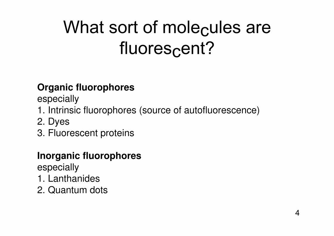
What sort of molecules are fluorescent?
Organic fluorophoresespecially1. Intrinsic fluorophores (source of autofluorescence)2. Dyes3. Fluorescent proteins
Inorganic fluorophoresespecially1. Lanthanides2. Quantum dots

5
What sort of molecules are fluorescent?1. Organic fluorophores
Chemical features:1. Conjugation2. Rigidity especially fused aromatic rings
3. Heteroatoms

6
Please rank these in order of fluorescence
2

7
What sort of molecules are fluorescent? 1. Endogenous organic fluorophores
Most common autofluorescent molecules:Flavins, NADH, NADPH, elastin, collagen, lipofuscin
Avoiding autofluorescence:choose dye emitting in red with big Stokes shiftadd quencher (Crystal violet)add reducing agent to react with autofluorescent moleculestime-gate fluorescence
8
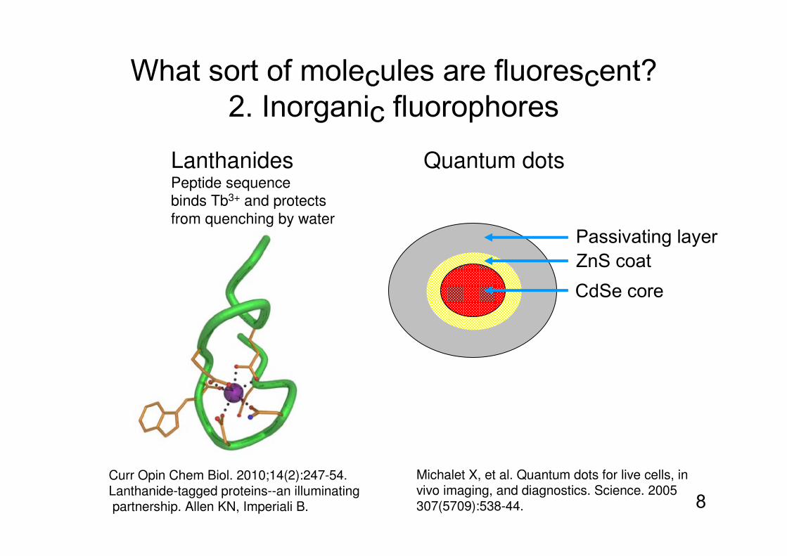
What sort of molecules are fluorescent? 2. Inorganic fluorophores
Lanthanides Quantum dotsPeptide sequencebinds Tb3+ and protects from quenching by water
CdSe coreZnS coatPassivating layer
Curr Opin Chem Biol. 2010;14(2):247-54. Lanthanide-tagged proteins--an illuminatingpartnership. Allen KN, Imperiali B.
Michalet X, et al. Quantum dots for live cells, in vivo imaging, and diagnostics. Science. 2005 307(5709):538-44.
9
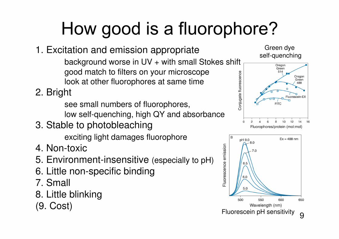
How good is a fluorophore?1. Excitation and emission appropriate
background worse in UV + with small Stokes shift
good match to filters on your microscopelook at other fluorophores at same time
2. Brightsee small numbers of fluorophores,
low self-quenching, high QY and absorbance
3. Stable to photobleachingexciting light damages fluorophore
4. Non-toxic5. Environment-insensitive (especially to pH)
6. Little non-specific binding7. Small8. Little blinking(9. Cost)
Fluorescein pH sensitivity
Green dye self-quenching
10
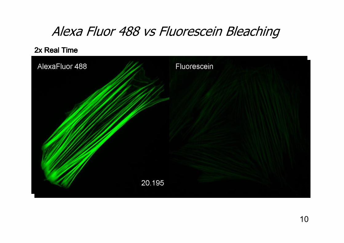
Alexa Fluor 488 vs Fluorescein Bleaching
2x Real Time2x Real Time2x Real Time2x Real Time

11
Laser-scanningcytometryEL4 cells biotin-anti-CD44+ streptavidin conjugates
Alexa Fluor Dyes – Photostability
Fluorescein is the commonest dye but has poor photostability.Also consider Atto dyes (Sigma) and Dyomics dyes

12
Protecting the fluorescence signal -Antifade Reagents for fixed cells
Scavenge and prevent reactive oxygen species from forming.
For fixed cells:Home made: 0.3% p-phenylene-diamine (Sigma)
or Propyl GallateVectashield: Proprietary, very effective all round, affects psfDabco Prolong Gold®
+ Prolong Gold
Untreated
13
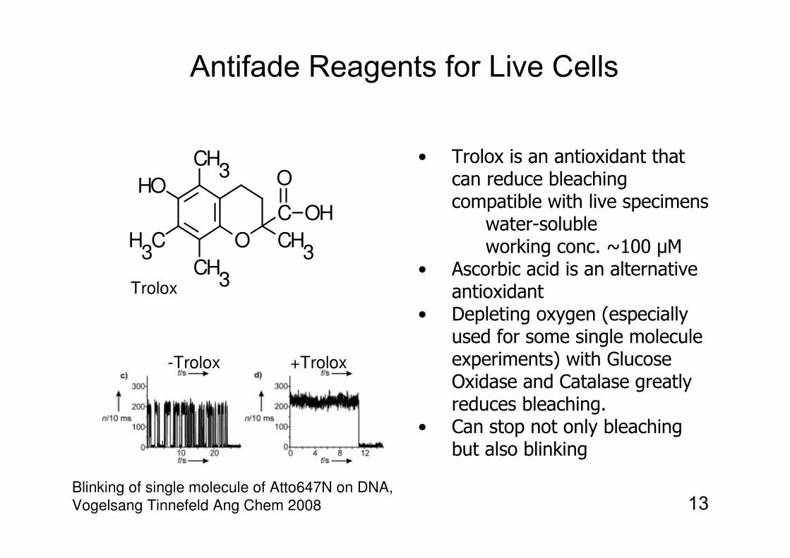
Antifade Reagents for Live Cells
O
CH3
HO
H3C
CH3
CH3
C OH
O• Trolox is an antioxidant that
can reduce bleaching compatible with live specimens
water-solubleworking conc. ~100 µM
• Ascorbic acid is an alternative antioxidant
• Depleting oxygen (especially used for some single molecule experiments) with Glucose Oxidase and Catalase greatly reduces bleaching.
• Can stop not only bleaching but also blinking
Trolox
-Trolox +Trolox
Blinking of single molecule of Atto647N on DNA, Vogelsang Tinnefeld Ang Chem 2008

14
Microsecond! fluorescent measurementswith Trolox + cysteamine
Oxygen helps stop triplet-state build-upBUT oxygen promotes photobleachingFor rapid photon cycling-1. leave oxygen in2. add Trolox to further quench triplet state3. include cysteamine (a thiol) to protect from singlet oxygen and hydroxyl radicals
Green/Red Alexa dye FRETon rapid folding protein V. Munoz Nat Meth 2011

15
Multiplexing- four main colours
Emission Emission Emission Emission wavelengths:wavelengths:wavelengths:wavelengths: Blue green orange/red fBlue green orange/red fBlue green orange/red fBlue green orange/red far redar redar redar red
Alexa FluorAlexa FluorAlexa FluorAlexa Fluor® 594594594594Texas Red, Cy3.5Texas Red, Cy3.5Texas Red, Cy3.5Texas Red, Cy3.5
Alexa FluorAlexa FluorAlexa FluorAlexa Fluor® 647647647647Cy5, APCCy5, APCCy5, APCCy5, APC
Alexa FluorAlexa FluorAlexa FluorAlexa Fluor® 555555555555Rhodamine,Rhodamine,Rhodamine,Rhodamine,TAMRA, TRITCTAMRA, TRITCTAMRA, TRITCTAMRA, TRITCCy3Cy3Cy3Cy3
Alexa FluorAlexa FluorAlexa FluorAlexa Fluor® 488488488488Fluorescein (FITC)Fluorescein (FITC)Fluorescein (FITC)Fluorescein (FITC)Cy2Cy2Cy2Cy2
Alexa FluorAlexa FluorAlexa FluorAlexa Fluor® 350350350350CoumarinCoumarinCoumarinCoumarin, AMCA, AMCA, AMCA, AMCA
350 400 450 500 550 350 400 450 500 550 350 400 450 500 550 350 400 450 500 550 600 650 700 600 650 700 600 650 700 600 650 700
DAPI/UVDAPI/UVDAPI/UVDAPI/UV FITCFITCFITCFITC TRITCTRITCTRITCTRITC FAR REDFAR REDFAR REDFAR RED
Colour Selection Colour Selection Colour Selection Colour Selection ♦♦♦♦ Brightness Brightness Brightness Brightness ♦♦♦♦ PhotostabilityPhotostabilityPhotostabilityPhotostability
350 488 555 350 488 555 350 488 555 350 488 555 647647647647ExcitationExcitationExcitationExcitationwavelengths:wavelengths:wavelengths:wavelengths:

16
Overview
1. What kind of structures are fluorescent
2. How to make and target fluorescent probes
3. Fluorescent probes for cellular structure and function
4. Using light to control cells

17
X GFP
GFP
X
?organic
fluorophores
quantumdots
photoaffinityprobes
Major bottleneck to using new probes is difficulty targeting them
fluorescent proteinseasy to target
other probes hardto target
18
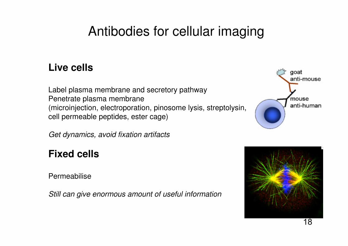
Antibodies for cellular imaging
Live cells
Label plasma membrane and secretory pathwayPenetrate plasma membrane(microinjection, electroporation, pinosome lysis, streptolysin, cell permeable peptides, ester cage)
Get dynamics, avoid fixation artifacts
Fixed cells
Permeabilise
Still can give enormous amount of useful information

19
Not just antibodies for targeting
Other types of targeting agents:
Proteins (especially antibodies, but also transferrin, insulin, EGF etc.)Peptides (MHC class I pathway, proteasome function)RNA (mRNA, molecular beacons, aptamers, siRNA)DNAlipids, lipoproteinsdrugs
?

20
How to dye: it is easy
Multiple ways to modify proteins(see Molecular Probes catalogue)
Most common ways are to modify:
1. Lysine
or
2. Cysteine
A Add dye to protein for 3 hr B 1cm Sephadex column to remove most free dye (10 min)C Dialyse away rest of free dye (24 hr)
maleimide-dye Thioether bondto dye
NHS-dye Amide bondto dye
Protein target

21
Site-specific protein labelling methods1. Binding domainSNAP-tag (NEB), HaloTag (Promega)
2. Binding peptideFlAsH (Invitrogen)
3. Enzymatic ligation to peptidePRIME AY Ting PNAS 2010
Chen & Ting, Curr. Opin. Biotech. 2005

22
Overview
1. What kind of structures are fluorescent
2. How to make and target fluorescent probes
3. Fluorescent probes for cellular structure and function
4. Using light to control cells

23
(follow DNA even when
nucleus breaks down)
Fixed cells:
Intercalate into DNA
DAPI
(well away from other channels)
Hoechst 33342
Live cells:
histone H2B-GFP
Putting the signal in context: nuclear labelling
24
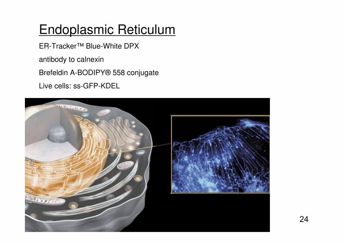
Endoplasmic ReticulumER-Tracker™ Blue-White DPX
antibody to calnexin
Brefeldin A-BODIPY® 558 conjugate
Live cells: ss-GFP-KDEL
25
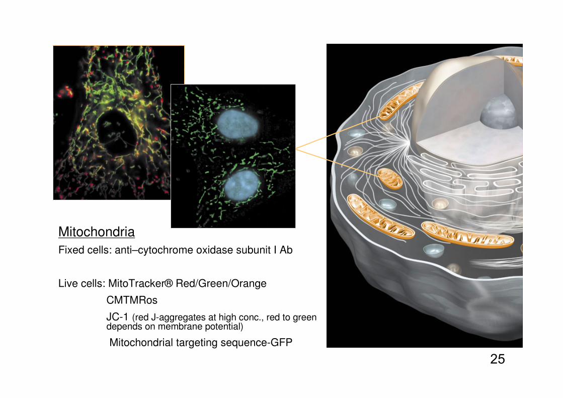
Mitochondria
Fixed cells: anti–cytochrome oxidase subunit I Ab
Live cells: MitoTracker® Red/Green/Orange
CMTMRos
JC-1 (red J-aggregates at high conc., red to green depends on membrane potential)
Mitochondrial targeting sequence-GFP

26
Lysosomes
Fixed cells: anti-LAMP1
Live cells: LysoTracker® Red /Green (weakly basic amines can accumulate in lysosomes)
LysoSensor™ Yellow/Blue DND-160, LAMP1-GFP

27
Lipid Rafts
BODIPY® FL C5-ganglioside GM1
Fluorescent Cholera Toxin subunit B (CT-B)

28Fixed cells: phalloidin-dye
Live cells: Lifeact-GFP (17 aa peptide binding actin)
Putting the signal in context: actin labelling
29
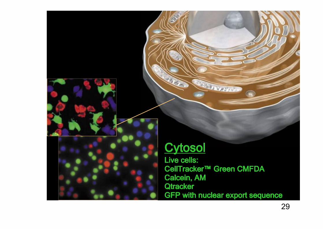
CytosolCytosolCytosolCytosolLive cells:Live cells:Live cells:Live cells:CellTrackerCellTrackerCellTrackerCellTracker™™™™ Green Green Green Green CMFDACMFDACMFDACMFDACalceinCalceinCalceinCalcein, AM, AM, AM, AMQtrackerQtrackerQtrackerQtrackerGFP with nuclear export sequenceGFP with nuclear export sequenceGFP with nuclear export sequenceGFP with nuclear export sequence

30
The breakthrough of fluorescent proteinsfrom jellyfish
How much did he need?
OsamuShimomura
Aequoreavictoria

31
X GFP
GFP
X
GreenFluorescentProtein
The breakthrough of fluorescent proteinsfor live cell imaging
GFP foldβ-can
GFP chromophorefrom Ser-Tyr-Gly
Link GFP sequence to gene ofyour favourite protein
GFP foldsand becomesfluorescent
GFP lights up your favourite protein in cell

32
Fluorescent proteins are
more than just labels
Photoactivation/PhotoswitchingPA-GFP, Dronpa, Eos
Reporting on environmentCa2+, phosphorylation, cAMP, cGMP, pH, neurotransmitters, voltage, cell cycle, redox
Reporting on protein-protein interactionCFP/YFP FRET, split fluorescent proteins
Modifying environmentSinglet oxygen generation, Channelrhodopsin
Targeting advantageto defined compartment,cell-type,
developmental stage

33
GreenFluorescentProtein
Chromophores in switching
GFP foldβ-can
PA-GFP, PS-CFP2
Dendra, Eos
Dronpa
Link GFP sequence to gene ofyour favourite protein
GFP foldsand becomesfluorescent
34
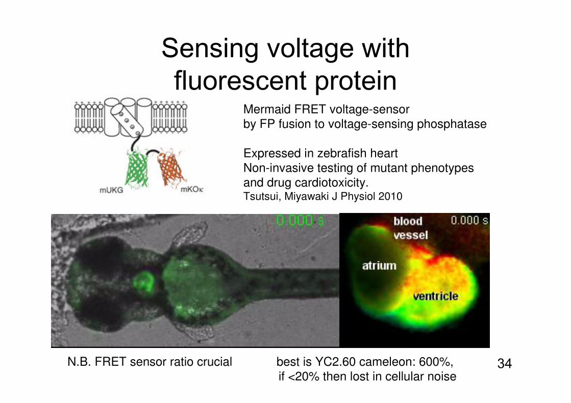
Sensing voltage with fluorescent protein
Mermaid FRET voltage-sensorby FP fusion to voltage-sensing phosphatase
Expressed in zebrafish heart Non-invasive testing of mutant phenotypesand drug cardiotoxicity.Tsutsui, Miyawaki J Physiol 2010
N.B. FRET sensor ratio crucial best is YC2.60 cameleon: 600%,if <20% then lost in cellular noise

35
Small molecule fluorescent sensors
Metal ions: calcium, magnesium, zinc, sodium, potassium, chloride,
mercury
pH (also dyes to conjugate to proteins, CyPher from GE, SNARF from
Invitrogen)
Reactive oxygen species, nitric oxideTransmembrane potential
Fura-2 sensing calcium

36
Why use small molecule rather than genetically-encoded probes?1. No need to transfect
hard for some organisms and primary cellseasier to titratepotential clinical application-e.g. image-guided surgery
2. Probes often brighter, with bigger signal to noisestruggle to make GFP-based calcium reporter as good as fura-like dyes
3. Probes with entirely different fluorescent properties QD photostability, probes with long fluorescence lifetimes, photouncaging
4. Smallere.g. calcium conc. right next to pore of ion channel
MMP-activated Cy5 peptidelabels tumour (RY Tsien 2010)

37
How good is a fluorescent protein?
A. victoria GFP is good for jellyfish,but not great for cell biologists!

38
How good is a fluorescent protein?
A. victoria GFP is terrible! EGFP is OK, but there are now better...1. Excitation and emission λ good match to filters on your microscope
look at other fluorophores at same time
2. Bright ε x QY Clover, YPet 2.5 x EGFP
mRuby2 3x mCherry
3. Stable to photobleaching EBFP bad, mCherry and YPet good
4. Non-toxic attach on right part of your protein
all make H2O2, FPs can transfer electrons
5. Environment-insensitive especially to pH, chloride
CyPet does not fold at 37°C, all need O2
Photoactivatable FP did not work in ER
6. Little non-specific binding fully monomeric, A206K non-dimerising
7. Fast Maturation Venus 2 min. Red FPs can start off green!
half-time ~15 min mCherry, 100 min TagRFP

39
You MUST worry about FP multimerization!
Tag multimerizing protein with FP and sometimes see foci-are these real or caused by the tag?
With hexameric barrel involved in E. coli protein degradation,many commonly used FPs induce artifactual foci(no cluster with Ab or SNAP-Tag)
as well as affecting daughter cell inheritance of proteolysis ability
mCherry, sfGFP, mYPet poor!mGFPmut3, Dronpa OKD. Landgraf et al. Nature Meth 2012

40
Problems with GFP in cells• GFP with light can donate electrons GFP with light can donate electrons GFP with light can donate electrons GFP with light can donate electrons
to different acceptorsto different acceptorsto different acceptorsto different acceptors(FMN, FAD, NAD+, cyt. c)GFP reddens after transfer: photobleaching and phototoxicityuse DMEM lacking e- acceptors
(riboflavin or all vitamins) for less bleaching(HEK 293T happy for 1 week)effect for EGFP and PA-GFP, not RFPsLukyanov Nat Meth 2009
• EGFP not good in secretory pathwayEGFP not good in secretory pathwayEGFP not good in secretory pathwayEGFP not good in secretory pathwaymixed disulfide oligomers in ER andnon-fluorescent in E. coli periplasm(superfolder GFP behaves fine)Erik Snapp, Traffic 2011

41
Fluorescent RNA imaging
See single mRNA: MS2 mRNA stem-loops bound by MCP-YFPSee product of translation: mRNA encodes CFP-SKL which goes to peroxisomes
S.M.Janicki et al. Cell 2004
Spinach RNA 60 nt aptamer binds cell-permeable fluorogenic dyePhotostable. Used to label 5S RNA in HEK cells. Samie Jaffrey Science 2011
MCP-YFP

42
Overview
1. What kind of structures are fluorescent
2. How to make and target fluorescent probes
3. Fluorescent probes for cellular structure and function
4. Using light to control cells

Why use light to control biology?Light control allows extreme temporal and spatial control.
Temporal controlgenes< chemicals < lightmin-hr s-min µs-s
Spatial controlchemicals / genes < light (note micropipettes for preciseone or many cells 1 μm part of cell small molecule delivery)
(often combine chemical/light control or gene/light control)optogenetics/chemogenetics
Limitations of light? $$$$$and usually data on one cell at a time

44
Controlling biology with light:light-gated ion channels
Channelrhodopsin from an alga, like rhodopsin, undergoes retinal isomerisationin response to light, and changes conformation, but opens a Na+ channel. This allows light to control membrane voltage and trigger neuron firing.
to understand neuronal firing patternsto control secretion in diabetespotentially in fixing neural diseases?e.g. damping down overactivity in epilepsy
trains of electrical firing flashes of light

LOV domains react and switch conformation with light
FMN cofactorpresent in all cells.Light polarises to increasereactivity to Cys attack.
LOV domains: light, oxygen, voltage respondersones responding to blue light in bacteria, plants and fungi
New pattern ofprotein-protein
interaction...

Genetically-encoded photoactivation
1. Constitutively active Rac mutant2. Optimise LOV-Rac junction,3. knockout GTP hydrolysis and GAP/GNDI/GEF interactionsKd for PAK 2 μM in dark, 200nM in light 10-fold ratioInteraction of Rac with PAK stimulates cell protrusion and migration.
K.Hahn et al. Nature Sept. 2009
458 or 473nm light
spontaneousreversion at RT
t1/2 43s for
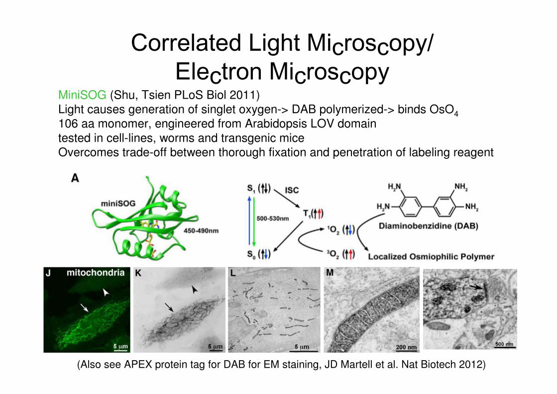
Correlated Light Microscopy/Electron Microscopy
MiniSOG (Shu, Tsien PLoS Biol 2011)Light causes generation of singlet oxygen-> DAB polymerized-> binds OsO4
106 aa monomer, engineered from Arabidopsis LOV domaintested in cell-lines, worms and transgenic miceOvercomes trade-off between thorough fixation and penetration of labeling reagent
(Also see APEX protein tag for DAB for EM staining, JD Martell et al. Nat Biotech 2012)

48
Conclusions
Choosing the right dye or fluorescent protein can make a big difference for:
sensitivitysignal stabilitymodification to molecule/cell function
by size or multimerization
Fluorescent probes allow more than
just following location:reporting cellular eventsuncaging biomolecule functioncontrolling interactions and ion flux
49
References
Fluorescence probesMolecular Probes Handbook, free from Invitrogen.Principles of Fluorescence Spectroscopy 2nd edition, by Joseph R. Lakowicz.
Protein modificationBioconjugate Techniques, 2nd Edition by Greg T. Hermanson.Chemical labeling strategies for cell biology, Marks KM, Nolan GP. Nat Methods. 2006 Aug;3(8):591-6.
Fluorescent proteins
(i) as labels: Nat Methods. 2012;9:1005-12. Improving FRET dynamic range with bright green and red fluorescent proteins. Lam AJ et al. Poster: http://www.nature.com/nrm/posters/fluorescent/index.html
(ii) as sensors: Designs and applications of fluorescent protein-based biosensors.Ibraheem A, Campbell RE.Curr Opin Chem Biol 2010;14:30-6