flowering locus t protein may act as the long … · flowering locus t protein may act as the...
TRANSCRIPT

FLOWERING LOCUS T Protein May Act as the Long-DistanceFlorigenic Signal in the Cucurbits W
Ming-Kuem Lin,a Helene Belanger,b Young-Jin Lee,c Erika Varkonyi-Gasic,b Ken-Ichiro Taoka,a Eriko Miura,a
Beatriz Xoconostle-Cazares,a,d Karla Gendler,e Richard A. Jorgensen,e Brett Phinney,c Tony J. Lough,b
and William J. Lucasa,1
a Section of Plant Biology, College of Biological Sciences, University of California, Davis, California 95616b AgriGenesis BioSciences, Auckland 1140, New Zealandc Proteomics Core Facility, Genome Center, University of California, Davis, California 95616d Departmento de Biotecnologia y Bioingenieria, Centro de Investigacion y Avanzados del Instituto Politecnico
Nacional, Zacatenco 07360, Mexicoe Department of Plant Sciences, University of Arizona, Tucson, Arizona 85721-0036
Cucurbita moschata, a cucurbit species responsive to inductive short-day (SD) photoperiods, and Zucchini yellow mosaic
virus (ZYMV) were used to test whether long-distance movement of FLOWERING LOCUS T (FT) mRNA or FT is required for
floral induction. Ectopic expression of FT by ZYMV was highly effective in mediating floral induction of long-day (LD)–treated
plants. Moreover, the infection zone of ZYMV was far removed from floral meristems, suggesting that FT transcripts do not
function as the florigenic signal in this system. Heterografting demonstrated efficient transmission of a florigenic signal
from flowering Cucurbita maxima stocks to LD-grown C. moschata scions. Real-time RT-PCR performed on phloem sap
collected from C. maxima stocks detected no FT transcripts, whereas mass spectrometry of phloem sap proteins revealed
the presence of Cm-FTL1 and Cm-FTL2. Importantly, studies on LD- and SD-treated C. moschata plants established that
Cmo-FTL1 and Cmo-FTL2 are regulated by photoperiod at the level of movement into the phloem and not by transcription.
Finally, mass spectrometry of florally induced heterografted C. moschata scions revealed that C. maxima FT, but not FT
mRNA, crossed the graft union in the phloem translocation stream. Collectively, these studies are consistent with FT
functioning as a component of the florigenic signaling system in the cucurbits.
INTRODUCTION
Leaf-to-apex communication initiates flowering in response to
environmental cues such as photoperiod. Grafting, phloem-
girdling, timed removal of leaves, and fractional and photoperiodic
induction experiments have been used to show that photoperiod
is perceived in the leaves and a phloem-mobile floral stimulus
(florigen), or inhibitor, is transported through the phloem trans-
location stream to the shoot apical meristem (Chailakhyan, 1936;
Zeevaart, 1962, 1976; Lang, 1965, 1977; Bernier, 1988; Colasanti
and Sundaresan, 2000; Bernier and Perilleux, 2005).
The molecular genetics of photoperiodic floral induction are
best characterized in Arabidopsis thaliana (Mouradov et al.,
2002; Bastow and Dean, 2003; Searle and Coupland, 2004;
Corbesier and Coupland, 2005). Key elements involved in inte-
grating the input signal(s) from the photoperiodic pathway are
the zinc-finger protein CONSTANS (CO) (Putterill et al., 1995) and
the RAF kinase inhibitor–like protein FLOWERING LOCUS T (FT)
(Kardailsky et al., 1999; Kobayashi et al., 1999). Promoter
b-glucuronidase analyses have revealed vascular- rather than
meristem-specific expression patterns for these genes, findings
consistent with long-distance signaling roles for both CO and FT
in floral induction (Takada and Goto, 2003; An et al., 2004).
A signaling role for CO was confirmed using tissue-specific
promoters in which it was demonstrated that its expression,
within companion cells located in source leaves, promoted
flowering in the co mutant background (An et al., 2004). Grafting
studies confirmed that CO controls the production of a phloem-
borne substance (presumably florigen) essential for floral induc-
tion. Assignment of CO, per se, as florigen was discounted as
companion cell-specific expression of a CO:green fluorescent
protein (GFP) construct did not reveal a capacity for CO-GFP to
traffic beyond the cells in which it was transcribed (An et al.,
2004; Ayre and Turgeon, 2004). However, the CO-GFP fusion
protein was still able to rescue flowering in the co mutant back-
ground.
A role for FT and/or FT mRNA as the phloem-mobile florigenic
signaling agent(s) was tested using a range of tissue-specific
promoters. In contrast with CO, FT promoted flowering not only
when it was expressed in source companion cells, but also in
numerous other tissues, including those of the meristem, a tissue
in which it is not normally detected by in situ hybridization (An
et al., 2004). Given that FT expression is controlled by CO (Samach
et al., 2000) and is required to promote flowering (Kardailsky et al.,
1999; Kobayashi et al., 1999), these results strongly implicated
FT as a component of the florigenic signaling system.
1 To whom correspondence should be addressed. E-mail [email protected]; fax 530-752-5410.The author responsible for distribution of materials integral to thefindings presented in this article in accordance with the policy describedin the Instructions for Authors (www.plantcell.org) is: William J. Lucas([email protected]).W Online version contains Web-only data.www.plantcell.org/cgi/doi/10.1105/tpc.107.051920
The Plant Cell, Vol. 19: 1488–1506, May 2007, www.plantcell.org ª 2007 American Society of Plant Biologists
As the phloem sap has been demonstrated to contain a range
of mRNA species and RNA binding proteins (Ruiz-Medrano et al.,
1999; Xoconostle-Cazares et al., 1999; Yoo et al., 2004; Gomez
et al., 2005), some of which appear to influence events taking
place in the meristem (Kim et al., 2001; Haywood et al., 2005),
the possibility exists that FT mRNA could act as the signaling
molecule. Lifschitz et al. (2006) conducted a series of hetero-
grafting studies to test this possibility. Although expression of
SINGLE FLOWER TRUSS (SFT), a tomato (Solanum lycopersi-
cum) FT homolog, in a clade orthologous to the FT/TWIN SISTER
OF FT (TSF) clade, in the tomato stock could rescue flowering in
the mutant scion, analysis of RNA extracted from scion tissues
failed to detect SFT mRNA in grafts in which floral induction had
occurred. These results did not support a role for the long-
distance trafficking of FT mRNA in floral induction.
Two recent studies conducted on Arabidopsis and rice (Oryza
sativa) examined whether FT protein and the rice ortholog
Hd3a participate in long-distance signaling to induce flowering
(Corbesier et al., 2007; Tamaki et al., 2007). Here, transgenic
plant lines expressing FT-GFP and Hd3a-GFP in vascular tissues
were used to test whether FT and Hd3a move in the phloem to the
shoot apical meristem (SAM) to induce flowering. Evidence con-
sistent with such long-distance movement was presented, but
neither study provided definitive proof against a role for FT mRNA.
In this study, we used a combination of Cucurbita moschata, a
cucurbit (squash) species responsive to inductive short-day (SD)
photoperiods, and a potyvirus vector, Zucchini yellow mosaic
virus (ZYMV), to test whether long-distance movement of FT
mRNA and/or FT is required for floral induction. The choice of a
potyvirus was important, as these plant viruses are polycistronic,
encoding for a polyprotein and, hence, do not produce sub-
genomic RNA species. Ectopic expression of FT by ZYMV was
highly effective in mediating floral induction of long-day (LD)–
treated C. moschata plants. Analysis of such induced plants
revealed that the infection zone of ZYMV was not coincident with
the floral meristems, indicating that FT transcripts are unlikely to
function as the florigenic signal in this system. Heterografting
studies demonstrated the efficient transmission of a florigenic
signal from flowering pumpkin (Cucurbita maxima) stocks to
LD-grown C. moschata scions. Real-time RT-PCR performed on
phloem sap collected from these flowering C. maxima stocks
failed to detect the presence of FT transcripts, whereas mass
spectrometry analysis revealed the presence of FT protein in the
translocation stream. Importantly, parallel mass spectrometry
analysis of the phloem sap from florally induced C. moschata
plants and scions also detected the presence of FT proteins.
These studies are consistent with FT functioning as the florigenic
signal in photoperiodic induction of flowering.
RESULTS
Screening Cucurbits for Photoperiodically
Controlled Flowering
Ideally, to test for the presence of FT mRNA and/or FT protein in
the phloem translocation stream, a photoperiodically inducible
model plant system should be employed in which it is possible to
gain direct access to the phloem sap. Given that the cucurbits
have been widely used to investigate the phloem translocation
stream (Golecki et al., 1998, 1999; Lough and Lucas, 2006),
we performed a photoperiodic screen of 97 cucurbit acces-
sions, obtained from Plant Genetic Resources Conservation Unit
(www.ars-grin.gov/ars/SoAtlantic/Griffin/pgrcu). One accession,
C. moschata PI441726, representing an undomesticated squash
species, remained vegetative under LD conditions but flowered
when grown under SD growth conditions. Seed collected from
C. moschata PI441726 was rescreened and, as shown in Figure
1A, floral induction was confirmed under SD conditions; plants
grown under LD conditions, by use of daylength extension,
remained vegetative and failed to develop floral buds (Figure 1B).
Seed stock obtained from this second screening was used for
subsequent viral expression and C. maxima (stock):C. moschata
PI441726 (scion) heterografting studies.
Cucurbit FT Homologs Activate Flowering in Arabidopsis
The basic tenet of the long-distance florigenic signal is that in
some form it will be common to all plant species (Chailakhyan,
1937; Lifschitz et al., 2006). This would suggest that cucurbit FT
homologs would function in Arabidopsis and vice versa. To test
this principle, we first employed C. moschata PI441726 (hereafter
referred to as simply C. moschata) and C. maxima vascular strip
cDNA libraries to isolate related cDNA clones that were being
expressed in flowering plants. Two full-length clones wereobtained
for each species and were named C. moschata FLOWERING
LOCUS T-LIKE1 (Cmo-FTL1) and Cmo-FTL2, and C. maxima
FLOWERING LOCUS T-LIKE1 (Cm-FTL1) and Cm-FTL2. The
Arabidopsis FT, Cm-FTL1, Cm-FTL2, Cmo-FTL1, and Cmo-FTL2
conceptual amino acid sequences (see Supplemental Figure
1 online) all exhibited an extremely high level of conservation.
A molecular phylogenetic analysis of the FT family of proteins
was performed, and the results demonstrate that Cmo-FTL1/
Cm-FTL1 and Cmo-FTL2/Cm-FTL2 form clades that are orthol-
ogous to a clade formed by At-FT and At-TSF (Figure 1C) (i.e.,
there is no one-to-one orthologous relationship between the C.
moschata and C. maxima genes and either Arabidopsis gene).
Rather, there exists a two-for-two relationship in which the
cucurbit FTL1/FTL2 clade is orthologous to the Arabidopsis FT/
TSF clade. In other words, we infer that there was an FT homolog
in the most recent common ancestor of cucurbits and Arabi-
dopsis inherited by each lineage and then was duplicated inde-
pendently in the lineages leading to both the cucurbits and
Arabidopsis. Thus, subsequent to duplication, each gene pair
had the potential for subfunctionalization and/or neofunctional-
ization; if either occurred in either lineage, an exact functionally
equivalent relationship would not exist between either cucurbit
gene and FT (or TSF). FT and TSF are known to be partially
redundant, but although they differ functionally, their differences
may be limited to transcriptional patterns in response to other
flowering regulators, and there may be no difference in func-
tion of their mRNA or protein products (Michaels et al., 2005;
Yamaguchi et al., 2005).
A functional analysis was next performed, using the com-
panion cell-specific Arabidopsis SUC2 promoter (Truernit et al.,
1996), to determine whether Cm-FTL1 and Cm-FTL2 were
able to induce early flowering in Arabidopsis. In contrast with
FT Protein as the Florigenic Signal? 1489

the vector control, transgenic Arabidopsis plants expressing
At-SUC2pro:Cm-FTL1 or At-SUC2pro:Cm-FTL2 exhibited early
flowering phenotypes (Figure 1D). The timing of floral induction
was determined by counting the number of rosette leaves at
the time of bolting. These studies established that the Cm-FTL1
and Cm-FTL2 gene products accelerated floral initiation in a
manner equivalent to that observed in Arabidopsis lines ex-
pressing CO or FT under the SUC2 promoter (Figure 1E).
Viral Vector-Mediated Induction of Flowering in
LD-Grown C. moschata
A cucurbit-infecting potyvirus, ZYMV, was used as a vector (Hsu
et al., 2004) to ectopically express At-FT in C. moschata plants
(Figures 2A and 2B). ZYMV was employed because it encodes
for a polyprotein (Figure 2A) and does not produce subgenomic
RNAs; thus, only FT protein would be free to move ahead of the
Figure 1. Analysis of Cucurbit FT-Like Orthologs.
(A) and (B) Flowering in C. moschata PI441726 was induced when plants were grown under SD (A) but repressed under LD (B) conditions. White box
represents daylight, black box represents darkness, and red box represents daylength extension interrupting the dark period. Bars ¼ 1 cm.
(C) Phylogenetic analysis of the relationship between Cmo-FTL1/FTL2, Cm-FTL1/ FTL2, and the FT-TFL family in angiosperms. The tree was constructed
based on a nucleic acid alignment of ;500 protein coding nucleotides using the neighbor-joining method implemented in MEGA 3.1. Numbers at each
branch point are the bootstrap values for percentages of 1000 replicate trees. The portions of the tree corresponding to clusters that include Arabidopsis
MOTHER OF FT (MFT), Antirrhinum majus CENTRORADIALIS (CEN), and two clusters of rice genes are compressed and can be viewed in Supplemental
Figure 1 online (see Supplemental Figures 2 and 3 online for additional details). The CEN cluster includes Arabidopsis FT homologs TFL1, BFT, and ATC.
Gene accession numbers are in Supplemental Table 1 online. Abbreviations for species are as follows: Arabidopsis thaliana (At), Cucurbita maxima (Cm),
Cucurbita moschata (Cmo), Solanum lycopersicum (Sl), Malus 3 domestica (Md), Oryza sativa (Os), and Populus trichocarpa (Pt).
(D) The SUC2 promoter was employed to drive companion cell–specific expression of CO, FT, Cm-FTL1, and Cm-FTL2 in transgenic Arabidopsis plants
grown under noninductive SD conditions.
(E) Timing of floral initiation in transgenic Arabidopsis plants was determined by counting the number of rosette leaves formed at the time of bolting
(mean 6 SE, n ¼ 20 plants per treatment).
1490 The Plant Cell

Figure 2. ZYMV-Mediated Expression of At-FT Induces Flowering in C. moschata PI441726 Grown under LD Noninductive Conditions.
(A) Genome arrangement of the ZYMV vector indicating the sites of insertion of GFP and At-FT.
(B) ZYMV-At-FT conceptual peptide sequence. Residues fused to the N and C terminus of At-FT are underlined, and arrows indicate the positions of
autocatalytic NIa-mediated cleavage.
(C) ZYMV-mediated expression of GFP did not induce flowering in LD-grown C. moschata plants. Bar ¼ 10 cm.
FT Protein as the Florigenic Signal? 1491

viral infection front. In addition, the ZYMV infectious clone used
was earlier mutated to produce an attenuated strain that caused
only very mild symptoms (Hsu et al., 2004). Plants of C. moschata
were grown under noninductive LD conditions using a 10-h
photoperiod and a 14-h night period that was interrupted by
15-min light breaks every hour. Microprojectile bombardment
was used to initiate infection with either the ZYMV (empty vector),
or ZYMV clones carrying GFP (ZYMV-GFP; control) or At-FT
(ZYMV-At-FT), and plants were then observed for the vegetative-
to-floral transition. Infection by ZYMV-At-FT led to floral induc-
tion, whereas ZYMV-GFP or ZYMV (empty vector) treated plants
remained vegetative under these LD noninductive conditions
(Figures 2C to 2E).
Time-course experiments for floral induction, mediated by
ZYMV-At-FT or an inductive SD photoperiod, were next per-
formed. As illustrated in Figures 2F and 2G, by 21 d after
inoculation (DAI), early bud formation was detected on ZYMV-
At-FT–inoculated C. moschata plants grown under either LD or
SD conditions. Upon floral induction, bud formation was ob-
served at all subsequent nodes. Importantly, under LD condi-
tions, plants inoculated with ZYMV (vector-only) or ZYMV-GFP
did not form buds at 28 or 40 DAI (Figure 2F). However, under SD
conditions, these control plants underwent the vegetative-
to-floral transition 1 week and four to six nodes later than the
ZYMV-At-FT–inoculated C. moschata plants (Figure 2G). Finally,
the percentage of plants exhibiting floral initiation, at each node,
was always highest in the ZYMV-At-FT–inoculated plants.
ZYMV Infection Domains Do Not Include Meristems
The observed ZYMV-At-FT–mediated floral induction may have
resulted from cell autonomous activation in the SAM and/or
lateral meristems. To test for this possibility, a series of exper-
iments was next performed to establish the physical relationship
between the ZYMV infection front and the SAM/lateral meristems
on the C. moschata plants. The infection pathway was first de-
fined in ZYMV-GFP–infected plants grown under LD conditions.
Analysis of GFP fluorescence in freehand sections, taken 30 DAI,
indicated that the virus was present in all tissues except for the
last four nodes located at the shoot apex. A fluorescent signal
was not detected in the C. moschata apical or lateral meristems.
Immunolocalization studies were next performed using poly-
clonal antibodies directed against the ZYMV coat protein (CP).
As shown in Figures 3A and 3B, CP accumulation, associated
with ZYMV-GFP and ZYMV-At-FT infection, could be detected in
young developing sink leaves located at the fifth or sixth node
back from the apex. Note that uninfected control plants were
devoid of CP signal (Figure 3C). An examination of longitudinal
sections, derived from the apices of ZYMV-GFP (Figure 3D) and
ZYMV-At-FT–infected plants (70 sections examined from 10
apices per treatment; Figure 3E), confirmed that the virus had
not entered into these meristematic tissues. Uninfected control
plants were similarly devoid of CP staining (Figure 3F). Analysis of
lateral meristems of ZYMV-At-FT and ZYMV-GFP (Figure 3G)
infected C. moschata plants identified the presence of virus in
developing leaves, but signal was again absent from the apical
tissues. Collectively, these studies are consistent with the hy-
pothesis that ZYMV-At-FT infection induces the floral transition
in the C. moschata meristem by means of non-cell-autonomous
signaling through the phloem. As the FT sequence is present only
in the ZYMV infectious RNA, our inability to detect virus in the
apex and lateral meristems suggested that FT mRNA, per se,
may not be directly involved in floral induction. However, based
on these experiments alone, we cannot exclude the possibility
that a very low level of ZYMV went undetected and that this was
the causal agent for floral induction.
Heterograft Experiments Confirm Phloem Delivery of
Florigenic Signal
The nature of florigen as a phloem-mobile signal was earlier
established using grafting experiments performed with florally
induced stocks and noninduced scions (Chailakhyan, 1937;
Zeevaart, 1976; Lang, 1977). As C. maxima (a day-neutral spe-
cies) flowers under LD conditions, whereas C. moschata remains
vegetative under this same growth regime, heterograft ex-
periments were next preformed using these two plant species.
As illustrated in Table 1, in the control experiment in which
C. moschata scions were grafted onto C. moschata stocks, the
scions remained vegetative when grown under LD noninductive
conditions. By contrast, C. moschata scions grafted onto flower-
ing C. maxima stocks were all induced to produce floral buds.
Hence, the phloem sap of flowering C. maxima carries a com-
patible florigenic signal that mediates the vegetative-to-floral
transition in C. moschata.
Figure 2. (continued).
(D) ZYMV-mediated expression of At-FT–induced flowering in LD-grown C. moschata plants. Bar ¼ 10 cm.
(E) Flowering rate was scored as the percentage of plants in each treatment having floral buds/flowers at 40 DAI (mean 6 SE, n ¼ 30 plants per
treatment).
(F) C. moschata plants, maintained under noninductive LD conditions, were inoculated with ZYMV-At-FT (red bars), ZYMV-GFP (gray bars), or mock
treatment (blue bars). Bud formation was first observed at 21 DAI on ZYMV-At-FT plants and occurred from the 4th and all subsequent nodes. Mock-
inoculated and ZYMV-GFP plants did not undergo floral initiation under these noninductive LD conditions.
(G) C. moschata plants, maintained under inductive SD conditions, were inoculated with ZYMV-At-FT (red bars), ZYMV-GFP (gray bars), or mock
treatment (blue bars). Note that mock-inoculated and ZYMV-GFP plants were induced to flower ;1 week later and four to six nodes higher on the plant
than ZYMV-At-FT plants.
Data in (F) and (G) are expressed as percentage of plants (n ¼ 5 to 9) forming stage 2 floral buds as a function of nodal position and DAI. White box
represents daylight, black box represents darkness, and white lines represent hourly night breaks of 15 min with incandescent (white) light.
1492 The Plant Cell

FT mRNA Was Not Detected in Phloem Sap of Florally
Induced C. maxima Plants
To further investigate the role of FT mRNA and/or FT protein as
a component of the florigenic long-distance signaling system,
we next examined the relationship between expression of the
C. maxima FT/TSF orthologs and their capacity to enter the
phloem translocation stream. For these experiments, LD-grown
nonflowering (4-week-old) and flowering (6- to 12-week-old)
C. maxima plants were used to obtain mRNA from stem vascular
tissue. Real-time RT-PCR analysis was employed, and in these
experiments, Cm-FTL1 and Cm-FTL2 mRNA levels were nor-
malized to Cm-PP16, as this mRNA species was earlier shown to
be expressed in companion cells prior to its entry into the sieve
elements for long-distance translocation (Ruiz-Medrano et al.,
1999; Xoconostle-Cazares et al., 1999; Haywood et al., 2005).
Furthermore, the Cm-PP16 mRNA levels were found to be
equivalent over the range of growth conditions employed. As
shown in Figure 4A, the levels of Cm-FTL1 and Cm-FTL2 mRNA
were low in 4-week-old pumpkin plants. Under the growth
conditions employed for our studies, C. maxima plants produced
their first floral buds ;5 weeks after germination. Consistent with
this observation, the levels of Cm-FTL1 and Cm-FTL2 mRNA
increased in stem vascular tissue of 6- to 8-week-old plants.
Based on these findings, all subsequent analyses were per-
formed on 6-week-old LD-grown pumpkin plants.
A time course for Cm-FTL1 and Cm-FTL2 expression was next
established to ascertain the appropriate period to collect phloem
sap for RNA analysis. Vascular tissues were collected over a 24-h
period and real-time RT-PCR performed. In contrast with LD-
grown Arabidopsis, where the levels of FT mRNA undergo a
considerable increase upon the onset of darkness (Suarez-
Lopez et al., 2001; Searle and Coupland, 2004), neither Cm-
FTL1 nor Cm-FTL2 transcript levels exhibited marked peaks at
the end of the photoperiod (Figure 4B). Based on these findings,
we selected four time points for the collection of phloem sap to
be used for RNA (and protein) analysis.
Our RNA analysis of the phloem sap confirmed that, as in pre-
vious studies (Ruiz-Medrano et al., 1999; Xoconostle-Cazares
et al., 1999; Haywood et al., 2005), Cm-PP16 mRNA was readily
detected (Figure 4C). However, by contrast, Cm-FTL1 and Cm-
FTL2 transcripts were undetectable in the phloem sap collected
from flowering pumpkin plants. Note that Cm-rbcS transcripts
were detected at a level 10,000 times below that for Cm-PP16.
These results suggested that, in pumpkin, Cm-FTL1 and Cm-FTL2
mRNA may not be components of the phloem long-distance
signaling system involved in floral induction.
Cm-FTL1 and Cm-FTL2 Are Present in Phloem Sap of
Flowering Pumpkin
To test for the presence of Cm-FTL1 and Cm-FTL2 in the vas-
cular tissue and phloem sap of flowering pumpkin plants, pro-
teins were extracted and then separated by fast protein liquid
chromatography (FPLC) methods. Conceptual translations for
Cm-FTL1 and Cm-FTL2 predicted positively charged proteins.
Consequently, proteins were dialyzed, clarified by centrifugation,
and loaded onto a HiTrap SP cation-exchange column, con-
nected to an Amersham Biosciences FPLC system; proteins
were eluted with a linear NaCl gradient. Figures 5A and 5C il-
lustrate the profiles of the FPLC-fractionated vascular tissue and
phloem sap proteins, respectively.
A combination of liquid chromatography–tandem mass spec-
trometry (LC-MS/MS) was used to interrogate the FPLC-frac-
tionated proteins for the presence of Cm-FTL1 and/or Cm-FTL2.
To this end, vascular tissue and phloem sap fractions were
separated on SDS-PAGE gels, the 20-kD regions excised, and
the proteins then trypsin digested, in-gel, for LC-MS/MS analysis.
Figure 3. ZYMV Infection Domains Do Not Include Meristems.
Virus accumulation and localization were determined by immunohisto-
chemical staining of sections using antisera specific for the ZYMV coat
protein. Tissues were collected 35 to 40 DAI, and images are represen-
tative of those observed from six plants (>30 sections per plant) per
treatment. Bars ¼ 500 mm.
(A) to (C) Transverse sections of lamina from young expanding leaves
located at the ninth node of plants having 12 to 14 visible nodes. Plants
were inoculated with ZYMV-GFP (A), ZYMV-At-FT (B), or mock inocu-
lated (control) (C). Note the presence of CP (dark brown stain; see
arrowheads) in plants inoculated with ZYMV-GFP or ZYMV-At-FT.
(D) to (F) Longitudinal sections from apical meristems of the main shoot
of ZYMV-GFP (D), ZYMV-At-FT (E), and mock-inoculated (F) plants.
(G) Longitudinal section of an apical meristem from a lateral shoot of a
ZYMV-GFP–infected plant. Note the presence of CP in the developing
leaves (arrowheads).
FT Protein as the Florigenic Signal? 1493

Our experiments established that, for both vascular tissue and
phloem sap, Cm-FTL1 and Cm-FTL2 were located predomi-
nantly in fractions E9 and E5, respectively (Figure 5). Figure 6 pre-
sents tandem MS spectra of doubly protonated VIGDVVDSFSR
and VIGDVIDSFTK, representative tryptic peptides for Cm-FTL1
and Cm-FTL2, respectively. These two peptides have very similar
amino acid sequences and happened to have the same nominal
mass (mass-to-charge ratio [m/z] of 597 for doubly protonated
ions). However, their theoretical accurate masses (m/ztheor
597.3116 and m/ztheor 597.3242) are 21 ppm apart and could
be readily distinguished with a high-accuracy mass spectrometer
(LTQ-FT), as shown in the inset mass spectra (m/zexp 597.3128
and m/zexp 597.3257, respectively). The two tandem MS spectra
have many common fragment ions, y6;y10 and b2;b5 ions, due
to their close sequence homology. However, some fragment ions,
such as y5 of VIGDVVDSFSR at m/z 611.4, clearly distinguish the
two peptides in the tandem MS spectra. Additional mass spec-
trometry data are presented in Supplemental Figure 5 online.
FTL1/FTL2 Entry in the C. moschata Phloem Translocation
Stream Is under Photoperiodic Not Transcriptional Control
Parallel studies were next performed on 6-week-old C. moschata
plants grown under either LD noninductive or SD inductive con-
ditions. As illustrated in Figure 7A, real-time RT-PCR performed
on mRNA extracted from stem vascular tissue established that
Cmo-FTL1 and Cmo-FTL2 were present in both LD- and SD-
grown plants. Interestingly, the Cmo-FTL1 mRNA level was
found to be as high, or higher, in LD as in SD plants. In situ hy-
bridization experiments confirmed that the Cmo-FTL1 and Cmo-
FTL2 transcripts were confined to the phloem in mature petioles
and stems. Supplemental Figure 6 online illustrates in situ ex-
periments performed on sections taken from the same region of
the plant as used for the mRNA analyses presented in Figure 7A.
Protein was also extracted from stem vascular tissue and
phloem sap collected from the same C. moschata plants used for
the RNA analysis. Although the Cmo-FTL1 mRNA levels were
nearly equivalent in stems of LD and SD plants (Figure 7A), our
tandem MS experiments performed on protein samples extracted
from these same tissues revealed that, based on the levels of
the representative tryptic peptide, VIGDVIDSFSR, Cmo-FTL1
was some fivefold higher in SD- compared with LD-grown plants
(Figure 7B). Quantification of the levels of the C. moschata
VIGDVIDSFSR, VIGDVIDSFTR (representative tryptic peptide for
Cmo-FTL2), and VEIGGTDLR (common tryptic peptide for
both Cmo-FTL1 and Cmo-FTL2) peptides was achieved using
isotopically labeled synthetic peptides as internal standards
(Kirkpatrick et al., 2005). As might be expected, based on the
observed levels of Cmo-FTL2 mRNA, only an extremely low level
of the VIGDVIDSFTR peptide was detected in stem vascular
tissue sampled from LD-grown plants. However, under SD con-
ditions, the level of this VIGDVIDSFTR peptide, and thus Cmo-
FTL2, increased some 40-fold (Figure 7B).
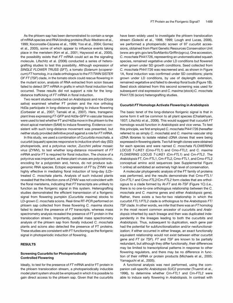
Analysis of the phloem sap collected from SD- and LD-grown
C. moschata revealed that Cmo-FTL1 and Cmo-FTL2 could be
detected only in SD plants. Again, for these analyses, isotopically
labeled synthetic peptides, VIGDVIDSFSR*, VIGDVIDSFTR*, and
VEIGGTDLR*, were employed as internal standards. Three rep-
licate experiments were conducted and representative MS data
are presented in Figure 8, while Figure 7C contains the summa-
tion for all the data collected. In all cases, signal associated with
the endogenous VIGDVIDSFSR peptide from the phloem sap
was extremely low, and for this reason, individual scans were not
included in Figure 8. Single reaction monitoring experiments per-
formed on phloem sap collected from SD-grown plants clearly
identified VIGDVIDSFTR and VEIGGTDLR peptides (cf. Figures
8A and 8B). By contrast, no VIGDVIDSFSR, VIGDVIDSFTR, or
VEIGGTDLR peptides could be detected in phloem sap collected
from LD-grown plants (cf. Figures 8C and 8D). Confirmation of
the identities for the endogenous VIGDVIDSFTR and VEIGGTDLR
peptides was obtained by tandem MS analysis at retention times
of 32 and 62.7 min, respectively (Figures 8E and 8F). Note that the
VIGDVIDSFTR and VEIGGTDLR peptides have exactly the same
MS/MS spectra as VIGDVIDSFTR* and VEIGGTDLR* except that
they have a 10-D shift for y series fragments. Taken together,
these experiments establish that the entry of Cmo-FTL1 and
Cmo-FTL2 into the phloem translocation stream is controlled by
daylength and correlates with flowering. Finally, based on our
quantitative analysis of the phloem sap collected from SD-grown
flowering C. moschata, the level of Cmo-FTL2 is ;10-fold higher
than that of Cmo-FTL1 (Figure 7C).
Detection of Pumpkin FT in Phloem Sap of Florally
Induced C. moschata Scions
To further investigate the relationship between FT and floral in-
duction, we next performed experiments, under LD conditions, in
which C. moschata scions were grafted onto flowering pumpkin
Table 1. Graft Transmission of Florigenic Signal from C. maxima Induces Flowering in C. moschata PI441726 Scions
Stock:Sciona C. moschata PI441726:C. moschata PI441726
Nodeb 1 2 3 4 5 6 7 8 9 10
Floral Budsc 0 (6) 0 (6) 0 (6) 1 (6) 1 (6) 1 (6) 1 (6) 0 (6) 0 (6) 0 (6)
Stock:Scion C. maxima:C. moschata PI441726
Node 1 2 3 4 5 6 7 8 9 10
Floral Buds 1 (6) 2 (6) 2 (6) 3 (6) 6 (6) 6 (6) 6 (6) 6 (6) 5 (6) 5 (6)
a All grafted plants (six per treatment) were grown under noninducing LD conditions.b Nodes 1 and 2 were present at time of grafting; nodes 3 to 10 developed after grafting.c Floral buds/flowers were scored as a function of nodal position using the same criteria used for Figures 2F and 2G.
1494 The Plant Cell

stocks. Upon induction to flowering (Figures 9A and 9B), each
scion was harvested to collect phloem sap and both mRNA and
protein extracted. RT-PCR experiments were performed using
primers specific for Cm-FTL1 and Cm-FTL2, and as shown in
Figure 9C, we obtained no evidence for import of these tran-
scripts across the graft union.
Single reaction monitoring experiments were next performed
on the phloem proteins obtained from each individual C. moschata
scion. As illustrated in Figures 9D and 9E, no VIGDVIDSFTR or
VEIGGTDLR peptides derived from Cmo-FTL2 could be detected.
Additionally, no VIGDVIDSFSR peptide derived from Cmo-FTL1
could be detected (data not shown). This result was expected,
based on our previous finding that these proteins do not enter the
phloem translocation stream when C. moschata is maintained
under LD conditions. However, as shown in Figure 9F, our sin-
gle reaction monitoring data detected a strong signal for the
VIGDVIDSFTK peptide derived from Cm-FTL2. Tandem MS anal-
ysis unambiguously identified this VIGDVIDSFTK peptide as
being derived from Cm-FTL2 present within the phloem proteins
collected from these florally induced C. moschata scions (Figure
9G). The VIGDVIDSFTK peptide was clearly identified in five out
of the six scion phloem protein preparations analyzed.
DISCUSSION
The role of the phloem in coordination of developmental events,
at the whole-plant level, has long been known (Oparka and
Turgeon, 1999; Oparka and Santa Cruz, 2000; Lucas and Lee,
2004; Lough and Lucas, 2006). However, molecular information
on the nature of the signaling agents that move through the
translocation stream has remained elusive. An excellent example
is florigen, the hormonal signal that transmits photoperiodic
information from mature leaves to the SAM to regulate flowering
time. Seven decades after Chailakhyan (1936) coined the term
florigen, molecular genetic studies have now begun to identify
the key players in this pathway. Studies on Arabidopsis earlier
implicated FT mRNA as florigen (Huang et al., 2005), but this
work was recently retracted (Bohlenius et al., 2007). Further-
more, experiments with tomato and tobacco (Nicotiana taba-
cum) provided results that failed to support a role for FT mRNA in
phloem long-distance signaling (Lifschitz et al., 2006). Here, we
provide direct evidence that FT, but not FT mRNA, is present in
the phloem sap of flowering cucurbits and correlates with floral
induction.
The Gene Pair Cm-FTL1 and Cm-FTL2 Is Orthologous to
the Arabidopsis Pair FT and TSF
We isolated two FT homologs from C. maxima and C. moschata,
which we named FTL1 and FTL2. Molecular phylogenetic anal-
ysis shows that these gene pairs are orthologous to the Arabi-
dopsis gene pair FT and TSF. Each gene duplication occurred
independently (i.e., after divergence of the lineages leading to
Arabidopsis, C. maxima, and C. moschata), and so neither
cucurbit gene is strictly orthologous to FT or TSF. Similarly, our
analysis shows that the clade with two poplar FT homologs,
known as FT1 and FT2, is most likely orthologous to the Arabi-
dopsis FT/TSF clade as well as the C. maxima and C. moschata
FTL1/FTL2 clades (i.e., there is a two-for-two relationship be-
tween the poplar gene pair and the Arabidopsis gene pair and
between the poplar gene pair and the cucurbit gene pairs).
Importantly, there is no one-for-one orthologous relationship
between either Arabidopsis gene or either cucurbit gene and any
other plant FT homolog (including tomato SFT) in our analysis.
Interestingly, rice has 10 FT homologs that group strongly with
Figure 4. Cm-FTL1 and Cm-FTL2 Transcripts Are Detectable in Stem
Vascular Tissue but Not in Phloem Sap of Flowering C. maxima Plants.
(A) Real-time RT-PCR analysis of Cm-FTL1, Cm-FTL2, Cm-rbcS, and
Cm-PP16 transcript levels in vascular tissue excised from mature stems
of 4- to 12-week-old pumpkin plants. Note that floral buds were first
detected on 5-week-old plants. Transcript abundance is expressed as a
ratio with Cm-PP16, which was set arbitrarily at 100. RNA was extracted
from vascular tissue collected from 2 to 3 PM. Data represent means 6 SE
for three independent replicate experiments.
(B) Time course of Cm-FTL1, Cm-FTL2, Cm-rbcS, and Cm-PP16 ex-
pression in vascular tissue excised from mature stems of 6-week-old
pumpkin plants, determined by real-time RT-PCR analysis. Transcript
abundance is expressed as a ratio with Cm-PP16, which was set
arbitrarily at 100. Data represent means 6 SE for three independent
replicate experiments.
(C) Real-time RT-PCR analysis of Cm-FTL1, Cm-FTL2, Cm-rbcS, and
Cm-PP16 levels in phloem sap collected from mature stems of 6-week-
old C. maxima plants. Transcript abundance is expressed as a ratio with
Cm-PP16, which was set arbitrarily at 1000. Note that Cm-FTL1 and Cm-
FTL2 transcripts were not detected in phloem sap at a level 10 times
lower than represented by Cm-rbcS mRNA contamination. Data repre-
sent means 6 SE for three independent replicate experiments.
FT Protein as the Florigenic Signal? 1495

these dicot FT homologs (Figure 1C), suggesting the possibility
of greater diversification of FT genes and possibly FT function in
rice than in Arabidopsis, cucurbits, or poplar.
Parallel experiments in which we ectopically expressed
Cm-FTL1, Cm-FTL2, FT, or CO in Arabidopsis companion cells
yielded early flowering phenotypes (Figures 1D and 1E). A similar
situation was recently reported in which expression of poplar FT2
in Arabidopsis resulted in early flowering phenotypes (Bohlenius
et al., 2006; Hsu et al., 2006).
Cucurbits Function as a Model System for the Dissection
of Florigenic Signaling
In this study, a novel cucurbit system was developed to inves-
tigate the molecular nature of florigen. By screening a large
population of cucurbit accessions, we identified C. moschata
PI441726 that flowers only under inductive SD conditions (Fig-
ures 1A and 2G). Here, we should note that this SD attribute
allowed us to perform photoperiodic induction experiments
without significantly altering the carbon budget of plants grown
under either SD or LD conditions. This is an important feature, as
the level of photosynthate has also been implicated as a com-
ponent of the florigenic signaling pathway (Bernier, 1988; Bernier
and Perilleux, 2005). An additional feature of this cucurbit system
is the ease with which grafting experiments can be performed
(Golecki et al., 1998, 1999; Ruiz-Medrano et al., 1999). Finally, as
a host for the ZYMV vector, C. moschata allowed us to test the
influence of heterologous/foreign genes on floral induction.
As viral infection can, by itself, alter the host flowering time, it
was important to establish whether the ZYMV vector, or ZYMV-
GFP, would alter the flowering response in C. moschata. We
obtained clear evidence that neither ZYMV (vector) nor ZYMV-
GFP had any effect on flowering in C. moschata plants being
grown under noninductive LD conditions: all infected plants re-
mained vegetative (Figures 2C, 2E, and 2F). By contrast, equiv-
alent plants inoculated with ZYMV-At-FT were all induced to
flower (Figures 2D to 2F). These results establish that, when
ectopically expressed, At-FT has a similar effect in C. moschata
as it does in Arabidopsis. Parallel studies performed on plants
grown under inductive SD conditions (Figure 2G) confirmed that
ZYMV-At-FT–inoculated plants flowered earlier than ZYMV-GFP
and mock-inoculated plants. Interestingly, ZYMV-GFP inocu-
lated plants were found to begin flowering two to four nodes
earlier than mock-inoculated plants. This suggests that under SD
conditions, ZYMV-GFP infection may have altered the time and/
or level of Cmo-FTL1/Cmo-FTL2 expression. In any event, our
Figure 5. Pumpkin FPLC-Fractionated Proteins Interrogated for Cm-FTL1 and Cm-FTL2 by MS.
(A) and (C) Proteins extracted from C. maxima stem vascular tissue (A) and phloem sap (C) were separated using cation-exchange FPLC, and aliquots
of each elution fraction (E1 to E11) were separated by SDS-PAGE. The 20-kD region associated with each elution fraction was excised, from individually
run gels, for in-gel tryptic digest, followed by tandem MS analysis.
(B) and (D) Fractions E4 to E10 from vascular tissue (B) or phloem sap (D) were pulled and then resolved on SDS-PAGE. Rectangular boxes indicate the
regions that were excised for in-gel tryptic digest, followed by tandem MS analysis for analysis of Cm-FTL1 and Cm-FTL2.
1496 The Plant Cell

findings are consistent with the notion that the long-distance
florigenic signal, either FT mRNA or FT, is common to all
flowering plants (Chailakhyan, 1937; Lifschitz et al., 2006).
ZYMV-mediated At-FT protein expression is based on auto-
catalytic processing of a high molecular weight viral polyprotein
precursor (Carrington et al., 1993). The processed At-FT protein
product has N-terminal S-A-C and C-terminal G-T-H-H-H-H-H-
H-L-V-D-T-V-M-L-Q amino acid residues fused to the At-FT
(Figure 2B). As this processed At-FT product was highly effective
at inducing flowering, it would appear that At-FT is tolerant of
N- and C-terminal extensions. This result will be of importance
for future studies on the trafficking of FT within the meristem.
Figure 6. MS Characterization of Cm-FTL1– and Cm-FTL2–Derived
Peptides.
(A) Tandem MS spectrum of doubly protonated VIGDVVDSFSR (m/z
597.3116) from the Cm-FTL1 protein. Almost all y and b series fragments
were observed. Specific fragment peaks (e.g., y2;y5 and b6;b10)
clearly differentiate this spectrum from that obtained for VIGDVLDSFTK
presented in (B). Note that the y5 fragment at m/z 611.4 is very intense
and explicable only on the basis of this peptide fragment. Inset mass
spectrum shows parent ion scan obtained with the LTQ-FT. Here, the
mass difference (Dm) between the measured and theoretical mass (m/z
597.3116) was only 1.9 ppm, which is within the 5-ppm experimental
error range typical for the LTQ-FT. Note that the mass difference with the
theoretical mass of VIGDVLDSFTK (m/z 597.3242) was 19 ppm, well
beyond the instrument error.
(B) Tandem MS spectra of doubly protonated VIGDVLDSFTK (m/z
597.3242) from the Cm-FTL2 protein. All the b and y series fragment
ions were clearly observed, except for b6 (m/z 597.4) and y5 (m/z 597.3),
which have almost the same mass as the parent ion and are unstable in
the tandem MS condition, and b1 and y1 ions, which must have been
discriminated out due to low mass cutoff by the ion trap mass spec-
trometer. Inset mass spectrum shows parent scan obtained with LTQ-
FT. Dm with the theoretical mass was only 2.5 ppm, which is within the
typical experimental error range of 5 ppm of this instrument, confirming
the sequence is correct, while the Dm with the theoretical mass of
VIGDVVDSFSR (m/z 597.3116) was 25 ppm, well beyond the instrument
error.
Figure 7. Comparative Analysis of Cmo-FTL1/FTL2 Transcript and
Protein Levels in Vascular Tissue and Phloem Sap Collected from Plants
Grown under Inductive SD and Noninductive LD Conditions.
(A) Real-time RT-PCR analysis of Cmo-FTL1, Cmo-FTL2, Cmo-rbcS,
and Cmo-PP16 transcript levels in vascular tissue excised from mature
stems of C. moschata plants subjected to SD and LD conditions. Tissue
samples were collected at 3 PM. Transcript abundance is expressed as a
ratio with Cmo-PP16, which was set arbitrarily at 100. Values represent
means 6 SE from five independent replicate experiments.
(B) Levels of Cmo-FTL1 and Cmo-FTL2 in excised vascular tissue
extracted from mature stems of C. moschata grown under SD and LD
conditions. Protein quantification was achieved using the Protein-AQUA
method (Kirkpatrick et al., 2005). Values represent means 6 SE from
three independent replicate experiments.
(C) Levels of Cmo-FTL1 and Cmo-FTL2 in phloem sap collected from
mature stems of C. moschata grown under SD and LD conditions. Protein
quantification was achieved using the Protein-AQUA method (Kirkpatrick
et al., 2005). Values represent means 6 SE from three independent
replicate experiments.
FT Protein as the Florigenic Signal? 1497
Figure 8. Photoperiodic Control of Cmo-FTL1/FTL2 Entry into the Phloem Translocation Stream.
Phloem sap was collected from mature stems of C. moschata grown under inductive SD or noninductive LD conditions. Proteins contained within the
18- to ;22-kD region of SDS-PAGE gels were in-gel digested and total peptides extracted for analysis by MS. Peptide aliquots (7 mg) for each
treatment were subjected to LC-single reaction monitoring (SRM). Arrows indicate the reaction monitored for each trace ion chromatogram. All LC-SRM
traces are from the same data set. The x and y axes in LC-SRM traces are retention times, in minutes, and ion counts, respectively.
(A) LC-SRM traces of isotopically labeled synthetic peptides (AQUA peptides; 100 fmol; R* represents Arg[13C615N4]) added to the 7-mg aliquot of
peptides derived from phloem sap collected from SD-grown plants. The AQUA peptides served as internal standards for measuring the levels of
VIGDVIDSFTR (TR) and VEIGGTDLR (LR) peptides derived from Cmo-FTL2.
(B) LC-SRM traces of Cmo-FTL2 native peptides, VIGDVIDSFTR and VEIGGTDLR, derived from phloem sap collected from SD-grown plants. Peptide
quantification was made by comparing peak abundances at the same retention times between the traces presented in (A) and (B).
(C) LC-SRM traces of AQUA peptides (100 fmol) added to the 7-mg aliquot of peptides derived from phloem sap collected from LD-grown plants.
(D) LC-SRM traces of Cmo-FTL2 native peptides derived from phloem sap collected from LD-grown plants. Note the absence of peaks at the same
retention times illustrated for the traces in (C).
(E) Tandem MS spectra of AQUA (top) and native (bottom) peptides of VEIGGTDLR from SD-grown plant phloem sap (SDPS) at the retention time of 32
min. Native peptides have the same tandem MS spectra as the AQUA peptides, except for a 10-D shift for y series fragments.
(F) Tandem MS spectra of AQUA (top) and native (bottom) peptides of VIGDVIDSFTR from SDPS at the retention time of 62.7 min.
1498 The Plant Cell

Our ZYMV immunolocalization studies (Figure 3) confirmed
that potyvirus infection domains do not include meristematic
tissues (Matthews, 1991; Jones et al., 1998). Here, we should
stress that, in our studies, the CP served as an effective reporter
for the presence of the viral RNA within the tissues of interest.
Thus, the absence of ZYMV from the plant apex (Figures 3D and
3E) proves that the influence of At-FT on floral induction must be
non-cell-autonomous in nature. The likelihood that At-FT RNA
sequences, embedded within the ;9-kb viral vector, contribute
to floral induction appears remote. A more likely scenario is that
At-FT was the non-cell-autonomous (mobile) signal or it acted
to propagate a signal that then mediated floral induction in the
C. moschata meristem(s).
Cucurbit Heterografts Confirm Phloem Transfer of
Florigenic Stimulus
Grafting experiments are a simple, but powerful, method for
establishing the transmission of a floral stimulus through the
phloem. In this regard, our heterografting experiments performed
Figure 9. Analysis of Phloem Sap from Florally Induced C. moschata Scions Grafted onto Pumpkin Failed to Detect Cm-FTL1 or Cm-FTL2 Transcripts,
Whereas Cm-FTL Peptides Were Present and Correlated with Floral Induction.
(A) and (B) Photographs of a C. moschata scion that was grafted onto a flowering C. maxima plant. Note the presence of the floral buds (red arrows);
yellow arrow in (A) indicates the graft union. All grafted plants were grown under noninductive LD conditions. Scion leaves were excised as they
approached maturity to ensure delivery of photosynthate from the pumpkin stock. Bars ¼ 10 cm.
(C) RT-PCR analysis failed to detect C. maxima FT mRNA in phloem sap collected from florally induced C. moschata scions. Gene-specific primers were
employed to amplify Cm-FTL1, Cm-FTL2, and Cm/Cmo-PP16. Expected product sizes are indicated on the left. cDNAs prepared from poly(A)þ RNA
extracted from C. maxima vascular tissue (Cm VS; positive control), phloem sap cDNAs prepared from poly(A)þ RNA extracted from C. moschata grown
under LD conditions (Cmo PS; negative control), and two individual florally induced C. moschata scions grafted on C. maxima were used as templates.
Amplification of Cm/Cmo-PP16 transcripts served as an internal control (bottom panel). PCR conditions were as follows: 2 min at 948C (one cycle), 30 s
at 948C; 30 s at 608C and 50 s at 728C (40 cycles). Specific tissues analyzed are indicted by an asterisk.
(D) to (G) Presence of Cm-FTL peptides in the phloem sap of C. moschata scions, grafted onto C. maxima stocks, correlate with floral induction. Phloem
sap was collected from flowering C. moschata scions grown under noninductive LD conditions. Proteins contained within the 18- to ;22-kD region of
SDS-PAGE gels were in-gel digested and total peptides extracted for analysis by mass spectrometry. Peptide aliquots (7 mg) for each treatment were
subjected to LC-SRM. Arrows indicate the reaction monitored for each trace ion chromatogram. All LC-SRM traces are from the same data set. The x
and y axes in LC-SRM traces are retention times, in minutes, and ion counts, respectively.
(D) LC-SRM traces of isotopically labeled synthetic peptides (AQUA peptides; 100 fmol, R* represents Arg[13C615N4]) added to the 7-mg aliquot of
phloem-derived peptides. The AQUA peptides served as internal standards for measuring the levels of VIGDVIDSFTR (TR) and VEIGGTDLR (LR)
peptides present in the scion phloem sap derived from Cmo-FTL2.
(E) LC-SRM traces of Cmo-FTL2 native peptides, VIGDVIDSFTR and VEIGGTDLR, derived from phloem sap collected from florally induced C.
moschata scions. Note the absence of peaks at the expected retention times.
(F) LC-SRM trace of Cm-FTL2 native peptide derived from phloem sap collected from florally induced C. moschata scions. Note the presence of the
VIGDVLDSFTK peptide indicating the import of Cm-FTL2 across the graft union.
(G) Tandem MS spectrum at the retention time corresponding to VIGDVLDSFTK confirms the presence of the C. maxima FT peptide.
FT Protein as the Florigenic Signal? 1499

with C. moschata and C. maxima demonstrated the delivery
of a long-distance florigenic signal that induced flowering in
C. moschata plants being grown under noninductive LD condi-
tions (Table 1). The C. moschata homografted plants grown
under exactly the same LD conditions served as a control for this
study. Here, five out of the six plants remained vegetative, with
one plant forming aborted floral buds at nodes 4 through 7. It is
important to note that removal of the mature leaves, from the
C. moschata scions, resulted in highly efficient floral induction at
nodes 5 to 10; nodes 1 and 2 existed at the time of grafting, while
3 to 10 developed after grafting. Our results indicated that the
phloem translocation stream in C. maxima stock plants should
contain Cm-FTL1/Cm-FTL2 mRNA and/or Cm-FTL1/Cm-FTL2,
if this is indeed a component of the florigenic signaling system.
FT mRNA Could Not Be Detected in Phloem Sap of
Flowering C. maxima
The diurnal expression patterns for both Cm-FTL1 and Cm-FTL2
indicated that transcript levels were highest during the daylight
hours and at their lowest in the early morning (Figure 4B). As there
was no pronounced peak in expression, phloem sap was col-
lected at four discrete times to assay for the presence of Cm-
FTL1/Cm-FTL2 mRNA. Our real-time RT-PCR experiments,
performed on phloem sap–extracted RNA, indicated that neither
Cm-FTL1 nor Cm-FTL2 transcripts could be detected (Figure
4C). Hence, if these transcripts were to function in florigenic
signaling, they must be present in the phloem sap at extremely
low levels. Such a scenario could explain why Lifschitz et al.
(2006) were unable to detect the tomato SFT transcripts in scions
whose phenotype(s) was rescued by a graft-transmissible sig-
nal(s). However, a more likely scenario is that, for the cucurbits,
tomato, and tobacco, the florigenic signal is not RNA based.
FTL1 and FTL2 in the Phloem Sap of C. maxima Is
Consistent with a Role in Long-Distance Signaling
Analytical quantities of phloem sap can be routinely collected
from the stems of flowering C. maxima, which made this plant an
ideal system to explore whether FT is present in the phloem
translocation stream. As the cucurbit phloem sap is a highly
complex mixture of proteins (Figure 5; Golecki et al., 1998, 1999;
Yoo et al., 2004), a combination of FPLC methods and MS was
used to identify tryptic peptides of Cm-FTL1 and Cm-FTL2. The
spectral data obtained from these studies provided direct evi-
dence for the presence of both Cm-FTL1 and Cm-FTL2 in phloem
sap collected from the long-distance translocation stream of
flowering C. maxima (Figure 6; see Supplemental Figure 5 online).
Thus, our results are consistent with the hypothesis that, in the
cucurbits, FTL1 and FTL2 can enter the phloem translocation
stream.
Photoperiodic Control of FTL1/FTL2 Entry into the
C. moschata Phloem Is Consistent with a Role in
Floral Induction
Our comparative analysis of Cmo-FTL1 and Cmo-FTL2 tran-
script and protein levels, under inductive SD and noninductive
LD conditions, indicated the operation of a complex control
mechanism acting on FTL1 and FTL2 synthesis and trafficking
into the phloem translocation stream (Figures 7 and 8). Although
high Cmo-FTL1 mRNA levels were detected in stem vascular
tissue of LD-grown plants, the level of protein in this same tissue
was low compared with the SD situation (Figures 7A and 7B). For
Cmo-FTL2, transcript levels were low under LD conditions and
increased ;100-fold in plants treated with SD inductive condi-
tions; a near parallel increase in FTL2 levels was observed under
SD conditions. These findings support the notion that Cmo-FTL2
may be more important in terms of contributing to the long-
distance floral stimulus. Our discovery that Cmo-FTL1 and Cmo-
FTL2 are present only in the phloem sap of SD-induced plants
is consistent with a role in florigenic signaling in this species.
Here, it is interesting to note that Brassica napus FT homologs
have also been detected in exudate obtained from inflorescence
stems (Giavalisco et al., 2006).
Pumpkin FTL2 Passes the Graft Union to Induced
Flowering in C. moschata Scions
The value of using cucurbits to examine the molecular nature
of the florigenic signal is exemplified by the results obtained
using the C. maxima:C. moschata grafting system. By main-
taining heterografted plants under noninductive LD conditions,
we could observe pumpkin-mediated floral induction in the C.
moschata scions (Figures 9A and 9B) and examine, by MS, the
contents of the scion phloem sap for the presence of pumpkin
FTL1 and FTL2 (Figures 9D to 9G). The fact that we could detect a
clear VIGDVLDSFTK peptide signal from Cm-FTL2, in the scion
phloem sap, implicates it as a signaling component that gives
rise to floral induction of C. moschata scions maintained under
noninductive LD conditions. The absence of Cm-FTL1 and Cm-
FTL2 mRNA in the phloem sap of these scions adds further
evidence against a role for the mRNA in this particular long-
distance signaling process.
Generality of FT Acting as a Long-Distance Signal?
Our results obtained with C. maxima and C. moschata are
consistent with those recently reported for rice and Arabidopsis.
In the rice study, Tamaki et al. (2007) used an Hd3a-GFP reporter
to compare the accumulation pattern of Hd3a with that of Hd3a
mRNA. The Hd3a-GFP was detected in the apex, where endog-
enous Hd3a transcript levels were extremely low, suggesting
movement of the reporter protein from the leaves; however, no
direct evidence was obtained for the actual trafficking of Hd3a in
the phloem translocation stream. In addition, no tests were per-
formed to ascertain whether the Hd3a:GFP transcripts actually
move, long-distance, from the presumptive site of synthesis in
leaves to the plant apex. Hence, a role for Hd3a mRNA in floral
induction, while seemingly unlikely, cannot be discounted
(Tamaki et al., 2007). Lastly, as the expression level of the
Hd3a:GFP transgene was ;10-fold higher than that measured
for endogenous Hd3a mRNA, caution may be called for in the
interpretation of these data.
In the Arabidopsis study, Corbesier et al. (2007) presented
further evidence indicating that FT may function as a phloem-
transported component of the florigenic signaling pathway.
1500 The Plant Cell

As the endogenous level of FT expression is known to be low,
Corbesier et al. (2007) used the strong SUC2 companion cell–
specific promoter to drive the expression of an FT:GFP reporter
system in an ft-7 background. Unfortunately, the SUC2 promoter
is active not only in the loading zones, but extends along the
long-distance transport pathway and has also been shown to be
active in various sink tissues (Martens et al., 2006). Consistent
with this activity in sink regions, parallel in situ hybridization
experiments probing for FT:GFP transcripts and confocal mi-
croscopy analyzing FT-GFP fluorescence clearly showed con-
gruence in the two signals at a distance of some 50 to 100 mm
from the SAM. Thus, detection of FT-GFP in the apex does not
provide compelling evidence for the hypothesis that FT-GFP
induction of flowering results from its import from distant tissues.
The SUC2:FT:GFP ft-7 transgenic plant line was also used in
heterografting experiments with wild-type Arabidopsis plants to
test for the passage of FT-GFP across the graft union. Analysis of
wild-type roots grafted onto SUC2:FT:GFP ft-7 transgenic scions
indicated that FT-GFP, but not FT:GFP mRNA, move long
distance into the rootstock. Y-graft experiments also demon-
strated import of FT-GFP into the receiver shoot apex, but the
caveat to these experiments is that FT:GFP was overexpressed
compared with endogenous FT, and even so, the effect on floral
induction was quite weak. It is also unfortunate that the Y-graft
system was not used to test for the delivery of FT:GFP transcripts
into this scion system. In any event, the conclusion drawn that
FT:GFP transcripts are not phloem mobile needs to be consid-
ered in light of our previous work indicating the presence of a
phloem-based RNA surveillance system to control transcript
import into specific sink tissues (Haywood et al., 2005).
Mechanism of Cucurbit FTL1 and FTL2 Trafficking into the
Sieve Tube System
Two modes of transport are possible for FTL1 and FTL2 to enter
the cucurbit phloem translocation stream (Oparka and Santa
Cruz, 2000; Lucas and Lee, 2004). Plasmodesmata connecting
functional sieve elements to their neighboring companion cells
allow for the exchange diffusion of the 27-kD GFP (Imlau et al.,
1999). When expressed under the SUC2 promoter, GFP enters
the phloem translocation stream and is delivered to sink tissues
where it can pass out through plasmodesmata into the surround-
ing cells (Imlau et al., 1999; Oparka et al., 1999; Stadler et al.,
2005). Thus, as FTL1 and FTL2 are 20 kD, they too would have
the potential to diffuse from their site of synthesis in the com-
panion cells into the sieve elements.
The companion cell–sieve element plasmodesmata also me-
diate the selective trafficking of proteins. In this situation, pro-
tein–protein interaction is required and specific mutations in the
non-cell-autonomous protein can inhibit cell-to-cell trafficking
(Lucas and Lee, 2004). That Cmo-FTL1 and Cmo-FTL2 are pres-
ent in the stem vascular tissues of plants maintained under LD
conditions, but undetectable in the phloem sap collected from
these same plants (Figures 7 and 8), implicates an additional
mechanism acting to control the trafficking of these two proteins
from the companion cells into the sieve tube system. Determi-
nation as to the nature of this control process and the actual
mode of trafficking employed by FTL1 and FTL2 to enter the
phloem will be an important next step in unraveling the mysteries
of florigen. Of equal importance will be the elucidation of the
mechanism by which FT egress from the terminal phloem to
begin the signal cascade that must propagate all the way to the
SAM (Abe et al., 2005; Wigge et al., 2005).
Finally, although our results with the cucurbits are consistent
with FTL1 and FTL2 acting as a component in the long-distance
florigenic signaling pathway, a role for the FT transcripts in other
plant systems should not be discounted. Indeed, presently there
are excellent examples, such as Cm-PP16 (Ruiz-Medrano et al.,
1999; Haywood et al., 2005), where both protein and the encod-
ing mRNA have been demonstrated to move through the phloem
translocation stream. It will certainly be of considerable interest if
future studies do indeed identify species in which FT transcripts,
or both FT and FT mRNA, are present in the phloem sap and
move to the plant apex. Identifying the molecular events involved
in switching between these two forms of long-distance macro-
molecule signals would provide considerable insight into the
evolution of processes that regulate events at the whole-plant
level.
METHODS
Plant Materials
Arabidopsis thaliana (ecotype Columbia) plants were grown hydroponi-
cally using Grodan blocks (Grodania) in a pathogen-free controlled
environment chamber under LDs (250 mmol m�2 s�1 PAR, 25/208C day/
night temperatures, daylength of 16 h) or SDs (conditions as above, but
with a daylength of 10 h). Flowering time was measured by scoring the
number of rosette leaves at the time of bolting; at least 20 individual plants
were used per treatment. Data are expressed as mean 6 SE. Cucurbita
moschata PI441726 and Cucurbita maxima cv Big Max plants were grown
under both a pathogen-free greenhouse environment (natural daylight
conditions: 1000 mmol m�2 s�1 PAR, 30/208C day/night temperatures,
daylength of 16 h [LD]) and in a controlled environment (350 mmol m�2 s�1
PAR, 28/208C day/night temperatures, and either 10 h SD or LD [10 h
daylength and a dark period that was interrupted by a 15-min incandes-
cent light treatment each hour]).
Identification of a cucurbit species in which floral induction was
photoperiodically controlled was achieved through screening of seeds
for 97 accessions provided by the Agricultural Research Service Plant
Genetic Resources Conservation Unit (www.ars-grin.gov/ars/SoAtlantic/
Griffin/pgrcu). Ten seedlings for each accession were grown in green-
houses under winter conditions, when the daylength was <12 h (i.e., SD
conditions), or in equivalent greenhouses in which incandescent lighting
was used to provide daylength extension such that the plants were
exposed to a 16-h photoperiod (i.e., LD conditions). Ninety-six acces-
sions were found to flower under both SD and LD conditions, whereas
plants for one accession, C. moschata PI441726, flowered only under SD
conditions. Fruit from these plants were collected and the resultant seed
rescreened under both LD and SD conditions to confirm that floral
induction in C. moschata PI441726 was indeed under photoperiodic
control.
Cloning of Cucurbita maxima, C. moschata, and Arabidopsis Genes
Vascular tissue, excised from mature (6-week-old) C. maxima or
C. moschata PI441726 stems, was used in the cloning of the respective
FT homologs. High molecular weight RNA was obtained using either an
FT Protein as the Florigenic Signal? 1501

RNAqueous kit (Ambion) or TRIZOL (Invitrogen). Aliquots (1 mg) of this
RNA were then used for RT-PCR with SuperScript RT III (Invitrogen)
according to the manufacturer’s instructions. Samples were treated with
DNase I (Invitrogen) for 15 min, at room temperature, to remove any
possibility of genomic DNA contamination, diluted 10-fold, and aliquots
(1 mL) used for PCR amplification. The same general procedure was used
for cloning FT and CO from Arabidopsis leaf tissue. The following gene-
specific primers were used: Cm-FTL1, forward 59-ATATGCATGCAT-
GCCGAGAAATCGTGAC-39 and reverse 59-GGTTGATAATATAATGTGA-
AGTGAAGTGT-39; Cm-FTL2, forward 59-TTGGGAGAGTCATCGGCGA-
CGTTA-39 and reverse 59-TCTTCTCCTTCCACCAGACCCAC-39; Cmo-FTL1,
forward 59-AGGTCCATCTCGATTAGGGTTGCTTACAACTCG-39 and re-
verse 59-CGTAGCACACTATCTCTTGTCCAAAGGTTGCCT-39; Cmo-FTL2,
forward 59-CACGAGGTCCATTTCCATTAGGGCTACTTACAACAAT-39 and
reverse 59-CATAGCACACGATCTCTTGACCAAAGGTCGCTC-39; FT, for-
ward 59-ATATGCATGCATGTCTATAAATATAAGAGAC-39 and reverse
59-ATATGGTACCAAGTCTTCTTCCTCCGCA-39; CO, forward 59-ATATG-
CATGCATGTTGAAACAAGAGAGTAAC-39 and reverse 59-ATATGGTAC-
CGAATGAAGGAACAATCCCA-39. PCR conditions were as follows: 2 min
at 948C (one cycle), 10 s at 948C; 30 s at 588C and 60 s at 688C (36 cycles).
FT Phylogenetic Analysis
Public databases, including The Institute for Genomic Research and Joint
Genome Initiative, were searched using various Arabidopsis and rice
(Oryza sativa) flowering-time genes as queries. Gene names and their
nucleotide accessions can be found in Supplemental Table 1 online. Os-
FT-L5 was not included in this study because it has been identified as
being a pseudogene. Nucleotide sequences were aligned based on a
protein sequence alignment generated using Muscle 3.52 (Edgar, 2004).
The alignments were edited using GeneDoc (http://www.psc.edu/
biomed/genedoc/) to remove nonaligning regions (see Supplemental
Figure 4 online). Neighbor-joining trees were constructed using the com-
puter program MEGA3.1 (Kumar et al., 2004). The Kimura two-parameter
substitution model was used, and a consensus tree was built from 1000
bootstrap replicates (see Supplemental Figure 2 online). In addition,
Bayesian phylogenetic analyses were performed using MrBayes 3.0B4
(Huelsenbeck and Ronquist, 2001). Searches were run using four different
Markov chains for 3,000,000 generations using the GTR model with a
substitution rate that varies according to the gamma distribution and
allows for a proportion of sites to be invariable (GTR þ IG). Sampling
occurred every 100 trees, and the first 10,000 trees were discarded to
build a consensus tree (see Supplemental Figure 3 online).
Plant Transformation
The SUC2 promoter sequence (Truernit et al., 1996) and the CO, FT, Cm-
FTL1, and Cm-FTL2 gene sequences were subcloned into the binary
vector pART27 (Gleave, 1992). All binary plasmids were introduced into
Agrobacterium tumefaciens strain GV3101 and transformed into Arabi-
dopsis (Columbia ecotype) plants by the floral dip method (Clough and
Bent, 1998). Seeds from primary transformed kanamycin-resistant trans-
genic control (vector only; three lines), CO (three lines), FT (six lines),
Cm-FTL1 (six lines), and Cm-FTL2 (12 lines) plants were used to generate
T1 progeny that were then employed for flowering-time assays.
ZYMV Constructs
The ZYMV-based viral vector, p35SZYMVNIbMCS (Lin et al., 2002), was
first digested with BglII and KpnI. Next, PCR fragments encoding FT, CO,
and eGFP full-length sequences were digested with SphI and KpnI
restriction enzymes followed by ligation into p35SZYMVNIbMCS. For
these studies, the eGFP template was obtained from a pEGFP plasmid
(BD Biosciences) using the following primer pair: forward 59-ATATG-
CATGCATGGTGAGCAAGGGCGAGGAGCT-39; reverse 59-ATATGGT-
ACCCTTGTACAGCTCGTCCATGCCGA-39. All viral constructs were
verified by sequence analysis.
Floral Induction Mediated by ZYMV Expression of At-FT
Viral vector inoculations were initiated by microprojectile bombardment
using a Helios gene gun system (Bio-Rad Laboratories). ZYMV-based
constructs were precipitated onto gold particles (DNA loading ratio of
2 mg/mg gold). Briefly, 1-mm gold particles (15 mg) were resuspended in
100 mL of 50 mM spermidine. DNA (25 mg) was then precipitated onto the
gold with 100 mL of 1 M CaCl2. This gold:DNA mixture was washed three
times with 500 mL of 100% ethanol, and the final pellet was resuspended
in 2.5 mL of 25 mg/mL polyvinylpyrrolidone in 100% ethanol for loading
into GoldCoat tubing at a ratio of 1 mL of DNA/gold particle suspension
per 15 3 0.5-inch cartridge. A helium pressure of 100 p.s.i., delivered
at a distance of 3 cm, was used to inoculate ZYMV constructs into
C. moschata PI441726 seedlings that had developed two true leaves.
The ZYMV vector was inoculated onto one true leaf and one cotyledon,
and plants were then monitored weekly for floral bud initiation at each
node. The stage of floral bud induction was recorded according to the
following scoring system: 0, no visible bud; 1, bud <1 mm; 2, bud 1 to
5 mm; 3, 5 to 15 mm; 4, bud >15 mm; and 5, open flowers. Flowering rate
was defined as the ratio of plants in each group with a stage $2.
The stability of the inserted sequence within the ZYMV vector was
monitored by RT-PCR. RNA was extracted (TRIZOL; Invitrogen) from
apical leaves of systemically infected plants 30 DAI. The RT reaction was
performed with 0.5 mg total RNA using Superscript RT III (Invitrogen) with
random primers (Invitrogen). The PCR conditions were as follows: 2 min at
948C (one cycle), 10 s at 948C; 30 s at 548C and 90 s at 708C (32 cycles)
using Taq DNA polymerase (Invitrogen) and ZYMV-specific primers flank-
ing the insertion site (forward 59-AGAGGCTATTTGCGCTGCGATG-39;
reverse 59-CTTTCACGCGTGGCAGTGACAT-39). The diagnostic size of
the amplified insert was evaluated using ethidium bromide–stained 1%
agarose gels. These assays established that the ZYMV-At-FT vector
stably expressed At-FT for up to 40 DAI.
Epifluorescence Microscopy
Spatial distribution of GFP in ZYMV-inoculated C. moschata PI441726
plants was determined using epifluorescence microscopy (Leica MZFLIII
stereomicroscope; Leica Microsystems). Plants 4 to 30 DAI were used to
obtain fresh, unfixed, sections of lamina, petiole, and stem tissues.
Images were captured and analyzed using a Leica DC300F camera
(excitation filter, 480 nm; emission filter, 510 nm) in conjunction with
Adobe Photoshop (Adobe Systems).
Immunolocalization of ZYMV Coat Protein
Tissues from C. moschata PI441726 plants were fixed in FAA (3.7%
formaldehyde, 5% acetic acid, and 50% ethanol) overnight at 48C,
dehydrated through an ethanol series and Neo-Clear (Merck), and finally
embedded in paraffin. Sections (10 mm) were cut and mounted on elec-
trostatically charged Superfrost Plus slides (British Drug House). Slides
were used immediately or stored at�208C. Sections were deparaffinized
in Neo-Clear (2 3 10 min), rehydrated through an ethanol series, and then
transferred into distilled water. Endogenous peroxidase activity was
blocked using a 10-min treatment in 0.5% H2O2 in methanol. Slides were
then pretreated in boiling 1 mM EDTA, pH 7.5, for 5 to 15 min, cooled to
room temperature, and then washed in distilled water for 5 min, followed
by TBST (0.01% Tween-20 in Tris-buffered saline, pH 7.5) for 5 min.
Sections were next incubated with blocking reagent (0.1% BSA in TBST)
for at least 10 min, excess blocking reagent was removed, and then they
were irrigated with anti-ZYMV antibody solution (Agdia) diluted 1:100 in
1502 The Plant Cell

blocking reagent. After overnight treatment in anti-ZYMV antibody solu-
tion, slides were washed in TBST (2 3 5 min) and sections covered with
the secondary antibody (anti-rabbit IgG; Sigma-Aldrich) diluted in block-
ing reagent (1:300). After a 1-h incubation, slides were washed in TBST
(2 3 5 min) and placed in a humid chamber, and sections were covered
with DAB substrate (Roche) and incubated until desired staining was
achieved (usually 5 to 15 min). Slides were rinsed with distilled water and
mounted and photographed on a Zeiss Axioskop mounted with a Zeiss
Axiocam MRc camera.
Grafting Protocol
Heterografting experiments were performed with C. moschata PI441726
and C. maxima plants essentially as previously described (Ruiz-Medrano
et al., 1999; Yoo et al., 2004). Each excised C. moschata PI441726 scion
(;15 cm in length with two nodes) was carefully inserted into an incision
made in the main stem of a 5-week-old stock (C. moschata PI441726
or C. maxima) plant. The graft site was then fastened and sealed with
Parafilm and the scion covered with a clear plastic bag that was removed
1 week later. As the scions grew, mature leaves were excised to maintain
the sink status of this tissue. Homo- and heterografted scions were ob-
served until they had developed a total of 10 visible nodes (i.e., eight new
leaves), and the scoring system for flowering was as described above.
Collection of RNA and Proteins from Phloem Sap and
Vascular Tissue
Phloem sap collection was performed as previously described (Ruiz-
Medrano et al., 1999; Yoo et al., 2004), with modifications. Briefly,
C. maxima or C. moschata PI441726 stems were excised with a sterile
razor blade and the cut surface blotted, several times, with sterile filter
paper (3 MM; Whatman). Phloem sap exuded thereafter was collected
using sterile micropipette tips and immediately mixed with 9 volumes of
protein sap collection buffer (7 M urea, 2 M thiourea, 4% CHAPS, and
67 mM DTT), or 200-mL aliquots were mixed with 500 mL of TRIZOL re-
agent (Invitrogen) for phloem sap RNA extraction. All buffers and samples
were kept on ice during phloem sap collection.
Vascular tissue was excised from the same region of the plant as used
for the collection of phloem sap. Stem sections (10 cm) were excised from
the plant and vascular bundles removed using forceps. Vascular tissue
was first ground under liquid nitrogen and then divided into two aliquots.
The first was mixed with TRIZOL reagent and processed for RNA ex-
traction according to the manufacturer’s protocol (Invitrogen). The sec-
ond aliquot was mixed with phloem sap collection buffer, followed by
sonication (eight pulses for 2 min using a Sonic Dismembrator; Fisher
Scientific) and centrifugation (10,000g for 30 min); the supernatant was
collected and filtered. All buffers and samples were kept on ice during
RNA and protein extraction.
Real-Time PCR
Transcript levels of C. maxima and C. moschata FTL1, FTL2, rbcS, and
PP16 were determined on a Bio-Rad iCycler IQ using POWER SYBR
Green PCR Master Mix (Applied Biosystems) in 25-mL reactions. An
aliquot (1 mL) of the RT reaction was used as template. PCR conditions
were as follows: 10 min at 958C (one cycle); 30 s at 958C, 30 s at 608C, and
50 s at 728C (50 cycles). Gene-specific primers used were as above and
as follows: Cm-rbcS, forward 59-AGGCACCAATGGCTTCCTCCATCGT-
CTC-39 and reverse 59-GCACTGGACTTGGCGGATGTTGTCGAAG-39;
Cm-PP16, forward 59-GTGGTAAAGGACTTCAAGCCCACGACC-39 and re-
verse 59-ATGGGTTTGAAGAAGCCAAGCCACTTA-39; Cmo-rbcS, forward
59-ATGGCTTCCTCCATCGTCTCATCC-39 and reverse 59-GAGCGGA-
GAAGGTACGCAATTTCC-39; Cmo-PP16, forward 59-ATGGGTTTGAA-
GAAGCCAAGCCAC-39 and reverse 59-AGTTGCTAAAAATGCAGGACC-
AGATCCATTATGGG-39. Each RT sample was assayed in triplicate.
Individual threshold cycles were located in 15 to 35 cycles, which is in
the linear range of PCR amplification for each gene. Data were further
analyzed in Excel. Transcript levels were normalized to C. maxima and C.
moschata PP16, respectively.
Protein Preparation and LC-MS/MS Analysis
Proteins isolated from C. maxima phloem sap and vascular tissue were
fractionated using chromatographic methods. Proteins were first di-
alyzed against buffer A (20 mM Na2HPO4/NaH2PO4, pH 7.0, 1 mM EDTA,
10% glycerol, and 1% 2-mercaptoethanol) and then clarified by centrif-
ugation (17,000g for 30 min). Samples were then loaded onto a buffer
A–equilibrated HiTrap SP column connected to an FPLC system (Amer-
sham Biosciences). After washing the column with 10 volumes of buffer A,
proteins were eluted with a linear gradient of 0 to 500 mM NaCl in buffer
A supplemented with 1 M NaCl. Proteins isolated from C. moschata
PI441726 phloem sap and vascular tissue were dialyzed against buffer B
(20 mM Tris, pH 7.5, 1 mM EDTA, and 1% 2-mercaptoethanol). FPLC-
fractionated vascular tissue and phloem sap proteins of C. maxima, as
well as dialyzed vascular tissue and phloem sap proteins of C. moschata
PI441726, were resolved on 12% SDS-PAGE gels. The 20-kD regions on
these gels were then excised, and the proteins therein were prepared
for MS analysis using standard reduction, alkylation, and tryptic digest
procedures (Rosenfeld et al., 1992).
All quantification data for Cmo-FTL1 and Cmo-FTL2 and semiquanti-
fication data for Cm-FTL1 and Cm-FTL2 were acquired using a Nano-LC-
2D system (Eksigent) coupled with an LTQ ion trap mass spectrometer
(Thermo Finnigan) with an in-laboratory fabricated fritless reverse phase
microcapillary column (75 mm 3 180 mm packed with Magic C18AQ,
3 mm 100 A; Michrom Bio Resources) and vented column configuration.
Each digested sample was transferred by an autosampler to the online
trap column (Zorbax 300SB-C18, 5 mm, 0.3 mm 3 5 mm; Agilent) and
desalted. Peptides were then eluted from the trap, separated by capillary
column by reverse phase gradient, at a flow rate of 300 nL per min, and
directly sprayed into the mass spectrometer. Buffer compositions used
for reverse phase chromatography were as follows: buffer A, 0.1% formic
acid in water; buffer B, 0.1% formic acid in 100% acetonitrile. A 120-min
(2 to 40% buffer B for 95 min, 40 to 80% buffer B for 12 min, and 80%
buffer B for 13 min) or 168-min gradient (2% buffer B for 3 min, 2 to 10%
buffer B for 7 min, 10 to 40% buffer B for 130 min, 40 to 80% buffer B for
15 min, and 80% buffer B for 13 min) was used for the Nano-LC-2D/LTQ
system.
A high-resolution mass spectrometer (LTQ-FT; Thermo Finnigan) was
used to acquire accurate mass spectra of VIGDVVDSFSR from Cm-FTL1
and VIGDVIDSFTK from Cm-FTL2.
Semiquantification of Cm-FTL1/FTL2 and Quantification
of Cmo-FTL1/FTL2
Semiquantitative information of Cm-FTL1 and Cm-FTL2 was obtained by
monitoring MS/MS of representative peptides VIGDVVDSFSR (m/z 597.3)
and VIGDVIDSFTK (m/z 597.3), respectively, plus peptide VEIGGTDLR
(m/z 480.3) that is common to both proteins. Cmo-FTL1 and Cmo-FTL2
were quantified using the Protein-AQUA method in which isotopically
labeled synthetic peptides were used (Kirkpatrick et al., 2005). Absolute
quantification (AQUA) peptides, VIGDVIDSFSR* (SR*; representative of
Cmo-FTL1), VIGDVIDSFTR* (TR*; representative of Cmo-FTL2), and
VEIGGTDLR* (LR*; common to Cmo-FTL1 and Cmo-FTL2) were pre-
pared by Sigma-Aldrich Genosys (http://www.sigmaaldrich.com/Brands/
Sigma_Genosys/Custom_Peptides.html) and used as internal standards
for tandem MS analyses. The R* represents Arg residues containing six
13C and four 15N; the synthetic peptides and their y series fragments
have 10-D higher mass than the corresponding native peptides and their
FT Protein as the Florigenic Signal? 1503

fragments. Prior to their use in AQUA experiments, all synthetic peptides
were tested to confirm that the preparations were devoid of contaminating
mono-isotopic peptides. Concentrations of in-gel–digested C. moschata
PI441726 proteins were determined on a NanoDrop ND-1000 (NanoDrop
Technologies) using the A280 method employing the extinction coeffi-
cient for BSA. Based on these measurements, 7 mg of total protein
containing 100 fmol of each synthetic peptide was injected into the Nano-
LC-2D/LTQ.
For quantification of Cmo-FTL1 and Cmo-FTL2, monitoring of tandem
MS and/or multiple reaction monitoring (MRM) of major fragments was
used for the native and synthetic peptides. Doubly protonated peptide
ions were selected for tandem MS monitoring: m/z 616.3 (TR*), 611.3 (TR),
609.3 (SR*), 604.3 (SR), 485.3 (LR*), and 480.3 (LR). Fragments monitored
in MRM were as follows: m/z 1019.5 (y9), 847.4 (y7), and 748.4 (y6) from
TR*; m/z 1009.5 (y9), 837.6 (y7), and 738.3 (y6) from TR; m/z 1005.4 (y9),
833.3 (y7), and 734.2 (y6) from SR*; m/z 995.4 (y9), 823.3 (y7), and 724.2 (y6)
from SR; m/z 741.3 (y7), 628.3 (y6), and 571.3 (y5) from LR*; and m/z
731.3 (y7), 618.4 (y6), and 561.3 (y5) from LR. Absolute quantification was
determined by comparing the abundance of the known internal standard
peptide with its native peptide.
For analysis of phloem proteins collected from heterografted C. moschata
scions, the following representative C. moschata and C. maxima peptides
were monitored in MRM: m/z 616.3 (TR*), 611.3 (TR), 609.3 (SR*), 604.3
(SR), 597.3 (VIGDVVDSFSR and VIGDVIDSFTK for Cm-FTL1 and Cm-FTL2,
respectively), 485.3 (LR*), and 480.3 (LR). Note VEIGGTDLR (LR) is common
in all C. moschata and C. maxima FTL proteins.
Database Search
Database matching was performed through the use of the Mascot search
algorithm (version 2.1; Matrix Science Limited). The sequence database
consisted of 155,976 ESTs derived from vascular tissues of C. maxima,
C. moschata PI441726, Cucumis sativus, and Sicyos angulatus assem-
bled into ;51,826 gene sets or consensi. Only statistically significant
peptides were considered as a reliable match and were pursued for
further analysis. SEQUEST search (Bioworks v2.1; Thermo Electron) was
performed to calculate sequence coverage of Cm-FTL1 and Cm-FTL2.
Parent mass tolerance of 2.5 D and fragment tolerance of 1 D were used.
Only fully tryptic peptides were accepted, and Xcorr cutoff values of 1.5,
2.0, and 2.5 were used for singly, doubly, and triply charged ions. Manual
validation was performed for every matched tandem mass spectrum.
Accession Numbers
Gene names and their nucleotide accessions can be found in Supple-
mental Table 1 online. Accession numbers for Populus trichocarpa genes
are gene model identifiers found at the Joint Genome Institute (http://
genome.jgipsf.org/Poptr1/Poptr1.home.html).
Supplemental Data
The following materials are available in the online version of this article.
Supplemental Methods.
Supplemental Table 1. Sequence Accession Numbers for Members
of the FT-TFL Family Analyzed.
Supplemental Figure 1. Comparison of Arabidopsis FT, Cm-FTL1,
Cm-FTL2, Cmo-FTL1, and Cmo-FTL2 Conceptual Amino Acid Se-
quences.
Supplemental Figure 2. Neighbor-Joining Phylogenetic Analysis of
40 FT-TFL Family Members in Angiosperms.
Supplemental Figure 3. Bayesian Phylogenetic Analysis of 40 FT-
TFL Family Members in Angiosperms.
Supplemental Figure 4. Nucleotide Sequence Alignment of 40 FT-
TFL Family Members in Angiosperms.
Supplemental Figure 5. Typical Sequence Coverage and Identified
Peptides for Cm-FTL1 and Cm-FTL2 from LC-MS/MS Data Analysis.
Supplemental Figure 6. Phloem-Specific Expression of Cmo-FTL1/
FTL2 in Plants Grown under Noninductive LD and Inductive SD
Conditions.
ACKNOWLEDGMENTS
We thank Shyi-Dong Yeh (National Chung Hsing University, Taiwan) for
kindly providing the ZYMV viral expression vector. We also thank
Prudence Grandison, Dieter Hermsmeier, and Justin Sweetman for
assistance during the early phase of this project. This work was
supported by grants from the National Science Foundation (IBN
03151174 to W.J.L. and DBI-0421679 to R.A.J.), the Department of
Energy, Division of Energy Biosciences (DE-FG02-94ER20134 to W.J.L.),
and the New Zealand Foundation for Research Science and Technology
(to T.J.L.). B.X.-C. was supported by a University of California Institute
for Mexico and the U.S.–Mexico National Council for Science and
Technology Sabbatical Leave Fellowship.
Received March 29, 2007; revised May 7, 2007; accepted May 18, 2007;
published May 31, 2007.
REFERENCES
Abe, M., Kobayashi, Y., Yamamoto, S., Daimon, Y., Yamaguchi, A.,
Ikeda, Y., Ichinoki, H., Notaguchi, M., Goto, K., and Araki, T.
(2005). FD, a bZIP protein mediating signals from the floral pathway
integrator FT at the shoot apex. Science 309: 1052–1056.
An, H., Roussot, C., Suarez-Lopez, P., Corbesier, L., Vincent, C.,
Pineiro, M., Hepworth, S., Mouradov, A., Justin, S., Turnbull, C.,
and Coupland, G. (2004). CONSTANS acts in the phloem to regulate
a systemic signal that induces photoperiodic flowering of Arabidopsis.
Development 131: 3615–3626.
Ayre, B.G., and Turgeon, R. (2004). Graft transmission of a floral
stimulant derived from CONSTANS. Plant Physiol. 135: 2271–2278.
Bastow, R., and Dean, C. (2003). Plant sciences. Deciding when to
flower. Science 302: 1695–1696.
Bernier, G. (1988). The control of floral evocation and morphogenesis.
Annu. Rev. Plant Physiol. Plant Mol. Biol. 39: 175–219.
Bernier, G., and Perilleux, C. (2005). A physiological overview of the
genetics of flowering time control. Plant Biotechnol. J. 3: 3–16.
Bohlenius, H., Eriksson, S., Parcy, F., and Nilsson, O. (2007). Re-
traction. Science 316: 367.
Bohlenius, H., Huang, T., Charbonnel-Campaa, L., Brunner, A.M.,
Jansson, S., Strauss, S.H., and Nilsson, O. (2006). CO/FT regulatory
module controls timing of flowering and seasonal growth cessation in
trees. Science 312: 1040–1043.
Carrington, J.C., Haldeman, R., Dolja, V.V., and Restrepo-Hartwig,
M.A. (1993). Internal cleavage and trans-proteolytic activities of the
VPg-proteinase (NIa) of tobacco etch potyvirus in vivo. J. Virol. 67:
6995–7000.
Chailakhyan, M.K. (1936). New facts for hormonal theory of plant
development. Dokl. Akad. Nauk SSSR 13: 79–83.
Chailakhyan, M.K. (1937). Concerning the hormonal nature of plant
development processes. Dokl. Akad. Nauk SSSR 16: 227–230.
Clough, S.J., and Bent, A.F. (1998). Floral dip: A simplified method
1504 The Plant Cell

for Agrobacterium-mediated transformation of Arabidopsis thaliana.
Plant J. 16: 735–743.
Colasanti, J., and Sundaresan, V. (2000). ‘Florigen’ enters the molec-
ular age: Long-distance signals that cause plants to flower. Trends
Biochem. Sci. 25: 236–240.
Corbesier, L., and Coupland, G. (2005). Photoperiodic flowering of
Arabidopsis: Integrating genetic and physiological approaches to
characterization of the floral stimulus. Plant Cell Environ. 28: 54–66.
Corbesier, L., Vincent, C., Jang, S., Fornara, F., Fan, Q., Searle, I.,
Giakountis, A., Farrona, S., Gissot, L., Turnbull, C., and Coupland,
G. (2007). FT protein movement contributes to long-distance signaling
in floral induction of Arabidopsis. Science 316: 1030–1033.
Edgar, R.C. (2004). MUSCLE: A multiple sequence alignment method
with reduced time and space complexity. BMC Bioinformatics
5: 1–19.
Giavalisco, P., Kapitza, K., Kolasa, A., Buhtz, A., and Kehr, J. (2006).
Towards the proteome of Brassica napus phloem sap. Proteomics 6:
896–909.
Gleave, A.P. (1992). A versatile binary vector system with a T-DNA
organisational structure conducive to efficient integration of cloned
DNA into the plant genome. Plant Mol. Biol. 20: 1203–1207.
Golecki, B., Schulz, A., Carstens-Behrens, U., and Kollmann, R.
(1998). Evidence for graft transmission of structural phloem proteins
or their precursors in heterografts of Cucurbitaceae. Planta 206:
630–640.
Golecki, B., Schulz, A., and Thompson, G.A. (1999). Translocation of
structural P proteins in the phloem. Plant Cell 11: 127–140.
Gomez, G., Torres, H., and Pallas, V. (2005). Identification of trans-
locatable RNA-binding phloem proteins from melon, potential com-
ponents of the long-distance RNA transport system. Plant J. 41:
107–116.
Haywood, V., Yu, T.S., Huang, N.C., and Lucas, W.J. (2005). Phloem
long-distance trafficking of GIBBERELLIC ACID-INSENSITIVE RNA
regulates leaf development. Plant J. 42: 49–68.
Hsu, C.H., Lin, S.S., Liu, F.L., Su, W.C., and Yeh, S.D. (2004). Oral
administration of a mite allergen expressed by zucchini yellow mosaic
virus in cucurbit species downregulates allergen-induced airway
inflammation and IgE synthesis. J. Allergy Clin. Immunol. 113: 1079–
1085.
Hsu, C.Y., Liu, Y., Luthe, D.S., and Yuceer, C. (2006). Poplar FT2
shortens the juvenile phase and promotes seasonal flowering. Plant
Cell 18: 1846–1861.
Huang, T., Bohlenius, H., Eriksson, S., Parcy, F., and Nilsson, O.
(2005). The mRNA of the Arabidopsis gene FT moves from leaf to
shoot apex and induces flowering. Science 309: 1694–1698.
Huelsenbeck, J.P., and Ronquist, F. (2001). MRBAYES: Bayesian
inference of phylogeny. Bioinformatics 17: 754–755.
Imlau, A., Truernit, E., and Sauer, N. (1999). Cell-to-cell and long-
distance trafficking of the green fluorescent protein in the phloem and
symplastic unloading of the protein into sink tissues. Plant Cell 11:
309–322.
Jones, A.L., Thomas, C.L., and Maule, A.J. (1998). De novo methyl-
ation and co-suppression induced by a cytoplasmically replicating
plant RNA virus. EMBO J. 17: 6385–6393.
Kardailsky, I., Shukla, V.K., Ahn, J.H., Dagenais, N., Christensen,
S.K., Nguyen, J.T., Chory, J., Harrison, M.J., and Weigel, D. (1999).
Activation tagging of the floral inducer FT. Science 286: 1962–1965.
Kim, M., Canio, W., Kessler, S., and Sinha, N. (2001). Developmental
changes due to long-distance movement of a homeobox fusion tran-
script in tomato. Science 293: 287–289.
Kirkpatrick, D.S., Gerber, S.A., and Gygi, S.P. (2005). The absolute
quantification strategy: A general procedure for the quantification of
proteins and post-translational modifications. Methods 35: 265–273.
Kobayashi, Y., Kaya, H., Goto, K., Iwabuchi, M., and Araki, T. (1999).
A pair of related genes with antagonistic roles in mediating flowering
signals. Science 286: 1960–1962.
Kumar, S., Tamura, K., and Nei, M. (2004). MEGA3: Integrated
software for Molecular Evolutionary Genetics Analysis and Sequence
Alignment. Brief. Bioinform. 5: 150–163.
Lang, A. (1965). Physiology of flower initiation. In Encyclopedia of Plant
Physiology, Vol. XV, W. Ruhland, ed (Berlin: Springer-Verlag), pp.
1380–1536.
Lang, A. (1977). Promotion and inhibition of flower formation in a day-
neutral plant in grafts with a short-day plant and a long-day plant.
Proc. Natl. Acad. Sci. USA 74: 2412–2416.
Lifschitz, E., Eviatar, T., Rozman, A., Goldshmidt, A., Amsellem, Z.,
Alvarez, J.P., and Esched, Y. (2006). The tomato FT ortholog triggers
systemic signals that regulate growth and flowering and substitute for
diverse environmental stimuli. Proc. Natl. Acad. Sci. USA 103: 6398–
6403.
Lin, S.-S., Hou, R.F., and Yeh, S.-D. (2002). Construction of in vitro and
in vivo infectious transcripts of a Taiwan strain of Zucchini yellow
mosaic virus. Bot. Bull. Acad. Sin. 43: 261–269.
Lough, T.J., and Lucas, W.J. (2006). Integrative plant biology: Role of
phloem long-distance macromolecular trafficking. Annu. Rev. Plant
Biol. 57: 203–232.
Lucas, W.J., and Lee, J.Y. (2004). Plasmodesmata as a supracellular
control network in plants. Nat. Rev. Mol. Cell Biol. 5: 712–726.
Martens, H.J., Roberts, A.G., Oparka, K.J., and Schulz, A. (2006).
Quantification of plasmodesmatal endoplasmic reticulum coupling
between sieve elements and companion cells using fluorescence
redistribution after photobleaching. Plant Physiol. 142: 471–480.
Matthews, R.E.F. (1991). Plant Virology. (San Diego, CA: Academic
Press).
Michaels, S.D., Himelblau, E., Kim, S.Y., Schomburg, F.M., and
Amasino, R.M. (2005). Integration of flowering signals in winter-
annual Arabidopsis. Plant Physiol. 137: 149–156.
Mouradov, A., Cremer, F., and Coupland, G. (2002). Control of
flowering time: Interacting pathways as a basis for diversity. Plant
Cell 14: S111–S130.
Oparka, K.J., Roberts, A.G., Boevink, P., Santa Cruz, S., Roberts, I.,
Pradel, K.S., Imlau, A., Kotlizky, G., Sauer, N., and Epel, B. (1999).
Simple, but not branched, plasmodesmata allow the nonspecific
trafficking of proteins in developing tobacco leaves. Cell 97: 743–754.
Oparka, K.J., and Santa Cruz, S. (2000). The great escape: Phloem
transport and unloading of macromolecules. Annu. Rev. Plant Physiol.
Plant Mol. Biol. 51: 323–347.
Oparka, K.J., and Turgeon, R. (1999). Sieve elements and companion
cells: Traffic control centers of the phloem. Plant Cell 11: 739–750.
Putterill, J., Robson, F., Lee, K., Simon, R., and Coupland, G. (1995).
The CONSTANS gene of Arabidopsis promotes flowering and en-
codes a protein showing similarities to zinc finger transcription fac-
tors. Cell 80: 847–857.
Rosenfeld, J., Capdevielle, J., Guillemot, J.C., and Ferrara, P.
(1992). In-gel digestion of proteins for internal sequence analysis
after one- or two-dimensional gel electrophoresis. Anal. Biochem.
203: 173–179.
Ruiz-Medrano, R., Xoconostle-Cazares, B., and Lucas, W.J. (1999).
Phloem long-distance transport of CmNACP mRNA: Implications for
supracellular regulation in plants. Development 126: 4405–4419.
Samach, A., Onouchi, H., Gold, S.E., Ditta, G.S., Schwarz-Sommer,
Z., Yanofsky, M.F., and Coupland, G. (2000). Distinct roles of
CONSTANS target genes in reproductive development of Arabidop-
sis. Science 288: 1613–1616.
Searle, I., and Coupland, G. (2004). Induction of flowering by seasonal
changes in photoperiod. EMBO J. 23: 1217–1222.
FT Protein as the Florigenic Signal? 1505

Stadler, R., Wright, K.M., Lauterbach, C., Amon, G., Gahrtz, M.,
Feuerstein, A., Oparka, K.J., and Sauer, N. (2005). Expression of
GFP-fusions in Arabidopsis companion cells reveals non-specific
protein trafficking into sieve elements and identifies a novel post-
phloem domain in roots. Plant J. 41: 319–331.
Suarez-Lopez, P., Wheatley, K., Robson, F., Onouchi, H., Valverde,
F., and Coupland, G. (2001). CONSTANS mediates between the
circadian clock and the control of flowering in Arabidopsis. Nature
410: 1116–1120.
Takada, S., and Goto, K. (2003). Terminal flower 2, an Arabidopsis
homolog of heterochromatin protein1, counteracts the activation of
flowering locus T by constans in the vascular tissues of leaves to
regulate flowering time. Plant Cell 15: 2856–2865.
Tamaki, S., Matsuo, S., Wong, H.L., Yokoi, S., and Shimamoto, K.
(2007). Hd3a protein is a mobile flowering signal in rice. Science 316:
1033–1036.
Truernit, E., Schmid, J., Epple, P., Illig, J., and Sauer, N. (1996). The
sink-specific and stress-regulated Arabidopsis STP4 gene: Enhanced
expression of a gene encoding a monosaccharide transporter by
wounding, elicitors, and pathogen challenge. Plant Cell 8: 2169–
2182.
Wigge, P.A., Kim, M.C., Jaeger, K.E., Busch, W., Schmid, M.,
Lohmann, J.U., and Weigel, D. (2005). Integration of spatial and
temporal information during floral induction in Arabidopsis. Science
309: 1056–1059.
Xoconostle-Cazares, B., Xiang, Y., Ruiz-Medrano, R., Wang, H.L.,
Monzer, J., Yoo, B.C., McFarland, K.C., Franceschi, V.R., and
Lucas, W.J. (1999). Plant paralog to viral movement protein that
potentiates transport of mRNA into the phloem. Science 283: 94–98.
Yamaguchi, A., Kobayashi, Y., Goto, K., Abe, M., and Araki, T. (2005).
TWIN SISTER OF FT (TSF) acts as a floral pathway integrator
redundantly with FT. Plant Cell Physiol. 46: 1175–1189.
Yoo, B.C., Kragler, F., Varkonyi-Gasic, E., Haywood, V., Archer-
Evans, S., Lee, Y.M., Lough, T.J., and Lucas, W.J. (2004). A sys-
temic small RNA signaling system in plants. Plant Cell 16: 1979–2000.
Zeevaart, J.A.D. (1962). Physiology of flowering. Science 137: 723–731.
Zeevaart, J.A.D. (1976). Physiology of flower formation. Annu. Rev.
Plant Physiol. Plant Mol. Biol. 27: 321–348.
1506 The Plant Cell

DOI 10.1105/tpc.107.051920; originally published online May 31, 2007; 2007;19;1488-1506Plant Cell
and William J. LucasMiura, Beatriz Xoconostle-Cázares, Karla Gendler, Richard A. Jorgensen, Brett Phinney, Tony J. Lough
Ming-Kuem Lin, Helene Belanger, Young-Jin Lee, Erika Varkonyi-Gasic, Ken-Ichiro Taoka, ErikoCucurbits
FLOWERING LOCUS T Protein May Act as the Long-Distance Florigenic Signal in the
This information is current as of July 29, 2018
Supplemental Data /content/suppl/2007/05/30/tpc.107.051920.DC1.html
References /content/19/5/1488.full.html#ref-list-1
This article cites 60 articles, 31 of which can be accessed free at:
Permissions https://www.copyright.com/ccc/openurl.do?sid=pd_hw1532298X&issn=1532298X&WT.mc_id=pd_hw1532298X
eTOCs http://www.plantcell.org/cgi/alerts/ctmain
Sign up for eTOCs at:
CiteTrack Alerts http://www.plantcell.org/cgi/alerts/ctmain
Sign up for CiteTrack Alerts at:
Subscription Information http://www.aspb.org/publications/subscriptions.cfm
is available at:Plant Physiology and The Plant CellSubscription Information for
ADVANCING THE SCIENCE OF PLANT BIOLOGY © American Society of Plant Biologists