the j-domain protein j3 mediates the integration of ... · the j-domain protein j3 mediates the...
TRANSCRIPT

The J-Domain Protein J3 Mediates the Integration of FloweringSignals in Arabidopsis W
Lisha Shen, Yin Ga Germain Kang, Lu Liu, and Hao Yu1
Department of Biological Sciences and Temasek Life Sciences Laboratory, National University of Singapore, 117543, Singapore
The timing of the switch from vegetative to reproductive development in Arabidopsis thaliana is controlled by an intricate
network of flowering pathways, which converge on the transcriptional regulation of two floral pathway integrators,
FLOWERING LOCUS T (FT) and SUPPRESSOR OF OVEREXPRESSION OF CONSTANS1 (SOC1). SHORT VEGETATIVE PHASE
(SVP) acts as a key flowering regulator that represses the expression of FT and SOC1. Here, we report the identification of
another potent flowering promoter, Arabidopsis DNAJ HOMOLOG 3 (J3), which mediates the integration of flowering signals
through its interaction with SVP. J3 encodes a type I J-domain protein and is ubiquitously expressed in various plant tissues.
J3 expression is regulated by multiple flowering pathways. Loss of function of J3 results in a significant late-flowering
phenotype, which is partly due to decreased expression of SOC1 and FT. We further show that J3 interacts directly with SVP
in the nucleus and prevents in vivo SVP binding to SOC1 and FT regulatory sequences. Our results suggest a flowering
mechanism by which J3 integrates flowering signals from several genetic pathways and acts as a transcriptional regulator
to upregulate SOC1 and FT through directly attenuating SVP binding to their regulatory sequences during the floral
transition.
INTRODUCTION
The transition from vegetative to reproductive development,
known as the floral transition, is tightly controlled by a complex
networkof genetic pathways in response tovariousdevelopmental
and environmental signals in Arabidopsis thaliana (Mouradov
et al., 2002; Simpson and Dean, 2002; Blazquez et al., 2003;
Boss et al., 2004). The photoperiod pathway monitors seasonal
changes in daylength,while the vernalization pathway senses the
prolonged exposure to low temperature. The gibberellin (GA)
pathway plays a particular promotive role in flowering under
noninductive photoperiods, while the autonomous pathway me-
diates flowering by perceiving plant developmental status. In
addition, the thermosensory pathway affects flowering through
mediating plant response to ambient temperature signaling. The
flowering signals from these multiple genetic pathways ulti-
mately converge on the regulation of two major floral pathway
integrators, FLOWERING LOCUS T (FT) and SUPPRESOR OF
OVEREXPRESSION OF CONSTANS1 (SOC1), which in turn
activate floral meristem identity genes, mainly APETALA1 (AP1)
and LEAFY (LFY), to initiate the generation of floral meristems
(Kardailsky et al., 1999; Kobayashi et al., 1999; Blazquez and
Weigel, 2000; Lee et al., 2000; Samach et al., 2000; Liu et al.,
2009a, 2009b; Wang et al., 2010).
The integration of flowering signals is regulated by a key
repressor complex that consists of two MADS box transcription
factors, FLOWERING LOCUS C (FLC) and SHORT VEGETATIVE
PHASE (SVP) (Michaels and Amasino, 1999; Hartmann et al.,
2000; Li et al., 2008). SVP expression is regulated by the
flowering signals perceived by the thermosensory, autonomous,
and GA pathways (Lee et al., 2007; Li et al., 2008), while FLC
expression is controlled by the signals from the vernalization and
autonomous pathways (Michaels and Amasino, 1999; Sheldon
et al., 1999). During the vegetative phase, the interaction of these
two potent repressors suppresses SOC1 expression in whole
seedlings and FT expression in leaves (Helliwell et al., 2006;
Searle et al., 2006; Lee et al., 2007; Li et al., 2008). During the
floral transition, stimulatory flowering signals from various flow-
ering pathways except for the photoperiod pathway downregu-
late the expression of FLC and SVP, which, in turn, derepresses
the expression of FT and SOC1 to allow the transformation of
vegetative shoot apical meristems into inflorescence meristems.
Although considerable efforts so far have beenmade to elucidate
the flowering regulatory hierarchy involving FLC and SVP, the
underlying mechanism mediating their role in transcriptional
regulation of target genes is largely unknown.
In Arabidopsis, there is a large and diverse family of molecular
chaperones, called J-domain proteins (Miernyk, 2001; Rajan
and D’Silva, 2009). Based on the secondary structural assign-
ments for J-domain, which is characterized by four a-helices and
an invariable tripeptide of His, Pro, and Asp (HPD motif) after
the second helix, a total of 120 J-domain proteins have been
identified in Arabidopsis and are classified into four types (I, II, III,
and IV) (Rajan and D’Silva, 2009). Type I J-domain proteins have
a modular sequence containing a J-domain, a Gly/Phe-rich
domain (G/F), a CXXCXGXG zinc finger domain, and a less con-
served C-terminal domain, whereas the other types of J-domain
proteins lack one or more of these domains. The sequential
domain organization in type I J-domain proteins is similar to the
1Address correspondence to [email protected] author responsible for distribution of materials integral to the findingspresented in this article in accordance with the policy described in theInstructions for Authors (www.plantcell.org) is: Hao Yu ([email protected]).WOnline version contains Web-only data.www.plantcell.org/cgi/doi/10.1105/tpc.111.083048
This article is a Plant Cell Advance Online Publication. The date of its first appearance online is the official date of publication. The article has been
edited and the authors have corrected proofs, but minor changes could be made before the final version is published. Posting this version online
reduces the time to publication by several weeks.
The Plant Cell Preview, www.aspb.org ã 2011 American Society of Plant Biologists 1 of 16
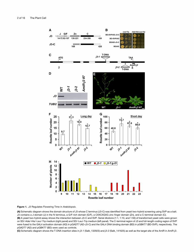
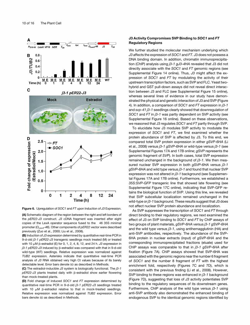
Figure 1. J3 Regulates Flowering Time in Arabidopsis.
(A) Schematic diagram shows the domain structure of J3 whose C terminus (J3-C) was identified from yeast two-hybrid screening using SVP as a bait.
J3 contains a J-domain (J) in the N terminus, a G/F-rich domain (G/F), a CXXCXGXG zinc finger domain (Zn), and a C-terminal domain (C).
(B) A yeast two-hybrid assay shows the interaction between J3-C and SVP. Serial dilutions (1:1, 1:10, and 1:50) of transformed yeast cells were grown
on SD/-Ade/-His/-Leu/-Trp medium (right panel) and SD/-Leu/-Trp medium (left panel). The C-terminal region of J3 and full-length coding region of SVP
were fused to the GAL4 activation domain (AD) in pGADT7 (AD-J3-C) and the GAL4 DNA binding domain (BD) in pGBKT7 (BD-SVP), respectively. The
pGADT7 (AD) and pGBKT7 (BD) were used as controls.
(C) Schematic diagram shows the T-DNA insertion sites in j3-1 (Salk_132923) and j3-2 (Salk_141625) as well as the target site of the AmiR in AmiR-j3.
2 of 16 The Plant Cell

modular structure of DnaJ/Hsp40 that was originally identified as
a 41-kD heat shock protein from Escherichia coli (Georgopoulos
et al., 1980).
Previous studies have shown that DnaJ interacts with Hsp70,
DnaK, and the nucleotide exchange factor, GrpE, to constitute a
molecular chaperone complex that functions in many cellular
processes (Liberek et al., 1991; Georgopoulos, 1992; Scidmore
et al., 1993; Cyr et al., 1994; Bukau and Horwich, 1998; Goffin and
Georgopoulos, 1998). Apart from the chaperone activity, DnaJ also
functions asaprotein disulfide isomerase to catalyze the formation,
reduction, or isomerization of disulfide bonds (de Crouy-Chanel
et al., 1995; Wang and Tsou, 1998). Furthermore, it has been
suggested that DnaJ function is conserved throughout evolution
(Caplan et al., 1993; Silver and Way, 1993; Qiu et al., 2006).
In plants, J-domain proteins have been reported to localize in
different subcellular compartments and participate in various
biological processes (Miernyk, 2001; Rajan and D’Silva, 2009).
However, as molecular chaperones are traditionally considered
as important components involved in cellular homeostasis under
stress conditions, previous studies on plant J-domain proteins
have been mainly focused on their functions in plant stress sig-
naling pathways (Wang et al., 2004; Ham et al., 2006; Yang et al.,
2010). While there are a few studies reporting the involvement of
plant J-domain proteins in developmental processes (Tamura
et al., 2007; Kneissl et al., 2009), the molecular basis for their
biological functions in plant growth and development is still
enigmatic.
In this study, we report that Arabidopsis DNAJ HOMOLOG 3
(J3), which encodes a type I J-domain protein (Zhou andMiernyk,
1999; Rajan and D’Silva, 2009), plays an essential role as a
transcriptional regulator in mediating the integration of flowering
signals. Loss of function of J3 significantly delays flowering,
which partly results from reduced expression ofSOC1 and FT. J3
interacts with SVP in the nucleus and attenuates the capacity of
SVP binding to the regulatory sequences of SOC1 and FT. Our
results suggest that J3 perceives flowering signals from several
genetic pathways and promotes flowering through directly an-
tagonizing SVP activity in repressing the transcription of SOC1
and FT during the floral transition.
RESULTS
Loss of Function of J3Delays Flowering Time inArabidopsis
To elucidate SVP function in Arabidopsis reproductive develop-
ment, we previously performed yeast two-hybrid screening to
identify its interacting partners (Liu et al., 2009b). Five out of over
50 interactors included the C-terminal part of J3 protein, which
contains the 252 amino acid residues from169 to 420 (Figure 1A).
We cloned this C-terminal fragment and confirmed its interaction
with SVP by yeast two-hybrid assay (Figure 1B). J3 encodes a
DnaJ-like heat shock protein, which contains a typical modular
sequence of type I J-domain proteins (Figure 1A) (Zhou and
Miernyk, 1999; Rajan and D’Silva, 2009; Yang et al., 2010). A
sequence comparison revealed that J3 homologs shared high
sequence similarity across plant species and other eukaryotic
organisms, such as yeast (Saccharomyces cerevisiae), zebra fish
(Danio rerio), mouse (Musmusculus), and human (Homo sapiens)
(see Supplemental Figure 1 online).
To investigate the biological function of J3, we isolated two
T-DNA insertional mutants, j3-1 and j3-2, from The Arabidopsis
Information Resource (TAIR; http://www.Arabidopsis.org). j3-1
and j3-2 contained a T-DNA insertion in the fourth and last
exons, respectively (Figure 1C). There was no detectable full-
length J3 transcript in either homozygousmutant line (Figure 1D).
Partial J3 transcripts upstream of the T-DNA insertion sites were
significantly downregulated, while no transcripts could be de-
tected spanning or downstream of the T-DNA insertion sites in
j3-1 and j3-2 (see Supplemental Figure 2A online). Under long
days (LDs) and short days (SDs), both j3-1 and j3-2 showed late
flowering, while the former possessed a stronger phenotype
(Figures 1E to 1G). The F1 progenies from the cross between
j3-1 and j3-2 flowered later than wild-type plants with an inter-
mediate flowering phenotype compared with the single mutants
(see Supplemental Figure 2B online), further indicating that j3-1
and j3-2 are allelic. Since j3-1 was likely a strong mutant allele,
we used it for further analyses. To test whether the late-flowering
phenotype of j3-1 can be attributed to the loss of J3 function,
we transformed j3-1 with a genomic construct (gJ3) harboring a
4.5-kb J3 genomic region that includes 2.2 kb of upstream
sequence, the 1.9-kb full coding sequence plus introns, and 0.4
kb of the 39 untranslated region. Most j3-1 gJ3 T1 transformants
exhibited comparable flowering time to wild-type plants (Figure
1H), demonstrating that J3 is responsible for the late-flowering
phenotype observed in j3-1.
To confirm J3 function in the regulation of flowering time, we
also created J3 knockdown transgenic plants by artificial micro-
RNA (AmiR) interference (Schwab et al., 2006). We generated 25
AmiR-j3 independent lines that expressed an AmiR specifically
targeting at the 39 region of the J3 mRNA (Figure 1C), among
which 19 lines exhibited different levels of late flowering under
LDs. We measured J3 expression in five selected late-flowering
lines and found that the degrees of late flowering in AmiR-j3
Figure 1. (continued).
Exons and introns in the coding region are indicated by black and white boxes, respectively. The start codon (ATG) and stop codon (TAA) are labeled.
Arrowheads indicate the position of primers used for amplifying full-length J3 transcript as shown in (D).
(D) RT-PCR shows that the full-length J3 transcript is undetectable in j3-1, j3-2, and AmiR-j3 #7. TUB2was amplified as an internal control. WT, wild type.
(E) j3-1 and AmiR-j3 exhibit late flowering under long days. Bar = 1 cm.
(F) and (G) Flowering time of j3mutants and AmiR-j3 #7 transgenic plants under LDs (F) and SDs (G). Values were scored from at least 15 plants of each
genotype. Mean values of rosette leaf number are indicated on top of bars. Error bars denote SD. Asterisks indicate significant difference in flowering
time of j3-1, AmiR-j3 #7, and j3-2 compared with that of wild-type plants (Student’s t test, P < 0.05).
(H) Distribution of flowering time in T1 transgenic plants harboring the J3 genomic fragment in j3-1 background.
J3 Regulates Flowering in Arabidopsis 3 of 16
plants were closely related to the levels of downregulation of J3
expression (see Supplemental Figures 2C to 2E online), suggest-
ing that downregulation of J3 has a dosage-dependent effect on
flowering. The strongest line, AmiR-j3 #7, which had the lowest
level of J3 expression, showed late flowering in both LDs and
SDs as j3-1 and j3-2 (Figures 1D to 1G). In contrast with the
significant late-flowering phenotype of J3 loss-of-function mu-
tants, transgenic plants overexpressing J3 showed normal
flowering time (see Supplemental Figure 3 online), implying that
the excessive amount of J3 might not affect flowering.
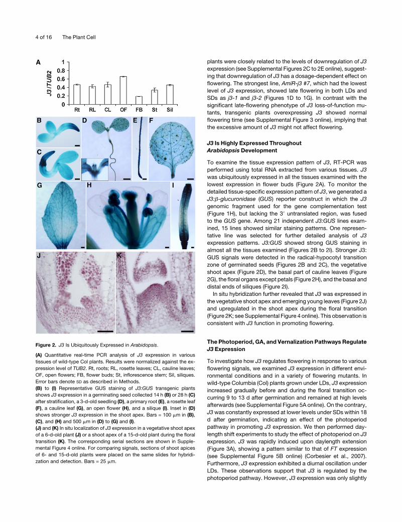
J3 Is Highly Expressed Throughout
Arabidopsis Development
To examine the tissue expression pattern of J3, RT-PCR was
performed using total RNA extracted from various tissues. J3
was ubiquitously expressed in all the tissues examined with the
lowest expression in flower buds (Figure 2A). To monitor the
detailed tissue-specific expression pattern of J3, we generated a
J3:b-glucuronidase (GUS) reporter construct in which the J3
genomic fragment used for the gene complementation test
(Figure 1H), but lacking the 39 untranslated region, was fused
to the GUS gene. Among 21 independent J3:GUS lines exam-
ined, 15 lines showed similar staining patterns. One represen-
tative line was selected for further detailed analysis of J3
expression patterns. J3:GUS showed strong GUS staining in
almost all the tissues examined (Figures 2B to 2I). Stronger J3:
GUS signals were detected in the radical-hypocotyl transition
zone of germinated seeds (Figures 2B and 2C), the vegetative
shoot apex (Figure 2D), the basal part of cauline leaves (Figure
2G), the floral organs except petals (Figure 2H), and the basal and
distal ends of siliques (Figure 2I).
In situ hybridization further revealed that J3 was expressed in
the vegetative shoot apex and emerging young leaves (Figure 2J)
and upregulated in the shoot apex during the floral transition
(Figure 2K; see Supplemental Figure 4 online). This observation is
consistent with J3 function in promoting flowering.
The Photoperiod, GA, and Vernalization Pathways Regulate
J3 Expression
To investigate how J3 regulates flowering in response to various
flowering signals, we examined J3 expression in different envi-
ronmental conditions and in a variety of flowering mutants. In
wild-type Columbia (Col) plants grown under LDs, J3 expression
increased gradually before and during the floral transition oc-
curring 9 to 13 d after germination and remained at high levels
afterwards (see Supplemental Figure 5A online). On the contrary,
J3was constantly expressed at lower levels under SDs within 18
d after germination, indicating an effect of the photoperiod
pathway in promoting J3 expression. We then performed day-
length shift experiments to study the effect of photoperiod on J3
expression. J3 was rapidly induced upon daylength extension
(Figure 3A), showing a pattern similar to that of FT expression
(see Supplemental Figure 5B online) (Corbesier et al., 2007).
Furthermore, J3 expression exhibited a diurnal oscillation under
LDs. These observations support that J3 is regulated by the
photoperiod pathway. However, J3 expression was only slightly
Figure 2. J3 Is Ubiquitously Expressed in Arabidopsis.
(A) Quantitative real-time PCR analysis of J3 expression in various
tissues of wild-type Col plants. Results were normalized against the ex-
pression level of TUB2. Rt, roots; RL, rosette leaves; CL, cauline leaves;
OF, open flowers; FB, flower buds; St, inflorescence stem; Sil, siliques.
Error bars denote SD as described in Methods.
(B) to (I) Representative GUS staining of J3:GUS transgenic plants
shows J3 expression in a germinating seed collected 14 h (B) or 28 h (C)
after stratification, a 3-d-old seedling (D), a primary root (E), a rosette leaf
(F), a cauline leaf (G), an open flower (H), and a silique (I). Inset in (D)
shows stronger J3 expression in the shoot apex. Bars = 100 mm in (B),
(C), and (H) and 500 mm in (D) to (G) and (I).
(J) and (K) In situ localization of J3 expression in a vegetative shoot apex
of a 6-d-old plant (J) or a shoot apex of a 15-d-old plant during the floral
transition (K). The corresponding serial sections are shown in Supple-
mental Figure 4 online. For comparing signals, sections of shoot apices
of 6- and 15-d-old plants were placed on the same slides for hybridi-
zation and detection. Bars = 25 mm.
4 of 16 The Plant Cell

Figure 3. J3 Is Regulated by the Photoperiod, Vernalization, and GA Pathways.
(A) J3 expression in response to a SD to LD shift determined by quantitative real-time PCR. Wild-type Col seedlings were grown under SDs for 11 d
before they were transferred to LDs. J3 expression in these seedlings was examined at 4 h intervals for 3 d comprising one SD followed by two LDs,
while its expression in plants grown under SDs was examined as a control. Bars below the graph indicate the duration of day (white) and night (black) for
the shift experiment (top) and the control experiment (bottom). Error bars denote SD as described in Methods.
(B) Comparison of J3 expression in the GA-deficient mutant ga1-3 (Ler background) and wild-type Ler plant determined by quantitative real-time PCR.
Four-day-old (D4) and 7-d-old (D7) seedlings grown under SDs were harvested for expression analysis. Error bars denote SD as described in Methods.
J3 expression in (B) to (F) was normalized to TUB2 expression.
(C) Effect of GA treatment on J3 expression in ga1-3 plants determined by quantitative real-time PCR. Exogenous GA (100 mM) or 0.1% ethanol (mock)
was applied daily onto 3-week-old ga1-3 seedlings grown under SDs for two consecutive days. J3 expression was examined either before (0 h) or 48 h
after (48 h) the first GA treatment. Error bars denote SD as described in Methods.
J3 Regulates Flowering in Arabidopsis 5 of 16

downregulated in loss-of-function mutants of two regulators in
the photoperiod pathway, FT and GIGANTEA, in the Col back-
ground but not affected by another key regulator, CONSTANS
(CO) (Figure 3F). Thus, the precise mechanism by which photo-
period modulates J3 needs to be further elucidated. J3 expres-
sion was consistently lower in the GA-deficient mutant ga1-3
than in the wild-type plant grown under SDs (Figure 3B). More-
over, GA treatment for two consecutive days upregulated J3
expression in ga1-3 grown under SDs (Figure 3C), indicating that
the GA pathway regulates J3. Similarly, J3 expression was also
upregulated inwild-type plants by vernalization treatment (Figure
3D). However, dramatic downregulation of FLC in FRI FLC after
vernalization did not enhance the upregulation of J3 in response
to vernalization, indicating that vernalization regulates J3 ex-
pression in an FLC-independent manner.
Like wild-type plants, j3-1 exhibited similar levels of delayed
flowering at 168C versus 238C (see Supplemental Figure 5C
online), suggesting that J3 is not directly involved in the thermo-
sensory pathway. J3 expression was also not affected in the
various mutants of the autonomous pathway (Figure 3E), indi-
cating that the autonomous pathway does not control J3 ex-
pression. In addition, several other flowering-time regulators,
such as SOC1, AGAMOUS-LIKE24 (AGL24), and SVP, which
mediate flowering signals from various genetic pathways, also
did not affect J3 expression (see Supplemental Figure 6 online).
Taken together, these results suggest that J3 perceives flower-
ing signals from photoperiod, GA, and vernalization pathways
but does not act downstream of floral pathway integrators (i.e.,
SOC1 and FT).
We further analyzed the genetic interaction between J3 and
other flowering time genes that function downstream of multiple
floral pathways (Figure 3G; see Supplemental Figure 7 online). A
comparison of flowering time between j3-1 and other flowering
mutants demonstrated that the effect of J3 on flowering in LDs
and SDs was comparable to that of SOC1. Overexpression of
SOC1 or FT significantly suppressed the late-flowering pheno-
type of j3-1 (Figure 3G), implying that these two major floral
pathway integrators act either downstream of or in parallel with
J3. Similarly, overexpression of CO, an upstream promoter of FT
and SOC1, also significantly suppressed the j3-1 phenotype.
Either ft-10 or soc1-2 enhanced late flowering of j3-1, whereas
ft-10 soc1-2 j3-1 triple mutants exhibited much delayed flower-
ing compared with single or double mutants (Figure 3G). These
results indicate that besides the potential regulatory hierarchies
among these genes, J3, SOC1, and FT also regulate flowering
through other independent pathways.
J3 Interacts with SVP
The genetic interaction between J3 and FTorSOC1 and the initial
identification of a J3 fragment as an interacting partner of SVP
(Figures 1A and 1B) aroused our interest in further understanding
the interaction between J3 and SVP, which is a direct transcrip-
tional regulator of FT and SOC1 (Lee et al., 2007; Li et al., 2008).
To this end, we first compared the expression patterns of J3 and
SVP using their GUS reporter lines. There was a comparable
spatial pattern of GUS staining in the aerial part of developing J3:
GUS and SVP:GUS (Li et al., 2008) seedlings, although J3:GUS
displayed much stronger GUS signals (Figure 4A). Second, we
compared the subcellular localization of J3 and SVP in tobacco
(Nicotiana tabacum) leaves using the red fluorescent protein
(RFP) and green fluorescent protein (GFP) fusion constructs 35S:
RFP-J3 and 35S:SVP-GFP. BothRFP-J3 andSVP-GFP localized
in the cytoplasm and nucleus, which is similar to the pattern
observed for 35S:RFP and 35S:GFP (Figures 4B to 4E). To verify
the subcellular localization of J3 and SVP inArabidopsis cells, we
then generated a functional j3-1 gJ3-4HA transgenic line (see
Supplemental Figure 8A online) and the specific anti-SVP anti-
body (see Supplemental Figure 9 online). Immunoblot analyses
revealed that J3 and SVP were present in both of the cytosolic
and nuclear fractions of j3-1 gJ3-4HA and 35S:SVP plants,
respectively (see Supplemental Figures 8B and 8C online). These
results demonstrate overlapping gene expression patterns and
subcellular protein localization of J3 and SVP.
Our yeast two-hybrid assay has shown that SVP interacts
with the C-terminal region of J3 that contains both part of the
zinc finger domain and the entire C-terminal domain (Figures 1A
and 1B). To determine which domain is responsible for J3
interaction with SVP, we cloned the sequences of the zinc finger
domain (J3-C1) and C-terminal domain (J3-C2), respectively,
for further yeast two-hybrid assays (Figure 4F; see Supplemental
Figure 10A online). The results demonstrated that the C-terminal
domain of J3 was required for the interaction with SVP. On
the contrary, none of the individual SVP domains (see Supple-
mental Figure 10B online) was sufficient for SVP interaction with
J3 (Figure 4G; see Supplemental Figure 10C online). In vitro
glutathione S-transferase (GST) pull-down assays further dem-
onstrated that both GST-J3 and GST-J3-C2 could bind to in
Figure 3. (continued).
(D) Effect of vernalization treatment on J3 expression determined by quantitative real-time PCR. For vernalization treatment, seeds were grown on MS
medium and vernalized at 48C under low-light conditions for 8 weeks. The 9-d-old seedlings grown under LDs were harvested for expression analysis.
Error bars denote SD as described in Methods.
(E) J3 expression determined by quantitative real-time PCR in 9-d-old mutants of the autonomous pathway. Error bars denote SD as described in
Methods.
(F) J3 expression determined by quantitative real-time PCR in 9-d-old mutants of the photoperiod pathway. Error bars denote SD as described in
Methods.
(G) Flowering time of various mutants or transgenic plants (Col background) grown under LDs. Values were scored from at least 15 plants of each
genotype. Mean values of rosette leaf number are indicated on top of bars. Error bars denote SD. Asterisks indicate significant difference in flowering
time of plants with various genetic backgrounds compared with that of j3-1 (Student’s t test, P < 0.05). Open triangle indicates no statistically significant
difference in flowering time of j3-1 compared with that of soc1-2 (Student’s t test, P > 0.5).
6 of 16 The Plant Cell
Figure 4. J3 Interacts with SVP.
(A) GUS staining of developing J3:GUS (top panels) and SVP:GUS seedlings. Bars = 1 mm.
(B) and (C) Subcellular localization of RFP-J3 in tobacco leaves. RFP localization was observed in tobacco leaves infiltrated with either 35S:RFP-J3 (B)
or a 35S:RFP control (C). Bars = 10 mm.
(D) and (E) Subcellular localization of SVP-GFP in tobacco leaves. GFP localization was observed in tobacco leaves infiltrated with either 35S:SVP-GFP
(D) or a 35S:GFP control (E). Bars = 10 mm.
(F) Yeast two-hybrid assay of the interaction between SVP and J3-C1 or J3-C2. Top panel shows the schematic diagram of J3 truncated proteins that
were fused to AD and used for yeast two-hybrid assays with SVP fused to BD. Transformed yeast cells were grown on SD/-Ade/-His/-Leu/-Trp medium
(bottom left panel) and SD/-Leu/-Trp medium (bottom right panel). Empty refers to AD- or BD-containing vector only.
(G) Yeast two-hybrid assay of the interaction between J3-C2 and SVP truncated proteins. The schematic diagram of SVP truncated proteins is
J3 Regulates Flowering in Arabidopsis 7 of 16
vitro–translated full-length myc-SVP, while the affinity between
GST-J3-C2 andmyc-SVPwasmuch stronger (Figure 4H). To test
the in vivo interaction between J3 and SVP, we performed
bimolecular fluorescence complementation (BiFC) experiments
(Ohad et al., 2007), which monitors the protein–protein inter-
action through detecting the fluorescence signals emitted by
reconstitution of an enhanced yellow fluorescent protein (EYFP)
from two fragments (N- and C-terminal halves) fused to two
interacting proteins. This revealed a direct interaction between
J3 and SVP in the nuclei of living tobacco cells (Figure 4I).
Furthermore, coimmunoprecipitation analysis of nuclear extracts
from j3-1 gJ3-4HA confirmed the in vivo interaction of J3 and
SVP in Arabidopsis (Figure 4J). These results provide evidence
that J3 interacts with SVP in the nucleus.
We next examined the biological significance of the interaction
of J3 and SVP through genetic analysis. As J3 and SVP have
opposite effects on the control of flowering time, j3-1 and svp-41
showed completely different flowering phenotypes (Figure 4K).
However, svp-41 j3-1 double mutants exhibited early flowering
similar to svp-41, demonstrating that SVP is genetically epistatic
to J3. This observation, together with the results showing in vitro
and in vivo protein interaction of J3 and SVP, suggests that J3
exerts its function in regulating flowering time via SVP.
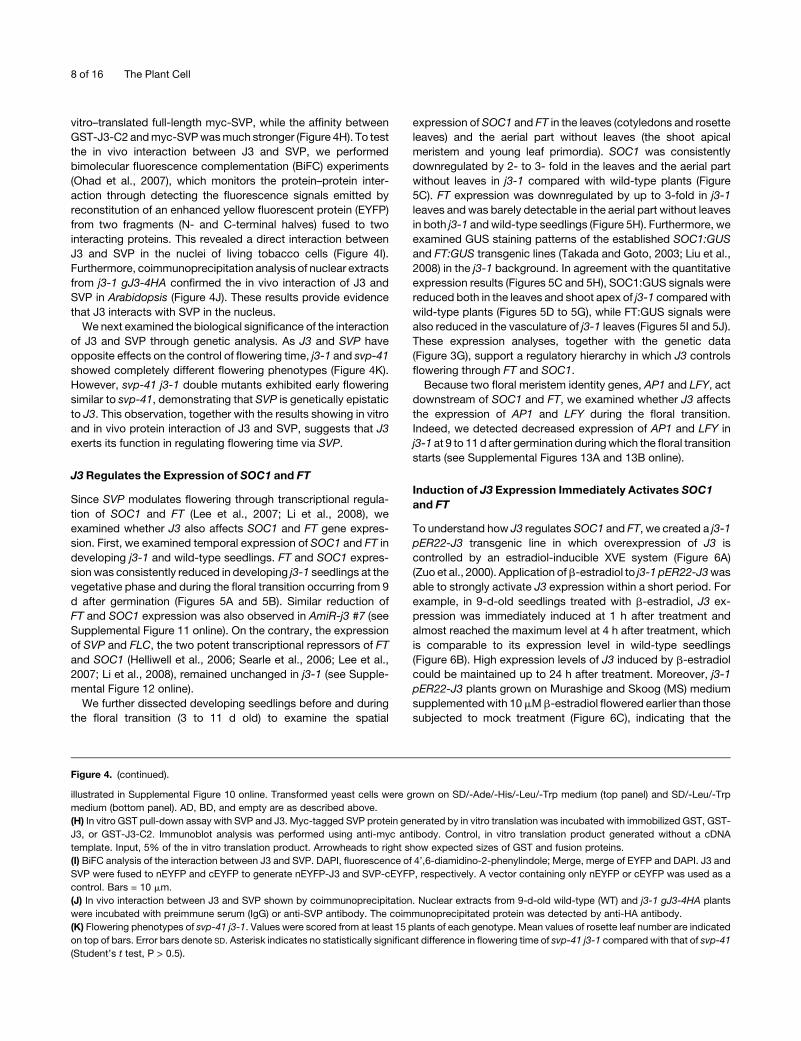
J3 Regulates the Expression of SOC1 and FT
Since SVP modulates flowering through transcriptional regula-
tion of SOC1 and FT (Lee et al., 2007; Li et al., 2008), we
examined whether J3 also affects SOC1 and FT gene expres-
sion. First, we examined temporal expression of SOC1 and FT in
developing j3-1 and wild-type seedlings. FT and SOC1 expres-
sion was consistently reduced in developing j3-1 seedlings at the
vegetative phase and during the floral transition occurring from 9
d after germination (Figures 5A and 5B). Similar reduction of
FT and SOC1 expression was also observed in AmiR-j3 #7 (see
Supplemental Figure 11 online). On the contrary, the expression
of SVP and FLC, the two potent transcriptional repressors of FT
and SOC1 (Helliwell et al., 2006; Searle et al., 2006; Lee et al.,
2007; Li et al., 2008), remained unchanged in j3-1 (see Supple-
mental Figure 12 online).
We further dissected developing seedlings before and during
the floral transition (3 to 11 d old) to examine the spatial
expression of SOC1 and FT in the leaves (cotyledons and rosette
leaves) and the aerial part without leaves (the shoot apical
meristem and young leaf primordia). SOC1 was consistently
downregulated by 2- to 3- fold in the leaves and the aerial part
without leaves in j3-1 compared with wild-type plants (Figure
5C). FT expression was downregulated by up to 3-fold in j3-1
leaves andwas barely detectable in the aerial part without leaves
in both j3-1 andwild-type seedlings (Figure 5H). Furthermore, we
examined GUS staining patterns of the established SOC1:GUS
and FT:GUS transgenic lines (Takada and Goto, 2003; Liu et al.,
2008) in the j3-1 background. In agreement with the quantitative
expression results (Figures 5C and 5H), SOC1:GUS signals were
reduced both in the leaves and shoot apex of j3-1 comparedwith
wild-type plants (Figures 5D to 5G), while FT:GUS signals were
also reduced in the vasculature of j3-1 leaves (Figures 5I and 5J).
These expression analyses, together with the genetic data
(Figure 3G), support a regulatory hierarchy in which J3 controls
flowering through FT and SOC1.
Because two floral meristem identity genes, AP1 and LFY, act
downstream of SOC1 and FT, we examined whether J3 affects
the expression of AP1 and LFY during the floral transition.
Indeed, we detected decreased expression of AP1 and LFY in
j3-1 at 9 to 11d after germination duringwhich the floral transition
starts (see Supplemental Figures 13A and 13B online).
Induction of J3 Expression Immediately Activates SOC1
and FT
To understand how J3 regulates SOC1 and FT, we created a j3-1
pER22-J3 transgenic line in which overexpression of J3 is
controlled by an estradiol-inducible XVE system (Figure 6A)
(Zuo et al., 2000). Application ofb-estradiol to j3-1 pER22-J3was
able to strongly activate J3 expression within a short period. For
example, in 9-d-old seedlings treated with b-estradiol, J3 ex-
pression was immediately induced at 1 h after treatment and
almost reached the maximum level at 4 h after treatment, which
is comparable to its expression level in wild-type seedlings
(Figure 6B). High expression levels of J3 induced by b-estradiol
could be maintained up to 24 h after treatment. Moreover, j3-1
pER22-J3 plants grown on Murashige and Skoog (MS) medium
supplementedwith 10mMb-estradiol flowered earlier than those
subjected to mock treatment (Figure 6C), indicating that the
Figure 4. (continued).
illustrated in Supplemental Figure 10 online. Transformed yeast cells were grown on SD/-Ade/-His/-Leu/-Trp medium (top panel) and SD/-Leu/-Trp
medium (bottom panel). AD, BD, and empty are as described above.
(H) In vitro GST pull-down assay with SVP and J3. Myc-tagged SVP protein generated by in vitro translation was incubated with immobilized GST, GST-
J3, or GST-J3-C2. Immunoblot analysis was performed using anti-myc antibody. Control, in vitro translation product generated without a cDNA
template. Input, 5% of the in vitro translation product. Arrowheads to right show expected sizes of GST and fusion proteins.
(I) BiFC analysis of the interaction between J3 and SVP. DAPI, fluorescence of 4’,6-diamidino-2-phenylindole; Merge, merge of EYFP and DAPI. J3 and
SVP were fused to nEYFP and cEYFP to generate nEYFP-J3 and SVP-cEYFP, respectively. A vector containing only nEYFP or cEYFP was used as a
control. Bars = 10 mm.
(J) In vivo interaction between J3 and SVP shown by coimmunoprecipitation. Nuclear extracts from 9-d-old wild-type (WT) and j3-1 gJ3-4HA plants
were incubated with preimmune serum (IgG) or anti-SVP antibody. The coimmunoprecipitated protein was detected by anti-HA antibody.
(K) Flowering phenotypes of svp-41 j3-1. Values were scored from at least 15 plants of each genotype. Mean values of rosette leaf number are indicated
on top of bars. Error bars denote SD. Asterisk indicates no statistically significant difference in flowering time of svp-41 j3-1 compared with that of svp-41
(Student’s t test, P > 0.5).
8 of 16 The Plant Cell

induced J3 activity in j3-1 pER22-J3 is biologically functional and
responsible for promoting flowering. Using this established
estradiol-inducible system, we tested the expression of SOC1
and FT in response to induced J3 expression (Figure 6D). SOC1
and FT expression was immediately upregulated by J3within 4 h
after b-estradiol treatment. After 4 h of treatment, SOC1 and FT
expression increased by 2.2- and 1.7-fold, respectively. Al-
though the levels of upregulation slightly decreased afterwards,
the expression of SOC1 and FT remained upregulated up to 24 h
after treatment. These observations suggest that J3 modulates
flowering time partly through activating the expression of two
floral pathway integrators, SOC1 and FT.
Figure 5. SOC1 and FT Expression Is Regulated by J3.
(A) and (B) Temporal expression of SOC1 (A) and FT (B) determined by quantitative real-time PCR in developing j3-1 and wild-type (WT) seedlings under
LDs. Error bars denote SD as described in Methods. The levels of gene expression normalized to TUB2 expression are shown as relative values to the
maximal expression level set at 100%.
(C) Fold change of SOC1 expression determined by quantitative real-time PCR in leaves and the aerial part without leaves in the wild type against that in
j3-1 seedlings under LDs. SOC1 expression was normalized to TUB2 expression. Error bars denote SD as described in Methods.
(D) to (G) GUS staining of 9-d-old seedlings of SOC1:GUS ([D] and [F]) and j3-1 SOC1:GUS ([E] and [G]). (D) and (E) show GUS staining of the aerial
part of the seedlings, while (F) and (G) show GUS staining in the shoot apex. Bars = 1 mm in (D) and (E) and 100 mm in (F) and (G).
(H) Fold change of FT expression determined by quantitative real-time PCR in leaves and the aerial part without leaves in the wild type against that in
j3-1 seedlings under LDs. FT expression was normalized to TUB2 expression. Asterisks indicate that FT transcript levels are barely detectable in the
aerial part without leaves using quantitative real-time PCR. Error bars denote SD as described in Methods.
(I) and (J) GUS staining of the first true leaves of 11-d-old seedlings of pFT:GUS (I) and j3-1 pFT:GUS (J). The bottom panels show a higher
magnification of an area of the middle half of the leaves. Bars = 1 mm.
J3 Regulates Flowering in Arabidopsis 9 of 16
J3 Activity Compromises SVP Binding to SOC1 and FT
Regulatory Regions
We further studied the molecular mechanism underlying which
J3 affects the expression ofSOC1 and FT. J3 does not possess a
DNA binding domain. In addition, chromatin immunoprecipita-
tion (ChIP) analysis using j3-1 gJ3-4HA revealed that J3 did not
directly associate with the SOC1 and FT genomic regions (see
Supplemental Figure 14 online). Thus, J3 might affect the ex-
pression of SOC1 and FT by modulating the activity of their
upstream transcription factors, such asSVP and FLC. Yeast two-
hybrid and GST pull-down assays did not reveal direct interac-
tion between J3 and FLC (see Supplemental Figure 15 online),
whereas several lines of evidence in our study have demon-
strated the physical and genetic interaction of J3 andSVP (Figure
4). In addition, a comparison of SOC1 and FT expression in j3-1
and svp-41 j3-1 seedlings clearly showed that downregulation of
SOC1 and FT in j3-1 was partly dependent on SVP activity (see
Supplemental Figure 16 online). Based on these observations,
we reasoned that J3 regulates SOC1 and FT partly through SVP.
To elucidate how J3 modules SVP activity to modulate the
expression of SOC1 and FT, we first examined whether the
protein abundance of SVP is affected by J3. To this end, we
compared total SVP protein expression in either gSVP-6HA (Li
et al., 2008) versus j3-1 gSVP-6HA or wild-type versus j3-1 (see
Supplemental Figures 17A and 17B online; gSVP represents the
genomic fragment of SVP). In both cases, total SVP expression
remained unchanged in the background of j3-1. We then mea-
sured nuclear SVP expression in both gSVP-6HA versus j3-1
gSVP-6HA and wild type versus j3-1 and found that nuclear SVP
expression was not altered in j3-1 background (see Supplemen-
tal Figures 17A and 17B online). Furthermore, we established a
35S:SVP-GFP transgenic line that showed late flowering (see
Supplemental Figure 17C online), indicating that SVP-GFP re-
tains the biological function of SVP. Using this line, we revealed
that SVP subcellular localization remained unchanged in the
wild-type or j3-1 background. These results suggest that J3 does
not affect nuclear SVP protein abundance and localization.
As SVP suppresses the transcription of SOC1 and FT through
direct binding to their regulatory regions, we next examined the
effect of J3 on SVP binding to SOC1 and FT by ChIP assays of
two groups of plant materials, gSVP-6HA versus j3-1 gSVP-6HA
and the wild type versus j3-1, using antihemagglutinin (HA) and
anti-SVP antibodies, respectively. The abundance of the SVP-
6HA protein in nuclear extracts (input) of gSVP-6HA and the
corresponding immunoprecipitated fractions (eluate) used for
ChIP assays was comparable to that in j3-1 gSVP-6HA after
fixation (Figure 7A). ChIP assays showed that SVP-6HA was
associatedwith the genomic regions near the number 6 fragment
of SOC1 and the number 8 fragment of FT with the highest
enrichment fold, respectively (Figures 7C and 7D), which is
consistent with the previous finding (Li et al., 2008). However,
SVP binding to these regions was enhanced in j3-1 background
(Figure 7D), suggesting that loss of J3 activity potentiates SVP
binding to the regulatory sequences of its downstream genes.
Furthermore, ChIP analysis of the wild type versus j3-1 using
anti-SVP antibody also demonstrated the enhanced binding of
endogenous SVP to the identical genomic regions identified by
Figure 6. Upregulation of SOC1 and FT upon Induction of J3 Expression.
(A) Schematic diagram of the region between the right and left borders of
the pER22-J3 construct. J3 cDNA fragment was inserted after eight
copies of the LexA operator sequence fused to the �46 35S minimal
promoter (OLexA-46). Other components of pER22 vector were described
previously (Zuo et al., 2000; Liu et al., 2008).
(B) Induction of J3 expression determined by quantitative real-time PCR in
9-d-old j3-1 pER22-J3 transgenic seedlings mock treated (M) or treated
with 10 mM b-estradiol (E) for 0, 1, 2, 4, 8, 12, and 24 h. J3 expression in
j3-1 pER22-J3 induced by b-estradiol was compared with that in 9-d-old
wild-type (WT) seedlings. Relative expression was normalized against
TUB2 expression. Asterisks indicate that quantitative real-time PCR
analysis of J3 RNA obtained very high Ct values because of its barely
detectable level. Error bars denote SD as described in Methods.
(C) The estradiol-inducible J3 system is biologically functional. The j3-1
pER22-J3 plants treated daily with b-estradiol show earlier flowering
than mock-treated plants.
(D) Fold change of induced SOC1 and FT expression determined by
quantitative real-time PCR in 9-d-old j3-1 pER22-J3 seedlings treated
with 10 mM b-estradiol relative to that in mock-treated seedlings.
Relative expression was normalized against TUB2 expression. Error
bars denote SD as described in Methods.
10 of 16 The Plant Cell

Figure 7. Loss of J3 Activity Enhances SVP Binding to SOC1 and FT Regulatory Regions.
(A) Measurement of SVP-6HA protein abundance in nuclear extracts or immunoprecipitated fractions of 9-d-old gSVP-6HA and j3-1 gSVP-6HA
seedlings. Nuclear extracts from various plants served as the input (I), while immunoprecipitated fractions by anti-HA antibody were used as the eluate
(E). Immunoblot analysis was performed using anti-HA antibody. WT, wild type.
(B) Measurement of SVP protein abundance in nuclear extracts or immunoprecipitated fractions of 9-d-old wild-type and j3-1 seedlings. Nuclear
extracts from various plants served as the input (I), while immunoprecipitated fractions by anti-SVP antibody were used as the eluate (E). Immunoblot
analysis was performed using anti-SVP antibody.
(C) Schematic diagrams show the SOC1 (top panel) and FT (bottom panel) genomic regions. Exons are represented by black boxes, while introns and
J3 Regulates Flowering in Arabidopsis 11 of 16

ChIP analysis of gSVP-6HA in the absence of J3 activity (Figures
7B, 7C, and 7E). These results, together with the fact that J3
activity promotes the expression of SOC1 and FT, suggest that
J3 regulates flowering time, at least in part, through compromis-
ing SVP binding to the regulatory regions of SOC1 and FT.
We further examined the capacity of SVP binding in response
to induced J3 activity using the established functional estradiol-
inducible system (j3-1 pER22-J3) (Figures 6A to 6C). To this end,
ChIP analyses were performed on 9-d-old j3-1 pER22-J3 seed-
lings treated with 10 mM b-estradiol for 4 h, in which J3 expres-
sion was induced to a similar level as that in wild-type seedlings
(Figure 6B). The abundance of endogenous SVP in nuclear
extracts (input) of j3-1 and j3-1 pER22-J3 mock treated or
treated with b-estradiol and in their corresponding immunopre-
cipitated fractions (eluate) used for ChIP assays was almost
comparable (Figure 8A), suggesting that both estradiol treatment
and the resulting change in J3 expression do not affect SVP
expression in the nucleus. Estradiol treatment of j3-1 did not
affect SVP binding to the genomic regions near the number 6
fragment of SOC1 and the number 8 fragment of FT, respectively
(Figures 8B and 8C). However, estradiol treatment of j3-1 pER22-
J3, which induced J3 expression, decreased SVP binding to
these regions (Figures 8B and 8C). These results demonstrate
that induced J3 activity is able to rapidly compromise SVP
binding capacity in planta.
DISCUSSION
Intensive studies of flowering in Arabidopsis have identified a
number of regulators involved in the dynamic process of the floral
transition in response to various environmental and develop-
mental cues perceived by several flowering genetic pathways. A
group of MADS box transcription factors, such as FLC, SVP,
SOC1, and AGL24 (Michaels and Amasino, 1999; Hartmann
et al., 2000; Lee et al., 2000; Yu et al., 2002;Michaels et al., 2003),
act as essential regulators at the convergence point of multiple
flowering pathways. They integrate flowering signals and further
regulate the spatial and temporal expression of floral meristem
identity genes to determine the transition from vegetative shoot
apical meristems to inflorescence meristems. Among these
MADS box regulators, SVP serves as a central flowering repres-
sor that functions together with another potent repressor, FLC, in
maintaining the duration of the vegetative phase by repressing
the expression of two major floral pathway integrators, FT and
SOC1 (Hartmann et al., 2000; Lee et al., 2007; Li et al., 2008).
Previous studies have suggested that various flowering path-
ways promote flowering through regulating either SVP mRNA
abundance or SVP protein abundance. The flowering signals
perceived by the thermosensory, autonomous, and GA path-
ways suppress SVP expression (Lee et al., 2007; Li et al., 2008).
Although the photoperiod pathway does not affect SVP expres-
sion, two essential circadian clock components, LATE ELON-
GATED HYPOCOTYL and CIRCADIAN CLOCK ASSOCIATED1,
regulate the abundance of SVP protein under continuous light
(Fujiwara et al., 2008). In this study, we report a flowering reg-
ulator, J3, which promotes flowering through attenuating SVP
binding to the regulatory sequences of FT and SOC1 rather than
affecting the abundance of SVP mRNA or protein (Figure 8D).
Several lines of evidence have demonstrated that J3 is an
essential regulator that is required for the integration of flowering
signals. First, the late-flowering phenotype of J3 loss-of-function
mutants in both LDs andSDs is similar to or evenmore significant
than that exhibited by loss of function of other key flowering
promoters, such as SOC1 and AGL24 (Figure 3; see Supple-
mental Figure 7 online). Second, J3 expression is regulated by
photoperiod, GA, and vernalization pathways (Figure 3), indicat-
ing that it affects flowering in response to both environmental and
developmental cues. Third, J3 interacts directly with SVP and
attenuates its in vivo binding to the regulatory sequences of
SOC1 and FT in developing seedlings (Figures 4 and 7). The
effect of J3 activity on SVP binding capacity is closely related to
SOC1 and FT expression in either j3mutants or j3-1 pER22-J3 in
which J3 expression is induced (Figures 5 to 8). These observa-
tions support that J3 acts an important regulator mediating the
floral transition through SVP. This is consistent with the genetic
evidence showing that svp-41 greatly suppresses the late-
flowering phenotype of j3-1 (Figure 4K). As SVP functions as a
dosage-dependent repressor of flowering (Hartmann et al.,
2000), modulation of its activity by increased J3 activity in re-
sponse to multiple flowering signals emerges as another impor-
tant mechanism for promoting the transition from vegetative to
reproductive growth in Arabidopsis.
Although our data support that J3 promotes flowering largely
through its interaction with SVP and regulation of SOC1 and FT,
there is evidence indicating that J3-mediated flowering process
involves other unknown regulators. The expression of SOC1 and
FT in svp-41 j3-1 was different from that in svp-41 (see Supple-
mental Figure 16 online), implying that regulation ofSOC1 and FT
by J3 is notmerely through SVP. Furthermore, J3 likely promotes
flowering throughother pathways independently ofSOC1andFT.
This is supported by two pieces of evidence. First, ft-10 soc1-2
Figure 7. (continued).
upstream regions are represented by white boxes. Bent arrows denote translation start sites and stop codons. Arrowheads indicate the sites containing
either single mismatch or perfect match to the consensus binding sequence (CArG box) of MADS domain proteins. Nineteen and fifteen DNA fragments
spanning the SOC1 and FT genomic regions were examined by ChIP enrichment test as shown in (D) and (E), respectively.
(D) ChIP analysis of SVP-6HA binding to the regulatory regions of SOC1 (top panel) and FT (bottom panel) in gSVP-6HA and j3-1 gSVP-6HA. ChIP
analysis was performed using the input and eluate described in (A).
(E) ChIP analysis of SVP binding to the regulatory regions of SOC1 (top panel) and FT (bottom panel) in the wild type and j3-1. ChIP analysis was
performed using the input and eluate described in (B).
A TUB2 fragment was amplified as a negative control in (D) and (E). Significant differences of enrichment fold changes in comparison with respective
controls are indicated with asterisks (Student’s t test, P < 0.05). Error bars denote SD of triplicate assays.
12 of 16 The Plant Cell

j3-1 flowers much later than any single or double mutants
(Figure 3G), suggesting that another regulator(s) contributes to
J3-mediated flowering in parallel with SOC1 and FT. Second,
while the genetic result shows that svp-41greatly suppresses j3-1
(Figure 4K), the upregulation of SOC1 and FT in svp-41 j3-1 was
less than that in svp-41 (see Supplemental Figure 16 online),
implying that the interaction between J3 and SVP also controls
other downstream regulators in addition to SOC1 and FT.
A large number of J-domain proteins, which are traditionally
considered as molecular chaperones along with Hsp70, have
been identified in both prokaryotes and eukaryotes (Craig et al.,
2006).The Arabidopsis genome contains 120 J-domain proteins,
and their functions are mostly uncharacterized (Miernyk, 2001;
Rajan andD’Silva, 2009).Whilemany studies have proposed that
J-domain proteins are part of chaperone machineries involved in
maintaining protein homeostasis under environmental stress
conditions, our results have shed light on the identity of J-domain
proteins as transcriptional regulators that play an indispensable
role in mediating plant development. Interestingly, MADS do-
main proteins, including SVP, are also a group of conserved tran-
scription factors that determine developmental processes and
signal transduction in eukaryotes (Riechmann and Meyerowitz,
1997; Theissen, 2000; Ng and Yanofsky, 2001). Therefore, the
interaction between J3 and SVP revealed in this study may
represent an emerging molecular relationship between their
respective families of conserved regulators, which links funda-
mental cellular homeostasis in response to environmental stimuli
and developmental control in eukaryotes.
METHODS
Plant Materials and Growth Conditions
Arabidopsis thaliana ecotypes Col-0 or Landsberg erecta (Ler) were
grown on soil or MS plates under LDs (16 h light/8 h dark) or SDs (8 h light/
16 h dark) at 238C6 28C. Themutants co-1,gi-1, ft-1 (Ler ft-1 introgressed
into Col), ft-10, fve-3, soc1-2, agl24-1, svp-41, j3-1, and j3-2 are in the Col
background, while co-2, ft-1, fve-1, fca-1, fpa-1, and ga1-3 are in the Ler
background (Li et al., 2008). All transgenic plants were generated in the
Col background through Agrobacterium tumefaciens–mediated transfor-
mation. Except for the transformants harboring pER22-J3 that were
selected on MS medium supplemented with hygromycin, transgenic
plants with other constructs were selected by Basta on soil. b-Estradiol
induction of j3-1 pER22-J3 plants was performed as previously described
(Liu et al., 2008).
Plasmid Construction
For the complementation test, a 4.5-kb J3 genomic fragment (gJ3)
including 2.2-kb upstream sequence, 1.9-kb coding sequence plus
introns, and 0.4-kb 39 untranslated region was amplified using primers
Figure 8. J3 Regulates Flowering Time by Mediating SVP Activity to
Regulate SOC1 and FT Transcription.
(A) Measurement of SVP protein abundance in nuclear extracts or
immunoprecipitated fractions of 9-d-old j3-1 and j3-1 pER22-J3 seed-
lings mock treated or treated with 10 mM b-estradiol for 4 h. Nuclear
extracts from various plants served as the input (I), while immunopre-
cipitated fractions by anti-SVP antibody were used as the eluate (E).
Immunoblot analysis was performed using anti-SVP antibody.
(B) and (C) ChIP analysis of SVP binding to the regulatory regions of
SOC1 (B) and FT (C) upon induced expression of J3 in 9-d-old j3-1
pER22-J3 seedlings. ChIP analysis was performed using the input and
eluate described in (A). The DNA fragments amplified in ChIP assays are
illustrated in Figure 7C. A TUB2 fragment was amplified as a negative
control. Significant differences of enrichment fold changes in compar-
ison with respective controls are indicated with asterisks (Student’s t
test, P < 0.05). Error bars denote SD of triplicate assays.
(D) Proposed model of J3 function in mediating the integration of
flowering signals during the floral transition. J3 expression is promoted
by the photoperiod, vernalization, and GA pathways. J3 promotes
flowering partly through upregulating the expression of SOC1 and FT.
J3 interacts with SVP in the nucleus and attenuates SVP binding to the
regulatory sequences of SOC1 and FT, thus affecting their expression.
Stimulatory interactions are indicated by arrows, while repressive inter-
actions are indicated by T-bars. Asterisks indicate the confirmed direct
transcriptional regulation.
J3 Regulates Flowering in Arabidopsis 13 of 16

gJ3-F-PstI (59-AACTGCAGCCAACCTACTTGCATGTTAACAAG-39) and
gJ3-R-XmaI (59-CCCCCCGGGGTACATGTCATCGGAGTTAACAC-39). The
resulting PCR fragment was digested and cloned into pHY105 (Liu et al.,
2007). To construct gJ3-4HA, the J3 genomic fragment including 2.2-kb
upstream sequence and 1.9-kb coding sequence plus introns was
amplified with primers gJ3-F-PstI and gJ3-R2-XmaI (59-CCCCCCGGG-
CTGCTGGGCACATTGCACCC-39) and cloned into pGreen-4HA to ob-
tain an in-frame fusion of J3-4HA. The pGreen-6HA vector (Li et al., 2008)
was cut with BamHI and self-ligated to generate pGreen-4HA. To
construct J3:GUS, the J3 genomic region was amplified with primers
gJ3-F-PstI and gJ3-R2-XmaI and cloned into pHY107 (Liu et al., 2007). To
construct AmiR-j3, design of the AmiR was performed using the software
on the website (http://wmd2.weigelworld.org). Based on the J3 se-
quence, a set of four primers was generated and used for the PCR
amplification according to the published protocol (Schwab et al., 2006).
The resulting PCR fragment was digested with EcoRI and BamHI and
cloned into pGreen 0229-35S vector (Yu et al., 2004). To construct
pER22-J3 and 35S:J3, the J3 cDNA sequence was amplified and cloned
into pER22 (Liu et al., 2008) and pGreen 0229-35S (Yu et al., 2004)
vectors, respectively.
Expression Analysis
Total RNAwas extracted using RNeasy plantmini kit (Qiagen) and reverse
transcribed with the SuperScript III first-strand synthesis system (Invi-
trogen) according to the manufacturer’s instructions. Quantitative real-
time PCR was performed using three biological replicates with three
technical replicates each on 7900HT Fast real-time PCR systems with
SYBR Green PCR Master Mix (Applied Biosystems). The mean and SD
were determined from one representative biological replicate. The rela-
tive expression levels were determined as previously described (Li et al.,
2008). The difference between the cycle threshold (Ct) of target genes and
the Ct of control primers (DCt = Cttarget gene2Ctcontrol) was used to obtain
the normalized expression of target genes. RT-PCR, nonradioactive in
situ hybridization, and synthesis of RNA probes were performed as
previously described (Yu et al., 2002). Primers used for gene expression
analysis are listed in Supplemental Table 1 online. For J3:GUS reporter
construct, we checked 17 independent transgenic lines at the T3 gen-
eration and selected one representative line for further analysis. GUS
staining was performed as previously described (Yu et al., 2000).
Yeast Two-Hybrid Assay
To construct the vectors for yeast two-hybrid assays, the coding regions
of SVP and J3 and their truncated versionswere amplified and cloned into
pGADT7 or pGBKT7 (Clontech). The yeast two-hybrid assay was per-
formed using the Yeastmaker Yeast Transformation System 2 according
to the manufacturer’s instructions (Clontech). The library screening was
performed as previously described (Liu et al., 2009b).
In Vitro GST Pull-Down Assay
The cDNAs encoding J3 and J3-C2 were cloned into pGEX-6p-2 vector
(Pharmacia). The GST-J3 and GST-J3-C2 fusion proteins were produced
by inducing Escherichia coli BL21 cells harboring the corresponding
constructs with isopropyl b-D-1-thiogalactopyranoside at 168C. The
soluble GST fusion proteins were extracted and immobilized on glutathi-
one sepharose beads (AmershamBiosciences) for subsequent pull-down
assays. Myc-tagged SVP proteins were generated as previously de-
scribed (Li et al., 2008). To produce myc-tagged FLC proteins, the full-
length FLC cDNA was cloned into the pGBKT7 (Clontech). The resulting
plasmid was added to the TNT T7 Quick Coupled Transcription/Trans-
lation Systems (Promega) to synthesize myc-FLC. These epitope-tagged
proteins were incubated with the immobilized GST and GST fusion
proteins. Proteins retained on the beads were resolved by SDS-PAGE
and detected with anti-myc antibody (Santa Cruz Biotechnology).
BiFC Analysis
The full-length coding regions of SVP and J3 were cloned into primary
pSAT1 vectors. The resulting cassettes including fusion proteins and
the constitutive promoters were cloned into pHY105 and transformed
into Agrobacterium. The Agrobacteria were coinfiltrated into 3-week-
old tobacco (Nicotiana benthamiana) leaves as previously described
(Sparkes et al., 2006).
Production of SVP-Specific Antibody
Synthetic peptides corresponding to amino acid residues 218 to 233
of the SVP protein (STGAPVDSESSDTSLR) (Fujiwara et al., 2008) were
used to produce polyclonal rabbit antibodies (GenScript). Anti-SVP
antibody could specifically detect endogenous SVP but not the closest
homolog of SVP, AGL24 (see Supplemental Figure 9 online).
Coimmunoprecipitation Experiment
Wild-type and j3-1 gJ3-4HA plants were ground with mortar and pestle in
liquid nitrogen, and nuclear proteins were extracted as previously de-
scribed (Liu et al., 2009b). The protein extracts were then incubated with
Protein G PLUS-Agarose (Santa Cruz Biotechnology) and anti-SVP
antibody or preimmune serum at 48C for 2 h. The immunoprecipitated
proteins and the protein extracts as inputs were resolved by SDS-PAGE
and detected by anti-HA antibody (Santa Cruz Biotechnology).
ChIP Assay
Seedlings were fixed on ice for 40 min in 1% formaldehyde under
vacuum. Fixed tissues were homogenized. Chromatin was isolated and
sonicated to produce DNA fragments of;250 bp. SVP-6HA and endog-
enous SVP protein were immunoprecipitated by anti-HA agarose conju-
gate (Sigma-Aldrich) and anti-SVP antibody bound to Protein G PLUS
Agarose, respectively. Relative enrichment of each fragment was deter-
mined by quantitative real-time PCR as previously reported (Li et al.,
2008). Primer pairs used for ChIP assays are listed in Supplemental Table
1 online. All ChIP assays were repeated three times with similar results,
and representative data are presented in Figures 7 and 8.
Accession Numbers
Sequence data from this article can be found in the Arabidopsis Genome
Initiative under the following accession numbers: J3 (At3g44110), J2
(At5g22060), SVP (At2g22540), FT (At1g65480), SOC1 (At2g45660), FLC
(At5g10140), and TUB2 (At5g62690). The T-DNA insertion mutants j3-1
(Salk_132923) and j3-2 (Salk_141625) were provided by TAIR.
Supplemental Data
The following materials are available in the online version of this article.
Supplemental Figure 1. The Amino Acid Sequence of J3 Is Highly
Conserved among Eukaryotes.
Supplemental Figure 2. Downregulation of J3 Expression Delays
Flowering.
Supplemental Figure 3. Overexpression of J3 Does Not Significantly
Affect Flowering.
Supplemental Figure 4. In Situ Localization of J3 Expression in Serial
Sections of Vegetative Shoot Apices or Shoot Apices during the Floral
Transition.
14 of 16 The Plant Cell

Supplemental Figure 5. Regulation of J3 by Various Flowering
Genetic Pathways.
Supplemental Figure 6. AGL24, SOC1, and SVP Do Not Affect J3
Expression.
Supplemental Figure 7. Flowering Time of Various Mutants (Col
Background) Grown under Short Days.
Supplemental Figure 8. Subcellular Localization of J3 and SVP
Revealed by Immunoblot Analysis.
Supplemental Figure 9. Anti-SVP Antibody Specifically Detects SVP.
Supplemental Figure 10. The Interaction between J3 and SVP
Requires the C-Terminal Domain of J3 and Full-Length SVP Protein.
Supplemental Figure 11. Expression of SOC1 and FT in AmiR-j3
Seedlings.
Supplemental Figure 12. J3 Does Not Affect the Expression of SVP
and FLC.
Supplemental Figure 13. The Expression of AP1 and LFY Is Regu-
lated by J3.
Supplemental Figure 14. J3 Is Not Associated with the SOC1 and FT
Genomic Regions.
Supplemental Figure 15. J3 Does Not Interact with FLC.
Supplemental Figure 16. A Comparison of SOC1 and FT Expression
in j3-1, svp-41, and svp-41 j3-1 Seedlings.
Supplemental Figure 17. J3 Does Not Affect Total and Nuclear SVP
Protein Expression or SVP-GFP Subcellular Localization.
Supplemental Table 1. List of Primers Used in This Study.
ACKNOWLEDGMENTS
We thank K. Goto, D. Weigel, P. Huijser, I. Lee, R. Amasino, M.F.
Yanofsky, and the ABRC for materials. This work was supported by
Academic Research Fund T208B3113 from the Ministry of Education,
Singapore, and intramural research funds from Temasek Life Sciences
Laboratory.
Received January 7, 2011; revised January 7, 2011; accepted February 8,
2011; published February 22, 2011.
REFERENCES
Blazquez, M.A., Ahn, J.H., and Weigel, D. (2003). A thermosensory
pathway controlling flowering time in Arabidopsis thaliana. Nat. Genet.
33: 168–171.
Blazquez, M.A., and Weigel, D. (2000). Integration of floral inductive
signals in Arabidopsis. Nature 404: 889–892.
Boss, P.K., Bastow, R.M., Mylne, J.S., and Dean, C. (2004). Multiple
pathways in the decision to flower: enabling, promoting, and reset-
ting. Plant Cell 16 (suppl.): S18–S31.
Bukau, B., and Horwich, A.L. (1998). The Hsp70 and Hsp60 chaperone
machines. Cell 92: 351–366.
Caplan, A.J., Cyr, D.M., and Douglas, M.G. (1993). Eukaryotic homo-
logues of Escherichia coli dnaJ: A diverse protein family that functions
with hsp70 stress proteins. Mol. Biol. Cell 4: 555–563.
Corbesier, L., Vincent, C., Jang, S., Fornara, F., Fan, Q., Searle, I.,
Giakountis, A., Farrona, S., Gissot, L., Turnbull, C., and Coupland,
G. (2007). FT protein movement contributes to long-distance signaling
in floral induction of Arabidopsis. Science 316: 1030–1033.
Craig, E.A., Huang, P., Aron, R., and Andrew, A. (2006). The diverse
roles of J-proteins, the obligate Hsp70 co-chaperone. Rev. Physiol.
Biochem. Pharmacol. 156: 1–21.
Cyr, D.M., Langer, T., and Douglas, M.G. (1994). DnaJ-like proteins:
Molecular chaperones and specific regulators of Hsp70. Trends
Biochem. Sci. 19: 176–181.
de Crouy-Chanel, A., Kohiyama, M., and Richarme, G. (1995). A novel
function of Escherichia coli chaperone DnaJ. Protein-disulfide isom-
erase. J. Biol. Chem. 270: 22669–22672.
Fujiwara, S., Oda, A., Yoshida, R., Niinuma, K., Miyata, K., Tomozoe,
Y., Tajima, T., Nakagawa, M., Hayashi, K., Coupland, G., and
Mizoguchi, T. (2008). Circadian clock proteins LHY and CCA1
regulate SVP protein accumulation to control flowering in Arabidopsis.
Plant Cell 20: 2960–2971.
Georgopoulos, C. (1992). The emergence of the chaperone machines.
Trends Biochem. Sci. 17: 295–299.
Georgopoulos, C.P., Lundquist-Heil, A., Yochem, J., and Feiss, M.
(1980). Identification of the E. coli dnaJ gene product. Mol. Gen.
Genet. 178: 583–588.
Goffin, L., and Georgopoulos, C. (1998). Genetic and biochemical
characterization of mutations affecting the carboxy-terminal domain
of the Escherichia coli molecular chaperone DnaJ. Mol. Microbiol. 30:
329–340.
Ham, B.K., Park, J.M., Lee, S.B., Kim, M.J., Lee, I.J., Kim, K.J.,
Kwon, C.S., and Paek, K.H. (2006). Tobacco Tsip1, a DnaJ-type Zn
finger protein, is recruited to and potentiates Tsi1-mediated tran-
scriptional activation. Plant Cell 18: 2005–2020.
Hartmann, U., Hohmann, S., Nettesheim, K., Wisman, E., Saedler,
H., and Huijser, P. (2000). Molecular cloning of SVP: A negative
regulator of the floral transition in Arabidopsis. Plant J. 21: 351–360.
Helliwell, C.A., Wood, C.C., Robertson, M., James Peacock, W., and
Dennis, E.S. (2006). The Arabidopsis FLC protein interacts directly in
vivo with SOC1 and FT chromatin and is part of a high-molecular-
weight protein complex. Plant J. 46: 183–192.
Kardailsky, I., Shukla, V.K., Ahn, J.H., Dagenais, N., Christensen,
S.K., Nguyen, J.T., Chory, J., Harrison, M.J., and Weigel, D. (1999).
Activation tagging of the floral inducer FT. Science 286: 1962–1965.
Kneissl, J., Wachtler, V., Chua, N.H., and Bolle, C. (2009). OWL1: An
Arabidopsis J-domain protein involved in perception of very low light
fluences. Plant Cell 21: 3212–3225.
Kobayashi, Y., Kaya, H., Goto, K., Iwabuchi, M., and Araki, T. (1999).
A pair of related genes with antagonistic roles in mediating flowering
signals. Science 286: 1960–1962.
Lee, H., Suh, S.S., Park, E., Cho, E., Ahn, J.H., Kim, S.G., Lee, J.S.,
Kwon, Y.M., and Lee, I. (2000). The AGAMOUS-LIKE 20 MADS
domain protein integrates floral inductive pathways in Arabidopsis.
Genes Dev. 14: 2366–2376.
Lee, J.H., Yoo, S.J., Park, S.H., Hwang, I., Lee, J.S., and Ahn, J.H.
(2007). Role of SVP in the control of flowering time by ambient
temperature in Arabidopsis. Genes Dev. 21: 397–402.
Li, D., Liu, C., Shen, L., Wu, Y., Chen, H., Robertson, M., Helliwell,
C.A., Ito, T., Meyerowitz, E., and Yu, H. (2008). A repressor complex
governs the integration of flowering signals in Arabidopsis. Dev. Cell
15: 110–120.
Liberek, K., Marszalek, J., Ang, D., Georgopoulos, C., and Zylicz, M.
(1991). Escherichia coli DnaJ and GrpE heat shock proteins jointly
stimulate ATPase activity of DnaK. Proc. Natl. Acad. Sci. USA 88:
2874–2878.
Liu, C., Chen, H., Er, H.L., Soo, H.M., Kumar, P.P., Han, J.H., Liou, Y.
C., and Yu, H. (2008). Direct interaction of AGL24 and SOC1 inte-
grates flowering signals in Arabidopsis. Development 135: 1481–1491.
J3 Regulates Flowering in Arabidopsis 15 of 16

Liu, C., Thong, Z., and Yu, H. (2009a). Coming into bloom: The
specification of floral meristems. Development 136: 3379–3391.
Liu, C., Xi, W., Shen, L., Tan, C., and Yu, H. (2009b). Regulation of
floral patterning by flowering time genes. Dev. Cell 16: 711–722.
Liu, C., Zhou, J., Bracha-Drori, K., Yalovsky, S., Ito, T., and Yu, H.
(2007). Specification of Arabidopsis floral meristem identity by re-
pression of flowering time genes. Development 134: 1901–1910.
Michaels, S.D., and Amasino, R.M. (1999). FLOWERING LOCUS C
encodes a novel MADS domain protein that acts as a repressor of
flowering. Plant Cell 11: 949–956.
Michaels, S.D., Ditta, G., Gustafson-Brown, C., Pelaz, S., Yanofsky,
M., and Amasino, R.M. (2003). AGL24 acts as a promoter of
flowering in Arabidopsis and is positively regulated by vernalization.
Plant J. 33: 867–874.
Miernyk, J.A. (2001). The J-domain proteins of Arabidopsis thaliana: An
unexpectedly large and diverse family of chaperones. Cell Stress
Chaperones 6: 209–218.
Mouradov, A., Cremer, F., and Coupland, G. (2002). Control of
flowering time: interacting pathways as a basis for diversity. Plant
Cell 14 (suppl.): S111–S130.
Ng, M., and Yanofsky, M.F. (2001). Function and evolution of the plant
MADS-box gene family. Nat. Rev. Genet. 2: 186–195.
Ohad, N., Shichrur, K., and Yalovsky, S. (2007). The analysis of
protein-protein interactions in plants by bimolecular fluorescence
complementation. Plant Physiol. 145: 1090–1099.
Qiu, X.B., Shao, Y.M., Miao, S., and Wang, L. (2006). The diversity of
the DnaJ/Hsp40 family, the crucial partners for Hsp70 chaperones.
Cell. Mol. Life Sci. 63: 2560–2570.
Rajan, V.B., and D’Silva, P. (2009). Arabidopsis thaliana J-class heat
shock proteins: Cellular stress sensors. Funct. Integr. Genomics 9:
433–446.
Riechmann, J.L., and Meyerowitz, E.M. (1997). MADS domain pro-
teins in plant development. Biol. Chem. 378: 1079–1101.
Samach, A., Onouchi, H., Gold, S.E., Ditta, G.S., Schwarz-Sommer,
Z., Yanofsky, M.F., and Coupland, G. (2000). Distinct roles of
CONSTANS target genes in reproductive development of Arabidop-
sis. Science 288: 1613–1616.
Schwab, R., Ossowski, S., Riester, M., Warthmann, N., and Weigel,
D. (2006). Highly specific gene silencing by artificial microRNAs in
Arabidopsis. Plant Cell 18: 1121–1133.
Scidmore, M.A., Okamura, H.H., and Rose, M.D. (1993). Genetic
interactions between KAR2 and SEC63, encoding eukaryotic homo-
logues of DnaK and DnaJ in the endoplasmic reticulum. Mol. Biol. Cell
4: 1145–1159.
Searle, I., He, Y., Turck, F., Vincent, C., Fornara, F., Krober, S.,
Amasino, R.A., and Coupland, G. (2006). The transcription factor
FLC confers a flowering response to vernalization by repressing
meristem competence and systemic signaling in Arabidopsis. Genes
Dev. 20: 898–912.
Sheldon, C.C., Burn, J.E., Perez, P.P., Metzger, J., Edwards, J.A.,
Peacock, W.J., and Dennis, E.S. (1999). The FLF MADS box gene: A
repressor of flowering in Arabidopsis regulated by vernalization and
methylation. Plant Cell 11: 445–458.
Silver, P.A., and Way, J.C. (1993). Eukaryotic DnaJ homologs and the
specificity of Hsp70 activity. Cell 74: 5–6.
Simpson, G.G., and Dean, C. (2002). Arabidopsis, the Rosetta stone of
flowering time? Science 296: 285–289.
Sparkes, I.A., Runions, J., Kearns, A., and Hawes, C. (2006). Rapid,
transient expression of fluorescent fusion proteins in tobacco plants
and generation of stably transformed plants. Nat. Protoc. 1: 2019–
2025.
Takada, S., and Goto, K. (2003). Terminal flower2, an Arabidopsis
homolog of heterochromatin protein1, counteracts the activation of
flowering locus T by constans in the vascular tissues of leaves to
regulate flowering time. Plant Cell 15: 2856–2865.
Tamura, K., Takahashi, H., Kunieda, T., Fuji, K., Shimada, T., and
Hara-Nishimura, I. (2007). Arabidopsis KAM2/GRV2 is required for
proper endosome formation and functions in vacuolar sorting and
determination of the embryo growth axis. Plant Cell 19: 320–332.
Theissen, G. (2000). Plant biology. Shattering developments. Nature
404: 711–713, 713.
Wang, C.C., and Tsou, C.L. (1998). Enzymes as chaperones and
chaperones as enzymes. FEBS Lett. 425: 382–384.
Wang, W., Vinocur, B., Shoseyov, O., and Altman, A. (2004). Role of
plant heat-shock proteins and molecular chaperones in the abiotic
stress response. Trends Plant Sci. 9: 244–252.
Wang, Y., Liu, C., Yang, D., Yu, H., and Liou, Y.C. (2010). Pin1At
encoding a peptidyl-prolyl cis/trans isomerase regulates flowering
time in Arabidopsis. Mol. Cell 37: 112–122.
Yang, Y., Qin, Y., Xie, C., Zhao, F., Zhao, J., Liu, D., Chen, S.,
Fuglsang, A.T., Palmgren, M.G., Schumaker, K.S., Deng, X.W.,
and Guo, Y. (2010). The Arabidopsis chaperone J3 regulates the
plasma membrane H+-ATPase through interaction with the PKS5
kinase. Plant Cell 22: 1313–1332.
Yu, H., Ito, T., Wellmer, F., and Meyerowitz, E.M. (2004). Repression
of AGAMOUS-LIKE 24 is a crucial step in promoting flower develop-
ment. Nat. Genet. 36: 157–161.
Yu, H., Xu, Y., Tan, E.L., and Kumar, P.P. (2002). AGAMOUS-LIKE 24,
a dosage-dependent mediator of the flowering signals. Proc. Natl.
Acad. Sci. USA 99: 16336–16341.
Yu, H., Yang, S.H., and Goh, C.J. (2000). DOH1, a class 1 knox gene, is
required for maintenance of the basic plant architecture and floral
transition in orchid. Plant Cell 12: 2143–2160.
Zhou, R.G., and Miernyk, J.A. (1999). Cloning and analysis of AtJ3
gene in Arabidopsis thaliana. Acta Bot. Sin. 41: 597–602.
Zuo, J., Niu, Q.W., and Chua, N.H. (2000). Technical advance: An
estrogen receptor-based transactivator XVE mediates highly inducible
gene expression in transgenic plants. Plant J. 24: 265–273.
16 of 16 The Plant Cell
DOI 10.1105/tpc.111.083048; originally published online February 22, 2011;Plant Cell
Lisha Shen, Yin Ga Germain Kang, Lu Liu and Hao YuArabidopsisThe J-Domain Protein J3 Mediates the Integration of Flowering Signals in
This information is current as of July 2, 2018
Supplemental Data /content/suppl/2011/02/22/tpc.111.083048.DC1.html
Permissions https://www.copyright.com/ccc/openurl.do?sid=pd_hw1532298X&issn=1532298X&WT.mc_id=pd_hw1532298X
eTOCs http://www.plantcell.org/cgi/alerts/ctmain
Sign up for eTOCs at:
CiteTrack Alerts http://www.plantcell.org/cgi/alerts/ctmain
Sign up for CiteTrack Alerts at:
Subscription Information http://www.aspb.org/publications/subscriptions.cfm
is available at:Plant Physiology and The Plant CellSubscription Information for
ADVANCING THE SCIENCE OF PLANT BIOLOGY © American Society of Plant Biologists