first evaluation of a clinical pathway using …
TRANSCRIPT
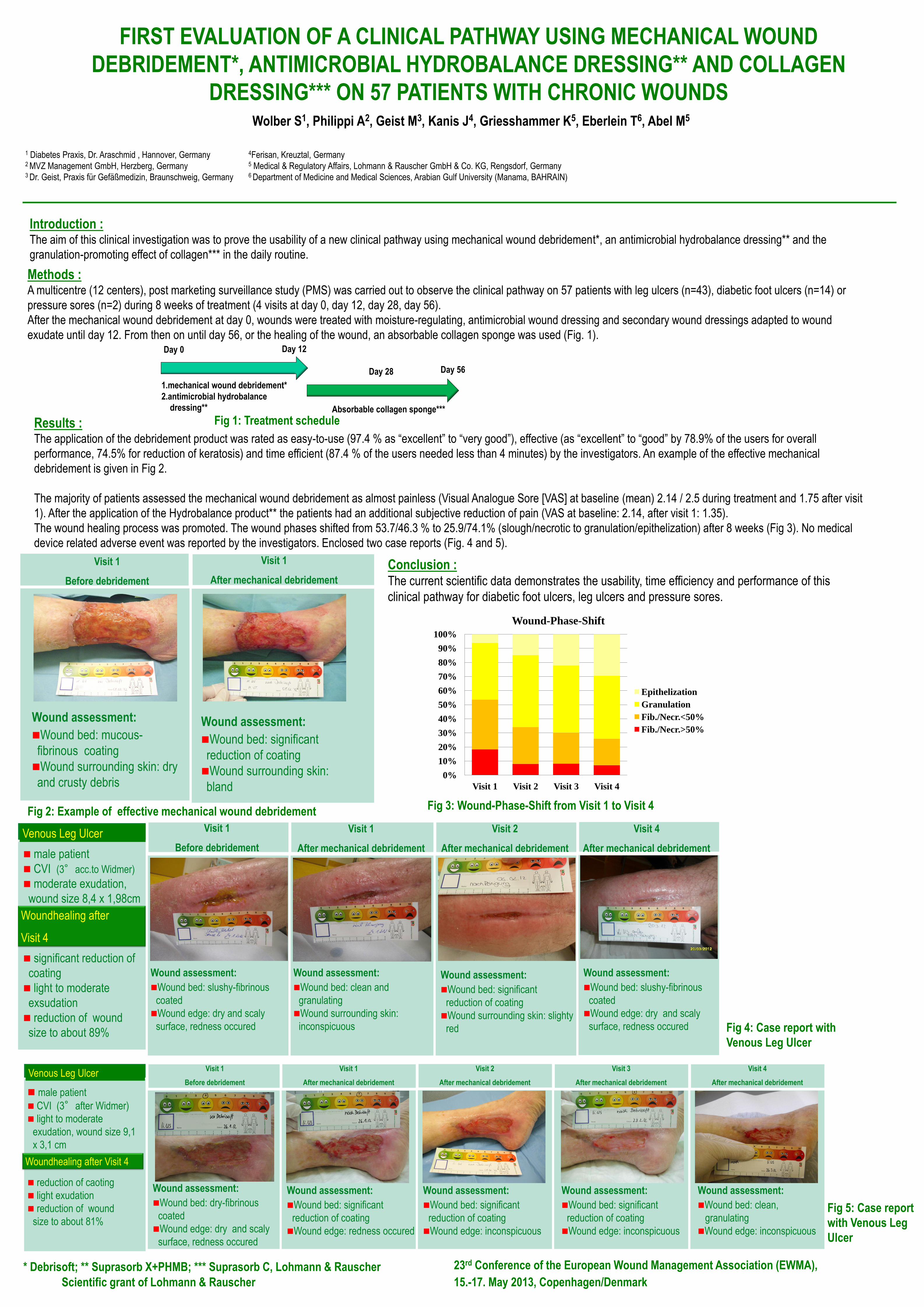
FIRST EVALUATION OF A CLINICAL PATHWAY USING MECHANICAL WOUND
DEBRIDEMENT*, ANTIMICROBIAL HYDROBALANCE DRESSING** AND COLLAGEN
DRESSING*** ON 57 PATIENTS WITH CHRONIC WOUNDS Wolber S1, Philippi A2, Geist M3, Kanis J4, Griesshammer K5, Eberlein T6, Abel M5
1 Diabetes Praxis, Dr. Araschmid , Hannover, Germany 4Ferisan, Kreuztal, Germany 2 MVZ Management GmbH, Herzberg, Germany 5 Medical & Regulatory Affairs, Lohmann & Rauscher GmbH & Co. KG, Rengsdorf, Germany 3 Dr. Geist, Praxis für Gefäßmedizin, Braunschweig, Germany 6 Department of Medicine and Medical Sciences, Arabian Gulf University (Manama, BAHRAIN)
Scientific grant of Lohmann & Rauscher
* Debrisoft; ** Suprasorb X+PHMB; *** Suprasorb C, Lohmann & Rauscher
Fig 2: Example of effective mechanical wound debridement
Introduction : The aim of this clinical investigation was to prove the usability of a new clinical pathway using mechanical wound debridement*, an antimicrobial hydrobalance dressing** and the
granulation-promoting effect of collagen*** in the daily routine.
Methods : A multicentre (12 centers), post marketing surveillance study (PMS) was carried out to observe the clinical pathway on 57 patients with leg ulcers (n=43), diabetic foot ulcers (n=14) or
pressure sores (n=2) during 8 weeks of treatment (4 visits at day 0, day 12, day 28, day 56).
After the mechanical wound debridement at day 0, wounds were treated with moisture-regulating, antimicrobial wound dressing and secondary wound dressings adapted to wound
exudate until day 12. From then on until day 56, or the healing of the wound, an absorbable collagen sponge was used (Fig. 1).
Results : The application of the debridement product was rated as easy-to-use (97.4 % as “excellent” to “very good”), effective (as “excellent” to “good” by 78.9% of the users for overall
performance, 74.5% for reduction of keratosis) and time efficient (87.4 % of the users needed less than 4 minutes) by the investigators. An example of the effective mechanical
debridement is given in Fig 2.
The majority of patients assessed the mechanical wound debridement as almost painless (Visual Analogue Sore [VAS] at baseline (mean) 2.14 / 2.5 during treatment and 1.75 after visit
1). After the application of the Hydrobalance product** the patients had an additional subjective reduction of pain (VAS at baseline: 2.14, after visit 1: 1.35).
The wound healing process was promoted. The wound phases shifted from 53.7/46.3 % to 25.9/74.1% (slough/necrotic to granulation/epithelization) after 8 weeks (Fig 3). No medical
device related adverse event was reported by the investigators. Enclosed two case reports (Fig. 4 and 5).
Wound assessment:
Wound bed: mucous-
fibrinous coating
Wound surrounding skin: dry
and crusty debris
Wound assessment:
Wound bed: significant
reduction of coating
Wound surrounding skin:
bland
Visit 1
Before debridement
Visit 1
After mechanical debridement
male patient
CVI (3°acc.to Widmer)
moderate exudation,
wound size 8,4 x 1,98cm
significant reduction of
coating
light to moderate
exsudation
reduction of wound
size to about 89%
Venous Leg Ulcer Visit 1
Before debridement
Wound assessment:
Wound bed: slushy-fibrinous
coated
Wound edge: dry and scaly
surface, redness occured
Visit 1
After mechanical debridement
Wound assessment:
Wound bed: clean and
granulating
Wound surrounding skin:
inconspicuous
Conclusion : The current scientific data demonstrates the usability, time efficiency and performance of this
clinical pathway for diabetic foot ulcers, leg ulcers and pressure sores.
Wundbeurteilung:
Wundgrund: sauber,
granulierend
Wundumgebung: unauffällig
23rd Conference of the European Wound Management Association (EWMA),
15.-17. May 2013, Copenhagen/Denmark
1.mechanical wound debridement*
2.antimicrobial hydrobalance
dressing**
Day 12
Day 56
Absorbable collagen sponge***
Day 28
Day 0
Fig 1: Treatment schedule
Woundhealing after
Visit 4
Visit 2
After mechanical debridement
Wound assessment:
Wound bed: significant
reduction of coating
Wound surrounding skin: slighty
red
Visit 4
After mechanical debridement
0%
10%
20%
30%
40%
50%
60%
70%
80%
90%
100%
Visit 1 Visit 2 Visit 3 Visit 4
Wound-Phase-Shift
Epithelization
Granulation
Fib./Necr.<50%
Fib./Necr.>50%
Fig 3: Wound-Phase-Shift from Visit 1 to Visit 4
Fig 4: Case report with
Venous Leg Ulcer
Wound assessment:
Wound bed: slushy-fibrinous
coated
Wound edge: dry and scaly
surface, redness occured
male patient
CVI (3°after Widmer)
light to moderate
exudation, wound size 9,1
x 3,1 cm
reduction of caoting
light exudation
reduction of wound
size to about 81%
Venous Leg Ulcer
Woundhealing after Visit 4
Fig 5: Case report
with Venous Leg
Ulcer
Visit 1
Before debridement
Visit 1
After mechanical debridement
Visit 2
After mechanical debridement
Visit 3
After mechanical debridement
Visit 4
After mechanical debridement
Wound assessment:
Wound bed: dry-fibrinous
coated
Wound edge: dry and scaly
surface, redness occured
Wound assessment:
Wound bed: significant
reduction of coating
Wound edge: redness occured
Wound assessment:
Wound bed: significant
reduction of coating
Wound edge: inconspicuous
Wound assessment:
Wound bed: significant
reduction of coating
Wound edge: inconspicuous
Wound assessment:
Wound bed: clean,
granulating
Wound edge: inconspicuous