fgf signaling inhibits chondrocyte proliferation and...
TRANSCRIPT

RESEARCH COMMUNICATION
FGF signaling inhibitschondrocyte proliferation andregulates bone developmentthrough the STAT-1 pathwayMalika Sahni,1 Davide-Carlo Ambrosetti,1
Alka Mansukhani,1 Rachel Gertner,2
David Levy,2 and Claudio Basilico1,3
Departments of 1Microbiology and 2Pathology, New YorkUniversity School of Medicine, New York, New York10016 USA
Several genetic forms of human dwarfism have beenlinked to activating mutations in FGF receptor 3, indi-cating that FGF signaling has a critical role in chondro-cyte maturation and skeletal development. However, themechanisms through which FGFs affect chondrocyteproliferation and differentiation remain poorly under-stood. We show here that activation of FGF signalinginhibits chondrocyte proliferation both in a rat chondro-sarcoma (RCS) cell line and in primary murine chondro-cytes. FGF treatment of RCS cells induces phosphoryla-tion of STAT-1, its translocation to the nucleus, and anincrease in the expression of the cell-cycle inhibitorp21WAF1/CIP1. We have used primary chondrocytesfrom STAT-1 knock-out mice to provide genetic evi-dence that STAT-1 function is required for the FGF me-diated growth inhibition. Furthermore, FGF treatment ofmetatarsal rudiments from wild-type and STAT-1−/−
murine embryos produces a drastic impairment of chon-drocyte proliferation and bone development in wild-type, but not in STAT-1−/− rudiments. We propose thatSTAT-1 mediated down regulation of chondrocyte pro-liferation by FGF signaling is an homeostatic mecha-nism which ensures harmonious bone development andmorphogenesis.
Received March 17, 1999; revised version accepted April 13,1999.
FGFs are a large family of fibroblast growth factors thatsignal through their binding to specific tyrosine kinasereceptors (FGFRs), which also constitute a four-membergene family (Basilico and Moscatelli 1992). FGF signalinghas a major role in a variety of developmental processes,and recent results have highlighted its role in bone mor-phogenesis (for review, see Goldfarb 1996). Long bonegrowth results from endochondral ossification, a strictlyregulated process that requires proliferation and differ-entiation of chondrocytes. Based on the evidence linkinggenetic forms of human dwarfism such as achondropla-
sia (ACH), thanatophoric dysplasia (TD), and hypochon-droplasia to activating mutations in FGFR3, as well asfrom the study of FGF transgenic (Coffin et al. 1995) orFGFR3 knockout mice (Colvin et al. 1996; Deng et al.1996), it has been suggested that FGFs act as negativeregulators of bone growth (Goldfarb 1996; Webster andDonoghue 1997; Burke et al. 1998; Naski and Ornitz1998). However, the downstream events through whichFGFs influence the proliferation or differentiation of os-teogenic chondrocytes remain to be elucidated. In mostcell types FGFs have a proliferative effect (Basilico andMoscatelli 1992), and in vitro studies have shown thatFGF treatment of primary chondrocytes leads to an in-crease in cell proliferation and an inhibition of their dif-ferentiation (Kato and Iwamoto 1990; Hill et al. 1991;Wroblewski and Edwall-Arvidsson 1995; Legeai-Malletet al. 1998), a finding that appears to be at variance withthe evidence from human genetics. We therefore studiedthe biological response and the signal transduction path-ways activated by FGF treatment of primary murinechondrocytes, as well as chondrocytic cell lines.
Results and Discussion
We used RCS cells, a rat chondrosarcoma cell line thatexhibits most of the properties of proliferating chondro-cytes. Among FGFRs these cells express exclusivelyFGFR3 as well as other chondrocyte markers, such ascollagen II (not shown). We treated RCS cells with FGF1,a high affinity ligand for all known FGFR isoforms (Or-nitz et al. 1996). Stimulation by FGF1 induces autophos-phorylation of endogenous FGFR3 within 30 sec (Fig. 1a)and leads to the phosphorylation of downstream mol-ecules such as MAPK (Fig. 1c) and Shp-2 (not shown),which have been reported to be activated in other celltypes upon FGF stimulation (Saxton et al. 1997). Surpris-ingly, FGF1 treatment did not stimulate proliferation ofthese cells but resulted in a drastic inhibition of growth,reflected in the frequency of DNA-synthesizing cells ob-served in the cultures (Fig. 1b). DNA synthesis inhibi-tion was rapid and reached its maximum ∼1 day aftertreatment. No evidence of increased apoptosis was ob-served (not shown).
It has been reported that a mutated form of FGFR3,carrying the strongly activating TDII mutation, can in-duce STAT-1 phosphorylation and DNA binding in tran-sient transfection assays in 293 cells (Su et al. 1997).STAT-1, originally identified as a signal transducingmolecule in the IFN pathway, is activated by tyrosinephosphorylation and translocated to the nucleus whereit then acts as a transcription factor (Darnell 1997). Be-cause STAT-1 function has been linked to anti-prolifera-tive effects, we tested whether FGF treatment of RCScells activated the STAT-1 pathway. As shown in Figure1, c and d, a significant increase in STAT-1 phosphory-lation, as well as increased nuclear translocation of thisfactor, was observed following FGF treatment. FGF treat-ment had no effect on STAT-3 phosphorylation and
[Key Words: FGF signaling; chondrocytic maturation; bone development;STAT-1]3Corresponding author.E-MAIL [email protected]; FAX (212) 263-8714.
GENES & DEVELOPMENT 13:1361–1366 © 1999 by Cold Spring Harbor Laboratory Press ISSN 0890-9369/99 $5.00; www.genesdev.org 1361
Cold Spring Harbor Laboratory Press on February 8, 2020 - Published by genesdev.cshlp.orgDownloaded from

nuclear translocation (data not shown). The activation ofSTAT-1 in chondrocytes could lead to the induction ofexpression of STAT-1 target genes such as IRF1 andp21WAF/CIP1, which are involved in inhibition of cell
growth (Abdollahi et al. 1991; Chin et al. 1996). Wetherefore studied the effect of FGF1 treatment of RCScells on the activity of a transiently transfected tk pro-moter–luciferase reporter construct containing four cop-ies of STAT binding sites derived from the IRF1 gene.Treatment with FGF1 results in sevenfold induction ofluciferase activity within the first 12 hr (Fig. 1e). In ad-dition, analysis of the levels of expression of p21 byWestern blotting shows that FGF1 treatment results inan increase in p21 expression, while the levels of MAPKremained unchanged (Fig. 1f). The p21 increase uponFGF-1 treatment correlates well with inhibition of cellproliferation, in agreement with previous reports (Chinet al. 1996) showing that activation of STAT-1 leads toincrease in p21 expression and to growth arrest in non-chondrocytic cell lines. Taken together, our results indi-cate that activation of FGFR3 and possibly STAT-1 ac-tivation mediate the inhibitory effect of FGF on chon-drocyte proliferation in vitro.
It has been reported that signaling through F6FR1 can-not induce STAT-1 activation (Silvennoinen et al. 1993).Thus, we considered it possible that STAT-1 activationwith its consequent inhibition of proliferation is specificto FGFR3. To address this question, we used NIH-3T3cells, which express FGFR1 and FGFR2, either untrans-fected or stably transfected with the FGFR3 ACH mu-tant (NIH-3T3 ACH). FGF1 has a proliferative effect onthese cells (Li et al. 1997; data not shown). Immunopre-cipitation of FGFR3 from RCS and NIH-3T3 ACH cellsshows that the level of expression of FGFR3 is similar inboth cell types (Fig. 2a). As expected, no FGFR3 could beimmunoprecipitated from untransfected NIH-3T3 cells.Autophosphorylation of FGFR3 is partially constitutivebut can be increased further by ligand in NIH-3T3 ACH,whereas in RCS autophosphorylation occurs only in thepresence of the ligand (Fig. 2a). Although the level ofphosphorylation of FGFR3 is similar in RCS and NIH-3T3 ACH cells, the extent of phosphorylation of STAT-1is quite different (Fig. 2b). In untransfected NIH-3T3cells that do not express FGFR3, there is weak phos-phorylation of immunoprecipitated STAT-1 followingFGF1 addition, possibly because of the activation ofFGFR1 and/or FGFR2. This weak degree of activation isnot enhanced in NIH-3T3 ACH cells (Fig. 2b). Similarresults were obtained with NIH-3T3 cells expressingwild-type FGFR3 (not shown). On the other hand,STAT-1 was phosphorylated much more strongly in RCScells than in untransfected NIH-3T3 or NIH-3T3 cellsexpressing FGFR3 (Fig. 2b). Thus, the introduction ofFGFR3 in NIH-3T3 fibroblasts does not lead to a signifi-cant increase in FGF-induced STAT-1 activation. Thissuggests that rather than FGFR3 being an inhibitory re-ceptor, the specific cell environment of chondrocytes fa-vors STAT-1 activation and growth inhibition. A conclu-sive demonstration of this point will, however, requirefurther research.
To determine whether STAT-1 activation was directlyrelated to the inhibitory effects of FGFs on chondrocyteproliferation, we studied primary growth plate chondro-cytes isolated from 10-day-old wild-type and STAT-1
Figure 1. Activation of FGFR3 phosphorylates STAT-1 and in-hibits RCS proliferation. (a) Immunoprecipitation of FGFR3 (IPFGFR3) from RCS cells untreated (0) or treated with 100 ng/mlFGF1 (0.5, 3, 15 min) followed by Western blotting with phos-photyrosine antibody 4G10. (b) RCS cells were incubated in thepresence (j) or absence (h) of FGF1 (10 ng/ml) and labeled withBrdU for the times indicated after FGF addition. Each histogramrepresents the average of three experiments; error bars representS.D.. (c) RCS cells were stimulated with FGF1 (100 ng/ml) andphosphorylation of STAT-1 (a-phospho-STAT-1, top) andMAPK (a-phospho-MAP kinase, bottom) was determined byWestern blotting. (d) Translocation of STAT-1 to the nucleus ofRCS cells following FGF stimulation. Cells were treated withFGF1 (100 ng/ml) for the times indicated and fractionated intonuclear and cytoplasmic fractions, which were subjected toSDS-PAGE and Western blotted with anti-STAT-1 antibodies.INFg was used as control for STAT-1 nuclear translocation(top). (e) Expression of an IRF promoter-driven plasmid is up-regulated by FGF in RCS cells. Cells were transfected with 0.4µg/106 cells of a plasmid expressing the luciferase gene underthe control of the tk promoter and four copies of the STAT-1binding sequences derived from the IRF-1 promoter (j) or withthe same plasmid lacking the IRF-1-derived elements (h). Thehistograms represent the ratio of luciferase activity betweencells treated with FGF1 (10 ng/ml) and untreated cells. FGF wasadded for the indicated times after washing of the calcium–phosphate/DNA precipitate. (f) Protein levels of p21WAF1/CIP1
protein increase upon treatment with 10 ng/ml FGF1 for 6, 12,and 24 hr (top). Western blotting of the same cell lysates withaERK2 antibody (bottom) was used as a control for the amountof protein loading.
Sahni et al.
1362 GENES & DEVELOPMENT
Cold Spring Harbor Laboratory Press on February 8, 2020 - Published by genesdev.cshlp.orgDownloaded from
knockout (−/−) mice (Durbin et al. 1996). These cellsexpress both FGFR1 and FGFR3 (Fig. 3a). Primary chon-drocytes were treated with FGF1 and the rate of DNA
synthesis measured by the frequency of cells incorporat-ing BrdU. Double staining with Alcian blue, a marker forchondrocytic cells, allowed us to distinguish chondro-cytes from nonchondrocytic cells such as fibroblasts andosteoblasts. FGF1 treatment of wild-type chondrocyteresulted in a significant decrease in chondrocyte DNAsynthesis within 24 hr (Fig. 3b). In contrast, contaminat-ing cells such as fibroblasts, which typically represent∼15% of cultures, responded to FGF treatment by an in-crease in their proliferation rate (data not shown). DNAsynthesis in STAT-1 knockout chondrocytes was unaf-fected by FGF treatment, although these cells expressthe same FGFRs as wild-type cells (Fig. 3).
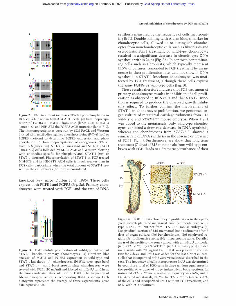
These results therefore indicate that FGF treatment ofprimary chondrocytes results in inhibition of cell prolif-eration as observed in RCS cells and that STAT-1 func-tion is required to produce the observed growth inhibi-tory effect. To further confirm the involvement ofSTAT-1 in chondrocyte proliferation, we performed or-gan culture of metatarsal cartilage rudiments from E15wild-type and STAT-1−/− mouse embryos. When FGF1was added to the metatarsals, only wild-type chondro-cytes exhibited a dramatic decrease in DNA synthesis,whereas the chondrocytes from STAT-1−/− showed asimilar rate of DNA synthesis in the absence or presenceof FGF1 (Fig. 4). Furthermore, we show that long-termtreatment (7 days) of E15 metatarsals from wild-type em-bryos with FGF1 leads to a dramatic perturbance of their
Figure 2. FGF treatment increases STAT-1 phosphorylation inRCS cells but not in NIH-3T3 ACH cells. (a) Immunoprecipi-tation of FGFR3 (IP FGFR3) from RCS (lanes 1–3), NIH-3T3(lanes 4–6), and NIH-3T3 the FGFR3 ACH mutation (lanes 7–9).The immunoprecipitates were run by SDS-PAGE and Westernblotted with antibodies against phosphotyrosine (P-Tyr) (top) orFGFR3 (bottom) to determine FGFR3 expression and phos-phorylation. (b) Immunoprecipitation of endogenous STAT-1from RCS (lanes 1–3), NIH-3T3 (lanes 4–6), and NIH-3T3 ACH(lanes 7–9) cells followed by SDS-PAGE and Western blottingwith antibodies specific for phosphorylated STAT-1 (top) orSTAT-1 (bottom). Phosphorylation of STAT-1 in FGF-treatedNIH-3T3 and in NIH-3T3 ACH cells is much weaker than inRCS cells, particularly when the total amount of STAT-1 pre-sent in the cell extracts (bottom) is considered.
Figure 3. FGF inhibits proliferation of wild-type but not ofSTAT-1 knockout primary chondrocytes. (a) Northern blotanalysis of FGFR1 and FGFR3 expression in wild-type andSTAT-1 knockout (−/−) chondrocytes. (b) Wild-type (open bars)and STAT-1−/− (solid bars) growth plate chondrocytes weretreated with FGF1 (10 ng/ml) and labeled with BrdU for 6 hr atthe times indicated after addition of FGF1. The frequency ofAlcian blue-positive cells incorporating BrdU is shown. Eachhistogram represents the average of three experiments; errorbars represent S.D..
Figure 4. FGF inhibits chondrocyte proliferation in the epiph-yseal growth plates of metatarsal bone rudiments from wild-type (STAT-1+/+) but not from STAT-1−/− mouse embryos. (a)Longitudinal section of E15 metatarsal bone rudiments after 2days of organ culture: (Pe) Perichondrium; (Ep) epiphyseal re-gion; (Pr) proliferative zone; (Hy) hypertrophic zone. Detailedareas of the proliferative zone stained with anti-BrdU antibody:(b,c) STAT-1+/+; (d,e) STAT-1−/−. (b,d) Untreated; (c,e) treatedmetatarsals with 200 ng/ml FGF1. FGF was present in the cul-ture for 2 days, and BrdU was added for the last 6 hr of culture.Cells that incorporated BrdU were visualized as described in thetext. The frequency of cells incorporating BrdU was determinedby counting a total of 1000 cells in three random equal areas inthe proliferative zone of three independent bone sections. Inuntreated STAT-1+/+ metatarsals the frequency was 76%, and inFGF-treated metatarsals, 24.7%. In STAT-1−/− metatarsals 90%of the cells had incorporated BrdU without FGF treatment, and88% with FGF treatment.
Growth inhibition of chondrocytes by FGF via STAT-1
GENES & DEVELOPMENT 1363
Cold Spring Harbor Laboratory Press on February 8, 2020 - Published by genesdev.cshlp.orgDownloaded from

development (Fig. 5). When E15 wild-type metatarsals,which are composed primarily of undifferentiated chon-drocytes, are cultured for 7 days they undergo consider-able longitudinal growth and development. They show awell-organized growth plate composed of orderly col-umns of proliferating chondrocytes that have undergonedifferentiation to prehypertrophic and hypertrophic cellspositive for type X collagen (Col X), a specific marker forhypertrophic chondrocytes (Fig. 5b). In contrast to thecontrol metatarsals, the treatment of E15 cartilage rudi-ments with FGF1 for 7 days results in the formation ofbones that are shorter and wider in their proximal anddistal extremities (Fig. 5c). These metatarsals exhibit noorganization of the growth plate, an absence of columnarchondrocytes, and a considerable reduction of the hyper-trophic zone, as shown by staining for Col X. This dem-onstrates that in addition to inhibiting proliferation, FGFaffects the process of the differentiation of chondrocytes.
To verify whether STAT-1 also mediates the effect ofFGF signaling on bone development, we cultured E15metatarsals from STAT-1−/− embryos for the same lengthof time. In contrast to wild-type metatarsals, FGF treat-
ment had no effect on growth plate chondrocytes. Inboth untreated and treated metatarsals, chondrocytesunderwent proliferation, prehypertrophy, and terminaldifferentiation to hypertrophic cells that expressed Col X(Fig. 5e,f). However, FGF-treated STAT-1−/− metatarsalsexhibited some increase in length and width comparedto controls, which could be due to the unmasking of thegrowth stimulatory effects of FGF signaling when theSTAT-1-mediated growth inhibitory pathway is blocked.Thus, treatment with FGF of E15 bone rudiments ap-pears to mimic the inhibition of endochondral ossifica-tion and bone development observed in human fetuseswith homozygous ACH or TD and in mouse models(Shah et al. 1973; Stanescu et al. 1990; Naski et al. 1998;Li et al. 1999). We show that this effect also requiresSTAT-1 function.
It is interesting to note that in these long-term experi-ments, FGF treatment had no significant inhibitory ef-fect on cells of the osteoblastic lineage and, rather,seemed to increase their proliferation. This is quite evi-dent in the FGF-treated metatarsals shown in Figure 5c,which display a dramatic thickening of the periosteum.This phenomenon was not observed in STAT-1−/− meta-tarsals (Fig. 5f), suggesting either that the increased os-teoblast proliferation is mediated by STAT-1 or, morelikely, this effect is at least partially indirect and mayrequire inhibition of formation of the hypertrophic zone.
The data presented in this report show that activationof FGF signaling in primary chondrocytes, chondrocyticcell lines, and cartilage bone rudiments from murine em-bryos results in significant inhibition of cell proliferationand bone development. This result is in line with theeffect of activating FGFR3 mutations in several forms ofhuman dwarfism, particularly ACH and TD, but con-trasts with previous reports (Kato and Iwamoto 1990;Hill et al. 1991; Wroblewski and Edwall-Arvidsson 1995;Legeai-Mallet et al. 1998) indicating that FGF treatmentof primary chondrocytes led to stimulation of cell pro-liferation. This discrepancy could be due to the heterog-eneous nature of the cell cultures studied. A recent re-port (Mancilla et al. 1998) showed that FGF2 treatmentof rat E20 metatarsal rudiments resulted in inhibition ofDNA synthesis of proliferative and epiphyseal chondro-cytes. Furthermore, we show that FGF-mediated inhibi-tion of proliferation in chondrocytes requires STAT-1function and that the STAT-1 requirement may be re-lated to its ability to induce antiproliferative genes, suchas p21WAF/CIP1. Studies of signal transduction in a vari-ety of cell systems have shown that although many sig-nal transduction pathways are activated simultaneouslyby receptor stimulation, it has been difficult to link aspecific downstream target with a specific biological re-sponse. Thus, this report presents one of the few ex-amples in which a signal transduction pathway, STAT-1activation, has been shown to be required for a specificcellular response, inhibition of chondrocyte prolifera-tion. The mechanism of STAT-1 activation by FGFR3 iscurrently being investigated. Our data suggest that theability to activate STAT-1 depends on the cellular con-text in which FGFR3 is expressed, as the introduction of
Figure 5. Long-term treatment of metatarsal rudiments byFGF causes inhibition of longitudinal growth and disruption ofthe growth plate in wild-type but not STAT-1−/− rudiments. E15metatarsals from STAT-1+/+ and STAT-1−/− mice were culturedfor 7 days in the presence or absence of 200 ng/ml FGF1. Lon-gitudinal sections of E.15 (a,d) and 7-day cultured metatarsals(b,c,e,f) were stained with a specific antibody against Col X. (a–f)Micrographs have the same magnification. (Insets) A higherpower view of the areas boxed in the low power adjacent mi-crographs, corresponding to the growth plate area.
Sahni et al.
1364 GENES & DEVELOPMENT
Cold Spring Harbor Laboratory Press on February 8, 2020 - Published by genesdev.cshlp.orgDownloaded from

FGFR3 or of its activating ACH mutation in fibroblastsdid not lead to growth inhibition or increased ability toactivate STAT-1 in response to FGF. It is interesting tonote that STAT-1−/− mice have not been reported to havebone defects, but this aspect has never been studied indetail, particularly during embryonic development orduring early life, and is currently under investigation.These and related studies on how FGF affects the differ-entiation program of cells of the chondrocytic lineageshould provide important information on the mecha-nisms of bone morphogenesis and on the way by whichunregulated FGF signaling causes bone morphogeneticdisorders.
Materials and methodsCell culture and proliferation assayRCS cells were maintained as monolayer cultures under conditions de-scribed previously (Mukhopadhyay et al. 1995). Primary chondrocyteswere isolated from long bone cartilages of 10-day-old wild-type andSTAT-1−/− mice. Growth plates were dissected and the chondrocytesisolated as described previously (Amling et al. 1997). The growth plateswere incubated with 0.1% collagenase type A (Sigma) for 30 min at roomtemperature with constant shaking. Thereafter, the supernatant was dis-carded, a fresh solution of 0.2% collagenase was added, and the incuba-tion was carried out for 3 hr at 37°C. Cells were filtered through a sterilenylon 0.45-µm mesh, centrifuged for 10 min at 1000 rpm. Then cellswere incubated at 37°C, in DMEM/F12 (1:1) medium supplemented with10% FCS, 50 µg/ml ascorbic acid (Sigma), and 100 µg/ml sodium pyru-vate. For proliferation assays, 5 × 10⁴ cells/400 µl were seeded on cover-slips; for RNA extraction, 8 × 10⁵ cells/3 ml were cultured in a 6-cmtissue culture dish. The medium was changed every other day and FGFtreatment was started at day 4 of the culture. For DNA synthesis, RCScells and primary chondrocytes were incubated in the presence of 1 µg/ml BrdU for 6 hr. Cells were fixed, permealized, and incubated withanti-BrdU antibody according to the manufacturer’s instructions (Boeh-ringer Mannheim). Cells incorporating BrdU were visualized and countedusing fluorescence microscopy.
Organ CulturesMetatarsal long bone rudiments from E15 wild-type and STAT−/− mouseembryos were dissected under sterile conditions. The cartilaginous longbones were left intact. Organ culture was carried out in a-MEM withoutnucleosides (GIBCO) supplemented with 50 µg/ml ascorbic acid, 300µg/ml L-glutamine, 50 µg/ml gentamicine, 250 µg/ml Fungizone, 1 mM
b-glycerophosphate, and 0.2% BSA Cohn fraction V (Sigma) (completemedium). Each long bone was cultured individually in 24-well platescontaining 400 µl of complete medium in the presence or absence of 200ng/ml of recombinant human FGF1 and 10 µg/ml heparin. The metatar-sal cultures were maintained at 37°C for either 48 hr or 7 days and themedium changed every other day. The experiments were set up for left/right paired observations, with one metatarsal serving as a control to theother. For the proliferation assay, the long bones were treated with FGFfor 48 hr and BrdU was added during the last 6 hr of treatment. The longbones were fixed at day 0, 2, and 7 of the organ culture.
ImmunohistochemistryMetatarsals were fixed in 4% paraformaldehyde overnight at 4°C andembedded in paraffin, and 4-µm tissue sections were performed. Sectionswere deparaffinized by treatment with xylene plus 100%, 95%, and 70%ethanol, followed by washes in TS buffer (100 mM Tris/HCl at pH 7.4,150 mM NaCl) and permealization with 0.25% Triton X-100. For local-ization of Col X, sections were treated with 1 mg/ml testicular hyaluron-idase at 37°C for 45 min in a humidified chamber. After overnight block-ing with 3% goat serum, the endogenous peroxidase was inactivated byincubating in 5% H2O2 in methanol for 10 min. Anti-BrdU monoclonal(Boehringer Mannheim) or anti-Col X polyclonal antibody was used witha Vectastain Elite ABC Kit (Vector Labs) to stain the cells. The colorreaction (dark brown) was deduced with Sigma Fast DAB peroxidase sub-strate according to the manufacturer’s manual.
Immunoprecipitation and Western blottingCells were stimulated with 100 ng/ml FGF1 and 10 µg/ml heparin at37°C for various times, and cells were lysed either in RIPA buffer orHNTG buffer (50 mM HEPES at pH 7.5, 150 mM NaCl, 1.5 mM MgCl2, 1mM EGTA, 10% glycerol, and 1% Triton X-100) in the presence of pro-tease and phosphatase inhibitors. Immunoprecipitations were performedwith polyclonal anti-FGFR3 and anti-STAT-1 antibodies (Santa Cruz).Immunoprecipitates and total protein lysates were separated by SDS-PAGE and analyzed by ECL detection system (Amersham). A polyclonalantibody specific for the tyrosine phosphorylated form of STAT-1 (NewEngland Biolabs) was used in Western blot analysis when indicated.
AcknowledgmentsWe thank Drs. G. Inghirami and F. Gonzalez for their help in histologicalpreparation, Dr. B.R. Olsen for providing the Col X antibody and Dr. R.Baron for the RCS cells. We thank Drs. M. Mohammadi and J. Schless-inger for providing human recombinant FGF1. This investigation wassupported by a fellowship from the Arthritis Foundation to M.S. and byU.S. Public Health Service grant CA42568 from the National CancerInstitute.
The publication costs of this article were defrayed in part by paymentof page charges. This article must therefore be hereby marked ‘advertise-ment’ in accordance with 18 USC section 1734 solely to indicate thisfact.
References
Abdollahi, A., K.A. Lord, B. Hoffman-Liebermann, and D.A. Liebermann.1991. Interferon regulatory factor 1 is a myeloid differentiation pri-mary response gene induced by interleukin 6 and leukemia inhibitoryfactor: Role in growth inhibition. Cell Growth Differ. 2: 401–407.
Amling, M., L. Neff, S. Tanaka, D. Inoue, K. Kuida, E. Weir, W.M. Phil-brick, A.E. Broadus, and R. Baron. 1997. Bcl-2 lies downstream ofparathyroid hormone-related peptide in a signaling pathway thatregulates chondrocyte maturation during skeletal development. J.Cell Biol. 136: 205–213.
Basilico, C. and D. Moscatelli. 1992. The FGF family of growth factorsand oncogenes. Adv. Cancer Res. 59: 115–165.
Burke, D., D. Wilkes, T.L. Blundell, and S. Malcolm. 1998. Fibroblastgrowth factor receptors: Lessons from the genes. Trends Biochem.Sci. 23: 259–262.
Chin, Y.E., M. Kitagawa, W.-C.S. Su, Z.-H. You, Y. Iwamoto, and X.-Y.Fu. 1996. Cell growth arrest and induction of cyclin-dependent ki-nase inhibitor p21WAF1/CIP1 mediated by STAT1. Science 272: 719–722.
Coffin, J.D., R.Z. Florkiewicz, J. Neumann, T. Mort-Hopkins, G.W. Dorn,P. Lightfoot, R. German, P.N. Howles, A. Kier, B.A. O’Toole, J. Sasse,A.M. Gonzalez, A. Baird, and T. Doetschman. 1995. Abnormal bonegrowth and selective translational regulation in basic fibroblastgrowth factor (FGF-2) transgenic mice. Mol. Biol. Cell 6: 1861–1873.
Colvin, J.S., B.A. Bohne, G.W. Harding, D.G. McEwen, and D.M. Ornitz.1996. Skeletal overgrowth and deafness in mice lacking fibroblastgrowth factor receptor 3. Nat. Genet. 12: 390–397.
Darnell, J.E., Jr. 1997. STATs and gene regulation. Science 277: 1630–1635.
Deng, C., A. Wynshaw-Boris, F. Zhou, A. Kuo, and P. Leder. 1996. Fibro-blast growth factor receptor 3 is a negative regulator of bone growth.Cell 84: 911–921.
Durbin, J.E., R. Hackenmiller, M.C. Simon, and D.E. Levy. 1996. Tar-geted disruption of the mouse Stat1 gene results in compromisedinnate immunity to viral disease. Cell 84: 443–450.
Goldfarb, M. 1996. Functions of fibroblast growth factors in vertebratedevelopment. Cytokine Growth Factor Rev. 7: 311–325.
Hill, D.J., A. Logan, and D. De Sousa. 1991. Stimulation of DNA andprotein synthesis in epiphyseal growth plate chondrocytes by fibro-blast growth factors. Interactions with other peptide growth factors.Ann. N.Y. Acad. Sci. 638: 449–452.
Kato, Y. and M. Iwamoto.. 1990. Fibroblast growth factor is an inhibitorof chondrocyte terminal differentiation. J. Biol. Chem. 265: 5903–5909.
Legeai-Mallet, L., C. Benoist-Lasselin, A.-L. Delezoide, A. Munnich, andJ. Bonaventure. 1998. Fibroblast growth factor receptor 3 mutations
Growth inhibition of chondrocytes by FGF via STAT-1
GENES & DEVELOPMENT 1365
Cold Spring Harbor Laboratory Press on February 8, 2020 - Published by genesdev.cshlp.orgDownloaded from

promote apoptosis but do not alter chondrocyte proliferation inthanatophoric dysplasia. J. Biol. Chem. 273: 13007–13014.
Li, C., L. Chen, T. Iwata, M. Kitagawa, X.-Y. Fu, and C.-X. Deng. 1999. ALys644Glu substitution in fibroblast growth factor receptor 3(FGFR3) causes dwarfism in mice by activation of STATs and ink4cell cycle inhibitors. Hum. Mol. Genet. 8: 35–44.
Li, Y., K. Mangasarian, A. Mansukhani, and C. Basilico. 1997. Activationof FGF receptors by mutations in the transmembrane domain. On-cogene 14: 1397–1406.
Mancilla, E.E., F. De Luca, J.A. Uyeda, F.S. Czerwiec, and J. Baron. 1998.Effects of fibroblast growth factor-2 on longitudinal bone growth.Endocrinology 139: 2900–2904.
Mukhopadhyay, K., V. Lefebvre, G. Zhou, S. Garofalo, J.H. Kimura, andB. de Crombrugghe. 1995. Use of a new rat chondrosarcoma cell lineto delineate a 119-base pair chondrocyte-specific enhancer elementand to define active promoter segments in the mouse Pro-a1(II) col-lagen gene. J. Biol. Chem. 270: 27711–27719.
Naski, M.C. and D.M. Ornitz. 1998. FGF signaling in skeletal develop-ment. Front. Biosci. 3: 781–794.
Naski, M.C., J.S. Colvin, J.D. Coffin, and D.M. Ornitz. 1998. Repressionof hedgehog signaling and BMP4 expression in growth plate cartilageby fibroblast growth factor receptor 3. Development 125: 4977–4988.
Ornitz, D.M., J. Xu, J.S. Colvin, D.G. McEwen, C.A. MacArthur, F.Coulier, G. Gao, and M. Goldfarb. 1996. Receptor specificity of thefibroblast growth factor family. J. Biol. Chem. 271: 15292–15297.
Saxton, T.M., M. Henkemeyer, S. Gasca, R. Shem, D.J. Rossi, F. Shalaby,G.-S. Feng, and T. Pawson. 1997. Abnormal mesoderm patterning inmouse embryos mutant for the SH2 tyrosine phosphatase Shp-2.EMBO J. 16: 2352–2364.
Shah, K., R. Astley, and A.H. Cameron. 1973. Thanatophoric dwarfism.J. Med. Genet. 10: 243–252.
Silvennoinen, O., C. Schindler, J. Schlessinger, and D.E. Levy. 1993. Ras-independent signal transduction in response to growth factors andcytokines by tyrosine phosphorylation of a common transcriptionfactor. Science 261: 1736–1739.
Stanescu, R., V. Stanescu, and P. Maroteaux. 1990. Homozygous achon-droplasia: Morphologic and biochemical study of cartilage. Am. J.Med. Genet. 37: 414–421.
Su, W.-C., M. Kitagawa, N. Xue, B. Xie, S. Garofalo, J. Cho, C. Deng, W.A.Horton, and X.-Y. Fu. 1997. Activation of Stat1 by mutant fibroblastgrowth-factor receptor in thanatophoric dysplasia type II dwarfism.Nature 386: 288–292.
Webster, M.K. and D.J. Donoghue. 1997. FGFR activation in skeletaldisorders: Too much of a good thing. Trends Genet. 13: 178–182.
Wroblewski, J. and C. Edwall-Arvidsson. 1995. Inhibitory effects of basicfibroblast growth factor on chondrocyte differentiation. J. BoneMiner. Res. 10: 735–742.
Sahni et al.
1366 GENES & DEVELOPMENT
Cold Spring Harbor Laboratory Press on February 8, 2020 - Published by genesdev.cshlp.orgDownloaded from

13:1999, Genes Dev. Malika Sahni, Davide-Carlo Ambrosetti, Alka Mansukhani, et al. development through the STAT-1 pathwayFGF signaling inhibits chondrocyte proliferation and regulates bone
References
http://genesdev.cshlp.org/content/13/11/1361.full.html#ref-list-1
This article cites 27 articles, 13 of which can be accessed free at:
License
ServiceEmail Alerting
click here.right corner of the article or
Receive free email alerts when new articles cite this article - sign up in the box at the top
Cold Spring Harbor Laboratory Press
Cold Spring Harbor Laboratory Press on February 8, 2020 - Published by genesdev.cshlp.orgDownloaded from