osteoarthritic degeneration chondrocyte anabolism and
TRANSCRIPT
Page 1/28
Histone Demethylase UTX Compromises ArticularChondrocyte Anabolism and AggravatesOsteoarthritic DegenerationWei-Shiung Lian
Kaohsiung Chang Gung Memorial HospitalRe-Wen Wu
Kaohsiung Chang Gung Memorial HospitalJih-Yang Ko
Kaohsiung Chang Gung Memorial HospitalYu-Shan Chen
Kaohsiung Chang Gung Memorial HospitalShao-Yu Wang
Kaohsiung Chang Gung Memorial HospitalChun-Ping Yu
Academia Sinica https://orcid.org/0000-0002-6806-9157Holger Jahr
University Hospital RWTH Aachen, GermanyFeng-Sheng Wang ( [email protected] )
Kaohsiung Chang Gung Memorial Hospital https://orcid.org/0000-0001-6025-6073
Article
Keywords: UTX, H3K27 trimethylation, PRC2, chondrocytes, osteoarthritis
Posted Date: May 18th, 2021
DOI: https://doi.org/10.21203/rs.3.rs-467396/v1
License: This work is licensed under a Creative Commons Attribution 4.0 International License. Read Full License

Page 2/28
AbstractHistone demethylase UTX removes repressive trimethyl groups at lysine 27 of histone 3 (H3K27me3) toregulate tissue integrity, while its role was not yet studied in articulating joint tissues in situ. We nowfound that UTX expression in articular chondrocytes positively correlated with human osteoarthritis. Utxoverexpression induced chondrocyte dysfunction, cartilage degeneration and osteophyte induction inmice. In contrast, chondrocyte-speci�c Utx knockout in mice promoted gross articular morphology anddelayed age- and collagenase-induced cartilage erosion, synovitis and osteophyte formation and largelyeliminated disease-associated joint pain. Additionally, pharmacological inhibition of Utx through GSK-J4preserved cartilage integrity. Our study is the �rst to suggest that Utx loss-mediated cartilage protectioninvolved a dysregulation of polycomb repressive complex 2 core components EZH2, EED, and SUZ12 toinduce H3K27 hypomethylation and a net anabolic effect. Speci�cally, Utx loss-of-function appears toinvolve, among others, Wnt10a signaling to reduce chondrocytic activities and an IGF-2-mediatedstimulation of extracellular matrix synthesis.
IntroductionOsteoarthritis (OA), characterized by a degenerative loss of articular cartilage, is the most common formof arthritis, causing joint pain, deformity, and disability in the elderly1. Severe degeneration of thecartilage extracellular matrix (ECM) causes a plethora of osteoarthritic symptoms, including synovialswelling, osteophyte formation, and subchondral plate sclerosis. Maintaining articular chondrocytehomeostasis is thus important to ensure articular tissue integrity and to protect the underlying bone fromweight bearing and athletic impact on joints1. Expanding evidence suggests that dysregulated expressionof, among others, chondrocyte key transcription factor SRY-box9 (SOX9), and canonical Wnt/β-cateninsignaling induce chondrocyte dysfunction which in turn accelerates OA development2–5. The underlyingmechanisms leading to this change in metabolic activity in osteoarthritic chondrocytes remains, however,poorly elucidated.
Epigenetic pathways chemically modify DNA-bound histones or DNA nucleotides directly, like CpGdinucleotides, to control transcription through altering promoter activities6. Of these modi�cations,histone methylation causes chromatin condensation that represses gene expression to change biologicalactivities6. Recent studies show that the histone methylation status of chondrocytes plays an importantrole in cartilage disorders. Hypomethylation of histone 3 at lysine 36 (H3K36) correlates withchondroblastomas7, while H3K9 methylation suppresses SOX9 transcription, slowing downchondrogenesis and skeletal morphogenesis in mice lacking AT-rich interactive domain 5b8. Moreover,aging-induced H3K4 methylation enhances cartilage loss in mice9 and loss of H3K36 methyltransferasedisruptor of telomeric silencing 1-like (DOTIL) in chondrocytes results in defective murine bonedevelopment and is further relevant to human OA10.

Page 3/28
Lysine (K)-speci�c histone demethylase 6A (UTX, KDM6A) and Jumonji domain containing 3 (Jmjd3)remove trimethyl groups from histone H3K27. In contrast, histone methyltransferase polycomb repressioncomplex 2 (PRC2) core components, including enhancer of zeste homolog 2 (EZH2), embryonic ectodermdevelopment (EED) and PRC 2 subunit (SUZ12), catalyze trimethylation of H3K2711. While UTX generallypromotes gene activation and appears essential during normal development and tissue-speci�cdifferentiation12, increased H3K27 trimethylation (H3K27me3) in cartilage correlates with human hipOA13. Mice de�cient in EZH1 and EZH2 in chondrocytes display poor chondrogenesis and skeletal tissueunderdevelopment along with low H3K27me3 abundance14. Interestingly, EZH2 deletion accelerates thedevelopment of OA15, while chondrocyte-speci�c EED knockout mice show a deformed skeleton anddecreased chondrocyte survival16. Recently, JMJD3 is shown to regulate in vitro chondrogenicdifferentiation of human mesenchymal stem cells in monolayer culture17. The role of UTX during articularcartilage homeostasis and progression of OA remains poorly understood.
This study aimed at elucidating how UTX activity affected H3K27 methylation state and how thissubsequently altered the articular cartilage phenotype. To this end, we used human OA specimens, RNAinterference and pharmacological inhibition in chondrocytes as well as chondrocyte-speci�c UTXknockout mice to study genome-wide chondrocyte-speci�c maker expression. Our analyses revealed thatUTX loss in primary chondrocytes suppressed PRC2-mediated H3K27 trimethylation in promoter regionsof key regulators of the chondrocytic phenotype, thereby facilitating transcription of Igf-2, butsuppressing that of Wnt10a, to promote cartilage metabolism and suppress OA progression.
ResultsUTX and H3K27 trimethyl marks correlates with human gonarthritis
Safranin-O-stained sections showed typical histopathology alterations in OA cartilage as compared tomacroscopically normal, non-OA regions in the knee. UTX expression was signi�cantly increased in OA ascompared to non-OA groups (Fig. 1a). Osteoarthritic chondrocytes further exhibited strong UTX (Fig. 1b)and H3K27me3 (Fig. 1c) immunostaining, respectively, con�rming this on protein level.
UTX inhibits ECM production in chondrocytesNext, we investigated Utx expression in relation to ECM expression in articular chondrocytes.Chondrocytes from knee joints of 7-day-old mice were transfected with Utx expressing vectors or UtxsiRNAs, respectively. Forced Utx expression increased H3K27me3 levels, but decreased Sox9 abundance(Fig. 1d). Chromatin immunoprecipitation (ChIP)-PCR analysis veri�ed that forced Utx expressionpromoted H3K27me3 enrichment at the proximal Sox9 promoter region (Fig. 1e) and reduced expressionof Sox9 and cartilage-speci�c key ECM markers Col2a1 and Acan (Fig. 1f). Consequently, Alcian bluestaining con�rmed ECM loss in micro-mass cultures as compared to scrambled controls (Fig. 1g). On thecontrary, silencing Utx expression by RNAi reduced overall H3K27me3 level and its occupancy at the Sox9promoter (Fig. 1d and e), but signi�cantly enhanced both aforementioned chondrocyte markers (Fig. 1f)and ECM production as compared to scrambled controls (Fig. 1g).
Page 4/28
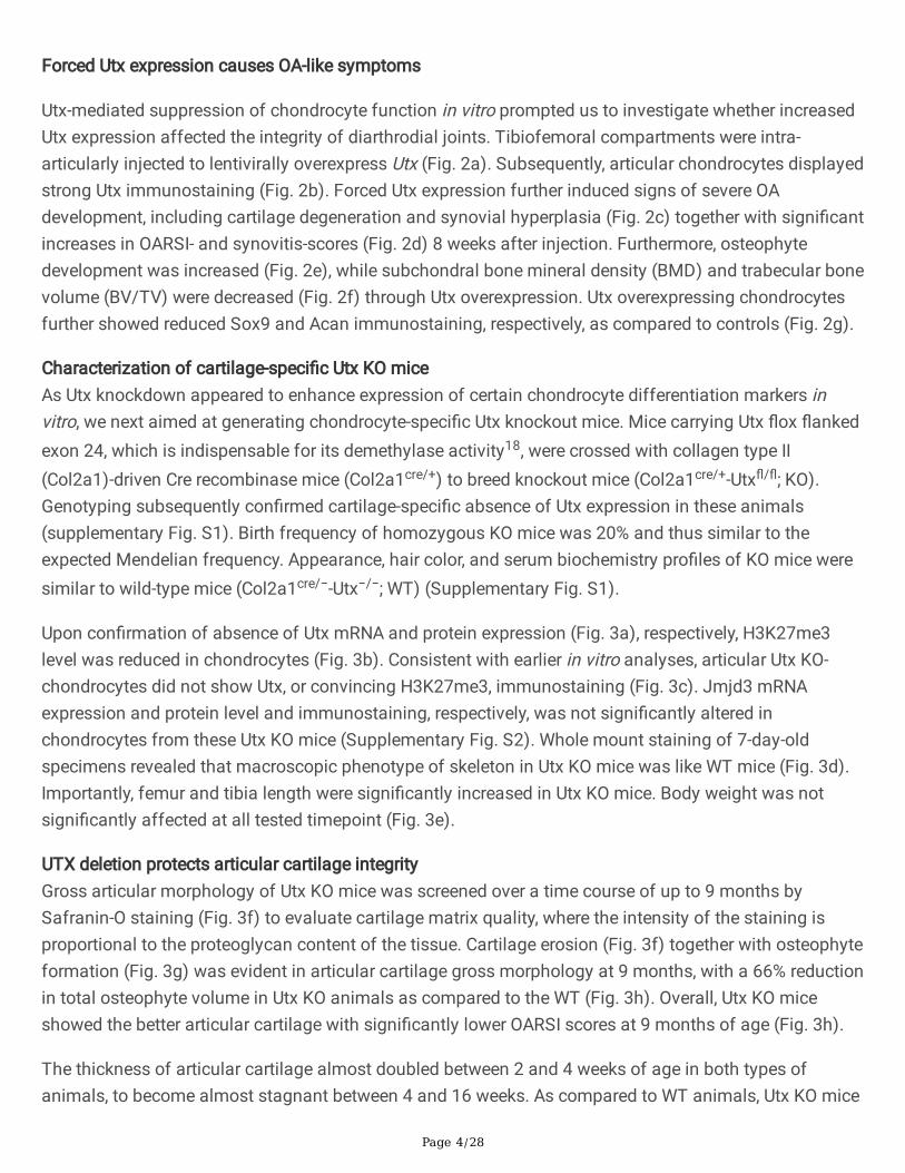
Forced Utx expression causes OA-like symptoms
Utx-mediated suppression of chondrocyte function in vitro prompted us to investigate whether increasedUtx expression affected the integrity of diarthrodial joints. Tibiofemoral compartments were intra-articularly injected to lentivirally overexpress Utx (Fig. 2a). Subsequently, articular chondrocytes displayedstrong Utx immunostaining (Fig. 2b). Forced Utx expression further induced signs of severe OAdevelopment, including cartilage degeneration and synovial hyperplasia (Fig. 2c) together with signi�cantincreases in OARSI- and synovitis-scores (Fig. 2d) 8 weeks after injection. Furthermore, osteophytedevelopment was increased (Fig. 2e), while subchondral bone mineral density (BMD) and trabecular bonevolume (BV/TV) were decreased (Fig. 2f) through Utx overexpression. Utx overexpressing chondrocytesfurther showed reduced Sox9 and Acan immunostaining, respectively, as compared to controls (Fig. 2g).
Characterization of cartilage-speci�c Utx KO miceAs Utx knockdown appeared to enhance expression of certain chondrocyte differentiation markers invitro, we next aimed at generating chondrocyte-speci�c Utx knockout mice. Mice carrying Utx �ox �ankedexon 24, which is indispensable for its demethylase activity18, were crossed with collagen type II(Col2a1)-driven Cre recombinase mice (Col2a1cre/+) to breed knockout mice (Col2a1cre/+-Utx�/�; KO).Genotyping subsequently con�rmed cartilage-speci�c absence of Utx expression in these animals(supplementary Fig. S1). Birth frequency of homozygous KO mice was 20% and thus similar to theexpected Mendelian frequency. Appearance, hair color, and serum biochemistry pro�les of KO mice weresimilar to wild-type mice (Col2a1cre/−-Utx−/−; WT) (Supplementary Fig. S1).
Upon con�rmation of absence of Utx mRNA and protein expression (Fig. 3a), respectively, H3K27me3level was reduced in chondrocytes (Fig. 3b). Consistent with earlier in vitro analyses, articular Utx KO-chondrocytes did not show Utx, or convincing H3K27me3, immunostaining (Fig. 3c). Jmjd3 mRNAexpression and protein level and immunostaining, respectively, was not signi�cantly altered inchondrocytes from these Utx KO mice (Supplementary Fig. S2). Whole mount staining of 7-day-oldspecimens revealed that macroscopic phenotype of skeleton in Utx KO mice was like WT mice (Fig. 3d).Importantly, femur and tibia length were signi�cantly increased in Utx KO mice. Body weight was notsigni�cantly affected at all tested timepoint (Fig. 3e).
UTX deletion protects articular cartilage integrityGross articular morphology of Utx KO mice was screened over a time course of up to 9 months bySafranin-O staining (Fig. 3f) to evaluate cartilage matrix quality, where the intensity of the staining isproportional to the proteoglycan content of the tissue. Cartilage erosion (Fig. 3f) together with osteophyteformation (Fig. 3g) was evident in articular cartilage gross morphology at 9 months, with a 66% reductionin total osteophyte volume in Utx KO animals as compared to the WT (Fig. 3h). Overall, Utx KO miceshowed the better articular cartilage with signi�cantly lower OARSI scores at 9 months of age (Fig. 3h).
The thickness of articular cartilage almost doubled between 2 and 4 weeks of age in both types ofanimals, to become almost stagnant between 4 and 16 weeks. As compared to WT animals, Utx KO mice

Page 5/28
have relatively thicker articular cartilage at all tested timepoints (Fig. 3i). However, 9 months old animalsin both groups showed a, possibly age-dependent, thinning of the articular cartilage, with that of Utx KOmice remaining relatively thicker than that of WT animals (Fig. 3i). In addition, the uncalci�ed portion ofthe articular cartilage remained relative thicker in Utx KO mice as compared to WT, too (Fig. 3j). While thenumber of chondrocytes in articular cartilage declined age-dependently steadily in both groups (Fig. 3k),their numbers stayed always relatively higher in age-matched Utx KO animals. Interestingly, in 9 monthsold animals the calci�ed zone was relatively thicker in Utx KO animals (Fig. 3l). Furthermore, articularcartilage of 16 weeks old Utx KO mice showed relatively stronger Sox9, Acan and Col2a1 immunostaining(Fig. 3m) as compared to WT, respectively, indicating better ECM homeostasis and an improved potentialto retard OA in Utx KO animals.UTX knockout retarded OA progression and improves pain scores
Given that high Utx expression was associated with human knee OA (Fig. 1) and articular cartilagedegeneration in mice (Fig. 2), we used intra-articular collagenase injections as an accepted model toinduce OA development in vivo19,20. In WT mice, collagenase-injected joints showed severe cartilage loss(Fig. 4a) and increased OARSI and synovitis scores (Fig. 4b), respectively, at 8 weeks after injections.Additionally, osteophytes were more prominently developed in WT than in Utx KO animals, revealingincreased overall volumes by quantitative µCT analyses upon OA induction in WT animals (Fig. 4c).Subchondral BMD and BV/TV were signi�cantly decreased in the injured joints (Fig. 4d). Of note,cartilage destruction, osteophyte formation, and subchondral bone loss was all less severe in Utx KOmice.
Pain and deregulated joint kinematics are prominent characteristic of OA and the former is a primarysymptom of OA in humans and the leading cause of disability21. We thus next conducted catwalkanalyses as a well-accepted model22 to assess pain and gait properties resulting from body-weightloading in animals of collagenase-treated and control legs. WT mice with OA knees showed deviatedbody axis (Supplementary Fig. S3) and irregular footprint (Fig. 4e) and gait pro�les, including decreasedfootprint area, maximum contact area, duration of stand, and duty cycles (Fig. 4f). Mobility parameters,like moving time and distance were also signi�cantly reduced as evident from open �eld assessment(Supplementary Fig. S3). In contrast, walking patterns, body posture and mobility of collagenase-injectedUtx KO animals were signi�cantly improved, which indicates that Utx loss of function alleviates joint painin this model. Likewise, Utx KO mice kept showing a prominent Sox9 and aggrecan, but a weakH3K27me3, immunoreactivity in chondrocytes within collagenase-treated articular tissue (Fig. 4g).UTX inhibition attenuates OA progression
In addition, one week after collagenase injection, Utx inhibitor GSK-J4 was intra-articularly injected toscreen for potential OA-modifying effects. Consistently, this treatment signi�cantly reduced cartilage lossand synovitis (Fig. 5a) together with decreases in OARSI and synovitis scores (Fig. 5b) in collagenase-injected joints. GSK-J4-treament further reduced osteophyte formation (Fig. 5c) and subchondral boneloss (Fig. 5d), respectively. The irregular footprint (Fig. 5e) and gait pro�les (Fig. 5f) from osteoarthritic

Page 6/28
joints were also partially reversed upon GSK-J4 injection. The treatment further revealed improved Sox9and aggrecan immunostaining, respectively, but reduced H3K27me3 immunoreactivity (Fig. 5g).
UTX loss decreases H3K27me3 enrichment at cartilage marker genesWe conducted genome-wide ChIP-Seq analyses to identify changes in the H3K27me3 binding epigenomiclandscape that might have contributed to the improved chondrocyte marker gene expression in Utx KOmice. Of 19531 H3K27me3 binding sites, 91.7% and 7.2% were enriched in genomes of WT and Utx KOanimals, respectively, (Fig. 6a). Loss of histone eraser Utx globally decreased the H3K27me3 occupancyin promoters preferably decreased within a 5 kb up- and downstream region of the respectivetranscriptional start sites (Fig. 6b), which consequently resulted in a higher normalized transcriptionalexpression (RPKM) in WT chondrocytes (Fig. 6c). The enrichment of H3K27me3 in exon region wasincreased in KO mice, which was indicative of gene transcription alteration upon Utx deletion (Fig. 6d).Pathway ontology analyses suggested that Utx knockout facilitated cellular processes related to cartilageand skeletal development, including ossi�cation and bone mineralization, as well as enhanced inhibitionof cellular senescence (Fig. 6e). Gene set enrichment analysis furthermore indicated transcription of Igf-2(Fig. 6f) and Wnt10a (Fig. 6g) together with signi�cant increases in Dkk1, sFRP1, Igf-R2, Wnt4, and Wnt7aexpression in Utx KO mice (Supplementary Fig. S4).
From the H3K27me3 repressive signatures, Igf-2 and Wnt10a but also Sox9 were selected for subsequentexperiments as they are known to regulate chondrocyte function and cartilage development23,24.Figure 7a shows the spatial distribution of H3K27me3 bindig sites in the coding sequence of Igf-2,Wnt10a, and Sox9, respectively, also indicating the relative position of the respective TSS, next to aquanti�ed representation of the relative H3K27me3 occupancy per gene (Fig. 7b). Upon Utx loss, mRNAexpression of Igf-2 and Sox9 was consequently increased, while that of Wnt10a was signi�cantly reduced(Fig. 7c).
Expression of Sox9, Col2a1 and Acan (Fig. 7d, IgG control) and glycosaminoglycan synthesis (Fig. 7e)were much higher in Utx KO cells than in WT cells. These effects were signi�cantly suppressed upon Igf-2antibody administration. Also, forced Wnt10a expression suppressed these cartilage marker genes(Fig. 7d) and ECM production (Fig. 7e) in Utx KO chondrocytes. Our data suggest that Igf-2 positivelystimulates chondrocyte anabolism, whereas Wnt10a-mediated signaling seems rather deleterious tocartilage ECM synthesis.
UTX loss-mediated H3K27 hypomethylation involves PRC2To understand how a de�ciency in histone eraser Utx caused a seemingly contradictory H3K27hypomethylation, we investigated whether PRC2 signaling was changed in chondrocytes. Increased Ezh2but reduced Eed, Suz12, and H3K27me3 abundances was evident in Utx KO cells (Fig. 8a). Additionally,Ezh2 RNAi decreased H3K27me3 levels (Fig. 8b) along with a signi�cant decrease in H3K27me3enrichment at the Sox9 promoter region (Fig. 8c), resulting in an upregulation of chondrocyte markers(Fig. 8d) and ECM production (Fig. 8e), respectively, in Utx KO chondrocytes. On the contrary, forced Eedand Suz12 expression increased H3K27me3 abundance (Fig. 8f) and the H3K27me3 occupancy at the

Page 7/28
Sox9 promoter in particular, relative to WT cells (Fig. 8g). This subsequently repressed chondrocytemarkers (Fig. 8h) and ECM synthesis (Fig. 8i). Experimental results suggested that loss-of-function of Utxderegulated PRC2 action, maintaining H3K27 relatively hypomethylated and therefore enhancedchondrocytic marker gene expression. Low Eed or Suz12 abundance appeared to inhibit Eeh2 and itsability to trimethylate H3K27.
DiscussionEpigenetic changes occur during OA and histone methylation, in particular, appear to imbalancechondrocyte metabolism25,26,. Our data now associate expression of histone eraser UTX with OAdevelopment in humans and correlate UTX and H3K27 trimethylation in articular cartilage withchondrocyte marker gene expression in vitro. This is in agreement with the only other studydemonstrating high H3K27me3 abundances in human hip osteoarthritic cartilage14. We further, for the�rst time, manipulated Utx activity through RNAi and lentiviral overexpression, established a murine UtxKO model and evaluated intraarticular effects of local Utx-speci�c inhibition in vivo. Our comprehensivestudy reveals a novel non-canonical role of the H3K27me2/3-speci�c demethylase Utx, in concert withPRC2 core components, in articular chondrocytes homeostasis and the progression of OA.
We used Chip-Seq to screen global changes in gene expression between WT and Utx KO chondrocytes toidentify molecular mechanisms protecting cartilage against degradation. GO terms already revealedmajor expressional changes occurring in gene regulation networks related to mineralization, ossi�cationand senescence. Candidates like Sox9, Igf-2 and Wnt10a arising from enrichment scores were thenfurther analyzed to reveal an aberrant H3K27me3 occupation at their respective promoter loci betweenWT and Utx KO chromatin. IGF-2 and Wnt signaling components were known to regulate chondrocytefunction and cartilage development23,24. Our enrichment scores now revealed upregulation of manymarkers associated with a proper articular chondrocyte phenotype, like relevant collagens of the cartilageECM (e.g., Col9a2) or the cartilage-speci�c acidic protein 1 (CRTAC1)27 and that of BMP antagonists likeGREM1. Col9a2 is considered to be a chondrocyte-speci�c marker gene, in addition to Col2a1, Col11a2and aggrecan28.
Sox9 is a prominent activator of Col2a1 transcription, making it a master regulator of the chondrocytephenotype29. To show a Utx-dependent compromised chondrocytic activity, we used key chondrocytemarker genes Sox9, Col2a1 and Acan as readouts, and quanti�ed proteoglycan production as a measureof ECM quality. Using UTX-speci�c inhibitor GSK-J4, shown to inhibit H3K27 demethylation of UTX targetgenes30, and con�rmed anabolic effects on ECM level in vitro and being in line with the observed overallimproved relative thickness of the articular cartilage in vivo. BMP signaling ultimately results in unwantedhypertrophy in articular chondrocytes. Leijten et al. already showed that decreased GREM1 and DKK1gene expression in cartilage were correlated with OA31. Harmonization of BMP and WNT signaling areimportant to maintain articular cartilage integrity. While BMPs32 and canonical WNTs33 may exert pro-hypertrophic actions under certain conditions, the parallel upregulation of BMP antagonists and WNT
Page 8/28
antagonists, like sFRP-1 or DKK1, may thus explain the overall chondroprotective net effect of Utx loss invitro and in vivo.
Interestingly, adult articular cartilage has been long considered a post-mitotic tissue with terminallydifferentiated chondrocytes34,35, but this textbook dogma has been questioned recently. A ‘phenotypicplasticity’ of articular chondrocytes has then been associated with OA36 in which chondrocytes de-differentiation towards a more �broblast-like phenotype37. To this end, Wnt signaling is an importantregulator of cell plasticity38 and the Wnt–frizzled–β-catenin pathway appears activated in OA39,40.Expression of Wnt4 and Wnt10, in particular, can stimulate osteogenesis41, and excessive Wnt signalingcan also lead to increased osteophyte formation42, which is in agreement with our �ndings. Recently,mouse genetic analysis also revealed that Wnt/β-catenin signaling components are important to regulateSox9 and Runx2 and Wnt signaling can thus suppress chondrogenic differentiation, while increasingosteoblastic differentiation43. In line with our hypothesis, H3K27me3 demethylase UTX has earlier beenshown to be essential to tissue development and stem cell plasticity44. In contrast, Wnt inhibitionattenuates OA development through anti-catabolic and anti-�brotic effects on chondrocytes and synovial�broblasts, respectively45. Wnt signaling plays a context-dependent role in the development of OA andupregulated expression of canonical Wnts, like Wnt10, and non-canonical, like Wnt4 and Wnt7,46, togetherwith Wnt inhibitors like sFRPs and Dkks hint towards a delicate balance of this system.
Currently developed Wnt inhibitors as disease-modifying osteoarthritis drugs (DMOAD)40 underscore theirpotential. Local intraarticular modulation of this pathway through Utx inhibition is another effectivemeans to change the course of this joint disease. To the best of our knowledge, no further information ona direct involvement of Utx in regulating the IGF cell signaling axis in OA exists, while IGF-2 is known toregulate cartilage development47. IGF-2 also compromises ECM underproduction in in�amedchondrocytes and preserves cartilage integrity even in a model of experimental osteoarthritis48. This may,partly, explain the bene�cial effects of Utx KO on ECM synthesis in chondrocytes. However, two groupsindependently reported that homologue Utx-1 in C. elegans regulates its life span through targeting IGF-1pathway49,50. This may link age-related changes in Utx activity to age-related chondrocyte senescenceand onset of primary OA2. IGF signaling also regulates cell proliferation, differentiation and apoptosis incartilage51. While IGF signaling is considered a promising drug target, complex negative feedbackregulation and the systemic importance of glucose homeostasis helps explaining the failures of singletarget therapies aiming at regulating IGF signaling51. Modulating IGF signaling indirectly andintraarticularly through pharmacological Utx modulation may thus be more promising.
Utx knockout largely protected against signs of cartilage degeneration in primary OA and in anexperimental murine model of induced secondary OA development. We then used a pharmacological Utxinhibitor in the latter model to demonstrate protection against gonarthrotic changes in the joint,con�rming anabolic effects on ECM level in vitro and being in line with the observed overall improvedrelative thickness of the articular cartilage in vivo. Pain is one of prominent symptoms of OA52. We thus

Page 9/28
used catwalk analyses as a well-accepted model to assess pain in rodents53, like that resulting from jointtissue degeneration due to deregulated joint kinematics20 in collagenase-treated limbs. Uponintraarticular injection, Utx inhibitor GSK-J4 not only protected against joint tissue degradation, but alsoagainst pain. The latter was evident from derived from restoration of normal gait pro�les and mobility inanimals with injured joints. To the best of our knowledge, only a single other study very recently studiedUtx in chondrocytes ex vivo, also using GSK-J417. These earlier results also hinting towards a cartilage-protective effect of Utx inhibition, but it is well accepted that the complex TGF-β signaling in adultcartilage in vivo cannot be fully appreciated by in vitro models of chondrogenesis used in that study.
UTX, a member of the Jumonji C family of histone erasers, usually removes di- and tri-methyl groups onH3K27 to promote target gene activation54. Surprisingly, loss-of-function of Utx now activatedchondroprotective pathways and mRNA expression of Igf2 and Sox9 in particular, while that of Wnt10awas signi�cantly reduced (Fig. 7). Additionally, Utx KO contra-intuitively reduced the H3K27me3occupation at the promoter loci of these genes. Apparently, other regulatory pathways potentiallycontribute and UTX loss can indeed enhance the EZH2-induced H3K27 trimethylation55. We thuspostulate that PRC2 core components participate in the UTX deletion-induced H3K27 hypomethylation inchondrocytes. Our data now reveal that upon UTX loss, EZH2 appears to curtail chondrocytic metabolismas EZH2 RNAi-mediated suppression maintained H3K27 hypomethylated and improves ECM synthesis,whereas downregulation of EED or SUZ12 appears to stimulate matrix anabolism, as restoring EED orSUZ12 results in H3K27 hypermethylation and compromised ECM synthesis. In agreement with our data,chondrocyte-speci�c EZH2 or EED knockout mice show poor cartilage development and defective bonegrowth13,15. EED thus appears functionally indispensable for trimethylation of H3K27 by EZH256. Ourstudy thus revealed a new paradigm in which opposing action of PRC2 components are responsible forUTX loss-mediated H3K27 hypomethylation to maintain proper ECM homeostasis in cartilage. Weak EEDand SUZ12 signaling blocked EZH2-mediated H3K27 trimethylation, driving chondrocytes in KO mice toproduce abundant extracellular matrices.
Utx is further a member of MLL2 H3K4 methyltransferase complex and got additional demethylaseindependent roles in chromatin remodeling through an interaction with SWI/SNF complex57,58. Whileactivation of non-canonical Wnt signaling may promote osteogenic differentiation through H3K9methylation, WNT10A facilitated β-catenin stabilization potentially also causes cartilage mineralization.In mammals, PRC2 core components EZH2 and EZH1 are important for writing trimethylation ofH3K2759. Of note, H3K27 methyltransferase EZH2 is known to repress Wnt signaling components60.PRC2 and H3K27me3 are involved in bivalent control of transcription activation and repression duringstem cell fate commitment61, in line with the earlier discussed plasticity of chondrocytes in OA. To thisend, our data is in agreement with studies demonstrating that UTX KO decreases H3K27 trimethylation toalter mesenchymal stem cells differentiation62. Although histone demethylase Jmjd3 is found to removetrimethyl group of H3K27 in various cell types11, its expression in chondrocytes was not changed in UTXKO mice. In general, Polycomb-group proteins together with their target genes control differentiationprogram in a dynamic manner. Co-localization of PRC2 with H3K27me3 is required to catalyze

Page 10/28
trimethylation61. As Polycomb-group proteins regulate gene silencing, repressing transdifferentiation in aH3K27me3 dependent manner63 and the latter appears to be the link between in�ammation andreprogramming of the epigenome63, this may - at least partly - explain our �ndings.
In conclusion, loss of function of Utx appears to be chondroprotective. However, a seeminglycontradictory trimethylation status was observed at the selected gene loci of key chondrocyte markers:loss of histone eraser Utx caused a depletion of H3K27me3 occupation at these domains. To this end, weidenti�ed a novel interaction between Utx and PRC2 core complex components. Our current model of theepigenetic regulation of cartilage metabolism is illustrated in Fig. 9; on bivalent loci, PRC2 activity acts inconcert with Utx to either stimulate cartilage anabolism or activate phenotypic de-differentiation and ECMdeterioration through activating Wnt signaling.
Our study sheds new light onto epigenetic causes of OA development. A better understanding of themolecular composition, but also of the interactions between, the different canonical and non-canonicalPRC complexes with histone erasers of the Jumonji family is still needed. Altogether, the complexity ofbiological functions assigned to Utx in general, and in cartilage in particular, has just started to emerge.We showed, for the �rst time, in a multimodal approach using human samples and animal models aswell as RNAi and pharmacological intervention that targeted manipulation of Utx activity appears to holda lot of potential for the development of future anti-OA therapies. While repetitive intraarticular injectionswith these inhibitors may be required to treat chronic diseases like OA, biomaterial-based controlleddelivery systems may be a realistic clinical treatment option.
MethodsHuman knee biopsies
Protocols for harvesting clinical specimens were in compliance with the local ethical guidelines andapproved by Chang Gung Medical Foundation Institutional Review Board (IRB a�davit number: 104-5248B and 106-2251C). Thirty-four patients in�icted with radiographic signs of end-stage knee OA wereincluded. Patients were fully informed and written consent obtained. During total knee arthroplasty, tissuespecimens were harvested from osteoarthritic regions and macroscopic healthy regions in the joint,lateral to the injured site (diagnosed by two orthopedic surgeons). Subsequent analyses were performedby investigators blinded to the grouping.Chondrocyte isolation and in vitro cultures
All breeding programs and experimental protocols for laboratory animals were in compliance withguidelines of animal wellbeing and approved by the Institutional Animal Care and Use Committee ofKaohsiung Chang Gung Memorial Hospital (IACUC A�davit No. 2017112701). Rodents had ad libitumaccess to chow and drinking water. Seven-day-old male C57BL/6 mice were euthanatized, chondrocyteswere isolated from knee joints and incubated in DMEM with 10% fetal bovine serum, as previouslydescribed64. For micro-mass cultures, 5 × 10⁵ cells were suspended in 10 µl of medium and gently
Page 11/28
pipetted to form a drop onto a culture plate prior to incubation for 7 days in a 37 C humidi�ed incubator.ECM synthesis was detected by Alcian blue staining, with the stain subsequently dissolved with 50 µl of 6M guanidine hydrochloride. The extracts were quanti�ed at 620 nm in a spectrophotometer andnormalized to total cellular protein. In some experiments, chondrocytes were incubated in basal mediumcontaining 10 ng/ml IGF-2 antibody (R&D Systems) or 10 ng/ml IgG for 7 days.Lentiviral transduction and RNAi experiments
Vectors (pMIF-cGFP-zeo; System Biosciences) encoding full length UTX, EED, SUZ12 or Wnt10a cDNAs,respectively, were transduced together with packaging vectors pPACKF1 (System Biosciences) into 293Tcells. In a subset of experiments, vectors encoding Utx (s75839) or Ezh2 (s65775) siRNAs were obtainedfrom Thermo Fisher Scienti�c. After incubating for 5 days in a humidi�ed cell culture incubator, culturesupernatants were harvested and centrifuged at 10,000 × g, 4 ℃ for 30 min. Titration of virus particlecondensate was performed using LentiX RT-qPCR Titration Kits (Clontech) and then suspended to resultin 107 infectious units/µl. Chondrocytes (5 × 10⁵ cells) were mixed with 100 ml of the lentivirus particlesuspension for 24 hours and suspension for 24 hours and then harvested for micro-mass cultures.Lentivirus-shuttled UTX gene transfer into knee joints
Lentivirus suspensions (1×10⁹/µl infectious unit) were prepared for in vivo studies. Male C57BL/6 mice(12 weeks old) were anesthetized, 10 µl UTX or mock virus particle suspension were intra-articularlyinjected (Digital Stereotaxic Instruments, RWD) into left knees under guidance of an ultrasonographysystem with a high frequency transducer (10–22 MHz, LOGIO™, GE Healthcare). At 8 weeks after injection,animals were euthanized and knee joints were dissected for µCT imaging and histological assessment.Chondrocyte-speci�c UTX knockout mice
Col2α1cre/+ C57BL/6 mice (Jackson Laboratory) were mated with Utx �ox mice (Jackson Laboratory) tobreed homozygous KO mice (Co12α1Cre/+ -UTX�/�) and WT mice (Col2α1Cre/−-UTX−/−). PureLink™genomic DNA Mini Kits (Invitrogen) were utilized to extract genomic DNA in tail tips. Genotypes werecon�rmed using primers for �ox (forward: 5’-TGAACGCTTACGGAAC-3’; reverse: 5’-AAATCATGCTGGAACCTAGAAC-3’), and Cre (forward: 5’-AGGTGACGTAATTCAGG-3’; reverse: 5’-CAATTGCTCATATGGACATGTAC-3’) and a thermal program ABI 7900 Detection System (AppliedBiosystems).RT-quantitative PCR assessment of mRNA expression
One µg of total RNA extracted from cell cultures were pipetted for reverse transcription. Twenty ng RTproducts were mixed with 2× TaqMan® Universal PCR Master Mix (Applied Biosystems) and 2.5 µMprimers for UTX, Jmjd3, Sox9, Col2a1, Acan, Igf-2, Igfr2, Wnt4, Wnt7a, Wnt10a, Dkk1, sFRP1, and 18SrRNA (Supplemental Table 1). PCR reactions were performed in an ABI 7900 Detection System. Thegradient thermal program, ampli�cation speci�city, and calculation of relative mRNA expression wereperformed as previously described19.Histomorphometric analyses

Page 12/28
Intact knee joints including tibiae and femurs were dissected and �xed in 10% PBS-bufferedformaldehyde, decalci�ed in 10% EDTA and then embedded in para�n wax. Sagittal sections at themedial mid-condylar region were subjected to Safranin-O staining. Histological examination and imageacquisition of proximal tibiae were performed using digital slide scanner (Pannoramic MIDI II, 3DHISTECH Ltd.), spanning anterior-to-posterior tibial areas and using six randomly selected section fromeach specimen in increments of 200 µm for histological evaluation. Average thickness of total articularcartilage (area between articular surface and cement line), uncalci�ed articular cartilage (area betweentidemark and articular surface), and calci�ed articular cartilage (area between tidemark and cement line)was measured, as previously described65,66. In some experiments, sections were stained withhematoxylin and eosin. Articular chondrocytes in each �eld (50 µm2) were counted; and six �elds in eachsection of each specimen were selected for study.Assessment of OA histopathology
Ten sections of knee joints spanning 200 µm were subjected to Safranin-O staining. The severity ofarticular cartilage damage, like Safranin-O staining intensity, cartilage �brillation, and erosion wasexamined using Zeiss light microscope and graded, according to the guideline of Osteoarthritis ResearchSociety International (OARSI score)67. Severity of synovitis, including cellularity and synovial membranethickness, in the region of interest was evaluated using a 0–3 scoring system, with 0 = normal, 1 = moderate, and 3 = severe degeneration.Immunohistochemistry
After removing wax and retrieving antigen, sections were probed using primary antibodies against UTX,Jmjd3, H3K27me3, SOX9, collagen II, aggrecan, respectively, and followed by Super Sensitive™ IHCDetection Systems (BioGenex Laboratories) containing non-biotin horseradish peroxidase-conjugated IgGand chromogenic substrate 3,3’-diaminobenzidine. Cell nuclei were counterstained with hematoxylin.Number of immuno-stained cells within each �eld (50 µm2) was counted; and 3 �elds in each section and3 sections of each specimen were selected for microscopic examination.Collagenase-induced OA in vivo model
Twelve weeks old male WT and KO mice were anesthetized, left knees were intra-articularly injected with1 unit of collagenase (Clostridium histolyticum, Aldrich-Sigma) or sterile saline19,20 underultrasonographic guidance. Weight-bearing activity was allowed throughout the study period. At 8 weeksafter collagenase injection, mice were subjected to euthanasia, knee joints, including proximal parts offemurs and tibiae, were excised for evaluating articular histopathology.Intra-articular UTX inhibitor administration
Male C57BL/6 mice were anesthetized, and 1 unit of collagenase was intra-articularly injected to induceOA. Two weeks after injection, animals were randomly divided into two equally sized groups (n = 5 or 6)and affected knees either received intra-articular injections with 250 µg/kg UTX inhibitor GSK-J4 (Tocris)or a vehicle (DMSO). Eight weeks after the initial injection, animals were euthanatized, and knee jointsdissected for further analyses.

Page 13/28
Gait and mobility analysesAt 8 weeks after collagenase injection, posture and gait pro�les of mice with affected knees wereexamined using a CatWalk system (Noldus Information Technology). Each mouse was analyzed 3 times.While mice walked through the passage, images and patterns of posture, movement, and footprints onthe walkway were synchronically recorded with highspeed cameras and highly-sensitive sensors of thesystem. Print area (mm2), maximum contact area (mm2), duration of stand (sec) and duty cycle (sec)were computed with CatWalk software 9.1 and CatWalk XT’s Automatic Footprint Classi�cation software.In some experiments, each mouse was placed in an open �eld area (30 cm × 30 cm × 30 cm) for 15 min.Distance and duration of mobility were computed automatically by the system.Micro-CT Analyses
The microstructure of mouse knee joints between metaphyseal region of proximal tibiae and proximalfemurs were evaluated using a Skyscan 1176 µCT scanner (Bruker). Each isotropic 9 µm voxel size X-rayscan was performed at 70 keV and 500 µA intensity for 69 ms. Reconstruction of 400 scanned sectionswas performed using SKYSCAN® CT-Analyser software. Osteophyte formation, like radiopaque tissueectopic around knee joints or protruded from skeletons, as well as trabecular morphology of subchondralbone in proximal tibiae were diagnosed, as previously described68. Osteophyte volume (mm3), bonemineral density (g/mm3), and total bone volume (BV/TV) were quanti�ed using the software.
ChIP sequencing (ChIP-seq) of H3K27me3 binding sites
H3K27me3 in 5×10⁶ chondrocyte cultures were immunoprecipitated using monoclonal antibodies forH3K27me3 (ab6002; Abcam) or IgG (Millipore). Chromatin in the immunocomplexes was extracted usingMagna ChIP A/G Kits (Millipores). Specimens were Proteinase K treated to remove protein residues andcondensed through spin columns. Whole genome sequencing and 2 × 150 paired-end reads wasperformed using an Illumina HiSeq4000 system (Illumina, Inc.). CLC Genomics Workbench (v10.) andTranscription Factor ChIP-Seq analysis pipeline were utilized for quality control (> 20M reads), trimming(average length of reads < 150 bp), mapping (mouse genome build 38/mm10, total mapped reads > 20M),and peak characterization (peak shape score, and p-value). Fold changes of RPM for peak positionbetween KO and WT groups were analyzed using DESeq software (R package version 1.16.0). Heatmapof read peaks annotating transcription start site (TSS) within upstream and downstream 5 kb werecharacterized using gplots R package (v. 2.17.0, https://CRAN.R-project.org/package=gplots). BAM �leswere also visualized by the Integrative Genomics Viewer (v.2.4.13) and imported to SeqMock (v.1.42.0)with pair distance cutoff 200 bp. Data are available at GEO database (accession number GSE #121698).Genome-wide expression pro�les and ontology of aligned gene set were veri�ed using gene setenrichment analysis (GSEA)69 and KEGG database.ChIP-PCR assessment of H3K27me3 enrichment in promoter regions
Aliquots of 0.1 ng DNA isolated from H3K27me3 immunoprecipates were subjected to probing thesequences of Sox9, Igf-2, and Wnt10a promoters proximal to transcription start site using speci�c Cy3-conjugated primers (Applied Biosystems) (Supplementary Table 1) and ABI 7900 Detection System. Ct

Page 14/28
values for serial 10-fold dilution of DNA were detected to verify ampli�cation e�ciency for PCR analysisof each gene. H3K27me3 occupancy in promoters of interest was expressed as % input.
Statistical analysesStudent’s t-test was employed to analyze the difference between clinical OA and non-OA cartilage ofspecimens. Wilcoxon test was utilized to verify the difference between WT and KO mice. ANOVA followedby Bonferroni post-hoc test was used to delineate difference between different groups of experimentalanimals or articular chondrocyte cultures. Signi�cant difference was de�ned as p values < 0.05.
AbbreviationsH3K27me2/me3, di-/trimethylated lysine 27 of histone 3; EZH2, enhancer of zeste homolog 2; EED,embryonic ectoderm development; SUZ12, polycomb repression complex 2 subunit, JMJD3, jumonjidomain containing 3; IGF-2, insulin-like growth factor-2; OA, osteoarthrisis; ECM, extracellular matrix; ACh,articular chondrocyte; AC, articular cartilage; DOTIL, disruptor of telomeric silencing 1-like; ChIP-PCR,chromatin immunoprecipitation-PCR; BMD, bone mineral density; BV/TV, bone volume/total volume; WT,wildtype; KO, knockout; IL-1β, interleukin-1β; ChIP-seq, chromatin immunoprecipitation-sequencing; TSS,transcriptional start sites.
DeclarationsAcknowledgements
This work was in part supported by grants [NHRI-EX110-1102SI] from National Health Research Institute;[MOST 107-2314-B-182A-038-MY3; MOST109-2628-B-182A-008] form Ministry of Science andTechnology, and [CMRPG8K0041-3] from Chang Gung Memorial Hospital, Taiwan.
Author contributions
Study conception and design: WS Lian, RW Wu, JY Ko, YS Chen, SY Wang, CP Yu, H Jahr, FS Wang
Acquisition of data: RW Wu, JY Ko diagnosed knee OA in patients and experimental animals; WS Lianperformed ChIP-seq assays and histomorphometry; YS Chen performed mCT analysis, cell cultures, RT-PCR, ChIP-PCR, genotyping & gene construction; SY Wang performed experimental OA study, gait analysis& immunoblotting; CP Yu performed epigenomic bioinformatics analysis.
Analysis and interpretation of data: WS Lian, RW Wu, JY Ko, H Jahr, FS Wang
Article drafting and revising: WS Lian, RW Wu, JY Ko, H Jahr, FS Wang
Article drafting and revising: WS Lian, JY Ko, H Jahr, FS Wang
Competing �nancial interests

Page 15/28
WS Lian, RW Wu, JY Ko, YS Chen, SY Wang, CP Yu, H Jahr, and FS Wang have no �nancial competinginterest.
References1. Sharma, L. Osteoarthritis of the Knee. Engl. J. Med. 384, 51-59 (2021).
2. Jeon, O. et al. Local clearance of senescent cells attenuates the development of post-traumatic osteoarthritis and creates a pro-regenerative environment. Med. 23, 775-781 (2017).
3. Monteagudo, S. & Lories, R. J. Cushioning the cartilage: a canonical Wnt restricting matter. Rev.Rheumatol.13, 670-681 (2017).
4. Sugita, S. et al. Transcription factor Hes1 modulates osteoarthritis development in cooperation withcalcium/calmodulin-dependent protein kinase 2. Natl. Acad. Sci. U. S. A.112, 3080-3085 (2015).
5. Kim, J. H. et al. Regulation of the catabolic cascade in osteoarthritis by the zinc-ZIP8-MTF1 axis. Cell156, 730-743 (2014).
�. Sen, P., Shah, P. P., Nativio, R. & Berger, S. L. Epigenetic mechanisms of longevity and aging. Cell 166,822-839 (2016).
7. Fang, D. et al. The histone H3.3K36M mutation reprograms the epigenome of chondroblastomas.Science 352, 1344-1348 (2016).
�. Hata, K. et al. Arid5b facilitates chondrogenesis by recruiting the histone demethylase Phf2 to Sox9-regulated genes. Commun.4, 2850 (2013).
9. Zhang, M. et al. Epigenetically mediated spontaneous reduction of NFAT1 expression causesimbalanced metabolic activities of articular chondrocytes in aged mice. Osteoarthritis Cartilage 24,1274-1283 (2016).
10. Monteagudo, S. et al. DOT1L safeguards cartilage homeostasis and protects against osteoarthritis.Commun.8,15889 (2017).
11. Wang, S. P. et al. UTX-MLL4-p300 transcriptional regulatory network coordinately shapes activeenhancer landscapes for eliciting transcription. Cell67, 308-321 (2017).
12. Tran, N., Broun, A. & Ge, K. Lysine demethylase KDM6A in differentiation, development, and cancer.Cell Biol. 40, e00341-20 (2020).
13. Kim, K. I., Park, Y. S. & Im, G. I. Changes in the epigenetic status of the SOX-9 promoter in humanosteoarthritic cartilage. Bone Miner. Res.28, 1050-1060 (2013).
14. Lui, J. C. et al. EZH1 and EZH2 promote skeletal growth by repressing inhibitors of chondrocyteproliferation and hypertrophy. Commun.7, 13685 (2016).
15. Du, X. et al. Ezh2 ameliorates osteoarthritis by activating TNFSF13B. J. Bone Miner. Res. 35, 956-965(2020).
1�. Mirzamohammadi, F. et al. Polycomb repressive complex 2 regulates skeletal growth by suppressingWnt and TGF-β signalling. Commun. 7, 12047 (2016).

Page 16/28
17. Yapp, C., Carr, A. J., Price, A., Oppermann, U. & Snelling, S. J. H3K27me3 demethylases regulate invitro chondrogenesis and chondrocyte activity in osteoarthritis. Arthritis Res. Ther. 18, 158 (2016).
1�. Wang, C. et al. UTX regulates mesoderm differentiation of embryonic stem cells independent ofH3K27 demethylase activity. Natl. Acad. Soc. U. S. A. 109, 15324-15328 (2012).
19. Ko, J. Y., Sun, Y. C., Li, W. C. & Wang, F. S. Chaperonin 60 regulation of SOX9 ubiquitination mitigatesthe development of knee osteoarthritis. Mol. Med. 94, 755-769 (2016).
20. Lorenz, J. & Grässel, S. Experimental osteoarthritis models in mice. Methods Mol. Biol. 1194, 401-419(2014).
21. Hunter, D. J. & Bierma-Zeinstra. S. Osteoarthritis. Lancet. 393, 1745-1759 (2019).
22. Ruan, M. Z., Petel, R. M., Dawson, B. C., Jiang, M. M. & Lee, B. H. Pain, motor and gait assessment ofosteoarthritis in a cruciate ligament transection model. Osteoarthritis Cartilage 21, 1355-1364 (2013).
23. Baron, J. et al. Short and tall stature: a new paradigm emerges. Rev. Endocrinol.11, 735-746 (2015).
24. Monteagudo, S. & Lories, R. J. Cushioning the cartilage: a canonical Wnt restricting matter. Rev.Rheumatol.13, 670-681 (2017).
25. McCulloch, K., Litherland, G. J. & Rai, T. S. Cellular senescence in osteoarthritis pathology. Aging Cell16, 210-218 (2017).
2�. Frank-Bertoncelj, M. et al. Epigenetically-driven anatomical diversity of synovial �broblasts guidesjoint-speci�c �broblasts functions. Commun. 8, 14852 (2017).
27. Steck, E., Bräun, J., Pelttari, K., Kadel, S., Kalbacher, H. & Richter, W. Chondrocytes secretes CRTAC1: aglycosylated extracellular matrix molecule of human articular cartilage. Matrix Biol. 26, 30-41 (2007).
2�. Etich, J., Holzer, T., Pitzler, L., Bluhm, B. & Brachvogel, B. MiR-26a modulates extracellular matrixhomeostasis in cartilage. Matrix Biol. 43, 27-34 (2015).
29. Yasuda, H., Oh, C. D., Chen, D., de Crombrugghe, B. & Kim, J. H. A novel regulatory mechanism of typeII collagen expression via a SOX9-dependent enhancer in intron 6. Biol. Chem. 292, 528-538 (2017).
30. Kruidenier, L. et al. A selective jumonji H3K27 demethylase inhibitor modulates the proin�ammatorymacrophage response. Nature 488, 404-408 (2012).
31. Leijten, J. C. et al. GREM1, FRZB and DKK1 mRNA levels correlate with osteoarthritis and areregulated by osteoarthritis-associated factors. Arthritis Res. Ther. 15, R126 (2013).
32. Deng, Z. H., Li, Y. S., Gao, X., Lei, G. H. & Huard, J. Bone morphogenetic proteins for articular cartilageregeneration. Osteoarthritis Cartilage. 26, 1153-1161 (2018)
33. Vincent, T. L. Of mice and men: converging on a common molecular understanding of osteoarthritis.Lancet Rheumatol. 2, e633-e645 (2020).
34. Dunn, S. L., Soul, J., Anand, S., Schwartz, J. M., Boot-Handford, R. P. & Hardingham, T. E. Geneexpression changes in damaged osteoarthritic cartilage identify a signature of non-chondrogenicand mechanical responses. Osteoarthritis Cartilage. 24, 1431-40 (2016).
35. Grogan, S. P., Miyaki, S., Asahara, H., D’Lima, D. D. & Lotz, M. K. Mesenchymal progenitor cell markersin human articular cartilage: normal distribution and changes in osteoarthritis. Arthritis Res. Ther. 11,

Page 17/28
R85 (2009).
3�. Vincent, T. L. & Wann, A. K. T. Mechanoadaptation: articular cartilage through thick and thin. Physiol.597, 1271-1281 (2019).
37. Charlier, E. et al. Chondrocyte differentiation and osteoarthritis (OA). Pharmacol. 165, 49-65 (2019).
3�. Zhan, T. et al. MEK inhibitors activate Wnt signaling and induce stem cell plasticity in colorectalcancer. Commun. 10, 2197 (2019).
39. Katz, J. N., Arant, K. R. & Loeser, R. F. Diagnosis and treatment of hip and knee osteoarthritis: areview. JAMA 325, 568-578 (2021)
40. Latourte, A., Kloppenburg, M. & Richette P. Emerging pharmaceutical therapies for osteoarthritis. Rev.Rheumatol. 16, 673-688 (2020).
41. Doolittle, M. L. et al. Genetic analysis of osteoblast activity identi�es Zbtb40 as a regulator ofosteoblast activity and bone mass. Genet. 16, e1008805 (2020).
42. Mason, J. B. et al. Wnt10b and Dkk-1 gene therapy differentially in�uenced trabecular bonearchitecture, soft tissue integrity, and osteophytosis in a skeletally mature rat model of osteoarthritis.Tissue Res. 58, 542-552 (2017).
43. Varela-Eirin, M. et al. Cartilage regeneration and ageing: Targeting cellular plasticity in osteoarthritis.Ageing Res. Rev. 42, 56-71 (2018).
44. Borensztein, M. et al. Contribution of epigenetic landscapes and transcription factors to X-chromosome reactivation in the inner cell mass. Commun.8, 1297 (2017).
45. Lietman, C. et al. Inhibition of Wnt/β-catenin signaling ameliorates osteoarthritis in a murine modelof experimental osteoarthritis. JCI Insight 3, e96308 (2018).
4�. Clevers, H. & Nusse, R. Wnt/β-catenin signaling and disease. Cell 149, 1192-1205 (2012).
47. Uchimura, T. et al. An essential role for IGF2 in cartilage development and glucose metabolism duringpostnatal long bone growth. Development 144, 3533-3546 (2017).
4�. Uchimura, T., Foote, A. T., Smith, E. L., Matzkin, E. G. & Zeng, L. Insulin-like growth factor II (IGF-II)inhibits IL-1β-induced cartilage matrix loss and promotes cartilage integrity in experimentalosteoarthritis. Cell. Biochem. 116, 2858-2869 (2015).
49. Jin, C. et al. Histone demethylase UTX-1 regulates C. elegans life span by targeting the insulin/IGF-1signaling pathway. Cell Metab. 14, 161-172 (2011).
50. Maures, T. J., Greer, E. L., Hauswirth, A. G. & Brunet, A. The H3K27 demethylase UTX-1 regulates C.elegans life in a germline-independent insulin-dependent manner. Aging Cell. 10,980-990 (2011).
51. Zhang, L., Smith, D. W., Gardiner, B. S. & Grodzinsky, A. J. Modeling the insulin-like growth factorsystem in articular cartilage. PLoS One. 8, e66870 (2013).
52. Hunter, D. J. & Bierma-Zwinstra, S. Osteoarthritis. Lancet 393, 1745-1759 (2019).
53. Xu, Y., Tian, N. X., Bai, Q. Y., Chen, Q., Sun, X. H. & Wang, Y. Gait assessment of pain and analgesics:comparison of the DigiGait™ and CatWalk™ gait imaging system. Bull. 35, 401-418 (2019).

Page 18/28
54. Lan, F. et al. A histone H3 lysine 27 demethylase regulates animal posterior development. Nature 449,689-694 (2007).
55. Ler, L. D. et al. Loss of tumor suppressor KDM6A ampli�es PRC2-regulated transcriptional repressionin bladder cancer and can be targeted through inhibition of EZH2. Transl. Med. 9, 378 (2017).
5�. Moody, J. D. et al. First critical repressive H3K27me3 marks in embryonic stem cells identi�ed usingdesigned protein inhibitor. Natl. Acad. Sci. U. S. A.114, 10125-10130 (2017).
57. van der Meulen, J., Speleman, F. & van Vlierberghe, P. The H3K27me3 demethylase UTX in normaldevelopment and disease. Epigenetics 9, 658-668 (2014).
5�. Lee, M. G. et al. Demethylation of H3K27 regulates polycomb recruitment and H2A ubiquitination.Science. 318, 447-50 (2007).
59. Laugesen, A., Højfeldt, J. W. & Helin, K. Molecular mechanisms directing PRC2 recruitment andH3K27 methylation. Cell. 74, 8-18 (2019)
�0. Hemming, S. et al. EZH2 deletion in early mesenchyme compromises postnatal bonemicroarchitecture and structural integrity and accelerates remodeling. FASEB J. 31, 1011-1027(2017).
�1. Di Croce, L. & Helin, K. Transcriptional regulation by Polycomb group proteins. Struct. Mol. Biol. 20,1147-1155 (2013).
�2. Shan, Y. et al. PRC2 speci�es ectoderm lineages and maintains pluripotency in primed but naïveESCs. Commun. 8, 672 (2017).
�3. De Santa, F., Totaro, M. G., Prosperini, E., Notarbartolo, S., Testa, G. & Natoli, G. The histone H3 lysine-27 demethylase Jmjd3 links in�ammation to inhibition of polycomb-mediated gene silencing. Cell130, 1083-1094 (2007).
�4. Gosset, M., Berenbaum, F., Thirion, S. & Jacques, C. Primary culture and phenotyping of murinechondrocytes. Proc. 3, 1253-1260 (2008).
�5. Jia, H., Ma, X., Tong, W., Doyran, B., Sun, Z., Wang, L. et al. EGFR signaling is critical for maintainingthe super�cial layer of articular cartilage and preventing osteoarthritis initiation. Nat. Acad. Sci. U. S.A. 113, 14360-14365 (2016).
��. Nomura, M., Sakitani, N., Iwasawa, I., Kohara, Y., Takano, S., Wakimoto, Y. et al. Thinning of articularcartilage after joint unloading or immobilization. An experimental investigation of the pathogenesisin mice. Osteoarthritis Cartilage 25, 727-736 (2017).
�7. Glasson, S. et al. The OARSI histopathology initiative-recommendations for histological assessmentof osteoarthritis in the mouse. Osteoarthritis Cartilage 18, S17-23 (2010).
��. Lian W. S. et al. MicroRNA-128a represses chondrocyte autophagy and exacerbates kneeosteoarthritis by disrupting Atg12. Cell Death Dis. 9, 919 (2018).
�9. Subramanian, A. et al. Gene set enrichment analysis: a knowledge-based approach for interpretinggenome-wide expression pro�les. Natl. Acad. Sci. U S. A. 102, 15545-15550 (2005).

Page 19/28
Figures
Figure 1
UTX-dependent reduction in chondrocytic activities. Safranin-O staining of macroscopically normalhuman articular cartilage (on left, non-OA) next to severely osteoarthritic tissue (OA). Quanti�cation ofUTX mRNA abundance in both tissues revealed elevated UTX expression in gonarthrotic cartilage (a);scale bar, 200 μm. Strong UTX (b) and H3K27me3 (c) immunostaining in osteoarthritic chondrocytes,next to quanti�ed protein expression from 34 donors; scale bar, 20 μm (low magni�cation), 10 μm (highmagni�cation). Forced UTX expression (d) increased H3K27me3 levels and reduced Sox9 abundance ofchondrocytic cells. Upregulated H3K27me3 enrichment in Sox9 promoter region (e), but reduced Sox9,Col2a1 and Acan expression (f), respectively, and glycosaminoglycan synthesis (g) quanti�ed by Alcianblue staining; scale bar, 500 μm. Culture experiments were repeated 4 times and data are expressed asmean ± standard error; *, p < 0.05; **, p < 0.001. SC, scrambled control; siUtx, Utx RNAi.

Page 20/28
Figure 2
Signs of OA upon forced expression of Utx in mice. Schematic drawing for intra-artcular injection oflentivirus Utx (a). Strong Utx immunostaining in articular cartilage upon intra-articular injection withlentiviruses expressing Utx (b); scale bar, 10 μm. Severe cartilage disintegration and synovial hypertrophyin Utx-treated murine knees (c), as evident from OARSI and synovitis scoring (d); scale bar, 500 μm (leftimage), 20 μm (upper right image), and 100 μm (lower right image). Forced Utx expression inducedosteophyte formation (arrows) (e) but reduced BMD and BV/TV (f) of subchondral bone; scale bar, 0.5mm. Weak Sox9 and Acan immunoreactivity upon Utx treatment (g); scale bar, 10 μm. Data are expressedas mean ± standard errors. *, p < 0.05; **, p < 0.001.

Page 21/28
Figure 3
Utx knockout promotes ECM synthesis and protects articular cartilage integrity. Utx mRNA expression andprotein abundance (a), respectively, con�rming absence of Utx and reduced H3K27me3 levels (b) in KOmice. Cell culture experiments were performed 3 times. Data are expressed as mean ± standard errors.Very faint Utx and H3K27me3 immunostaining in articular cartilage of KO mice (c); scale bar, 10 μm.Whole mount-stained skeleton in KO mice were like WT mice (d). Utx deletion did not signi�cantly changebody weight, but increased femur and tibia length (e). Safranin-O staining of articular cartilage (f; scalebar, 100 μm) and micro-CT images showing osteophyte formation (arrows) in 9-month-old KO and WTmice (g; scale bar, 0.5 mm). Decreased OARSI scores and osteophyte volume in 9-month-old KO mice (h).Signi�cantly increased relative thickness of articular cartilage (i), uncalci�ed (j) and density of articularchondrocytes (k), respectively, and calci�ed articular cartilage regions (l) in KO mice. Relatively strongerSox9, Acan, and Col2α1 immunostaining (m) in articular chondrocytes of 4-week-old Utx KO mice ascompared to WT mice; scale bar, 10 μm. Data are expressed as mean ± standard errors calculated from 6mice. *, p < 0.05; **, p < 0.001; n.s., no signi�cance.

Page 22/28
Figure 4
Utx KO in mice protects against gonarthritis. Intra-articular collagenase injections were used to induce OA-like symptoms (i.e. cartilage damage and synovitis) in murine knee joints. Animals received eithercollagenase (OA) or saline (NC) injections. Upon OA induction, 16-week-old Utx KO mice developedweaker symptoms than WT (WT-OA) animals as evident from Safranin-O staining (a) and quantitativeOARSI and synovitis scores (b); scale bar, 500 μm (left image), 20 μm (upper right image), and 100 μm(lower right image). Utx loss protected against osteophyte formation (c). Changes in bone mineral density(BMD) and subchondral bone volume fraction (BV/TV) of KO mice (d); scale bar, 500 μm. Footprinthistograms (e) and gait pro�les, including print area, maximum contact intensity, duration of sand andduty cycles (f) suggest largely normal load-bearing of collagenase-injected joints in Utx KO mice ascompared to limping WT animals. Prominent Sox9 and Acan immunostaining, but weak H3K27me3immunoreactivity, in chondrocytes of collagenase-treated knees in KO mice (g); scale bar, 10 μm.. *, p <0.05; **, p < 0.001.

Page 23/28
Figure 5
Pharmacological Utx inhibition protects against gonarthritis. Intra-articular treatment with GSK-J4reduced collagenase-induced articular cartilage loss and synovial hyperplasia (a) together with decreasesin OARSI and synovitis scores (b); scale bar, 500 μm (left image), 20 μm (upper right image), and 100 μm(lower right image). GSK-J4 further reduced osteophyte formation (c) and improved subchondral BMDand BV/TV (d) of joints upon OA induction; scale bar, 500 μm. The administration improved footprintpro�les (e) and print area and maximum contact duration in respective extremities (f), indicating painalleviation in the affected joints. GSK-J4-treated joints showed prominent Sox9 and Acanimmunostaining, but weak H3K27me3 immunoreactivity, in chondrocytes (g); scale bar, 10 μm., NC.Vehicle; OA collagenase injection.

Page 24/28
Figure 6
ChIP-seq analyses of H3K27me3 in chondrocytes. Venn diagram showing the overlap of H3K27me3peaks between KO and WT mice. Total numbers of H3K27me3-enriched binding sites and their relativeproportions (%) are indicated. Note that KO mice revealed fewer binding sites compared to WT animals(a). Heatmaps of ChIP-seq analyses showing H3K27me3 DNA occupancy within 5 kb upstream anddownstream, respectively, of transcriptional start site (TSS) (b). Scatter plots shows H3K27me3 ChIP-seqsignals distribution in KO and WT (c), next to relative distribution of H3K27me3 occupancy acrossgenomic regions in chondrocytes of WT and KO animal, respectively (d). Selected H3K27me3 enrichedGO terms in KO mice (p-value<0.001) (e). Gene set enrichment analysis of H3K27me3 marks revealedsigni�cant enhancement two speci�c pathways in Utx KO cells. Igf-2 (f) and Wnt10a (g) were selected asdifferentially regulated and earlier reported H3K27me3-mediated candidates controlling chondrocytedifferentiation (enlarged section). Data are calculated from 3 experiments.

Page 25/28
Figure 7
UTX-dependence of H3K27me3-mediated transcriptional repression. Gene track illustrating limitedH3K27me3 chromatin occupation at regulator loci and promoter regions (a) of three selected genes (i.e.,Igf-2, Wnt10a and master regulator Sox9) in Utx KO (red) as compared to WT chondrocytes (blue). Thiscon�rms highly signi�cant differences in the enrichment of H3K27me3 by ChIP-PCR assay (b). Utxknockout increased transcription of Igf-2 and Sox9, but suppressed that of Wnt10a (c). Antibody-mediated titration of Igf-2 and forced expression of Wnt10a both suppressed Sox9, Col2a1, and Acanexpression (d), as well as glycosaminoglycan production (e), in Utx KO chondrocytes. Cell cultureexperiments were performed 3 times. Data are expressed as mean ± standard errors. Scale bar, 500 μm. *,p < 0.05; **, p < 0.001.

Page 26/28
Figure 8
PRC2 core components contribute to Utx depletion-mediated anabolic chondrocytic activity. Loss of Utxfunction increased abundance of Ezh2, but reduced that of Eed and Suz12 (a). RNAi-mediated silencingof Ezh2 (siEzh2) reduced H3K27me3 levels (b) and its enrichment within Sox9 promoter regions of UtxKO chondrocytes (c). Ezh2 RNAi further increased Sox9, Col2a1, and Acan expression (d) and cartilageECM synthesis (e). Forced Eed and Suz12 expression increased H3K27me3 abundance (f) andH3K27me3 occupancy at the Sox9 promoter (g). Chondrocytic maker gene expression (h) and ECMproduction (i) by chondrocytes from Utx KO mice were suppressed by forced expression of bothcomponents. Cell culture experiments were performed 3 times. Data are expressed as mean ± standarderrors. Scale bar, 500 μm.*, p < 0.05; **, p < 0.001.

Page 27/28
Figure 9
Schematic illustration of how epigenetic changes affect cartilage ECM integrity. Age-dependent changesin articular chondrocytes that are leading to an increased UTX activity (i.e., mimicked by lentiviraloverexpression) are, together with histone writer PRC2 component EZH2, resulting in an elevated histonemethylation state in these cells. This consequently dysregulates cartilage homeostasis through, amongothers, facilitated canonical Wnt10a signaling. Also, this epigenetic signature suppresses SOX9 activity,which is crucial for maintaining ECM integrity. Ultimately, this culminates in the development of OA (onleft). In contrast, inhibiting UTX genetically or pharmacologically facilitates transcriptional activity atpromoter regions of certain cartilage key markers (like e.g., SOX9, Col2a1, ACAN) at least partially throughco-suppression of EZH2. This net stimulation of anabolic factors then aids in maintaining the ECMintegrity of the tissue to ensure proper homeostasis (on right).
Supplementary Files
This is a list of supplementary �les associated with this preprint. Click to download.
SupplementaryTable1.docx

Page 28/28
FigureS1.tif
FigureS2.tif
FigureS3.tif
FigureS4.tif