fever with rash - department of pediatrics · fever, chills, and a rash. the rash covers her neck,...
TRANSCRIPT

FEVER WITH RASH
LEARNING OBJECTIVES
Review high yield causes of fever with rash
Review the clinical presentation of Meningococcemia, Kawasaki Disease, GAS, and a few viral exams
Review management of the aforementioned clinical scenarios

GROUP A STREPTOCOCCUS
Aka Streptococcus pyogenes
Two major virulence factors Hyaluronic acid capsule prevents phagocytosis M Protein, a surface protein that prevents opsonization and and phagocytosis, facilitates
tissue invasion Streptococcal toxins; superantigens; stimulate massive cytokine release
Noninvasive infections Strep pharyngitis can lead to Acute Rheumatic Fever or PSGN Scarlett fever Impetigo Erysipelas and cellulitis can lead to PSGN
Invasive infections Streptococcal Toxic Shock Syndrome Acute Necrotizing Fasciitis

SCARLET FEVER
Caused by exotoxin commonly associated with pharyngitis, but can be due to skin infections (pyrogenic exotoxin/erythrogenic exotoxin) Less frequent in areas where antibiotics are commonly used
Rash begins on upper chest/neck 1-2 days after the onset of infection and spreads to trunk and extremities Diffuse erythema that blanches sandpaper texture of skin
Pinpoint areas of deeper red scattered petechiae w/o blanching Pastia’s lines
Other findings include circumoral pallor, strawberry tongue, eventual desquamation (after 3-4 days)
Treat the underlying infection PCN/amoxicillin, cephalexin, clarithromycin or clindamycin

Pastia’s Lines
Sandpaper Rash
Strawberry Tongue
KAWASAKI DISEASE
Acute febrile illness of childhood characterized by vasculitis of medium-sized, extraparenchymal arteries, with predilection for coronary arteries the reason for concern for coronary artery aneurysms
Children of Japanese ancestry are at highest risk of KD
Poor clinical outcomes associated with: Age (>6 months; <9 years)
Male
Asian/Pacific Islanders and Hispanic
KD – CLINICAL PRESENTATION
At least 5 days of fever and 4 principle criteria: Fever is abrupt in onset >39C that may not remit with antipyretics
>90% of children have bilateral conjunctivitis w/ limbic sparing
Erythematous oropharynx, cracked lips, strawberry tongue
Erythematous rash, not vesicular
Edema of extremities and erythema of palms and soles
Can progress to periungual peeling from fingers and toes 2-3 weeks after fever onset
Unilateral cervical LAD > 1.5 cm
KD Algorithm

L
LAB FINDINGS
KD - TREATMENT
If diagnosis is clear, treatment should not be delayed by echo Proximal LAD and RCA most commonly affected by coronary artery aneurysms
Larger baseline measurements predict development of worsening lesions overtime
Treatment is high dose IVIG (2g/kg) and medium to high dose ASA (ideally within first 7 days of illness)
Children with exceptionally large aneurysms may require additional anticoagulation
Problem of KD is not always the acute phase, but sequelae of coronary aneurysms stenosis, putting them at risk for MI events

MENINGOCOCCEMIA
Most common organisms S. pneumoniae (mostly reduced due to vaccination)
N. Meningitides
Hib (mostly eliminated due to vaccination)
GBS
Listeria monocytogenes
Clinical presentation Sudden onset of fever, N/V, headache, and myalgias
Progression of disease is very quick, between 12-24 hours depending on age the younger the patient, the quicker the progression
MENINGOCOCCEMIA
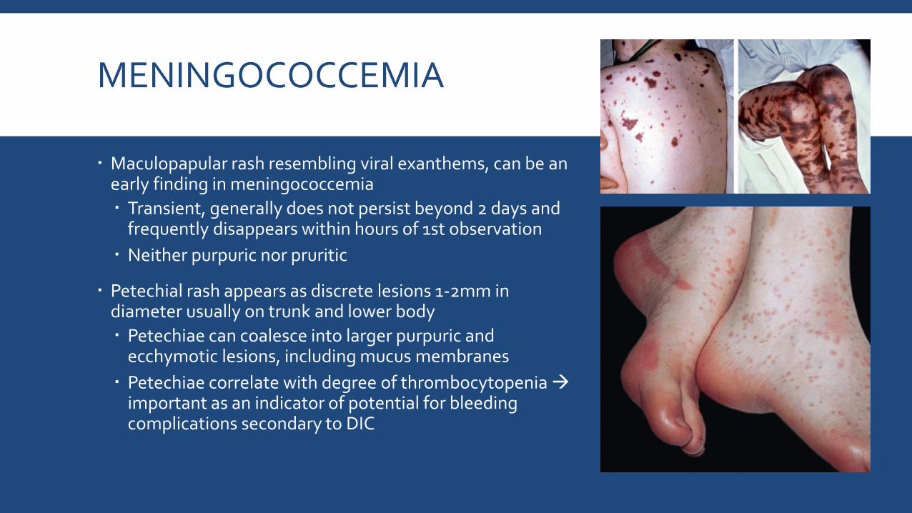
Maculopapular rash resembling viral exanthems, can be an early finding in meningococcemia
Transient, generally does not persist beyond 2 days and frequently disappears within hours of 1st observation
Neither purpuric nor pruritic
Petechial rash appears as discrete lesions 1-2mm in diameter usually on trunk and lower body
Petechiae can coalesce into larger purpuric and ecchymotic lesions, including mucus membranes
Petechiae correlate with degree of thrombocytopenia important as an indicator of potential for bleeding complications secondary to DIC

MENINGOCOCCEMIA – MANAGEMENT
Workup includes: CBC, BCx, LP (CSF gram stain, Cx, cell count, glucose & protein)
Initial management is massive fluid resuscitation patients can decompensate into shock and DIC
If not in shock, need to be aware of SIADH and may need to end up on fluid restriction
Elevated ICP is possible and may require mannitol and hyperventilation
Any generalized seizures that occur can be managed with standard seizure medications
Sequelae can include subdural empyema, which may require drainage

MENINGOCOCCEMIA – TREATMENT
Immediately after LP, before results, start antibiotics
Empiric treatment children >1 month involves vancomycin + 3rd gen cephalosporin If resistant can escalate to cefepime or meropenem
Narrow antibiotics after culture
Poor prognosis if young age, greater bacterial burden, and delayed CSF sterilization
Pretty much any organ system failure is possible with this bacteria

VIRAL EXANTHEMS
VERY Broad Topic – we will cover a few
Fifth Disease – Parvovirus Slapped-cheek appearance, bright red macules that
coalesce involving the cheeks with perioral sparing
In several days they are scattered on the limbs and trunk with central clearing
If fever, it’s mild
Can cause aplastic crisis

VIRAL EXANTHEMS
Roseola (HHV6) Rubelliform rash after 3-5 days of fever
Rose pink maculopapules throughout body
Fevers can be >40C, known to be associated
with febrile seizures
Enteroviruses (Echo and Coxsackie A&B) Wide variety of presentations, but frequently morbilliform
Primarily affects young children
Can include fevers

MEASLES
Early phase Low fever, cough, photophobia, coryza, and may include GI symptoms
A few days pass Koplik spots develop on buccal mucosa (highly contagious at this point)
Next few days spots fade, fever rises, and rash appears Red to purple red papules start on head and spread inferiorly
Coalescence of lesions occurs by 3rd day – may appear purpuric on fair skinned patients
Sequelae include encephalitis and bacterial superinfection
If not vaccinated, treatment is supportive along with Vitamin A, or can give IVIG or IMIG at time of exposure

PRACTICE #1
A 4-year-old Pacific Islander boy is diagnosed as having KD on day 6 of illness. He is admitted to the hospital for further management. Laboratory studies are obtained, and an intravenous peripheral line is placed. Which of the following is the most appropriate initial treatment regimen?
A. Aspirin (40 mg/kg per day) plus IVIG (2 g/kg per day).
B. Aspirin (100 mg/kg per day) plus solumedrol (30 mg/kg per day).
C. Aspirin (80 mg/kg per day) plus solumedrol (2 mg/kg per day).
D. IVIG (2 g/kg per day) plus cyclosporine (9 mg/kg per day).
E. IVIG (2 g/kg per day) plus infliximab (5 mg/kg per dose).

WHAT IS THE LIKELY DIAGNOSIS? #2
A 9 year old girl is brought to the clinic because she is suffering from a headache, fever, chills, and a rash. The rash covers her neck, chest, and under her armpits. The parents explain that the rash appeared today, and that for the past two days the patient had been complaining of a sore throat. The child has no allergies, her immunizations are all up to date, and she has no other past medical history. Her blood pressure is 115/70 mm Hg, pulse is 110/min, respirations are 22/min, and temperature is 101.2°F. A physical exam reveals a generalized erythematous rash that has a sandpaper-like texture, and it will also blanch when pressure is applied. The patient also has submandibular lymphadenopathy, and the throat is covered in gray-white exudates.

PRACTICE #3
A 5-year-old girl presents to the clinic with 7 days of fever that responds occasionally to acetaminophen. Her physical examination is significant for an erythematous tongue, bilateral conjunctival injection, a maculopapular rash, and cervical lymphadenopathy. She is diagnosed as having KD. Which of the following clinical findings on examination of her hands and feet is most likely to be seen during the acute phase of the disease in this patient?
A. Bruising of the fingers and toes.
B. Desquamation of the fingers and toes.
C. Firm swelling of the palms and soles.
D. Hemarthrosis of the ankles and wrists.
E. Lytic lesions in the long bones of the upper and lower extremities.

REFERENCES
Group A Streptococcus. Monika L. Dietrich and Russell W. Steele. Pediatrics in Review. 2018; 39 (8); 379 – 391
Group A Streptococcus. Stanford Shulman & Caroline Reuter. Nelsons Textbook of Pediatrics. Elselvier. 2020
Clinical manifestations of meningococcal infection. Meg Sullivan. November 2018. UpToDate
Incomplete (atypical) Kawasaki disease. Elizabeth TePas. September 2017. UpToDate
Infectious Exanthems. A. Howland Hartley and James E. Rasmussen. Pediatrics in Review. 1988; 9 (10); 321 - 329
Kawasaki Disease. Mary Beth F. Son and Jane W. Newburger. Pediatrics in Review. 2018;39 (2);78 – 90
Kawasaki disease: Clinical features and diagnosis. Elizabeth TePas. November 2018. UpToDate
Measles. Jan Drutz. Pediatrics in Review. 2016; 37 (2); 220-221
Meningitis. Douglas Swanson. Pediatrics in Review. 2015;36 (12);514 -526
Neisseria meningitides (Meningococcus). Andrew Pollard & Manish Sadarangani. Nelsons Textbook of Pediatrics. Elselvier. 2020
Roseola (Human Herpes Viruses 6 & 7). Brenda Tesini & Mary Caserta. Nelsons Textbook of Pediatrics. Elselvier. 2020
Answer Key: 1. A, 2. Scarlett Fever, 3. C