february 25th-march 1st, krynica-zdrój, polandnmr.cent3.uw.edu.pl/mmce2015/abstracts.pdf ·...
TRANSCRIPT

Magnetic Moments
in Central Europe 2015
February 25th-March 1st,Krynica-Zdrój, Poland

Magnetic Momentsin Central Europe 2015
Book of Abstracts
Krynica-Zdrój, Poland2015-02-25 / 2015-03-01

EditorsKrzysztof KosińskiMateusz UrbańczykSzymon Żerko
Cover DesignMateusz Urbańczyk
PublisherNobell Congressing sp. z o.o.Nadbrzeżna 405-850 Ożarów MazowieckiPolandwww.nobell.pl
PrintingMirage Hobbyul. Tyniecka 3602-621 WarszawaPoland
Typeset with XƎLATEX and TEX Live 2013
© 2015 University of Warsaw© 2015 Participants of Magnetic Moments in Central Europe 2015is book was prepared entirely with Free/Open Source Soware.

Organizing Committee
Prof. Wiktor Koźmiński CNBCh, University of WarsawJohn BreslinProf. Robert Konrat University of ViennaDr. Tibor Liptaj Slovak University of TechnologyProf. Predrag Novak University of ZagrebProf. Janez Plavec National Institute of Chemistry, SloveniaProf. Jan Schraml ICPF, Academy of Sciences of the Czech RepublicDr. Jan Sykora ICPF, Academy of Sciences of the Czech RepublicProf. Csaba Szantay Gedeon Richter Plc.Dr. omas Zellhofer Zellhofer Consulting
Local Organizers
Prof. Wiktor Koźmiński CNBCh-UWRupashree Dass CeNTAgata Jarzębowska CeNTDr. Krzysztof Kazimierczuk CeNTKrzysztof Kosiński CNBCh-UWDr. Karolina Madrak CNBCh-UWDr. Michał Nowakowski CNBCh-UWSaurabh Saxena CNBCh-UWDr. Alexandra Shchukina CeNTMateusz Urbańczyk CeNTSzymon Żerko CNBCh-UW
MMCE 2015 3

Contents
Organizers 3
Table of Contents 5
Sponsors 9
Foreword 10
Mission Statement 11
General Information 12
Program 13
Oral Presentations 19
On the human aspects of scientific thinking in NMR spectroscopyCsaba Szantay, Jr. . . . . . . . . . . . . . . . . . . . . . . . . . . . . . . . . . . . . . . . . 21New methods based on 13C direct detection to study intrinsically disordered proteinsIsabella C. Felli . . . . . . . . . . . . . . . . . . . . . . . . . . . . . . . . . . . . . . . . . . 22Fast-pulsing NMR techniques: general concepts, practical aspects, and selected applicationsBernhard Brutscher . . . . . . . . . . . . . . . . . . . . . . . . . . . . . . . . . . . . . . . . 23Direct monitoring of correlated ensemble fluctuations in intrinsically disordered proteinsDennis Kurzbach . . . . . . . . . . . . . . . . . . . . . . . . . . . . . . . . . . . . . . . . . 24MAP1B light chain and its interaction with microtubulesomas Schwarz . . . . . . . . . . . . . . . . . . . . . . . . . . . . . . . . . . . . . . . . . 25Simplifying proton-detected NMR spectra by spatially-selective excitationKlaus Zangger . . . . . . . . . . . . . . . . . . . . . . . . . . . . . . . . . . . . . . . . . . 26Precise measurement of heteronuclear coupling constants: novel applications of broadbandproton-proton decouplingKatalin E. Kövér . . . . . . . . . . . . . . . . . . . . . . . . . . . . . . . . . . . . . . . . . 27Visualisation of basic NMR: quantum and classical aspectsLars G. Hanson . . . . . . . . . . . . . . . . . . . . . . . . . . . . . . . . . . . . . . . . . . 28Application of AQARI (Accurate antitative NMR with Internal Substance) to quality con-trol of organic reagentsMichal Malon . . . . . . . . . . . . . . . . . . . . . . . . . . . . . . . . . . . . . . . . . . . 30New point of view in utilising tags for structural analysis of complex mixturesDušan Uhrín . . . . . . . . . . . . . . . . . . . . . . . . . . . . . . . . . . . . . . . . . . . 31From monosaccharides to polysaccharides: NMR applicationsSvetlana Simova . . . . . . . . . . . . . . . . . . . . . . . . . . . . . . . . . . . . . . . . . 32NMR spectroscopy in structural biology and for drug validation and development in neurode-generationChristian Griesinger . . . . . . . . . . . . . . . . . . . . . . . . . . . . . . . . . . . . . . . 33Investigating dynamic layers of cellular information transferHarald Schwalbe . . . . . . . . . . . . . . . . . . . . . . . . . . . . . . . . . . . . . . . . . 35
MMCE 2015 5

Pitfalls in RNA structure predictionZofia Gdaniec . . . . . . . . . . . . . . . . . . . . . . . . . . . . . . . . . . . . . . . . . . . 36Structural diversity of guanine rich sequences in genomes of human papillomavirusesMaja Marušič . . . . . . . . . . . . . . . . . . . . . . . . . . . . . . . . . . . . . . . . . . . 37A tetrahelical DNA fold not stabilized by G-quartetsVojč Kocman . . . . . . . . . . . . . . . . . . . . . . . . . . . . . . . . . . . . . . . . . . . 38Structural insights into aberrant splicing of CFTR exon 9Peter J. Lukavsky . . . . . . . . . . . . . . . . . . . . . . . . . . . . . . . . . . . . . . . . . 39Application of homonuclear mixing on 1H in 100 kHz magic angle spinningYusuke Nishiyama . . . . . . . . . . . . . . . . . . . . . . . . . . . . . . . . . . . . . . . . 402D and 3D CP-VC as tools for dynamics studyPiotr Paluch . . . . . . . . . . . . . . . . . . . . . . . . . . . . . . . . . . . . . . . . . . . . 41Real time J-scaling in nuclear magnetic resonanceSimon Glanzer . . . . . . . . . . . . . . . . . . . . . . . . . . . . . . . . . . . . . . . . . . 42Protein MAS DNP at 190 K and MAS triple-resonance spectroscopy of membrane proteinsHartmut Oschkinat . . . . . . . . . . . . . . . . . . . . . . . . . . . . . . . . . . . . . . . . 43NMR crystallographyMarek J. Potrzebowski . . . . . . . . . . . . . . . . . . . . . . . . . . . . . . . . . . . . . . 44Structural studies of metal-organic frameworks by solid-state NMR spectroscopy and first-principles calculationsGregor Mali . . . . . . . . . . . . . . . . . . . . . . . . . . . . . . . . . . . . . . . . . . . . 45Recent advances in orienting organic compounds for RDC structural analysisChristina M. iele . . . . . . . . . . . . . . . . . . . . . . . . . . . . . . . . . . . . . . . . 46NMR in paramagnetic systems in solutionJozef Kowalewski . . . . . . . . . . . . . . . . . . . . . . . . . . . . . . . . . . . . . . . . . 47And now to something completely different:Fast Field Cycling NMR RelaxometryBert Heise . . . . . . . . . . . . . . . . . . . . . . . . . . . . . . . . . . . . . . . . . . . . . 48CASE: computer assisted structure elucidationZoltán Béni . . . . . . . . . . . . . . . . . . . . . . . . . . . . . . . . . . . . . . . . . . . . 49High-dimensional 13C-detected experiments for assignment of intrinsically disordered pro-teinsAnna Zawadzka-Kazimierczuk . . . . . . . . . . . . . . . . . . . . . . . . . . . . . . . . . . 50Structural insights into disease-associated human prion protein mutants by NMRIvana Biljan . . . . . . . . . . . . . . . . . . . . . . . . . . . . . . . . . . . . . . . . . . . . 51High-dimensional NMR experiment for sequential assignment in 13C- labeled RNAsSaurabh Saxena . . . . . . . . . . . . . . . . . . . . . . . . . . . . . . . . . . . . . . . . . . 52Probing the conformation of (1→2)-C-disaccharides by NMRRadek Pohl . . . . . . . . . . . . . . . . . . . . . . . . . . . . . . . . . . . . . . . . . . . . 53Sparsity in NMR and aroundKrzysztof Kazimierczuk . . . . . . . . . . . . . . . . . . . . . . . . . . . . . . . . . . . . . 54Chemical exchange saturation transfer (CEST) MR imagingVladimír Mlynárik . . . . . . . . . . . . . . . . . . . . . . . . . . . . . . . . . . . . . . . . 55
6 MMCE 2015

Can we do mouse brain histology in vivo using MRI?Władysław P. Węglarz . . . . . . . . . . . . . . . . . . . . . . . . . . . . . . . . . . . . . . 56Non-uniform sampling meets DOSYMateusz Urbańczyk . . . . . . . . . . . . . . . . . . . . . . . . . . . . . . . . . . . . . . . . 57
Poster Presentations 59
T2* relaxometry of thalamus in multiple sclerosisEva Baranovičová . . . . . . . . . . . . . . . . . . . . . . . . . . . . . . . . . . . . . . . . . 61New point of view in utilising tags for structural analysis of complex mixturesNicholle G. A. Bell . . . . . . . . . . . . . . . . . . . . . . . . . . . . . . . . . . . . . . . . 62Binding abilities of new chiral reagents for separation of helicenesPetra Cuřínová . . . . . . . . . . . . . . . . . . . . . . . . . . . . . . . . . . . . . . . . . . 631H NMR, CD and UV study of duplex-quadruplex structural hybridKarolina Czajczyńska . . . . . . . . . . . . . . . . . . . . . . . . . . . . . . . . . . . . . . 64Analysis of complex reacting mixtures by time-resolved 2D NMRRupashree Dass . . . . . . . . . . . . . . . . . . . . . . . . . . . . . . . . . . . . . . . . . . 65High-dimensional 13C-detected experiments for assignment of intrinsically disordered pro-teinsPaweł Dziekański . . . . . . . . . . . . . . . . . . . . . . . . . . . . . . . . . . . . . . . . . 66Interaction of neuronal intrinsically disordered proteins with multiple binding partners stud-ied by NMRAndrea Flamm . . . . . . . . . . . . . . . . . . . . . . . . . . . . . . . . . . . . . . . . . . 67NMR detection of the tautomeric equilibria for the substituted β-diketonesPetra Galer . . . . . . . . . . . . . . . . . . . . . . . . . . . . . . . . . . . . . . . . . . . . 68Study of micellization of surfactants by DOSY NMRZuzana Grňová . . . . . . . . . . . . . . . . . . . . . . . . . . . . . . . . . . . . . . . . . . 69Processing of multidimensional data using multiple fixing SMFT methodKatarzyna Grudziąż . . . . . . . . . . . . . . . . . . . . . . . . . . . . . . . . . . . . . . . 70Spiro cycles from acridin-9-ylmethylamine: a NMR studyJán Imrich . . . . . . . . . . . . . . . . . . . . . . . . . . . . . . . . . . . . . . . . . . . . 71NMR characterisation of 1,5-bis(salicylidene)carbohydrazide in solution and solid stateTomislav Jednačak . . . . . . . . . . . . . . . . . . . . . . . . . . . . . . . . . . . . . . . . 72NMR spectroscopic studies of de/protonation mechanisms in thiosemicarbazide and azoben-zene based anion chemosensorsDamjan Makuc . . . . . . . . . . . . . . . . . . . . . . . . . . . . . . . . . . . . . . . . . . 73Faster and cleaner real time pure shi NMR experimentsJohannes Mauhart . . . . . . . . . . . . . . . . . . . . . . . . . . . . . . . . . . . . . . . . 74e solution structure of the MANEC-type domain from hepatocyte growth factor inhibitor1 reveals an unexpected PAN/apple domain-type foldMichał Nowakowski . . . . . . . . . . . . . . . . . . . . . . . . . . . . . . . . . . . . . . . 75Magnetic resonance access to transiently formed protein complexTomáš Sára . . . . . . . . . . . . . . . . . . . . . . . . . . . . . . . . . . . . . . . . . . . . 76Sparsity-constrained NUS reconstruction in NMR: possible pitfallsAlexandra Shchukina . . . . . . . . . . . . . . . . . . . . . . . . . . . . . . . . . . . . . . . 77
MMCE 2015 7

G-quadruplexes: formation of long-lived intermediatesPrimož Šket . . . . . . . . . . . . . . . . . . . . . . . . . . . . . . . . . . . . . . . . . . . . 78NMR detection of tautomeric equilibria for substituted β-diketonesUrška Slapšak . . . . . . . . . . . . . . . . . . . . . . . . . . . . . . . . . . . . . . . . . . . 79Residual dipolar coupling assisted NMR and DFT analysis of an exotic product of PovarovreactionMichal Šoral . . . . . . . . . . . . . . . . . . . . . . . . . . . . . . . . . . . . . . . . . . . 80Experimental determination of structural parameters in selected polycyclic aromatic com-poundsJan Sykora . . . . . . . . . . . . . . . . . . . . . . . . . . . . . . . . . . . . . . . . . . . . 81Electron paramagnetic resonance study on co-phthalocyanine ionic derivative spontaneouslyadsorbed on highly ordered pyrolitic graphite (HOPG)Ján Tarábek . . . . . . . . . . . . . . . . . . . . . . . . . . . . . . . . . . . . . . . . . . . . 82Kaempferol glycosides from the leaves of Lotus japonicusMária Vilková . . . . . . . . . . . . . . . . . . . . . . . . . . . . . . . . . . . . . . . . . . 83Solid state 13C NMR studies of modified poly(3-hydroxybutyrate)Peter Vrábel . . . . . . . . . . . . . . . . . . . . . . . . . . . . . . . . . . . . . . . . . . . . 84Application of alignment media in structural analysis of calix[4]arene derivativesLukas Vrzal . . . . . . . . . . . . . . . . . . . . . . . . . . . . . . . . . . . . . . . . . . . . 85An efficient approach to 6D HNCO(NCA)CONHSzymon Żerko . . . . . . . . . . . . . . . . . . . . . . . . . . . . . . . . . . . . . . . . . . 86Analysis of molecular mobility in pathogenic and protective mutants of human prion proteinfrom 15N relaxation dataIgor Zhukov . . . . . . . . . . . . . . . . . . . . . . . . . . . . . . . . . . . . . . . . . . . . 87
List of Participants 88
Author Index 93
8 MMCE 2015

Sponsors
MMCE 2015 9

Dear Colleagues and distinguished guests,
Welcome to the Magnetic Moments in Central Europe conference in Krynica, Poland.e biennial conference Magnetic Moments in Central Europe (MMCE) was established in 2007 with
the vision of providing a unique knowledge-sharing event for NMR scientists and students in the region.e current meeting is the fourth (fih if we include MMCE number 0 in Ljubljana in 2008) in a seriesof successful events. e meetings in Otočec in Slovenia (2009), Tatranská Lomnica in Slovakia (2011),and Semmering in Austria (2013) proved the concept.
e main aim of this conference is to provide an effective forum for discussions, at both formal andinformal levels. e number of registered participants should guarantee a stimulating environment, andthe conference program allows maximizing the time the participants spend together.
During the past years, since its inception, the conference was sponsored by Varian, and then Agilent.is year, the unexpected and disastrous decision of Agilent to leave the NMR business affected notonly the NMR community, but also made the organization of MMCE more demanding. anks to thegenerous support of Jeol, we were able to complete this task. We hope that this meeting will againprovide us possibilities to share scientific experience and continue the good traditions of MMCE.
Best wishes,
Wiktor Koźmiński
10 MMCE 2015

Mission Statement
e conference Magnetic Moments in Central Europe (MMCE) was conceived in 2007 with the visionof providing a unique knowledge-sharing event for NMR scientists and students in the region. ephilosophy behind MMCE is centered on the following main goals:
• e conference is didactic in its fundamental spirit. Rather than expecting scientists to communi-cate their latest results aimed to be published in a scientific journal, the conference wants to offer,in the form of so-called Tutorial Talks, scientists an opportunity to speak about their research,their intellectual and emotional struggles, their “a-ha!” moments, their provocative thoughts andtheir acquired wisdom in relation to their chosen topic in such an informal, conceptual, personaland edifying manner that would not normally be possible in the context of a “regular” scientificpresentation. Tutorial Talks are expected to project a kind of distilled wisdom on a topic thatcan potentially be more captivating and instructive to both students and seasoned NMR scientiststhan a new-result-centered “regular” lecture.
• In addition to being beneficial for specialists in a given field, Tutorial Talks should foster commu-nication and understanding between various branches of NMR.
• MMCE aims to address all walks of theoretical and applied NMR spectroscopy, ranging from fun-damental theory through small-molecule structure determination, new pulse sequences, sowareand hardware development, etc. to biological molecules.
• e talks offered by a speaker at MMCE need not necessarily be ”trendy”; old concepts addressedfrom a new, exciting, or eye-opening perspective are welcome.
• MMCE also accommodates the presentation of cuing-edge new results, with the speakers beingencouraged to add some didactic flavor to their talks.
• MMCE wishes to act as a dynamic NMR discussion forum in both an intellectual and social sense.Discussions are encouraged aer each presentation, and to that end sufficient room is providedin the program.
• It aims to become a well-consolidated platform for building social capital in the NMR communityof Central Europe and beyond.
• MMCE is dedicated to involving many young scientists and university students as participants.
MMCE is held every second year in varying locations within Central Europe. It usually lasts for fivedays, starting Wednesday evening and ending Sunday noon. Topics are divided into sessions. ere areno parallel sessions. As a general rule of thumb, each session starts with a Tutorial Talk lasting about 1 hr,followed by a few Invited Talkswhich are 30min long. Tutorial Speakers and Invited Speakers are invitedby the scientific board of MMCE. Each session also provides space for a few 20-minute talks deliveredby the regular participants of the conference. ere are also poster sessions and student presentationsessions.
MMCE 2015 11

General Information
LecturesOral presentations will take place in the Nubis Hall. Speakers should contact the technical assistant inthe conference room before the session to ensure proper display of their presentations. e chairpersonsare asked to ensure that presentations do not exceed the alloed time limit. Five-minute slots should bereserved for discussion and switching of presentations.
PostersPlease pin your posters to the boards in the Floral Hall (Sala Kwiatowa) on ursday, February 26, usingthe provided pins. Posters are numbered by the page they appear in the abstract book (in alphabeticalorder). ere are two poster sessions for odd (ursday, 17:15–19:30) and even-numbered posters (Friday,17:15–19:30). Presenting authors are asked to be present in front of their posters during the session.Posters should be removed aer the dinner on Friday, February 27. Aer this time, the remaining posterswill be discarded.
Emergency NumbersPolice 997 or 112Ambulance 999 or 112
Weather Forecastwww.meteo.pl numerical short-term forecast, select MODEL UM and click on
the map near x = 248, Y = 486www.pogodynka.pl official service of the Institute of Meteorology and Water Man-
agement, numerical and synoptic forecast
Conference WWW Pagenmr.cent3.uw.edu.pl/mmce2015
12 MMCE 2015

Program Outline
Wednesday, February 25
14:00 ArrivalRegistration
19:00 Wiktor Koźmiński19:15 Csaba Szantay
20:00 Dinner
ursday, February 26
7:30 Breakfast
8:30 Isabella Felli9:15 Bernhard Brutscher9:45 Dennis Kurzbach
10:00 omas Schwarz
10:15 Coffee break
10:45 Klaus Zangger11:30 Katalin Kövér12:00 Lars Hanson
12:30 Lunch
14:00 Hiroaki Sasakawa14:15 Michal Malon14:45 Dušan Uhrín15:15 Svetlana Simova
15:45 Coffee break
16:30 Christian Griesinger
17:15 Poster session
19:30 Dinner
Friday, February 27
7:30 Breakfast
8:30 Harald Schwalbe9:15 Zofia Gdaniec9:45 Maja Marušič
10:00 Vojč Kocman
10:15 Coffee break
10:45 Peter Lukavsky11:15 Yusuke Nishiyama12:00 Piotr Paluch12:15 Simon Glanzer
12:30 Lunch
14:00 Hartmut Oschkinat14:45 Marek Potrzebowski15:15 Gregor Mali
15:45 Coffee break
16:30 Christina iele
17:15 Poster session
19:30 Dinner
Saturday, February 28
7:30 Breakfast
8:30 Jozef Kowalewski9:15 Bert Heise9:45 Zoltán Béni
10:15 Coffee break
10:45 AnnaZawadzka-Kazimierczuk
11:15 Ivana Biljan11:45 Saurabh Saxena12:00 Radek Pohl
12:15 Lunch
14:00 Free time
19:30 Dinner
Sunday, Mar 1
7:30 Breakfast
9:00 Krzysztof Kazimierczuk9:45 Vladimír Mlynárik
10:15 Coffee break
10:45 Władysław Węglarz11:15 Mateusz Urbańczyk11:30 Poster prize winner11:45 Closing
12:15 Lunch
14:00 Departure
MMCE
201513

Detailed Program
Wednesday, February 2514:00 Arrival
Registration
19:00 Wiktor Koźmiński University of Warsaw, PolandOpening
19:15 Csaba Szantay Gedeon Richter Plc., HungaryOn the human aspects of scientific thinking in NMR spectroscopy
20:00 Dinner
14 MMCE 2015

ursday, February 2607:30 Breakfast
Chairperson: Harald Schwalbe
8:30 Isabella C. Felli University of Florence, ItalyNew methods based on 13C direct detection to study intrinsically disordered proteins
9:15 Bernhard Brutscher University of Grenoble, FranceFast-pulsing NMR techniques: general concepts, practical aspects, and selectedapplications
9:45 Dennis Kurzbach University of Vienna, AustriaDirect monitoring of correlated ensemble fluctuations in intrinsically disordered proteins
10:00 omas Schwarz University of Vienna, AustriaMAP1B light chain and its interaction with microtubules
10:15 Coffee break
Chairperson: Csaba Szantay
10:45 Klaus Zangger University of Graz, AustriaSimplifying proton-detected NMR spectra by spatially-selective excitation
11:30 Katalin E. Kövér University of Debrecen, HungaryPrecise measurement of heteronuclear coupling constants: novel applications of broad-band proton-proton decoupling
12:00 Lars Hanson Technical University of Denmark, DenmarkVisualisation of basic NMR: quantum and classical aspects
12:30 Lunch
Chairperson: Predag Novak
14:00 Hirosaki Sasakawa JEOL UK, United KingdomRecent developments in hardware: the most sophisticated spectrometer and probes
14:15 Michal Malon JEOL Resonance Inc., JapanApplication of AQARI (Accurate antitative NMR with Internal Substance) to qualitycontrol of organic reagents
14:45 Dušan Uhrín University of Edinburgh, United KingdomNew point of view in utilising tags for structural analysis of complex mixtures
15:15 Svetlana Simova Institute of Organic Chemistry with Centre of Phytochemistry, Bulgarian Academy ofSciences, BulgariaFrom monosaccharides to polysaccharides: NMR applications
15:45 Coffee break
Chairperson: Predag Novak
16:30 Christian Griesinger Max Planck Institute for Biophysical Chemistry, GermanyNMR spectroscopy in structural biology and for drug validation and development inneurodegeneration
17:15 Poster session, odd-numbered posters
19:30 Dinner
MMCE 2015 15

Friday, February 2707:30 Breakfast
Chairperson: Janez Plavec
8:30 Harald Schwalbe Goethe University Frankfurt, GermanyInvestigating dynamic layers of cellular information transfer
9:15 Zofia Gdaniec Institute of Bioorganic Chemistry, Polish Academy of Sciences, PolandPitfalls in RNA structure prediction
9:45 Maja Marušič Slovenian NMR Center, National Institute of Chemistry, SloveniaStructural diversity of guanine-rich sequences in genomes of human papillomaviruses
10:00 Vojč Kocman Slovenian NMR Center, National Institute of Chemistry, SloveniaA tetrahelical DNA fold not stabilized by G-quartets
10:15 Coffee break
Chairperson: Tibor Liptaj
10:45 Peter J. Lukavsky CEITEC, Masaryk University, Czech RepublicStructural insights into aberrant splicing of CFTR exon 9
11:15 Yusuke Nishiyama JEOL Resonance Inc, JapanApplication of homonuclear mixing on 1H in 100 kHz magic angle spinning
12:00 Piotr Paluch Centre of Molecular and Macromolecular Studies, PAS, Poland2D and 3D CP-VC as tools for dynamics study
12:15 Simon Glanzer University of Graz, AustriaReal time J-scaling in nuclear magnetic resonance
12:30 Lunch
Chairperson: TBA
14:00 Hartmut Oschkinat Leibniz-Institut ür Molekulare Pharmakologie, GermanyProtein MAS DNP at 190 K and MAS triple-resonance spectroscopy of membraneproteins
14:45 Marek Potrzebowski Centre of Molecular and Macromolecular Studies, PAS, PolandNMR crystallography
15:15 Gregor Mali National Institute of Chemistry, SloveniaStructural studies of metal-organic frameworks by solid-state NMR spectroscopy andfirst-principles calculations
15:45 Coffee break
Chairperson: TBA
16:30 Christina iele Technical University of Darmstadt, GermanyRecent advances in orienting organic compounds for RDC structural analysis
17:15 Poster session, even-numbered posters
19:30 Dinner
16 MMCE 2015

Saturday, February 2807:30 Breakfast
Chairperson: Christian Griesinger
8:30 Jozef Kowalewski Stockholm University, SwedenNMR in paramagnetic systems in solution
9:15 Bert Heise Spin-Doc, GermanyStructural studies of metal-organic frameworks by solid-state NMR spectroscopy andfirst-principles calculations
9:45 Zoltán Béni Gedeon Richter Plc., HungaryCASE: computer assisted structure elucidation
10:15 Coffee break
Chairperson: Marek Potrzebowski
10:45 Anna Zawadzka-Kazimierczuk University of Warsaw, PolandHigh-dimensional 13C-detected experiments for assignment of intrinsically disorderedproteins
11:15 Ivana Biljan Slovenian NMR Center, National Institute of Chemistry, SloveniaStructural insights into disease-associated human prion protein mutants by NMR
11:45 Saurabh Saxena University of Warsaw, PolandHigh-dimensional NMR experiment for sequential assignment in 13C labeled RNAs
12:00 Radek Pohl Institute of Chemical Technology, Czech RepublicProbing the conformation of (1→2)-C-disaccharides by NMR
12:15 Lunch
14:00 Free time
19:30 Dinner
MMCE 2015 17

Sunday, Mar 107:30 Breakfast
Chairperson: Wiktor Koźmiński
9:00 Krzysztof Kazimierczuk University of Warsaw, PolandSparsity in NMR and around
9:45 Vladimír Mlynárik Medical University of Vienna, AustriaChemical exchange saturation transfer (CEST) MR imaging
10:15 Coffee break
Chairperson: Wiktor Koźmiński
10:45 Władysław P. Węglarz Institute of Nuclear Physics, PAS, PolandCan we do mouse brain histology in vivo using MRI?
11:15 Mateusz Urbańczyk University of Warsaw, PolandNon-uniform sampling meets DOSY
11:30 Poster prize winner11:45 Closing
12:15 Lunch
14:00 Departure
18 MMCE 2015

O P


On the human aspects of scientific thinking in NMR spectroscopyCsaba Szantay, Jr.
Spectroscopic Research Division, Gedeon Richter Plc.H-1475 Budapest 10, P.O. Box 27, Hungary
cs.szantay richter.hu
In this somewhat philosophical, and therefore admiedly off-the-wall presentation I will outline a con-scious way of thinking, or an aitude, if you will, that our team has been cultivating for a while inour research facility, and which has proved to be highly useful not only in our everyday professionallife, but also in private life. e main theme of this aitude is to develop a keen mindfulness of howour human nature influences our thoughts in science, and on how this influence can secretly lead eventhe smartest and most knowledgeable scientists into what we call “Mental Traps”, resulting in cognitiveerrors ranging from widely held scientific misconceptions to faulty deductions in structure elucidation.ese Mental Traps are an intrinsic feature of how we, as humans, think both in science and in our ev-eryday lives, and by understanding and analyzing their nature, one can develop the enlightening facultyof detecting and avoiding them both in one’s own and others’ thoughts.
e topic was inspired by some conspicuous and long-standing misconceptions in the basic descrip-tions of the physics of NMR, and also by the pressing need never to misidentify a molecular structurein a competitive pharmaceutical R&D environment. In this largely non-technical presentation I willdiscuss several Mental Traps, mentioning some examples from both the theory and application of NMR.e presentation is also closely related to two other talks given by others in this conference, one on thequantum and classical aspects of basic NMR (Lars Hanson), and the other on computer-assisted structureelucidation (Zoltán Béni). e full version of this material is expected to be published in book form thisyear.
MMCE 2015 Wednesday 19:15 21

New methods based on 13C direct detection to study intrinsicallydisordered proteins
Isabella C. FelliMagnetic Resonance Center (CERM) and Department of Chemistry “Ugo Schi”
University of Florence, Via Luigi Sacconi 6, 50019 Sesto Fiorentino, Italyfelli cerm.unifi.it
Recent progress in NMR instrumentation, in parallel to the growing interest in understanding the func-tional role of protein intrinsic disorder and flexibility, have stimulated the development of a variety ofnew NMR methods to study intrinsically disordered proteins (IDPs). e high flexibility and largelysolvent exposed backbone typical of IDPs influence NMR parameters causing reduced chemical shi dis-persion and extensive broadening of amide proton resonances, in particular approaching physiologicalconditions. ese constitute general features of IDPs that need to be taken into account in the designof NMR experimental methods. 13C detected NMR experiments now offer a valuable tool to addressthese peculiar features of IDPs. e experimental variants to improve the performance of 13C detectedNMR experiments to study IDPs will be discussed [1–6]. ese open the way to characterize IDPs ofincreasing size and complexity and to in-cell studies. Several examples will be presented.
Acknowledgments: this work has been supported in part by the EC Projects IDPbyNMR (Contract no. 264257), INSTRUCT(contract no. 211252) and BioNMR (contract no. 261863).
References[1] I. Bertini et al., Angew. Chem. Int. Ed. 2011, 50, 2339–2341.[2] W. Bermel et al., J. Biomol. NMR 2012, 53, 293–301.[3] W. Bermel et al., J. Biomol. NMR 2013, 57, 353–361.[4] I. C. Felli, R. Pieraelli, J. Magn. Reson. 2014, 241, 115–125.[5] S. Gil et al., Angew. Chem. Int. Ed. 2013, 52, 11808–11812.[6] I. C. Felli, L. Gonnelli, R. Pieraelli, Nat. Protoc. 2014, 9, 2005–2016.
22 ursday 8:30 MMCE 2015

Fast-pulsing NMR teniques: general concepts, practical aspects, andselected applicationsBernhard Brutscher
Institute of Structural Biology, University of Grenoble 1, CEA, CNRS71 avenue des Martyrs, 38044 Grenoble Cedex 9, France
bernhard.brutscher ibs.fr
During the last decade, we have developed a series of fast-pulsing, multidimensional NMR techniques forthe study of biological macromolecules. ese experiments, namely SOFAST-HMQC, BEST-HSQC, andBEST-TROSY provide highest possible sensitivity in short experimental time by enhancing the steady-state 1H, and eventually also heteronuclear spin polarization. As a consequence, a simple “BEST- opti-mization” of a particular pulse scheme may lead to a significant gain in experimental sensitivity, andallows reducing the overall time required for data collection. Fast-pulsing techniques also open up newpossibilities for the NMR investigation of short-lived, or only transiently populated molecular states, asthey allow the recording of higher dimensional (2D, 3D) NMR spectra during the short lifetime.
In this lecture, I will present the basic concepts of SOFAST and BEST techniques, their experimentalimplementation, and discuss the potential benefits for selected applications. In addition, I will show anapplication of these techniques to investigate at atomic resolution the structural and dynamic featuresof the major folding intermediate of the amyloidogenic protein β2-microglobulin with a half-life of only20 minutes.
MMCE 2015 ursday 9:15 23

Direct monitoring of correlated ensemble fluctuations in intrinsicallydisordered proteins
Dennis Kurzbach, Gerald Platzer, omas C. Schwarz, Agathe Vanas,and Robert Konrat
Department of Structural and Computational BiologyMax F. Perutz Laboratories, University of Vienna
Campus Vienna Biocenter 5, A-1030 Vienna, Austriadennis.kurzbach univie.ac.at
Here we present a novel methodology, NMR-based paramagnetic relaxation interference (PRI), that al-lows for the first time to directly observe concerted motions and cooperatively folded sub-states in IDPs.e problem of experimental conformational averaging is circumvented by manipulation of electronrelaxation rates in doubly spin-labeled proteins leading to a reduction of the paramagnetic effect due tospatial proximity of an NMR-observed nucleus and the two spin labels. A statistical analysis of PRI datayields a graphical display of compact conformations and correlated fluctuations in the conformationalensemble of the IDP Osteopontin.
With this method we provide mechanistic insight into the subtleties of conformational averaging inIDPs under changing environmental conditions and the relevance of correlated structural fluctuationsof individual sub-domains in substrate binding of IDPs.
References[1] D. Kurzbach et al., Angew. Chem. Int. Ed. 2014, 53, 3840–3843.[2] D. Kurzbach et al., Biochemistry 2013, 52, 5167–5175.[3] D. Kurzbach, T. C. Schwarz, G. Platzer, A. Vanas, R. Konrat, 2015, in preparation.[4] G. Platzer et al., Biochemistry 2011, 50, 6113–6124.
24 ursday 10:00 MMCE 2015

MAP1B light ain and its interaction with microtubulesMarkus Huber,ᵃ omas Schwarz,ᵇ Zsuzsanna Orban-Nemeth,ᵇ Morkos Henen,ᵇ
Wiktor Koźmiński,ᶜ Szymon Żerko,ᶜ Saurabh Saxena,ᶜ Jan Stanek,ᶜLeonhard Geist,ᵇ Friedrich Propst,ᵃ and Robert Konratᵇ
a Department of Biochemistry and Cell BiologyMax F. Perutz Laboratories, University of Vienna
Campus Vienna Biocenter 5, A-1030 Vienna, Austriab Department of Structural and Computational Biology
Max F. Perutz Laboratories, University of ViennaCampus Vienna Biocenter 5, A-1030 Vienna, Austria
c Biological and Chemical Research CentreFaculty of Chemistry, University of WarsawŻwirki i Wigury 101, 02-089 Warsaw, Poland
robert.konrat univie.ac.at
Microtubule-associated protein MAP1B is a neuronal protein which has been shown to be essentialfor the maturation of synapses and network formation in murine brain development. Deregulation ofMAP1B has been associated with several neuronal diseases. MAP1B can bind to microtubules as wellas to F-actin and, thus, has been postulated to be responsible for coupling these two components ofthe cytoskeleton. MAP1B has been demonstrated to utilize its light chain for microtubule binding, butinsights into its function are limited.
Here we report our efforts to understand the structure of the MAP1B light chain and its mode of in-teraction with microtubules. A light chain fragment containing the NH2-terminal domain of the MAP1Blight chain and was expressed in E. coli. We used 5D and 4D pulse sequences and sparse multidimen-sional Fourier transformation protocols in order to gain backbone as well as side-chain assignments. eassignment obtained indicated only a slight tendency towards alpha-helical conformations at the NH2terminus of the protein. A 15N labeled sample was used to measure electron paramagnetic resonance(PRE) data on the construct. Here we gained information about a slight compaction of the protein at itsveryNH2 terminus. Both of these observations agreewithMeta Structure derived results on the protein’scompactness. First investigations into the binding mode of MAP1B indicate that the protein contactsmicrotubules with its NH2 terminus, and is capable to interact both with polymerized microtubules aswell as with tubulin dimers.
References[1] E. Tortosa et al., J. Biol. Chem. 2011, 286, 40638–40648.[2] A. Meixner et al., J. Cell Biol. 2000, 151, 1169–1178.[3] E. Allen et al., Nature 2005, 438, 224–228.[4] P. Opal et al., J. Biol. Chem. 2003, 278, 34691–34699.[5] M. Togel, G. Wiche, F. Propst, J. Cell Biol. 1998, 143, 695–707.[6] R. Noiges et al., J. Neurosci. 2002, 22, 2106–2114.[7] A. Zawadzka-Kazimierczuk et al., J. Biomol. NMR 2012, 52, 329–337.[8] K. Kazimierczuk, A. Zawadzka, W. Koźmiński, J. Magn. Reson. 2009, 197, 219–228.[9] Z. Orban-Nemeth et al., Biomol. NMR Assign. 2014, 8, 123–127.
[10] R. Konrat, Cell. Mol. Life Sci. 2009, 66, 3625–3639.
MMCE 2015 ursday 10:15 25

Simplifying proton-detected NMR spectra by spatially-selectiveexcitation
N. Helge Meyer, Simon Glanzer, Nina Gubensäk, and Klaus ZanggerInstitute of Chemistry, University of GrazHeinrichstraße 28, A-8010 Graz, Austriaklaus.zangger uni-graz.at
Protons are themost oen used nuclei for NMR structure elucidation of organic and biological molecules.Compared to other NMR detectable nuclei, 1H spectra typically suffer from low resolution and severesignal overlap, mainly due to extensive scalar coupling between protons. Homonuclear broadband de-coupling (pure shi NMR), which leads to a collapse of 1H signals into singlets, vastly increases theresolution, which in some cases corresponds to a theoretical signal dispersion of NMR spectrometers atseveral GHz [1]. One of the approaches for homonuclear broadband decoupling in the indirect dimen-sion of two- and multidimensional NMR spectra uses frequency-selective pulses during a weak gradientfield [2].
We recently reported an adaption of this method to achieve homonuclear broadband decoupling dur-ing acquisition [3]. Scalar coupling information, which is oen key in analyzing chemical structures,is of course completely lost in such experiments. Two methods, which constitute a compromise be-tween pure-shi spectra and fully coupled spectra will also be presented: real-time SERF spectra [4] andreal-time J-scaled proton spectra. With the first of these, 1D spectra are obtained which contain scalarcoupling to one selected signal only, while J-scaling allows the recording of proton spectra with reducedcoupling constants, reminiscent of off-resonance heteronuclear decoupling.
1.01.52.02.53.03.54.04.55.0
1d( H) [ppm]Figure 1: Regular and real-time pure-shi spectra of azithromycin
References[1] J. A. Aguilar et al., Angew. Chem. Int. Ed. 2010, 49 (23), 3901–3903.[2] K. Zangger, H. Sterk, J. Magn. Reson. 1997, 124, 486–489.[3] N.H. Meyer, K. Zangger, Angew. Chem. Int. Ed. 2013, 52, 7143–7146.[4] N. Gubensäk, W.M. F. Fabian, K. Zangger, Chem. Commun. 2014, 50, 12254–12257.
26 ursday 10:45 MMCE 2015

Precise measurement of heteronuclear coupling constants: novelapplications of broadband proton-proton decoupling
Katalin E. Kövér,ᵃ István Timári,ᵃ Lukas Kaltschnee,ᵇ Andreas Kolmer,ᵇRalph W. Adams,ᶜ Mathias Nilsson,ᶜ Tünde Z. Illyés,ᵃ László Szilágyi,ᵃ
Christina M. iele,ᵇ and Gareth A. Morrisᶜa Institute of Chemistry, University of Debrecen
Egyetem tér 1, H-4032 Debrecen, Hungaryb Clemens Schöpf Institute for Organic Chemistry and Biochemistry
Technical University of DarmstadtAlarich-Weiss-Straße 16, D-64287 Darmstadt, Germany
c University of Manchester, School of ChemistryOxford Road, Manchester, M13 9PL, United Kingdom
kover science.unideb.hu
A variety of novel methods have been developed in recent years that reduce the complexity of 1H NMRspectra by collapsing the multiplet structure of each resonance into a singlet. ese experiments deliverpure chemical shi information without the complication of homonuclear coupling interactions andwith significantly increased resolution in the direct proton dimension.
Implementing this broadband proton-proton decoupling methodology in the CLIP/CLAP-HSQC [1]and HSQMBC [2] experiments the undesired proton-proton spliings are removed from the heteronu-clear multiplets, thus the heteronuclear couplings of interest can be determined simply by measuringthe frequency differences between the peak maxima of pure doublets.
e potential of these experiments will be demonstrated on molecules featuring extended networksof mutually coupled protons. Additional multiplet fiing procedures would normally be required toextract long-range heteronuclear coupling constants when using standard HSQMBC experiments forsuch cases. Incorporation of the recent PSYCHE [3] pulse sequence element or of instant (real-time)homonuclear broadband decoupling [4] methodology in the HSQMBC sequence, resulting in significantimprovements in sensitivity, will also be presented.
Financial support from OTKA K 105459, OTKA NN 109671 and TÁMOP-4.2.2/A-11/1/KONV-2012-0025 is gratefully ac-knowledged.
References[1] I. Timári et al., J. Magn. Reson. 2014, 239, 130–138.[2] I. Timári et al., Chem. Eur. J. 2015, DOI 10.1002/chem.201405535.[3] M. Foroozandeh et al., Angew. Chem. Int. Ed. 2014, 53, 6990–6992.[4] N.H. Meyer, K. Zangger, Angew. Chem. Int. Ed. 2013, 52, 7143–7146.
MMCE 2015 ursday 11:30 27

Visualisation of basic NMR: quantum and classical aspectsLars G. Hanson
Danish Research Centre for Magnetic Resonance, Hvidovre Hospital, and DTU ElekroTechnical University of Denmark, Copenhagen, DenmarkDept. 714, Keegaard Alle 30, DK-2650 Hvidovre, Denmark
larsh drcmr.dk
Handwaving and semi-classical graphics are widely used to illustrate spin dynamics such as excitationand echoes. antum descriptions are oen accompanied by different figures such as level diagramsand cone figures. In this talk, the relation between quantum and classical mechanics is discussed witha particular focus on visualisation. It is argued that the classical and the quantum descriptions of basicNMR are more similar than they may first appear, and that “classical” illustrations can accurately depictessential aspects of quantum NMR [1]. In contrast, illustrations meant to convey quantum mechanicsoen do not [2]. Figures that aremisleading or divert aention from crucial aspects are abundant. Simplegraphical tools aimed at early NMR and MRI education are demonstrated.
Figure 1: Bloch vector vizualization [3] of thermal equilibrium which is equally valid from classical and quantumperspectives.
Figure 2: e Bloch Simulator [4] is a free Flash™application running directly in most browsers. It can be used toexplore basic NMR and MRI methods such as echo formation.
28 ursday 12:00 MMCE 2015

References[1] R. P. Feynman, F. L. Vernon Jr., R.W. Hellwarth, J. Appl. Phys. 1957, 28, 49–52.[2] L. G. Hanson, Concepts Magn. Reson. 2008, 32A (5), 329–340.[3] L. G. Hanson, Introductory NMR visualisations, 2015, http://www.drcmr.dk/MR[4] L. G. Hanson, Bloch Simulator, 2015, http://www.drcmr.dk/bloch
MMCE 2015 ursday 12:00 29

Application of AQARI (Accurate antitative NMR with InternalSubstance) to quality control of organic reagents
Toru Miura,ᵃ Shinji Nakao,ᵃ Shinya Takaoka,ᵃ Yuko Yamada,ᵃ Michal Malon,ᵇand Takako Suematsuᵇ
a Wako Pure Chemical Industries, Ltd., 1633 Matoba, Kawagoe-shi, 350-1101 Saitama, Japanb JEOL Resonance Inc., Musashino 1-2-3, Akishima-shi, 196-8558 Tokyo, Japan
mmichal jeol.co.jp
antitative HPLC (high performance liquid chromatography) analysis is typically carried out by us-ing a reference substance which is the same as the compound to quantify. However, certified refer-ence substances are sometimes unavailable due to strict requirements and high cost of their production.erefore, commercial reagents are oen used as reference substances in chromatographic assays, eventhough their absolute purity is unknown. To ensure the reliability of quantitative assay, absolute purityis crucial. qNMR (quantitative NMR) is known as one of absolute quantification methods suitable forpurity determination of organic compounds [1]. Recently, 1H qNMR has drawn much aention in manyfields as it provides accurate and precise quantitative values without the usage of a reference compoundthat is chemically identical to the analyte. In addition, qNMR analysis with SI (Le Système Internationald’Unités, International System of Units) traceability is possible by using appropriate protocol. ere-fore qNMR qualifies as a method to evaluate reference substances for other analytical methods, suchas HPLC. qNMR has already been employed for purity determination in Japanese regulatory standardssuch as Japanese Pharmacopoeia [2] and Japan Specification and Standard for Food Additives [3, 4].
In this study, we have developed a system for quality control of organic reagents by 1H qNMR. Inparticular, we have used AQARI (Accurate quantitative NMRwith internal substance) [5], which is one ofqNMR methods and provides highest precision and accuracy than other qNMR methods. e protocolfor AQARI includes not only NMR measurement but also sample preparation. We have evaluated alarge variety of reagents and here we discuss feasibility of AQARI for purity determination. We havefound that AQARI is able to determine the purity of reagents with accuracy of approximately 1% andcan be applied as an efficient quality control of organic reagents. By using reference substances withSI traceability and incorporating purity obtained by qNMR into calculation of quantitative values inquantitative analyses by HPLC and other methods, SI traceability is ensured. erefore, our approachsignificantly improves reliability of commonly used quantitative methods.
References[1] T. Ihara, T. Saito, N. Sugimoto, Synthesiology 2009, 2, 13–24.[2] Japanese Pharmacopoeia, 16th Edition, Supplement II, pp. 2628–2630, http://www.pmda.go.jp/english/
pharmacopoeia/online.html announced and enforced on February 28, 2014.[3] Ordinance for Partial Revision of the Ordinance for Enforcement of the Food Sanitation Act and the Ministerial Ordi-
nance on Milk and Dairy Products, Cabinet Office and Ministry of Health, Labour and Welfare Ordinance No. 5 of2011, announced on August 31, 2011 (in Japanese).
[4] Ordinance for Partial Revision of the Specifications and Standards of Food and Food Additives, Ministry of Health,Labour and Welfare Ordinance No. 307 of 2011 (in Japanese).
[5] J. Hosoe et al., Pharm. Med. Dev. Regulatory Sci. 2014, 45, 243–250.
30 ursday 14:15 MMCE 2015

New point of view in utilising tags for structural analysis of complexmixtures
Nicholle G. A. Bell,ᵃ Adam A. L. Michalchuk,ᵃ John W. T. Blackburn,ᵃMargaret C. Graham,ᵇ and Dušan Uhrínᵇa EastChem School of Chemistry, University of Edinburgh
King’s Buildings, David Brewster Rd, Edinburgh, EH9 3FJ, United Kingdomb School of Geosciences, University of Edinburgh
King’s Buildings, James Huon Rd, Edinburgh, EH9 3FE, United Kingdomdusan.uhrin ed.ac.uk
Even the most powerful multidimensional (nD) NMR methodologies alone cannot solve structures ofcompounds contained in mixtures of thousands of small molecules. Some form of “spectroscopic separa-tion” is therefore required. To achieve this, we have developed isotope-filtered nDNMR spectroscopy [1]that allows structural investigation of isotopically tagged molecules in complex, inseparable mixtures.
Figure 1: Polarisation transfer scheme
Incorporating isotopically labelled moietieswithin targeted functional groups of small or-ganic molecules opens a unique possibility forstructure characterisation of molecules in com-plex mixtures. Although isotopic tagging hasbeen used in the analysis of mixtures in the past,we propose a new paradigm. Rather than focus-ing on the chemical shis of the tags only, weuse the tags for accessing chemical shis and cou-pling constants of the parent molecules. We il-lustrate this approach by 13C-methylation of hy-droxyl and carboxyl groups in combination withpurpose-designed nD NMR experiments.
Scheme on the right shows the interactionsthat can be used to transfer the polarisation ina 13C-methylated phenol. ese couplings wereutilised in the design of nD NMR experimentsthat provide multiple correlated chemical shisand coupling constants of aromatic rings nuclei.In these experiments, the signals from unlabelledmolecules are eliminated, providing much needed simplification of NMR spectra of complex mixtures.e obtained information enabled the structures of derivatised compounds to be solved.
e developed methodology is aimed at the analysis of the aromatic moieties of humic substances(HS), the main organic component of soil and amongst the most complex mixtures on Earth. We haverecently applied it to characterise major substitution paerns of phenolic moieties of fulvic acid isolatedfrom a Scoish peaty soil [2].
Our approach is not limited to studies of HS, neither is the methylation as the way of introducinglabels to report on the parent molecules. We are currently exploring several different approaches basedon the principles outlined here.
References[1] N. G. A. Bell et al., Chem. Commun. 2014, 50, 1694–1697.[2] N. G. A. Bell, A. A. L. Michalchuk, J.W. T. Blackburn, M. C. Graham, D. Uhrín, Isotope-filtered 4D NMR for structure
determination of humic substances, submied.
MMCE 2015 ursday 14:45 31

From monosacarides to polysacarides: NMR applicationsSvetlana Simova
Institute of Organic Chemistry with Centre of PhytochemistryBulgarian Academy of Sciences, Acad. G. Bonchev bl. 9, 1113 Sofia, Bulgaria
sds orgchm.bas.bg
Carbohydrates, or saccharides, are the most abundant of the four major classes of biomolecules, alongwith proteins, nucleotides, and lipids. ere are various types of saccharides, including mono-, oligo-and polysaccharides [1]. Traditionally, NMR spectroscopy has been one of the major tools to fosterthe advance of carbohydrate chemistry [2]. e study of the structure, conformation and dynamics ofmono- and oligosaccharides is important because of their biological relevance. e high solubility andtheir generally good NMR properties, resulting from relatively small molecular sizes, narrow lines andsufficiently slow relaxation allow thorough investigation of a great variety of individual carbohydratesby relatively simple 1D and 2D NMR methods.
Recent results from the application of this knowledge and questions arising when studying problemslike chemical and metabolic profiling, carbohydrate complex formation and stucture of polysachharideswill be presented and discussed.
Figure 1: From chemical profiling to polysaccharide structure
References[1] H.-J. Gabius, e Sugar Code: Fundamentals of Glycosciences, Wiley-VCH, Weinheim, 2003.[2] J. Jiménez-Barbero, T. Peters, NMR Spectroscopy of Glycoconjugates, Wiley-Blackwell, 2009.
32 ursday 15:15 MMCE 2015

NMR spectroscopy in structural biology and for drug validation anddevelopment in neurodegeneration
Sergey Ryazanov,ᵃᵇ Han Sun,ᵃ Manuel Schmidt,ᵃ Michael Müller,ᵃNina Schützenmeister,ᵃ Johannes Levin,ᶜ Jens Wagner,ᶜ Song Shi,ᶜ
Ana Martinez Hernandez,ᵇᵈ Hope Y. Agbemenyah,ᵇᵉ Claudio O. Fernandez,ᶠAndré Fischer,ᵇᵉ Nasrollah Resaei-Ghaleh,ᵃᵇ Uwe M. Reinscheid,ᵃ
Armando Navarro-Vázquez,ᵈ Stefan Eimer,ᵇᵉ Jochen Weishaupt,ᵍ Herbert Jäckle,ᶜGregor Eichele,ᵇᵉ Armin Giese,ᵈ Stefan Becker,ᵃ Andrei Leonov,ᵃᵇ Adam Lange,ᵃ
Markus Zwecksteer,ᵃᵇ and Christian Griesingerᵃᵇa Deptartment for NMR-based Structural Biology
Max Planck Institute for Biophysical Chemistry, Göingen, Germanyb DFG-Center for the Molecular Physiology of the Brain, Göingen, Germanyc Center for Neuropathology and Prion Research, LMU, Munich, Germany
d Department of Genes and BehaviorMax Planck Institute for Biophysical Chemistry, Göingen, Germany
e European Neuroscience Institute, Göingen, Germanyf Instituto de Biología Molecular y Celular de Rosario
Universidad Nacional de Rosario Suipacha, Rosario, Argentinag Deptartment of Neurology, Medical University of Göingen, Germany
cigr nmr.mpibpc.mpg.de
An introduction will be given into the scope of NMR spectroscopy to address questions in natural prod-uct chemistry [1–19], membrane proteins [20–24] and signalling. en a more applied project in thedepartment will be discussed where small molecules play a decisive role in redirecting aggregation.Parkinson’s disease features aggregates of α-synuclein, so called Lewy bodies, which are connected toneuronal dysfunction and death. With NMR in liquid and solid state, we have characterized the poly-morphic forms of α-synuclein [25–31]: a) Monomers, that are considered to be the healthy form, areso called intrinsically disordered proteins lacking secondary structure but having partial tertiary struc-ture. e laer autoinhibits aggregation. In line with this we observe that mutants that destabilize thepartially folded form aggregate faster and exhibit higher toxicity in animal models. b) Oligomers ofrather well defined size are observed that have less β-structure than the fibrils. ey are on-pathwayto the fibrils. c) Fibrils are formed which could be characterized structurally by solid state NMR andare non-toxic. From the structure mutants have been predicted that don’t form fibrils but oligomers athigher concentration. ey have dramatically increased toxicity in animal models.
e “aggregation landscape” can also be modified by small molecules [32–34]. With screening andtargeted libraries we identified compounds that reduce the size of oligomers in vitro and in vivo. eyare off-pathway, since no fibrils are formed any more. Biophysically they prevent pore formation inmembranes, an activity of aggregates that is discussed as a reason for toxicity. Oral application ofthese highly bioavailable compounds to various animal models (C. elegans, fly, mouse) of PD rescuesthe phenotype, i.e. reduces the aggregates and leads to an improvement of the behavioural phenotypewith respect to locomotion in PD, learning and memory in AD and survival in CJD as well as PD andAD. Interestingly, the in vitro biophysical characteristics found first for α-synuclein are also observedfor proteins responsible for other diseases, namely Creutzfeld-Jacob and Alzheimer’s disease.
MMCE 2015 ursday 16:30 33

References[1] S. Bartoschek et al., Angew. Chem. Int. Ed. (2010, 49, 1426–1429.[2] J. Orts et al., Angew. Chem. Int. Ed. 2008, 47, 7736–7740.[3] M. Reese et al., Angew. Chem. Int. Ed. 2007, 46, 1864–1868.[4] V.M. Sánchez-Pedregal et al., Angew. Chem. Int. Ed. 2005, 44, 2–4.[5] R. Berger et al., Angew. Chem. Int. Ed. 2012, 51, 2–5.[6] H. Ge et al., Chem. Eur. J. 2012, 18, 5213–5221.[7] E. Whitson et al., J. Nat. Prod. 2012, 75, 394–399.[8] H. Sun et al., J. Am. Chem. Soc. 2011, 17, 1811–1817.[9] H. Sun et al., Chem. Eur. J. 2011, 17, 1811–1817.
[10] U.M. Reinscheid et al., Eur. J. Org. Chem. 2010, 36, 6900–6903.[11] A. Schuetz et al., Angew. Chem. Int. Ed. 2008, 47, 1–4.[12] A. Schuetz et al., J. Am. Chem. Soc. 2007, 129 (49), 15114–15115.[13] P. Haberz, J. Farjon, C. Griesinger, Angew. Chem. Int. Ed. 2005, 44, 427–429.[14] Z. X. Xie et al., Chinese J. Chem. 2008, 26, 1272–1276.[15] J. D. Knight, S. J. Sauer, D.M. Coltart, Org. Le. 2011, 13, 3118–3121.[16] W. P. Hems et al., Org. Proc. Res. Devel. 2012, 16, 461–463.[17] J.M. Karle et al., Exp. Parasitol. 1993, 76, 345–351.[18] M. Müller et al., Angew. Chem. Int. Ed. 2013, 52, 6047–6049.[19] N. Schützenmeister et al., Chem. Eur. J. 2013, 19, 17584–17588.[20] M. Bayrhuber et al., Proc. Nat. Acad. Sci. USA 2008, 105 (40), 15370–15375.[21] S. Villinger et al., Proc. Nat. Acad. Sci. USA 2010, 107 (52), 22546–22551.[22] R. Schneider et al., Angew. Chem. Int. Ed. 2010, 49 (10), 1882–1885.[23] S. Villinger et al., FEBS Journal 2012, 279 (Supplement 1), 433.[24] S. Villinger et al., J. Biol. Chem. 2014, 289, 13397–13406.[25] C. O. Fernandez et al., EMBO Journal 2004, 23, 2039–2046.[26] C.W. Bertoncini et al., Proc. Nat. Acad. Sci. USA 2005, 102, 1430–1435.[27] R.M. Rasia et al., Proc. Natl. Acad. Sci. USA 2005, 102, 4294–4299.[28] P. Bernado et al., J. Am. Chem. Soc. 2005, 127 (51), 17968–17969.[29] A. Binolfi et al., J. Am. Chem. Soc. 2006, 128 (30), 9893–9901.[30] C.W. Bertoncini et al., J. Mol. Biol. 2007, 327 (3), 708–722.[31] P. Karpinar et al., EMBO Journal 2009, 28, 3256–3268.[32] New drugs for inhibiting aggregation of proteins involved in diseases linked to protein aggregation and/or neurode-
generative diseases, EP 08010458.1, WO 2010/000372 A2, 2010-01-07, submied: 2008-06-09.[33] J. Wagner et al., Acta Neuropathol. 2013, 125, 795–813.[34] J. Levin et al., Acta Neuropathol. 2014, 127, 779–780.
34 ursday 16:30 MMCE 2015

Investigating dynamic layers of cellular information transferHarald Schwalbe
Center for Biomolecular Magnetic Resonance (BMRZ), Goethe UniversityMax-von-Laue-Straße 7, D-60438 Frankfurt am Main, Germany
schwalbe nmr.uni-frankfurt.de
In this lecture, I will discuss two examples where information transfer during transcription and trans-lation is dynamically regulated. e first example deals with riboswitches. Riboswitches are gene reg-ulatory elements located in the 5’-untranslated regions of messenger RNA (mRNA). Ligand bindingto an aptamer domain of riboswitches induces either the up- or down-regulation of the expression ofligand-associated genes. Riboswitch regulate gene expression either at the level of transcription or trans-lation. For transcriptional riboswitches, ligand binding supposedly induces a conformational switchbetween mutually exclusive antiterminator or terminator conformations that represent the on- and off-states of the switches. We show that in stark contrast to this classical model for riboswitch functionthree different transcriptional riboswitches adopt the terminator conformation (off-state) at thermody-namic equilibrium regardless of the presence or absence of the ligand. By investigating the guanine-and hypoxanthine-sensing xpt-pbuX riboswitch from Bacillus subtilis, we find that in contrast to thefull-length mRNA, transcription intermediates undergo ligand-dependent conformational changes. Im-portantly, in the absence of ligand, the RNAwas able to adopt the antiterminator conformation in a tran-scription intermediate that was kinetically trapped and did not refold fast enough to form the longer andmore stable terminator conformation in the limited time window available during transcription. iskinetic trapping allows the RNA-polymerase to escape from the termination site before the terminator isfolded and to maintain gene expression in the absence of ligand. In the presence of ligand, early ligandbinding stabilised the aptamer domain, suppressed the formation of the antiterminator conformation,and, thus, accelerated formation of the terminator conformation by at least two orders of magnitude,leading to gene repression. Transcriptional regulation therefore executes the genetic decision duringa single-round of transcription and is critically dependent on the kinetics of ligand binding and RNArefolding that constantly change as the length of the mRNA chain grows during transcription.
e second example deals with protein folding and reports that differential usage of synonymouscodons governs co-translational folding and final protein structure. e genetic code is degenerate,with up to six synonymous codons encoding a given amino acid in the protein. e occurrence ofsynonymous codons in open reading frames (ORFs) of genes is not random, suggesting the existence ofevolutionary constraints on codon choice.
In this lecture I will present data demonstrating that synonymous codon usage governs the kineticsof translation, co-and post-translational protein folding and final protein structure. e ribosome-boundnascent chains of the mammalian eye lens protein, gamma-B crystallin, expressed from two gene vari-ants with different synonymous codon composition but encoding the same polypeptide, aain differentconformations as indicated by altered in vivo stability and in vitro protease resistance. Our 2D NMRspectroscopic data suggest that the observed structural differences are associated with different cys-teine oxidation states of the synonymous variants. Synonymous codon usage was found to alter localand global rates of translation and affect the efficiency of co-translational folding of protein domains aswell as the ultimate stable conformation aained by the protein.
MMCE 2015 Friday 8:30 35

Pitfalls in RNA structure predictionZofia Gdaniec
Institute of Bioorganic Chemistry, Polish Academy of SciencesNoskowskiego 12/14, 61-704 Poznań, Polandzofia.gdaniec ibch.poznan.pl
RNA molecules play important roles in the regulation of gene expression at various levels. For example,RNAs are active participants in the regulation of gene expression by interference or by riboswitches,in the processing of RNA introns and in protein synthesis by the ribosome. Knowledge of the three-dimensional structures of these molecules is a fundamental prerequisite to a complete understanding ofRNA function.
New RNAs are being discovered much faster than their three-dimensional structures. Experimentaldetermination of sequences of gens and entire genoms, fromwhich the sequences of RNA can be reliablyinferred is much cheaper and faster than experimental determination of the structures. In a consequence,the disparity is increasing between known RNA 3D structures and known RNA sequences.
To provide insight into RNA function and to guide experimental work in biology and biochemistryseveral strategies has been identified, which now allow for reasonably accurate prediction of 2D and3D structures. However, it is important to remember that the prediction of any RNA structure is justan approximation based on a number of simplifying assumptions. Many RNAs contain both structuredand functionally important but flexible elements. Structure may also change because of environmentalfactors, e.g. binding of other molecules or the composition of the solution (salt, pH). At present, theprediction may be close to a structure found in nature or very far off, depending on the length andnucleotide sequence of an RNA molecule.
e advantage of NMR spectroscopy in studying RNA is an ability to monitor its structure anddynamics in physiologically relevant solution. Several examples of RNA molecules for which NMRspectroscopy has provided structural information inaccessible to the other methods will be presented.
36 Friday 9:15 MMCE 2015

Structural diversity of guanine ri sequences in genomes of humanpapillomaviruses
Maja Marušič,ᵃ Katarina Tlučková,ᵇ Petra Tóthová,ᵇ Lubos Bauer,ᵇ Primož Šket,ᵃViktor Viglasky,ᵇ and Janez Plavecᵃᶜᵈ
a Slovenian NMR Center, National Institute of ChemistryHajdrihova ulica 19, SI-1001 Ljubljana, Slovenia
b Department of Biochemistry, Institute of Chemistry, Faculty of SciencePavol Jozef Šafárik University, Moyzesova 11, 04154 Košice, Slovakia
c EN-FIST Centre of Excellence, Trg Osvobodilne fronte 13, 1000 Ljubljana, Sloveniad Faculty of Chemistry and Chemical Technology, University of Ljubljana
Večna pot 113, 1000 Ljubljana, Sloveniamarusic.maja ki.si
Human papillomaviruses (HPV) are a diverse group of viruses that are important human pathogens.Infection with certain low-risk types of HPV can lead to formation of warts and lesions, while infectionwith high-risk HPV can cause cancerous changes of head and neck, skin and anogenital area. Amongthose, cervical cancer represents one of the most common cancers worldwide and preceding infectionwith high-risk HPV is required for cancer development. Guanine-rich sequences in the genomes of HPVhave potential to fold into four-stranded DNA structures or G-quadruplexes that were shown to act ascontrollers of processes of replication, transcription and translation. G-quadruplex stabilization withligands could therefore make it possible to control HPV replication and/or gene expression.
We have analyzed G-quadruplex forming potential of guanine-rich sequences found in different HPVgenomes in the presence of K+ with help of electrophoresis, CD, UV and NMR spectroscopy [1]. Mostof them have been shown to fold into G-quadruplexes with polymorphic behaviour. Location of thesequences in regulatory long control region and in the regions coding for early (E1 and E4) and late(L2) proteins suggests a potential role in transcription, replication and alternative splicing. Our resultsrepresent a starting point for the design of specific ligands for viral G-quadruplex motifs and suggestyet unexploited pathways for the control of viral replication and transcription.
Figure 1: G-quartet, building block of G-quadruplexes (le) and locations of G-rich sequences in HPV genomes (right).
References[1] K. Tlučková et al., Biochemistry 2013, 52 (41), 7207–7216.
MMCE 2015 Friday 9:45 37
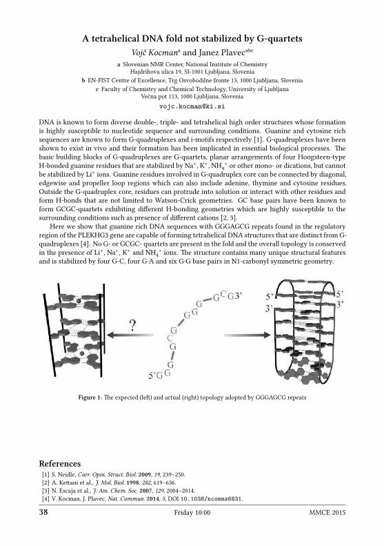
A tetrahelical DNA fold not stabilized by G-quartetsVojč Kocmanᵃ and Janez Plavecᵃᵇᶜ
a Slovenian NMR Center, National Institute of ChemistryHajdrihova ulica 19, SI-1001 Ljubljana, Slovenia
b EN-FIST Centre of Excellence, Trg Osvobodilne fronte 13, 1000 Ljubljana, Sloveniac Faculty of Chemistry and Chemical Technology, University of Ljubljana
Večna pot 113, 1000 Ljubljana, Sloveniavojc.kocman ki.si
DNA is known to form diverse double-, triple- and tetrahelical high order structures whose formationis highly susceptible to nucleotide sequence and surrounding conditions. Guanine and cytosine richsequences are known to form G-quadruplexes and i-motifs respectively [1]. G-quadruplexes have beenshown to exist in vivo and their formation has been implicated in essential biological processes. ebasic building blocks of G-quadruplexes are G-quartets, planar arrangements of four Hoogsteen-typeH-bonded guanine residues that are stabilized by Na+, K+, NH4
+ or other mono- or dications, but cannotbe stabilized by Li+ ions. Guanine residues involved in G-quadruplex core can be connected by diagonal,edgewise and propeller loop regions which can also include adenine, thymine and cytosine residues.Outside the G-quadruplex core, residues can protrude into solution or interact with other residues andform H-bonds that are not limited to Watson-Crick geometries. GC base pairs have been known toform GCGC-quartets exhibiting different H-bonding geometries which are highly susceptible to thesurrounding conditions such as presence of different cations [2, 3].
Here we show that guanine rich DNA sequences with GGGAGCG repeats found in the regulatoryregion of the PLEKHG3 gene are capable of forming tetrahelical DNA structures that are distinct from G-quadruplexes [4]. No G- or GCGC- quartets are present in the fold and the overall topology is conservedin the presence of Li+, Na+, K+ and NH4
+ ions. e structure contains many unique structural featuresand is stabilized by four G-C, four G∙A and six G∙G base pairs in N1-carbonyl symmetric geometry.
Figure 1: e expected (le) and actual (right) topology adopted by GGGAGCG repeats
References[1] S. Neidle, Curr. Opin. Struct. Biol. 2009, 19, 239–250.[2] A. Keani et al., J. Mol. Biol. 1998, 282, 619–636.[3] N. Escaja et al., J. Am. Chem. Soc. 2007, 129, 2004–2014.[4] V. Kocman, J. Plavec, Nat. Commun. 2014, 5, DOI 10.1038/ncomms6831.
38 Friday 10:00 MMCE 2015

Structural insights into aberrant splicing of CFTR exon 9Peter J. Lukavsky
Central European Institute of TechnologyMasaryk University, Brno, Czech Republicpeter.lukavsky ceitec.muni.cz
Alternative pre-mRNA splicing processes play a key role in creating the vast number of gene productsunderlying our complex organism. e processing of pre-mRNAs is tightly regulated and any imbal-ance can change the outcome of gene expression, oen leading to disease. Despite the importance ofalternative splicing regulation, our knowledge at the molecular level is still in its infancy.
We study the regulatory RNA-protein and protein-protein interaction networks surrounding theaberrantly spliced cystic fibrosis transmembrane conductance regulator (CFTR) exon 9. Skipping of thisexon 9 leads to the production of a non-functional chloride channel, which is associated with severeforms of cystic fibrosis. is unfavourable splicing event depends primarily on TDP-43 (43 kDa TARDNA binding protein) which binds to a UG-rich region upstream of the 3’ splice site (ss) of exon 9 andthis, in concert with other splicing factors, prevents the recognition of the 3’ss of exon 9. us exon 9 isexcluded from the spliced mRNA and this results in a non-functional protein product.
As a first step towards understanding the molecular basis of aberrant CFTR exon 9 splicing, wesolved the solution structure of TDP-43 RRMs in complex with UG-rich RNA. Ten nucleotides are boundbetween both RRMs with six being sequence-specifically recognized. Among those, a central guanosineis found interacting with both RRMs and stabilizing a novel tandem RRM arrangement. Mutations whicheliminate recognition of this key nucleotide or crucial inter-RRM interactions disrupt RNA binding andTDP-43 - dependent splicing regulation. In contrast, point mutations that affect base-specific recognitionin each RRM have weaker effects. Our findings reveal not only how TDP-43 recognizes UG repeats butalso how RNA-binding dependent inter-RRM interactions are crucial for TDP-43 function.
Currently, we are reconstituting the entire aberrant 3’ss complex and we are determining the struc-ture of two TDP-43 copies bound to the 3’ss RNA, a 50 kDa RNA-protein complex, using various se-lective isotope labelling approaches. Novel molecular insights into this regulatory RNA-protein andprotein-protein interaction network emerging from SAXS experiments and NMR spectroscopy as wellas challenges working on large RNA-protein complexes will be discussed.
MMCE 2015 Friday 10:45 39

Application of homonuclear mixing on 1H in 100 kHz magic anglespinning
Yusuke NishiyamaJEOL Resonance Inc., Musashino 1-2-3, Akishima-shi, 196-8558 Tokyo, JapanRIKEN CLST-JEOL collaboration center, Yokohama, 230-0045 Kanagawa, Japan
yunishiy jeol.co.jp
1H NMR of rigid samples becomes a major choice thanks to the inventions of ultrafast magic angle spin-ning (MAS), which now provides frequencies over 110 kHz [1, 2]. In this paper, we will introduce recentprogress of 1H-1H magnetization mixing at ultrafast MAS rate [3–7]. Although rapid spin diffusiondue to strong 1H-1H homonuclear dipolar interaction quickly mix the longitudinal 1H magnetization atstatic or moderate MAS rate (< 20 kHz), spin diffusion process becomes very slow due to the suppres-sion of 1H-1H dipolar interactions under the fast MAS regime. us it is needed to re-introduce 1H-1Hdipolar interactions for homonuclear correlations, homogenizing 1H magnetizations, transferring 1Hmagnetizations, etc.
First, we focused on the radio frequency driven dipolar recoupling (RFDR) sequence which is widelyused to re-introduce homonuclear interactions in zero-quantum form mostly for 13C [8]. We experimen-tally and numerically investigated the phase cycling in RFDR for 1H-1H mixing. While XY814 is knownas the best for 13C [9], XY414 is the best choice of phase cycling because of robustness towards r.f. fieldinhomogeneities and chemical shi offset, if the r.f. field strength is strong enough (> 400 kHz).
Secondly, we applied RFDR to reduce the repetition delay, thus shorten the measurement time. 1HT1 relaxation time is uniform in rigid-solid samples due to rapid spin diffusion at static or moderate MASconditions. However, at fast MAS rate, each 1H nucleus tends to show individual 1H T1 relaxation timedue to suppression of 1H-1H spin diffusion [10]. is is quite usual for most low gamma nuclei like 13C,29Si because of absence of spin diffusion. In that case, we need to wait long enough time to allow thenuclei of interest to recover to the thermal magnetization, even if the other nuclei are already recovered.Fortunately, in 1H system at ultrafast MAS, we can reintroduce 1H-1H spin diffusion by applying RFDRand bring the magnetization from 1H with short T1 to the other 1H with long T1. is can significantlyreduce the repetition delay, leading to time-reduction factor of 10 in the favorable case.
irdly, RFDR is utilized to correlate between 15N through 1H-1H mixing. Since the homonuclear15N-15N interactions are quite weak, it is not straightforward to achieve 15N-15N correlations [11]. Herewe transfer 15N magnetization via 1H-1H spin diffusion by RFDR to achieve 15N-15N correlation andobserve signal 1H detection. is involves four time CP transfer between 1H and 15N, however, dueto high CP efficiency and high sensitivity of 1H indirect detection, 15N-15N correlation is successfullymeasured.
References[1] S. Parthasarathy, Y. Nishiyama, Y. Ishii, Acc. Chem. Res. 2013, 46, 2127–2135.[2] T. Kobayashi et al., Angew. Chem. Int. Ed. 2013, 52, 14108–14111.[3] Y.-Q. Ye et al., J. Magn. Reson. 2014, 239, 75–80.[4] Y. Nishiyama, R. Zhang, A. Ramamoorthy, J. Magn. Reson. 2014, 243, 25–32.[5] Y. Nishiyama et al., J. Magn. Reson. 2014, 244, 1–5.[6] R. Zhang, Y. Nishiyama, P. Sun, A. Ramamoorthy, J. Magn. Reson., in press.[7] M. K. Pandey, Y. Nishiyama, submied.[8] A. E. Benne et al., J. Chem. Phys. 1998, 108, 9463–9479.[9] M. Shen et al., J. Magn. Reson. 2012, 223, 107–119.
[10] Y. Nishiyama et al., J. Magn. Reson. 2010, 202, 135–139.[11] J. R. Lewandowski et al., J. Am. Chem. Soc. 2009, 131, 5769–5776.
40 Friday 11:15 MMCE 2015

2D and 3D CP-VC as tools for dynamics studyPiotr Paluch,ᵃ Tomasz Pawlak,ᵃ Julien Trébosc,ᵇ Tatyana Polenova,ᶜ
Jean-Paul Amoureux,ᵇᵈ and Marek J. Potrzebowskiᵃa NMR Laboratory, Centre of Molecular and Macromolecular StudiesPolish Academy of Sciences, Sienkiewicza 112, 90-363 Łódź, Polandb Unit of Catalysis and Chemistry of Solids (UCCS), CNRS-8181University Lille North of France, 59652 Villeneuve d’Ascq, France
c Department of Chemistry and Biochemistry, University of Delaware, Newark, Delaware, USAd Physics Department & Shanghai Key Laboratory of Magnetic Resonance
East China Normal University, Shanghai 200062, Chinappaluch cbmm.lodz.pl
One of the biggest achievements of modern solid state NMR spectroscopy is its ability to determineaccurate inter-nuclear distances, which can be aerward used as structural restraints for reconstructionof three-dimensional structures of the condensed maer. e most common strategy is based on theanalysis of homo- and/or hetero-nuclear dipolar couplings, which are both inverse proportional to thecube of inter-nuclear distances. Among different interactions, C−H and N−H one-bond contacts areof great interest in the context of characterizing inter-molecular arrangement via hydrogen bonding aswell as backbone and side-chain dynamics in biological molecules. Indeed, the partial averaging of C−Hand/or N−H dipolar couplings gives information about the geometry and amplitude of the motionalprocesses in the solid state.
During the last decades, different methodological approaches, both for static samples and samplesunder magic angle spinning (MAS), have been introduced in order to improve the quality and reliabilityof obtained data. e big achievement in the field of measurements of X−1H dipolar couplings wasthe introduction of the PISEMA technique and its different variants, which allowed the determinationof dipolar interactions under MAS, e.g. PILGRIM. Two years ago we demonstrated that a very simpleexperiment, cross-polarization with variable contact time (CP-VC) [1], is very efficient under ultra-fastMAS (vRO = 60 kHz) to measure accurately the C−H and N−H distances, and to analyze the dynamicsof bio-molecules.
Very recently we developed newmultidimensional solid-state NMRmethodology, which permits theanalysis of 1H-13C dipolar spliings in a simple and accurate way and further scrutiny of the molecularmotions in side chains in nanocrystalline proteins. e power of the technique is demonstrated in 3DNMRCPVC-RFDR correlation experiments in two proteins, GB1 and DLC8. is presentedmethodologyis general and can be extended to other systems.
In my presentation I will briefly present some methodology and possibilities of advanced solid stateNMR. Aer that, I will discuss how to probe the dynamics of aromatic residues: phenylalanine, tyrosineand tryptophan using our new methodological approaches.
References[1] P. Paluch et al., J. Magn. Reson. 2013, 233, 56–63.
MMCE 2015 Friday 12:00 41

Real time J-scaling in nuclear magnetic resonanceSimon Glanzer and Klaus Zangger
Institute of Chemistry, University of GrazHeinrichstraße 28, A-8010 Graz, Austriasimon.glanzer uni-graz.at
NMR spectroscopy is one of the most frequently used methods for structural studies of small to mediumsized organic and biomolecules. Because of its high natural abundance, widespread occurrence andhigh sensitivity, 1H nuclei are oen used in this process. Resonance frequencies and scalar couplingconstants can provide important structural information. However, due to the restricted chemical shirange of protons, the signals are oen overlapped, rendering the extraction of structural informationdifficult or sometimes impossible. e application of pure-shi methods is one way to increase theresolution of 1H NMR spectra [1–3], which yield singlet-only spectra, reminiscent of proton broadbanddecoupled 13C NMR spectra.
While these experiments provide highest-resolution 1D proton NMR spectra, scalar coupling infor-mation, which is oen key in analyzing chemical structures, is completely lost in such experiments.Several techniques have been described in order to simplify spectra (increase their resolution) and stillallow the extraction of scalar coupling information. Most commonly two-dimensional E-COSY typespectra are employed, yielding multiplets where active and passive couplings are distinguished or 2DJ-resolved experiments, separating the chemical shi in the direct from J-coupling information in theindirect dimension. For complicated multiplets or larger molecules, these spectra still oen result incrowded multiplet paerns. e aim of this research is to provide a method which allows the extractionof homonuclear scalar coupling constants and chemical shis at the same time with high resolution.While in some situations higher resolution is needed for chemical shis, other cases require higher res-olution in scalar coupling interactions. erefore, it would be desirable to be able to continuously scalethe ratio of chemical shi over scalar coupling constants, preferably in the detection dimension of theNMR spectrum.
Here we present a pulse sequence [4] which allows the real-time (single scan) or expansion scaling ofhomonuclear coupled multiplets by a user-defined factor, whereas the chemical shi values are le un-touched. e down-scaling technique is based on a recently developed instant homonuclear decouplingmethod [2]. is experiment utilizes slice selective excitation, which can be achieved by selective pulsesduring a weak field gradient. e selective pulse hits all protons, but dependent on the position in thesample tube different signals are excited. By modifying this experiment, a total decoupling is replacedby a partial decoupling, dependent on the J-scaling factor λ. is method separates overlapping peakswithout losing multiplicity.
On the other hand, in less crowded spectral regions up-scaling achieves a major resolution improve-ment for scalar couplings. Furthermore, it has the potential to reveal spliings that are “buried” underthe spectral line width in regular spectra. For example, by increasing the coupling constant by a factorof 7 in n-propanol, it was possible to observe and measure a spliing difference of 0.7 Hz. is wasnot possible in the regular NMR spectrum, where the line width is on the order of 1.8 Hz. Additionally,the up-scaling sequence does not depend on slice-selective excitation and has therefore a sensitivitycomparable to regular NMR spectra.
References[1] J. A. Aguilar et al., Angew. Chem. Int. Ed. 2010, 49 (23), 3901–3903.[2] N.H. Meyer, K. Zangger, Angew. Chem. Int. Ed. 2013, 52, 7143–7146.[3] M. Foroozandeh et al., Angew. Chem. Int. Ed. 2014, 53, 6990–6992.[4] S. Glanzer, K. Zangger, 2015, in preparation.
42 Friday 12:15 MMCE 2015

Protein MAS DNP at 190 K and MAS triple-resonance spectroscopy ofmembrane proteins
Hartmut Oschkinat, Michel-Andreas Geiger, Marcella Orwick-Rydmark,Ümit Akbey, Katharina Märker, Edgar Specker, Marc Nazaré, Trent Franks,
Anja Voreck, Mahsheed Sorabi, Joren Retel, Barth-Jan van Rossum,and Andy Nieuwkoop
Leibniz-Institut ür Molekulare PharmakologieRobert-Rössle-Str. 10, D-13125 Berlin, Germany
Oschkinat fmp-berlin.de
Solid-state NMR enables the investigation of heterogeneous, complex biological samples at high reso-lution. In future, a major factor facilitating such investigations will be dynamic nuclear polarisation(DNP), which was introduced to increase signal-to-noise by one or two orders of magnitude. Duringthe DNP process, electron polarization is transferred to the surrounding ’core’ nuclei, subsequently tothe bulk nuclei, and then further on to the molecule of interest. is process depends on several factors,among them are the relaxation behavior of the electrons and protons in the sample. In order to improvethe quality of DNP spectra and to obtain maximum signal-to-noise, new radicals were synthesized andemployed in measurements of protein samples around 190K. Enhancements in the range of 15-20 wereobserved in this temperature range while acceptable spectral resolution is observed. In this context,mechanisms of polarisation propagation are analysed and schemes for optimization of samples will bepresented. Application of DNP to protein systems will be shown and discussed.
In the second part of the talk, investigations of membrane proteins are presented which include ex-tensive deuteration, and subsequent detection of protons under magic-angle spinning conditions. Vari-ous schemes for the usage of proton chemical shis for achieving sequence-specific assignments will bediscussed. ese studies involve an ABC-Transporter that imports positively charged amino acids intobacteria and outer membrane protein G form e.coli, OmpG.
MMCE 2015 Friday 14:00 43

NMR crystallographyMarek J. Potrzebowski
NMR Laboratory, Centre of Molecular and Macromolecular StudiesPolish Academy of Sciences, Sienkiewicza 112, 90-363 Łódź, Poland
marekpot cbmm.lodz.pl
NMRCrystallography is an ideawhich combines analysis of X-ray powder diffraction (PXRD) data, quan-tum mechanical calculations and NMR measurements. e key technique in this strategy is solid state.SS NMR provides rich set of constraints, which are extremely useful in structural analysis of condensedmaer. First, spectra provide a “fingerprint” of the local structure and represent the local electronicenvironment for each nucleus under investigation. NMR responds to the short-range environment ofrelevant atoms and is not directly influenced by long-range order. e most important NMR parameter,chemical shi, gives information about intermolecular interactions. Analysis of principal elements ofthe chemical shi tensor δii provides detailed information about the electronic distribution around eachindividual nucleus. Inter- and intra-molecular hydrogen bond linkages can be identified. Informationon crystallographic asymmetric units is especially readily available, usually merely by counting lines.Polymorphs are usually easily distinguished. Phase transitions can be monitored. Crystallographic dis-order is detectable, and distinctions between spatial and temporal disorder can bemade. Measurement ofdipolar coupling constants yields through-space inter-atomic (i.e. internuclear) distances, though thesewill be modulated by local mobility.
During the talk, a few applications of NMR crystallography approach will be presented. In particular,aention will be paid to the following problems:
1. Fine refinement of structure of material aer thermal or chemical transformations when singlecrystal is not available [1].
2. Refinement of structure when the deposited X-ray data are poor quality [2, 3].3. Fine refinement of structure when only X-ray data of powder are available (de novo refine-
ment) [4].4. Search for new crystallographic forms, e.g. polymorphs, host-guest complexes, and so on [5].
References[1] M. Jaworska et al., J. Phys. Chem. C 2012, 116, 12330–12338.[2] T. Pawlak, M. Jaworska, M. J. Potrzebowski, Phys. Chem. Chem. Phys. 2013, 3137–3145.[3] T. Pawlak, M. J. Potrzebowski, J. Phys Chem. B 2014, 118 (12), 3298–3309.[4] P. Paluch et al., Solid State NMR, 2015, in press.[5] T. Kobayashi et al., Angew. Chem. Int. Ed. 2013, 52, 14108–14111.
44 Friday 14:45 MMCE 2015

Structural studies of metal-organic frameworks by solid-state NMRspectroscopy and first-principles calculations
Gregor Mali, Matjaž Mazaj, Tomaž Kos, Andraž Krajnc, and Nataša Z. LogarNational Institute of Chemistry
Hajdrihova ulica 19, SI-1001 Ljubljana, Sloveniagregor.mali ki.si
Solid-state NMR spectroscopy has gained significant power as a tool for structural analysis of crystallinelaices since it became possible to predict NMR chemical shis ab-initio using the density-functionaltheory (DFT) with the gauge-including projector-augmented wave (GIPAW) approach. e class of ma-terials that benefied the most from these advances is probably the class of crystalline pharmaceuticals,for which 1H and 13C chemical shis can be predicted very accurately. For such materials the develop-ment of the ab-initio chemical shi prediction soware even lead to aempts of NMR crystallography,i.e., to the structure-determination approach in which for a material with an unknown structure thequality of trial structural model is evaluated by the comparison of the measured and calculated chemicalshis.
Metal-organic framework (MOF) structures can also be studied by NMR, because bothmajor buildingunits of these hybrid materials, organic linkers and metal-oxo clusters, contain NMR-active nuclei. Inthis contribution we will show on a series of magnesium-based MOFs that the combination of solid-stateNMR spectroscopy and first-principles calculations provides valuable information about the structureof MOFs, eventhough these materials are generally much more complex than the small-molecule phar-maceutical substances. We will show that 13C chemical shis and 25Mg quadrupolar coupling constantscalculated by the DFT/GIPAW and DFT/PAW approaches are very sensitive to the structural models, sothat the comparison of the calculated and the measured values can oen tell whether the diffraction-based structural models are correct or not.
Figure 1: 25Mg and 13C solid-state NMR spectra of Mg-based MOF
A particular advantage of NMR spectroscopy over diffraction techniques (or its complementarityto these techniques) is its ability for inspecting motifs that do not exhibit long-range order. In MOFssuch motifs are oen related to guest molecules located within the pores and also to various linkermolecules distributed within the frameworks of the so-called mixed-linker or multivariate MOFs. Usingmixed-linker DUT-5 MOF we shall demonstrate that relatively simple NMR techniques, accompaniedby numerical simulations and first principles calculations, can provide valuable information about theincorporation and distribution of different linker molecules within the framework, i.e. they can provideinformation that is usually not accessible to diffraction techniques.
MMCE 2015 Friday 15:15 45

Recent advances in orienting organic compounds for RDC structuralanalysis
Martin Leyendecker, Nils-Christopher Meyer, Yulia Moskalenko, Viktor Bagutski,Volker Schmidts, and Christina M. iele
Clemens Schöpf Institute for Organic Chemistry and BiochemistryTechnical University of Darmstadt
Alarich-Weiss-Straße 16, D-64287 Darmstadt, Germanycthiele thielelab.de
Residual dipolar couplings (RDCs), which belong to the class of anisotropic NMR parameters, can yieldinformation complementary to 3J couplings and NOE parameters for the determination of the threedimensional structure of organic or organometallic compounds by high-resolution solution state NMRspectroscopy. A lot of progress in the development of new alignment media compatible with organicsolvents has been made within the last 10 years [1].
is talk will focus on new columnar liquid crystals based on benzo-1,3,5-tricarboxamides [2] andchemically cross-linked PDMS gels [3, 4]. e laer even allowed the alignment and structure determi-nation of an unstable palladium intermediate [5].
Acknowledgements: Funding by the ERC (starting grant no. 257041 to C.M.T.) and the German Research Council (researchunit FOR 934) is gratefully acknowledged.
References[1] B. Böcher, C.M.iele, “Determining the Stereochemistry ofMolecules from Residual Dipolar Couplings (RDCs)”
[in:] Encyclopedia of Magnetic Resonance, Eds. R. K. Harris, R. E. Wasylishen, John Wiley & Sons, Chichester, 2012.[2] M. Leyendecker, N.-C. Meyer, C.M. iele, unpublished results.[3] J. C. Freudenberger et al., J. Am. Chem. Soc. 2004, 126, 14690.[4] Y. Moskalenko, V. Bagutski, C.M. iele, unpublished results.[5] L.-G. Xie, V. Bagutski, D. Audisio, L. Wolf, V. Schmidts, K. Hofmann, C. Wirtz, W. iel, C.M. iele, N. Maulide,
unpublished results.
46 Friday 16:30 MMCE 2015
NMR in paramagnetic systems in solutionJozef Kowalewski
Arrhenius Laboratory, Department of Materials and Environmental ChemistryStockholm University, SE-106 91 Stockholm, Sweden
jozef.kowalewski mmk.su.se
Paramagnetic systems contain unpaired electrons, which give rise to large magnetic moments. e elec-tronic magnetic moment interacts with the magnetic moments of nuclear spins through the hyperfineinteraction, further divided into dipolar and scalar parts. e hyperfine interaction has a profound effecton the NMR spectral properties (shis, relaxation and spliings). A brief overview of the theory [1, 2]will be provided and illustrative examples, concerned with paramagnetic proteins [3], MRI contrastagents [4] and free radicals in solution [5], will be presented.
References[1] I. Bertini, C. Luchinat, G. Parigi, Solution NMR of Paramagnetic Molecules, Elsevier, Amsterdam, 2001.[2] J. Kowalewski, D. Kruk, G. Parigi, Adv. Inorg. Chem. 2005, 57, 41–104.[3] I. Bertini et al., ChemBioChem 2005, 6, 1536–1549.[4] D. Kruk et al., J. Chem. Phys. 2011, 134, 024508.[5] D. Kruk et al., J. Chem. Phys. 2013, 138, 124506.
MMCE 2015 Saturday 8:30 47

And now to something completely different:Fast Field Cycling NMR Relaxometry
Bert Heiseᵃ and Gianni Ferranteᵇa Spin-Doc, Kiefernweg 13, D-58239 Schwerte, Germany
b Stelar SRL, Via E. Fermi 4, I-27035 Mede, Italybert.heise spin-doc.net
Fast Field Cycling (FFC) NMR relaxometry [1, 2] offers a convenient way to measure longitudinal (T1)relaxation constants over a large range of field strengths for all kinds of sample types. As T1 relaxation issensitive to molecular motions at the Larmor frequency, it can deliver insights into dynamic processes inthe sample. Unlike with NMR spectrometers and conventional low-field relaxometers, FFC relaxometersmeasure T1 relaxation not only at one fixed field strength but an entire nuclear magnetic relaxationdispersion (NMRD) profile can be obtained from a field strength of a few kHz up to tens of MHz. Hence,dynamic processes are accessible of in a large range of materials like polymers, metals, porous media,gels, food, biological and any liquid or solid sample in general – all of them having their sensitive areaof molecular motion in a different frequency regime typically coverec by the relaxation profile that FFCcan deliver.
References[1] R. Kimmich, E. Anoardo, Prog. Nucl. Magn. Reson. Spectr. 2004, 44, 257–320.[2] E. Anoardo, G. Galli, G. Ferrante, Appl. Magn. Reson. 2001, 20, 365–404.
48 Saturday 9:15 MMCE 2015

CASE: computer assisted structure elucidationZoltán Béni, Zsuzsanna Sánta, and Zoltán Szakács
Spectroscopic Research Division, Gedeon Richter Plc.H-1475 Budapest 10, P.O. Box 27, Hungary
z.beni richer.hu
e development of modern computer assisted structure elucidation (CASE) soware tools receivesmuch aention by the worldwide community of chemists and NMR spectroscopists. Whether artificialintelligence can eliminate human error, or even the need for human spectrum-interpretational knowl-edge, in structure elucidation is certainly an intriguing and important question in its own right, and isof particular interest within the conceptual and philosophical framework of Mental Traps, which willbe introduced in some detail in another talk at this conference (Csaba Szántay).
In this presentation, aer a short, non-technical introduction to CASE soware modules, we willaddress the above question by examining the capabilities of an expert system through some real-lifeexamples for structure elucidation problems where Mental Traps could be caught in the act.
MMCE 2015 Saturday 9:45 49

High-dimensional 13C-detected experiments for assignment ofintrinsically disordered proteins
Anna Zawadzka-Kazimierczuk,ᵃ Paweł Dziekański,ᵃᵇ Katarzyna Grudziąż,ᵃPatrik Jarvoll,ᶜ and Wiktor Koźmińskiᵃ
a Biological and Chemical Research CentreFaculty of Chemistry, University of WarsawŻwirki i Wigury 101, 02-089 Warsaw, Polandb Faculty of Physics, University of Warsaw
Pasteura 5, 02-093 Warsaw, Polandc Agilent Technologies, 10 Mead Road, OX5 1QU Yarnton, United Kingdom
anzaw chem.uw.edu.pl
High-dimensional 13C-detected NMR experiments have proven useful for resonance assignment of in-trinsically disordered proteins (IDPs). Acquired using non-uniform sampling [1], high-dimensional ex-periments provide spectra with extraordinary resolution. Carbon detection [2] facilitates assignmentwhen proline residues are abundant in a given protein or when the amide protons undergo fast chem-ical exchange. Here we present three such novel experiments: 4D (HACA)CON(COCA)NCO, 5D Hab-CabCO(CA)NCO and 5D HNCO(CA)NCO.
Data processing is performed using sparse multidimensional Fourier transform (SMFT) [3], basedon the concept of fixing some of spectral dimensions to the frequencies known from a so called basisspectrum, acquired in advance. e fixing protocol was in this study developed to so called multiple-fixing method, which facilitates access to spectral data. e experiments were tested on a 140-aa-longIDP, α-synuclein. Resonance assignment was performed automatically, using the TSAR program [4],yielding almost 93% of correctly assigned resonances.
References[1] K. Kazimierczuk et al., Prog. Nucl. Mag. Res. Spectr. 2010, 57 (4), 420–434.[2] W. Bermel et al., Prog. Nucl. Magn. Reson. Spectr. 2006, 48, 25–45.[3] K. Kazimierczuk, A. Zawadzka, W. Kozminski, J. Magn. Reson. 2009, 197, 219–228.[4] A. Zawadzka-Kazimierczuk, W. Kozminski, M. Billeter, J. Biomol. NMR 2012, 54, 81–95.
50 Saturday 10:45 MMCE 2015

Structural insights into disease-associated human prion protein mutantsby NMR
Ivana Biljan,ᵃᵇ Gregor Ilc,ᵃᶜ Gabriele Giachin,ᵈ Giuseppe Legname,ᵈand Janez Plavecᵃᶜᵉ
a Slovenian NMR Center, National Institute of ChemistryHajdrihova ulica 19, SI-1001 Ljubljana, Slovenia
b Department of Chemistry, Faculty of Science, University of ZagrebHorvatovac 102a, HR-10000 Zagreb, Croatia
c EN-FIST Centre of Excellence, Trg Osvobodilne fronte 13, 1000 Ljubljana, Sloveniad Laboratory of Prion Biology, Neurobiology SectorInternational School for Advanced Studies (SISSA)
via Bonomea 265, Trieste, Italye Faculty of Chemistry and Chemical Technology, University of Ljubljana
Večna pot 113, 1000 Ljubljana, Sloveniaibiljan chem.pmf.hr
Prion diseases are fatal neurodegenerative disorders which can be of sporadic, genetic and infectiousorigin. In genetic forms of prion diseases, misfolding of cellular prion protein, PrPC, into its pathologi-cal form, PrPSc, is caused by mutations in the human prion protein gene. Understanding of the earlieststages of the conformational changes leading to spontaneous generation of prions in genetic forms ofprion diseases may benefit from detailed structural characterization of various human (Hu) PrP variants.NMR spectroscopy is a powerful tool for providing information on three-dimensional (3D) structuralfeatures at the atomic level. erefore, we undertook solution-state NMR studies of HuPrPs with patho-logical Q212P [1] and V210I [2] mutations linked to Gerstmann-Sträussler-Scheinker (GSS) syndromeand genetic Creutzfeldt–Jakob disease (CJD), respectively and of HuPrP carrying naturally occurringE219K polymorphism [3] considered to protect against sporadic CJD (sCJD).
Nearly complete backbone and side-chain assignments along with a high number of NOE distancerestraints enabled high-resolution structure determination of HuPrP variants. None of the mutation af-fects the overall fold of HuPrP which highly resembles that of theWT protein consisting of unstructuredN-terminal tail and a globular C-terminal domain. However, pathological Q212P and V210I mutationsintroduce several local structural differences in comparison to the WT HuPrP. Variations are mostlyclustered at the α₂-α₃ interhelical interface and in the β₂-α₂ loop region leading to higher exposure of hy-drophobic residues to solvent what may facilitate intermolecular interactions involved in spontaneousgeneration of prions. Notably, hydrophobic interactions in the β₂-α₂ loop region are not interruptedin HuPrP with protective E219K polymorphism. e NMR structures of HuPrP variants provide newinsights into the possible key structural determinants underlying conversion of PrPC to PrPSc.
In addition, we analyzed the effect of pH on the structural features of HuPrP(V210I) [4]. Comparisonof NMR structures of HuPrP(V210I) at pH 5.5 and 7.2 revealed that the tertiary contacts are less perturbedat neutral pH conditions suggesting that spontaneous formation of prions may occur under acidic pHconditions in endosomal compartments.
References[1] G. Ilc et al., PLoS ONE 2010, 5, e11715.[2] I. Biljan et al., J. Mol. Biol. 2011, 412, 660–673.[3] I. Biljan et al., Biochem. J. 2012, 446, 243–251.[4] I. Biljan et al., Biochemistry 2012, 51, 7465–7474.
MMCE 2015 Saturday 11:15 51

High-dimensional NMR experiment for sequential assignment in 13C-labeled RNAs
Saurabh Saxena,ᵃ Jan Stanek,ᵃ Mirko Cevec,ᵇ Janez Plavec,ᵇᶜᵈand Wiktor Koźmińskiᵃ
a Biological and Chemical Research CentreFaculty of Chemistry, University of WarsawŻwirki i Wigury 101, 02-089 Warsaw, Poland
b Slovenian NMR Center, National Institute of ChemistryHajdrihova ulica 19, SI-1001 Ljubljana, Slovenia
c EN-FIST Centre of Excellence, Trg Osvobodilne fronte 13, 1000 Ljubljana, Sloveniad Faculty of Chemistry and Chemical Technology, University of Ljubljana
Večna pot 113, 1000 Ljubljana, Sloveniasaxena chem.uw.edu.pl
e determination of RNA structure by NMR spectroscopy is a challenging task, since it is oen associ-ated with severe spectral overlaps. e low proton density and less chemical-shi dispersion in RNAsmakes the task even more difficult. e sequential resonance assignment in RNA is achieved usingthrough-space NOE-type [1] or through-bond HCP-type experiments [2] and the efficacy of both typesof experiments is severely affected by spectral overlaps, which further increases with the size of RNA.
Here we propose a novel through bond, non-uniformly sampled, H4’/C4’ selective, four-dimensionalHC(P)CH experiment which provides sequential connectivity via H4’(i)-C4’(i)-P(i)-C4’(i+1)-H4’(i+1) links.e experiment is designed with an emphasis on achieving higher resolution, reducing sensitivity lossesand selective coherence transfer from 13C4’ to 31P and from 31P to 13C4’. For obtaining reasonable res-olution, non-uniform sampling is employed in indirect evolution of t1 (1H) and t2/t3 (13Cs). To reducesensitivity losses, multiple quantum coherences are preserved during evolution/transfer delays. Selec-tive inversion pulses are used for selective coherence transfer and prevent C-C coupling evolutions. Aninteresting aspect of the experiment is the suppression of auto (out-and-back) peaks and enhancementof the cross (out-and-stay) peaks, which further facilitates the unambiguous resonance assignments inRNAs. e performance of the experiment was tested on a fully 13C,15N-labeled 34-nt hairpin RNA con-sisting of two A-RNA form stems, one adenine bulge, an asymmetric internal loop and a GAAA terminalloop [3].
e proposed experiment complements the set of two recently reported high dimensional experi-ments, 5D-APSY HCNCH [4] and 4D-NUS C(aro),C(ribo)-NOESY [5] for resonance assignment in RNAs.
References[1] E. P. Nikonowicz, A. Pardi, J. Mol. Biol. 1993, 232 (4), 1141–1156.[2] J. P. Marino et al., J. Am. Chem. Soc. 1994, 116, 6472–6473.[3] M. Cevec, C. ibaudeau, J. Plavec, Nucleic Acids Res. 2010, 38 (21), 7814–7821.[4] B. Krähenbühl et al., J. Biomol. NMR 2012, 52 (2), 141–150.[5] J. Stanek et al., J. Biomol. NMR 2013, 57 (1), 1–9.
52 Saturday 11:45 MMCE 2015

Probing the conformation of (1→2)-C-disacarides by NMRRadek Pohl,ᵃ Beáta Oroszová,ᵇ Jan Choutka,ᵇ and Kamil Parkanᵇ
a Institute of Organic Chemistry and BiochemistryAcademy of Sciences of the Czech Republic
Flemingovo nám. 2, 166 10 Prague 6, Czech Republicb University of Chemistry and Technology, Prague
Technicka 5, 166 28 Prague 6, Czech Republicpohl uochb.cas.cz
eC-glycosides are compounds in which the interglycosidic oxygen atom is replaced with a methylenegroup to form glycoside analogs that are stable to enzyme or chemical hydrolysis. ey belong to an im-portant class of carbohydrate mimics that oen exhibit a range of interesting biological properties [1–3].Recently, we have developed a modular synthetic approach to a variety of (1→2)-C-glycosides (Figure 1)employing challenging sp2-sp3 Suzuki/Miyaura cross-coupling reaction [4].
Figure 1: Structure of of studied C-glycosides.
e structure of the prepared compounds was studied by NMR.e configuration and conformationof individual saccharide units arise from an analysis of the vicinal coupling constants of ring protons. Itwas found that both -mannopyranose and -glucopyranose unit retains the same 4C1 chair conforma-tion in all final C-disaccharides. e overall conformation of C-glycosides was explored using NMR andmolecular modeling [5]. Conformational space was explored by systematic variation of torsional anglesφ and ψ, reflecting mutual rotation of saccharide units. e most stable conformers were optimizedusing DFT B3LYP/6-31G* method in water (polarizable continuum model). Probability distributions ofindividual conformers were obtained from calculated energies (3 kcal/mol energy cuto) according tothe Boltzman distribution. Optimized geometries of conformers provided also theoretical 3JH,H of theprotons from methylene bridge encoding the C-disaccharide conformation. e weighted values of the3JH,H according the Boltzman distribution were in agreement with observed ones.
Acknowledgement: is work was supported by the Czech Science Foundation (Grant No. 13-24880S and P207/12/P713).
References[1] X. Li et al., Tetrahedron 2008, 64, 9911–9920.[2] J. J. Reina et al., ChemMedChem 2007, 2, 1030–1036.[3] P. Sears, C.-H. Wong, Angew. Chem. Int. Ed. 1999, 38, 2300–2324.[4] B. Oroszová, J. Choutka, R. Pohl, K. Parkan, 2015, submied.[5] J.-F. Espinosa et al., Chem. Eur. J. 1999, 5, 442–447.
MMCE 2015 Saturday 12:00 53

Sparsity in NMR and aroundKrzysztof Kazimierczuk
Centre of New Technologies, University of WarsawBanacha 2C, 02-097 Warsaw, Poland
k.kazimierczuk cent.uw.edu.pl
Many scientific or technical problems are solved according to the following scheme:1. Costly sampling of a signal2. Finding a “sparse” representation of a signal3. Signal compression.
NMR spectroscopy may serve as an example, where days-long measurements of millions of points offree induction decay signal are needed to find a few hundreds of spectral peaks, or costly sequencing ofgenome performed to find subtle changes caused by medical disorders. e following question arises: iscostly data collection (point 1) really required, if it is compressed later on (point 3)? Rapidly developingfield of numerical analysis known as “compressed sensing” provided an effective tool to solve theseand other problems. e review of key concepts of the theory and its applications in a broad range ofproblems from chemistry, biology and technology will be presented during the talk.
Acknowledgements: For the financial support, the National Centre of Science (SONATA BIS 2 grant) and Foundation forPolish Science (TEAM programme) are kindly acknowledged.
54 Sunday 9:00 MMCE 2015
Chemical exange saturation transfer (CEST) MR imagingVladimír Mlynárik
High Field MR Center, Department of Biomedical Imaging and Image-Guided erapyMedical University of Vienna, Währinger Gürtel 18-20, 1090 Vienna, Austria
vladimir.mlynarik meduniwien.ac.at
Methods of magnetic resonance imaging (MRI) have originally been designed to provide morphologicalimages with sufficient contrast between normal and pathological tissues. Like in other NMR experi-ments, signal intensities in traditional MRI scans are dimensionless relative numbers and can be affectedby various physical properties of free (mobile) water in tissues such as T1 and T2 relaxation times, ap-parent diffusion constants, or rates of magnetization transfer between free water and water bound tomacromolecules.
Nowadays, researchers and clinicians become interested not only in morphology of tissues but alsoin their physical and biochemical properties, which can be assigned to specific pathological conditions.It means that image signal intensities should correspond to physical or chemical quantities (parametermaps). e easiest way of obtaining parameter map is to measure a series of images with varyingparticular pulse sequence parameter, e.g. an echo time for T2 maps, an inversion time for the T1 maps,a diffusion gradient for diffusion maps, etc.
e traditional way of measuring biochemical parameters (metabolite concentration maps) is spec-troscopic imaging. However, MR signal intensity of important brain, muscle or liver metabolites is low,which results in long measurement times and low spatial resolution. Recently, an alternative way ofmeasuring metabolic maps was suggested [1]. It is based on magnetization transfer between exchange-able protons of metabolites or biomacromolecules (–OH or –NH) and free tissue water. If a specificexchanging proton is selectively saturated or inverted, its non-equilibrium magnetization is transferredby chemical exchange to water and causes a decrease of water magnetization, which is then used forimaging. Aer a proper calibration, the decrease in water signal coming from individual voxels can berelated to the concentration of the specific exchangeable protons. is CEST effect can be substantiallyenhanced by prolonged or repeated saturation so a measurable decrease in water signal can be observedeven at low concentrations of exchangeable protons.
Changes in creatine concentration in exercising muscle were measured in a high resolution CESTMRI experiment by saturating its –NH2 protons at 1.8 ppm downfield from water. e dynamic changesin the creatine concentration were in good agreement with the 31P spectroscopic data [2]. e fastchemical exchange between –OH protons of glycosaminoglycans resonating at about +1.4 ppm andwater enabled to perform a CEST experiment for determining concentrations of glycosaminoglycans inarticular cartilage of knee [3]. Depletion of glycosaminoglycans in articular cartilage is considered anearly marker of its degeneration.
References[1] V. Guivel-Scharen et al., J. Magn. Reson. 1998, 133 (1), 36–45.[2] F. Kogan, M. Haris, A. Singh et al., Magn. Reson. Med. 2014, 71 (1), 164–172.[3] C. Rehnitz, J. Kupfer, N. A. Streich et al., Osteoarthritis and Cartilage 2014, 22 (10), 1732–1742.
MMCE 2015 Sunday 9:45 55

Can we do mouse brain histology in vivo using MRI?Władysław P. Węglarz
Institute of Nuclear Physics, Polish Academy of SciencesRadzikowskiego 152, 31-342 Kraków, Polandwladyslaw.weglarz ifj.edu.pl
Animal models are widely used in biomedicine to investigate pathologies of the nervous system and tosearch for their effective treatments. Specifically, white maer (WM) degeneration is caused by manydisorders including Multiple Sclerosis. Among many constituents of WM, myelin damage is the mostpronounced and affects brain function. Myelin changes can be evaluated bymagnetic resonance imaging(MRI) through T2 and T1 relaxation times [1–4]. e inversion recovery ultra short echo time pulsesequence (IR-UTE) [3] has been shown to be well suited for T1 quantification measurements in vivo.However, imaging of the whole brain is challenging using these techniques due to long total acquisitiontime.
Recently, Bock et al. [4] suggested MP-RAGE pulse sequence for high resolution and time efficientimaging of mouse brain that yields high quality myelin maps including cortical white maer. Feasibilitystudy of application of the segmented MP-RAGE pulse sequence and cryo-coil for contrasting WM/GMand quantification of T1 in mouse brain in vivo (normal and with cuprizone induced demyelination), ascompared to room temperature brain surface coil will be discussed.
Fig. 1 shows the comparison of selected horizontal slices obtained at two different inversion times(TI), illustrating switching of contrast between GM and WM, due to nulling of the signal. Calculated T1map exhibiting fine details in different regions of the brain is presented as well.
Figure 1: MP-RAGE images (TI = 900 ms and 1000 ms) and the T1 map of the normal mouse brain obtained fromcryo-coil.
According to our experience so far, the MP-RAGE sequence used with a cryo-coil at 9.4 T, thanks to asignificantly higher SNR (2-3 times) than that achievable from a dedicated room temperature brain coil,allows one to obtain high resolution images of the whole mouse brain in vivo. WM/GM contrast is easilyadjustable through the choice of appropriate inversion time, and experimental time is very aractivefor in vivo experiments. e proposed MP-RAGE/cryo-coil setup is very promising for quantitativeassessment of myelination in mouse models and potentially for in vivo mouse brain histology.
References[1] M. Wilhelm, H. Ong, S. L. Wehrli et al., Proc. Nat. Acad. Sci. USA 2012, 109 (24), 9605–9610.[2] P. Larson, S. Conolly, J. Pauly et al., Magn. Reson. Med 2006, 56 (1), 94–103.[3] W. Piędzia et al., J. Neurosci. Methods 2014, 232, 30–35.[4] N. Bock et al., NeuroImage 2013, 65, 1–12.
56 Sunday 10:45 MMCE 2015

Non-uniform sampling meets DOSYMateusz Urbańczyk,ᵃᵇ Wiktor Koźmiński,ᵃ and Krzysztof Kazimierczukᵇ
a Biological and Chemical Research CentreFaculty of Chemistry, University of WarsawŻwirki i Wigury 101, 02-089 Warsaw, Poland
b Centre of New Technologies, University of WarsawBanacha 2C, 02-097 Warsaw, Polandmurbanczyk chem.uw.edu.pl
Multidimensional diffusion-ordered NMR spectroscopy is a useful tool for analysis of chemical mixtures.However, the method is limited by a long experimental time. In this study, we show how the limitationcan be circumvented by an application of joint sparse sampling of both time and gradient domain. esignal processing exploits the sparsity-enforcing properties of ℓ1-normminimization. e approach wastested on 3D HSQC-DOSY spectra of two model mixtures. For both experiments the use of sparse sam-pling shortened the experimental time and improved the quality of the reconstructed spectra comparedto the classical approach [1].
Acknowledgements: For the financial support, the National Centre of Science (HARMONIA grant) and Foundation for PolishScience (TEAM programme) are kindly acknowledged.
References[1] M. Urbańczyk, W. Koźmiński, K. Kazimierczuk, Angew. Chem. Int. Ed. 2014, 53 (25), 6464–6467.
MMCE 2015 Sunday 11:15 57


P P


T2* relaxometry of thalamus in multiple sclerosisEva Baranovičová, Petra Hnilicová, Eva Hečková, Michal Bišanský,
and Dušan DobrotaJessenius Faculty of Medicine in Martin
Comenius University in BratislavaMala Hora 4, 036 01 Martin, Slovakia
baranovicova jfmed.uniba.sk
ere is growing interest in the significane and impact of brain iron in multiple sclerosis. It has generallybeen shown that patients with MS oen show T2 hypointensity or other MRI changes suggestive of irondeposition in gray maer areas [1, 2]. A suitable parameter for monitoring tissue iron is T2*, which iscaused by a combination of spin-spin interaction and magnetic field inhomogenity [3]. We investigatedtwenty eight subjects with clinically definedMS (mean EDSS of 3.44, range 1–8) and fieen age-matchedhealthy controls on 1.5 T scanner with multi-echo multi-slice GRE sequences with the aim to observeiron deposits in the thalamus.
Figure 1: Distribution of T2* values in le thalamus in age matched: multiple sclerosis female patients with a) EDSS =3.5, b) EDSS = 1, and c) healthy volunteer.
We observed an increasing T2* distribution width with increasing EDSS. Significant correlation ofstandard deviation (p < 0.001) and interquartile range (between 25th and 75th percentile) (p < 0.001) withEDSS was found. On the other hand, we did not observe any correlation of T2* mean with EDSS. Ourresults indicate that instead of the commonly used averaging of T2* values, it is more relevant to lookat particular T2* distributions as a whole.
is work was supported by project “Competence Center for research and development in the field of diagnostics and therapyof oncological diseases”, ITMS: 26220220153, co-funded from EU sources and European Regional Development Fund.
References[1] R. Bakshi et al., Arch. Neurol. 2002, 59 (1), 62–68.[2] J. Stankiewicz et al., Neurotheraupetics 2007, 4 (3), 371–386.[3] G. B. Chavhan et al., Radiographics 2009, 29 (5), 1433–1449.
MMCE 2015 Friday 17:15 61

New point of view in utilising tags for structural analysis of complexmixtures
Nicholle G. A. Bell,ᵃ Adam A. L. Michalchuk,ᵃ John W. T. Blackburn,ᵃMargaret C. Graham,ᵇ and Dušan Uhrínᵇa EastChem School of Chemistry, University of Edinburgh
King’s Buildings, David Brewster Rd, Edinburgh, EH9 3FJ, United Kingdomb School of Geosciences, University of Edinburgh
King’s Buildings, James Huon Rd, Edinburgh, EH9 3FE, United Kingdomdusan.uhrin ed.ac.uk
Even the most powerful multidimensional (nD) NMR methodologies alone cannot solve structures ofcompounds contained in mixtures of thousands of small molecules. Some form of “spectroscopic separa-tion” is therefore required. To achieve this, we have developed isotope-filtered nDNMR spectroscopy [1]that allows structural investigation of isotopically tagged molecules in complex, inseparable mixtures.
Figure 1: Polarisation transfer scheme
Incorporating isotopically labelled moietieswithin targeted functional groups of small or-ganic molecules opens a unique possibility forstructure characterisation of molecules in com-plex mixtures. Although isotopic tagging hasbeen used in the analysis of mixtures in the past,we propose a new paradigm. Rather than focus-ing on the chemical shis of the tags only, weuse the tags for accessing chemical shis and cou-pling constants of the parent molecules. We il-lustrate this approach by 13C-methylation of hy-droxyl and carboxyl groups in combination withpurpose-designed nD NMR experiments.
Scheme on the right shows the interactionsthat can be used to transfer the polarisation ina 13C-methylated phenol. ese couplings wereutilised in the design of nD NMR experimentsthat provide multiple correlated chemical shisand coupling constants of aromatic rings nuclei.In these experiments, the signals from unlabelled
molecules are eliminated, providing much needed simplification of NMR spectra of complex mixtures.e obtained information enabled the structures of derivatised compounds to be solved.
e developed methodology is aimed at the analysis of the aromatic moieties of humic substances(HS), the main organic component of soil and amongst the most complex mixtures on Earth. We haverecently applied it to characterise major substitution paerns of phenolic moieties of fulvic acid isolatedfrom a Scoish peaty soil [2].
Our approach is not limited to studies of HS, neither is the methylation as the way of introducinglabels to report on the parent molecules. We are currently exploring several different approaches basedon the principles outlined here.
References[1] N. G. A. Bell et al., Chem. Commun. 2014, 50, 1694–1697.[2] N. G. A. Bell, A. A. L. Michalchuk, J.W. T. Blackburn, M. C. Graham, D. Uhrín, Isotope-filtered 4D NMR for structure
determination of humic substances, submied.
62 ursday 17:15 MMCE 2015

Binding abilities of new iral reagents for separation of helicenesJaroslav Žádný, Jan Storch, and Petra Cuřínová
Institute of Chemical Process FundamentalsAcademy of Sciences of the Czech Republic
Rozvojová 2/135, 165 02 Prague 6, Czech Republiccurinova icpf.cas.cz
For the separation of unsubstituted racemic helicenes, a series of chiral reagents was designed and syn-thesised. e main idea was the synthesis of a chiral receptor, which would be capable of the formationof charge transfer complexes with unsubstituted helicene [1]. To bind an electron-rich helicene throughthe π-π interaction, some electron withdrawing groups were aached to the aromatics contained inthe receptor [2]. For the subsequent industrial application, the synthesis shouldn’t be very expensive.erefore, some non-expensive or naturally chiral compounds were employed for chirality introductioninto the receptor.
Figure 1: Structures of the receptors
For the comparison of binding properties of the prepared receptors, complexation constants3 towardspure [6]helicene as well as some derivatives of [6]helicene and [7]helicene were evaluated [3]. Basedon preliminary 1H NMR and 19F NMR titration measurements, the receptors appeared to be capable ofcomplex formation with helicenes and, also, of some discrimination against the individual derivatives.
Acknowledgments: for the financial support, the Technology Agency of the Czech Republic (TA04010082) is gratefullyacknowledged.
References[1] C. A. Hunter, J. K.M. Sanders, J. Am. Chem. Soc. 1990, 112, 5525–5534.[2] C. R. Martinez, B. L. Iverson, Chem. Sci. 2012, 3, 2191–2201.[3] P. ordarson, Chem. Soc. Rev. 2011, 40, 1305–1323.
MMCE 2015 Friday 17:15 63

1H NMR, CD and UV study of duplex-quadruplex structural hybridKarolina Czajczyńska, Dorota Gudanis, and Zofia Gdaniec
Institute of Bioorganic Chemistry, Polish Academy of SciencesNoskowskiego 12/14, 61-704 Poznań, Poland
kczajczynska ibch.poznan.pl
Guanine-rich nucleic acid sequences fold into inter- and intramolecular structures known as G-quadruplexes. e core of G-quadruplex is composed of at least two stacking G-tetrad stabilizedby cations (central metal ions). adruplexes play a fundamental role in gene expression and areimplicated in important biological processes.
e main research objective of our work is the design of duplex-quadruplex structural hybrid. Usingoligonucleotides with a dual functionality we are planning to develop a novel strategy of gene silencing,schematically presented in Fig. 1. ese oligonucleotides will consist of two functionally independentdomains: antisense domain which will bind to the target mRNA at its proper binding site and the seconddomain with two or three units of contiguous guanine runs designed to assemble into quadruplex byincorporation of a guanine rich region of the target mRNAs. We expect that quadruplex motif willcontribute to the increased selectivity and stability of complex between antisense oligonucleotide andRNA target site [1, 2].
Figure 1: Schematic representation of duplex-quadruplex structural hybrid
Oligonucleotides will be chemically modified in order to improve the thermodynamic stability of theduplex motif and to promote the formation of quadruplex motif.
Our preliminary results obtained for antisense oligonucleotide where duplex part was modified with2’-OMe residue and abasic linker was introduced into guanosine rich region will be presented.
References[1] G.W. Collie, G. N. Parkinson, Chemical Society reviews 2011, 40, 5867–5892.[2] K.W. Lim, Z. J. Khong, A. T. Phan, Biochemistry 2014, 53, 247–57.
64 ursday 17:15 MMCE 2015

Analysis of complex reacting mixtures by time-resolved 2D NMRRupashree Dass,ᵃ Wiktor Koźmiński,ᵇ and Krzysztof Kazimierczukᶜ
a Faculty of Chemistry, University of WarsawPasteura 1, 02-093 Warsaw, Poland
b Biological and Chemical Research CentreFaculty of Chemistry, University of WarsawŻwirki i Wigury 101, 02-089 Warsaw, Poland
c Centre of New Technologies, University of WarsawBanacha 2C, 02-097 Warsaw, Polandrupashree chem.uw.edu.pl
Nuclear magnetic resonance (NMR) spectroscopy is a versatile tool for chemical analysis. Besides themost straightforward application to study a stable sample containing a single compound, NMR has beenalso used for the analysis of mixtures. In particular, the analyzed mixtures can undergo changes causedby chemical reactions. e multi-dimensional NMR techniques are especially effective in a case ofsamples containing many components. Unfortunately, they are usually too lengthy to be applied intime-resolved experiments performed to study mentioned changes in a series of spectral “snapshots”.Recently, time-resolved non-uniform sampling (NUS) has been proposed as a straightforward solutionto the problem. In this paper we discuss the features of time-resolved NUS and give practical recom-mendations regarding the temporal resolution and use of the time pseudo-dimension to resolve thecomponents. e theoretical considerations are exemplified by the application in challenging cases offermenting samples of wheat flour and milk [1–4].
References[1] M. Mayzel et al., J. Biomol. NMR 2014, 58 (2), 129–139.[2] W. Bermel, R. Dass, K.-P. Neidig, K. Kazimierczuk, ChemPhysChem 2014, accepted.[3] K. Kazimierczuk, V. Y. Orekhov, Angew. Chem. Int. Ed. 2011, 50 (24), 5556–5559.[4] R. Dass, W. Koźmiński, K. Kazimierczuk, Analytical Chemistry 2015, 87 (2), 1337–1343.
MMCE 2015 Friday 17:15 65

High-dimensional 13C-detected experiments for assignment ofintrinsically disordered proteins
Paweł Dziekański,ᵃᵇ Katarzyna Grudziąż,ᵃ Patrik Jarvoll,ᶜ Wiktor Koźmiński,ᵃand Anna Zawadzka-Kazimierczukᵃ
a Biological and Chemical Research CentreFaculty of Chemistry, University of WarsawŻwirki i Wigury 101, 02-089 Warsaw, Polandb Faculty of Physics, University of Warsaw
Pasteura 5, 02-093 Warsaw, Polandc Agilent Technologies, 10 Mead Road, OX5 1QU Yarnton, United Kingdom
anzaw chem.uw.edu.pl
High-dimensional 13C-detected NMR experiments have been proven useful in protein studies [1]. Inparticular, the research on intrinsically disordered proteins (IDPs) gained much from the application ofvarious techniques of this kind [1]. Due to high incidence of proline residues in IDPs the proton detectedexperiments are oen infeasible. High flexibility of an IDP chain and high incidence of sequential repeatscause low peak dispersion and high dimensional experiments can alleviate this problem.
Here we present three new 13C-detected experiments: 4D (HACA)CON(COCA)NCO, 5D Hab-CabCO(CA)NCO and HNCO(CA)NCO, which combined provide establishment of sequential links,recognition of glycine residues by peak signs and β chemical shis (for amino acid recognition),thereby enabling assignment process. e pulse sequences take advantage of non-uniform samplingto maximize the achievable resolution and minimize the experimental time [2]. e proposed set ofexperiments was successfully applied to the sample of a resolution-demanding IDP: α-synuclein.
References[1] I. C. Felli, B. Brutscher, ChemPhysChem 2009, 10, 1356–1368.[2] K. Kazimierczuk et al., Prog. Nucl. Mag. Res. Spectr. 2010, 57 (4), 420–434.
66 ursday 17:15 MMCE 2015

Interaction of neuronal intrinsically disordered proteins with multiplebinding partners studied by NMR
Andrea Flamm, Nicolas Coudevylle, and Robert KonratDepartment of Structural and Computational Biology
Max F. Perutz Laboratories, University of ViennaCampus Vienna Biocenter 5, A-1030 Vienna, Austria
andrea.flamm univie.ac.at
Intrinsically disordered proteins (IDPs) have aracted a lot of aention in recent years, in particular be-cause of the discovery of their importance in eukaryotic life and their central role in protein interactionnetworks. In contrast to their stably folded counterparts, IDPs lack stably folded tertiary structures. In-deed their intrinsic flexibility is of significant importance on their biological functionality. e samplingof a vast and heterogeneous conformational space endows themwith enormous potential to interact withand control multiple binding partners at once. Yet, the way of interaction with their partners remainselusive and is a very active field of research.
We investigate IDP interactions with multiple and different molecules and their consequences on acellular level. We focus on two proteins involved in neuronal plasticity, the GrowthAssociated Protein 43(GAP-43) and the Brain Acid Soluble Protein 1 (BASP1) [1, 2]. ese proteins are functionally relatedas they are both found in neurons where they sequester phosphatidylinositol-4,5-bisphosphate (PIP2)in lipid ras. ey undergo long-chain acylation, are substrates of Protein Kinase C (PKC), interactwith calmodulin (although by very distinct modes) and bind to membranes. Surprisingly, although theyshare so similar functionalities, GAP-43 and BASP1 share no significant sequence identity. ereforethese two proteins are models of choice in order to understand how different (but functionally related)IDPs exploit their various conformational ensembles for accommodation of their respective partners.Using changes in chemical shi and relaxation rates combined with PRE data and other biophysicalmethods, we show the binding of the two IDPs to membrane systems, cognate protein binding partnersas well as the impact of post-translational modifications (long-chain acylation and phosphorylation) onthe structural dynamics of IDPs.
References[1] L. Geist et al., Protein Sci. 2013, 22, 1196–1205.[2] J. Stanek et al., Angew. Chem. Int. Ed. 2013, 52, 4604–4606.
MMCE 2015 Friday 17:15 67
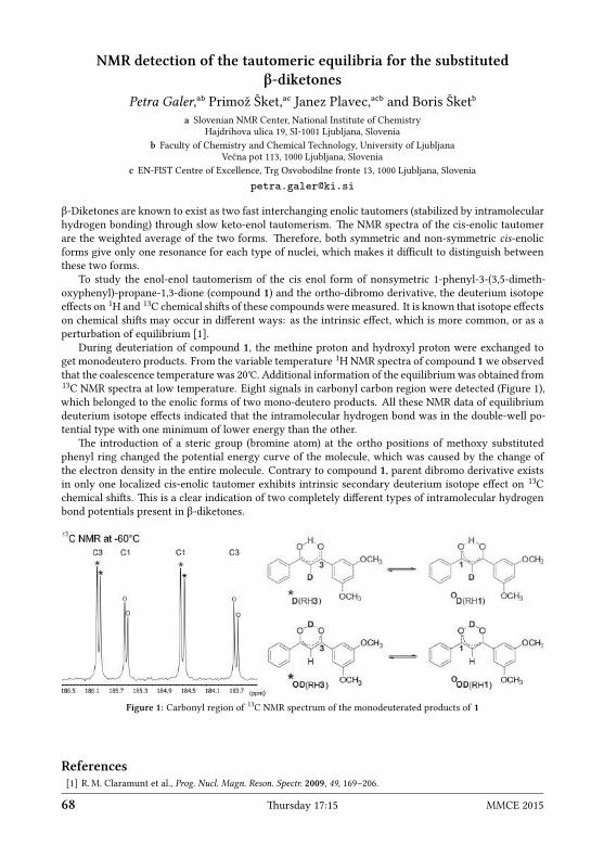
NMR detection of the tautomeric equilibria for the substitutedβ-diketones
Petra Galer,ᵃᵇ Primož Šket,ᵃᶜ Janez Plavec,ᵃᶜᵇ and Boris Šketᵇa Slovenian NMR Center, National Institute of Chemistry
Hajdrihova ulica 19, SI-1001 Ljubljana, Sloveniab Faculty of Chemistry and Chemical Technology, University of Ljubljana
Večna pot 113, 1000 Ljubljana, Sloveniac EN-FIST Centre of Excellence, Trg Osvobodilne fronte 13, 1000 Ljubljana, Slovenia
petra.galer ki.si
β-Diketones are known to exist as two fast interchanging enolic tautomers (stabilized by intramolecularhydrogen bonding) through slow keto-enol tautomerism. e NMR spectra of the cis-enolic tautomerare the weighted average of the two forms. erefore, both symmetric and non-symmetric cis-enolicforms give only one resonance for each type of nuclei, which makes it difficult to distinguish betweenthese two forms.
To study the enol-enol tautomerism of the cis enol form of nonsymetric 1-phenyl-3-(3,5-dimeth-oxyphenyl)-propane-1,3-dione (compound 1) and the ortho-dibromo derivative, the deuterium isotopeeffects on 1H and 13C chemical shis of these compounds were measured. It is known that isotope effectson chemical shis may occur in different ways: as the intrinsic effect, which is more common, or as aperturbation of equilibrium [1].
During deuteriation of compound 1, the methine proton and hydroxyl proton were exchanged toget monodeutero products. From the variable temperature 1HNMR spectra of compound 1we observedthat the coalescence temperature was 20℃. Additional information of the equilibriumwas obtained from13C NMR spectra at low temperature. Eight signals in carbonyl carbon region were detected (Figure 1),which belonged to the enolic forms of two mono-deutero products. All these NMR data of equilibriumdeuterium isotope effects indicated that the intramolecular hydrogen bond was in the double-well po-tential type with one minimum of lower energy than the other.
e introduction of a steric group (bromine atom) at the ortho positions of methoxy substitutedphenyl ring changed the potential energy curve of the molecule, which was caused by the change ofthe electron density in the entire molecule. Contrary to compound 1, parent dibromo derivative existsin only one localized cis-enolic tautomer exhibits intrinsic secondary deuterium isotope effect on 13Cchemical shis. is is a clear indication of two completely different types of intramolecular hydrogenbond potentials present in β-diketones.
Figure 1: Carbonyl region of 13C NMR spectrum of the monodeuterated products of 1
References[1] R.M. Claramunt et al., Prog. Nucl. Magn. Reson. Spectr. 2009, 49, 169–206.
68 ursday 17:15 MMCE 2015

Study of micellization of surfactants by DOSY NMRZuzana Grňová,ᵃ Juraj Filo,ᵇ and Tibor Liptajᵃ
a Faculty of Chemical and Food TechnologySlovak University of Technology
Radlinského 9, 812-37 Bratislava, Slovakiab Faculty of Natural Sciences, Comenius University in Bratislava
Mlynská dolina, 842-15 Bratislava, Slovakiazuzana.grnova stuba.sk
Diffusion-ordered spectroscopy (DOSY) NMR experiments represent an aractive non-invasive methodfor the analysis of mixtures and the study of diffusion properties of molecules in solution. In DOSY NMR,spectral separation of mixture components is based on their different diffusion properties and hence ondifferences in their molecular size, radius, shape and/or intermolecular interactions. DOSY NMR tech-niques provide multi-dimensional maps in which one axis corresponds to the diffusion coefficient andthe others to the chemical shi dimensions [1, 2]. Diffusion separation, as observed in pure solvent solu-tions, is, however, frequently not sufficient. One of the remedies is a modification of the solvent matrixby the addition of an appropriate inert agent, which can specifically interact with the solute moleculesand in this way alter their diffusion properties. ere are various classes of molecules exploited for thispurpose. Among them, an important role belongs to surfactants.
Surfactants have an amphiphilic type of molecule containing hydrophilic and hydrophobic moiety.ey self-associate in aqueous solutions to form aggregates of various types, shapes and sizes. At a cer-tain concentration, called critical micellar concentration (cmc), micelle formation occurs. e size ofsurfactant molecules and thus their self-diffusion behavior change uponmicellization [3]. e process ofmicellization can be monitored by the observation of concentration dependent changes of self-diffusioncoefficients.
In this work we have explored the micellization process of some surfactants, with the aim of theirexploitation for DOSY NMR spectroscopy.
Acknowledgement: e authors wish to thank VEGA 1/0770/15 for support.
References[1] C. Johnson, Prog. Nucl. Magn. Reson. Spectr. 1999, 34, 203–256.[2] B. Antalek, Concepts Magn. Reson. 2002, 14 (4), 225–258.[3] W. Al-Soufi, L. Piñeiro, M. Novo, J. Colloid Interf. Sci. 2012, 370, 102–110.
MMCE 2015 Friday 17:15 69

Processing of multidimensional data using multiple fixing SMFT methodKatarzyna Grudziąż,ᵃ Paweł Dziekański,ᵃᵇ Patrik Jarvoll,ᶜ Wiktor Koźmiński,ᵃ
and Anna Zawadzka-Kazimierczukᵃa Biological and Chemical Research CentreFaculty of Chemistry, University of WarsawŻwirki i Wigury 101, 02-089 Warsaw, Polandb Faculty of Physics, University of Warsaw
Pasteura 5, 02-093 Warsaw, Polandc Agilent Technologies, 10 Mead Road, OX5 1QU Yarnton, United Kingdom
anzaw chem.uw.edu.pl
e sparse multidimensional Fourier transform processing algorithm (SMFT) [1] allows one to obtain aset of two-dimensional cross-sections from a non-uniformly sampled multidimensional experiment. Itis possible by fixing frequencies of two (in case of 4D experiment) or three (in case of 5D experiment)types of nuclei. Fixed frequencies are collected from the spectrum (the “basis spectrum”) that containsat least part of the frequencies measured in the high-dimensional experiment.
Herewe present howfixing different frequencies from the basis spectrum, 4D (HACA)CON(COCA)NCO,enabled us to easily access information frommultidimensional experiments: 4D (HACA)CON(COCA)NCO,5D HNCO(CA)NCO and 5D HabCabCO(CA)NCO.is information was then used to find the sequentialconnectivities and assign peaks.
In addition, we compared the usefulness of various subsets of data obtained by multiple fixing of 4D(HACA)CON(COCA)NCO, 5D HNCO(CA)NCO and 5D HabCabCO(CA)NCO experiments in automaticchemical shis assignment for alfa-synuclein, a resolution-demanding, intrinsically disordered protein140 amino acids long, using the TSAR program [2].
References[1] K. Kazimierczuk, A. Zawadzka, W. Kozminski, J. Magn. Reson. 2009, 197, 219–228.[2] A. Zawadzka-Kazimierczuk, W. Kozminski, M. Billeter, J. Biomol. NMR 2012, 54, 81–95.
70 ursday 17:15 MMCE 2015

Spiro cycles from acridin-9-ylmethylamine: a NMR studyMária Vilková, Marianna Prokaiová, and Ján Imrich
NMR Laboratory, Institute of Chemistry, Faculty of SciencePavol Jozef Šafárik University, Moyzesova 11, 04154 Košice, Slovakia
jan.imrich upjs.sk
A novel type of spontaneous cyclization of 1-alkyl/aryl-3-(acridin-9-ylmethyl)thioureas obtained fromthe title amine and alkyl/aryl isothiocyanates led to 1-alkyl/aryl-spiro[dihydroacridine-9’(10’H),5-imi-dazolidine]-2-thiones further transformed with mesitylnitrile oxide to spiro[dihydroacridine-9’(10’H),5-imidazolidine]-2-ones that partly reopened to urea isomers. Forty target compounds in three series wereprepared to study their structure, mechanism of formation, and substituent effects by 1H, 13C, and 15NNMR spectroscopy. Whilst all thioureas except bis-acridine ones cyclized at the acridine C-9 carbonvia R2−N nitrogen, bis-acridine precursors (R2 = 9-Acr) cyclized via methylene carbon to give invertedspiro cycles. Transfer of substituent effects of para substituents on R2 to the spiro ring was differentbetween the thione and one derivatives, as it was demonstrated by induced shis of the correspondingsignals in 13C and 15N NMR spectra.
N
NH
R
R1
N
NHNR
S
R2
R1
NH
N
N
R
R2
S
R1
R = H, Pr, Bn R1 = H, CH3
R2=CH3, allyl, Ph, 4-CH3O-Ph, 4-Br-Ph, 4-F-Ph, 4-NO2-Ph, 9-Acr
NH
N
N
O
R2
R
R1
Alkyl/aryl-ITC
chloroform, rt, 6 h
N
NHNR
S N
NH
NH
N
NR
O
H1́
4́
8´
5́
1́ ´´
8´´´4
NH
NH
N
NR
S
H
rt, 0-30 min acetonitrilert, 5h
MNO
acetonitrilert, 5h
MNO
Figure 1: Reaction scheme
Acknowledgements: Grant VEGA 1/0672/11. e Slovak State Programme for Support of Research Infrastructure: NMR.
MMCE 2015 Friday 17:15 71

NMR aracterisation of 1,5-bis(salicylidene)carbohydrazide in solutionand solid state
Tomislav Jednačak,ᵃ Predrag Novak,ᵃ Janez Plavec,ᵇ Primož Šket,ᵇ Mirta Rubčić,ᵃand Nives Galićᵃ
a Department of Chemistry, Faculty of Science, University of ZagrebHorvatovac 102a, HR-10000 Zagreb, Croatia
b Slovenian NMR Center, National Institute of ChemistryHajdrihova ulica 19, SI-1001 Ljubljana, Slovenia
tjednacak chem.pmf.hr
Carbohydrazide derived Schiff bases are higher urea homologues with interesting biological properties,which together with their reactivity, led to a number of laboratory and industrial applications. esecompounds have been widely used as selective analytical reagents for ion binding [1], as precursors inthe syntheses of nitrogen containing heterocycles [2] and as multitopic ligands for the targeted construc-tion of coordination systems with biological activity [3]. ey are usually prepared by the condensationreaction between carbohydrazide and appropriate carbonyl compound.
e structure of 1,5-bis(salicylidene)carbohydrazide 1 has been characterised by NMR spectroscopyin solution and solid state. As shown in Figure 1, this molecule might exist in several tautomeric formsand has a few reactive groups able to participate in both intra- and intermolecular hydrogen bonds. Ithas been demonstrated that the tautomer 1a is the dominant form in solution and in all solid phases.Significant deshielding and line-broadening of the signals corresponding to NH and OH protons haveclearly indicated that both groups formed hydrogen bonds. Furthermore, the analysis of solid-state NMRspectra allowed the identification of five crystallographic forms of 1: three polymorphs (I, II and III)and two solvates (IV and V) [4]. e obtained results can further be exploited for beer understandingthe relationship between the structure, physico-chemical properties and bioactivity of carbohydrazidederivatives.
Figure 1: Tautomeric forms of 1
References[1] K. Užarević et al., Angew. Chem. Int. Ed. 2008, 47, 7022–7025.[2] A.H. Corvin, J. D. Reinheimer, J. Am. Chem. Soc. 1951, 73 (3), 1184–1186.[3] R. N. Patel, Inorg. Chim. Acta 2010, 363, 3838–3846.[4] M. Rubčić et al., Crystal Growth & Design 2014, 14, 2900–2912.
72 ursday 17:15 MMCE 2015

NMR spectroscopic studies of de/protonation meanisms inthiosemicarbazide and azobenzene based anion emosensors
Damjan Makuc,ᵃᵇ Kristina N. Farrugia,ᶜ Maria Cardona,ᶜ David C. Magri,ᶜand Janez Plavecᵃᵇ
a Slovenian NMR Center, National Institute of ChemistryHajdrihova ulica 19, SI-1001 Ljubljana, Slovenia
b EN-FIST Centre of Excellence, Trg Osvobodilne fronte 13, 1000 Ljubljana, Sloveniac Department of Chemistry, Faculty of Science, University of Malta
damjan.makuc ki.si
Four thiosemicarbazide anion chemosensors containing three N−H groups, substituted with phenyland/or 4-nitrophenyl units, were synthesized and studied for their anion binding abilities with OH–, F–,CH3COO–, H2PO4
– and Cl– anions. Noteworthy changes were observed in the UV-visible absorptionspectra upon addition of all anions, except for chloride, accompanied by dramatic color changes visible tothe naked eye. ese changes were determined to be due to the deprotonation of the central N−Hprotonand not due to hydrogen bonding, based on 1H-15NNMR titration studies with acetate in DMSO-d6/0.5%water. Deprotonation was confirmed by the disappearance of NMR signal for the central thiourea H2proton and the formation of acetic acid. In addition, long-range 1H-15N NMR experiments allowedunambiguous assignment of 15N chemical shis, where aromatic N1 atom was strongly deshielded fromδ 106 to 158 ppm. erefore, the thiosemicarbazide anion chemosensors operate by a deprotonationmechanism of the central N−H proton rather than by hydrogen bonding as is oen reported [1].
Figure 1
ree azobenzene based pH indicators with amino[bis(ethanesulfonate)] substituents were synthe-sized and preliminary studied by UV-visible absorption spectroscopy in aqueous solution. e azoben-zene indicators exhibit brilliant and distinct color changes with transitions between pH 1 and 4. eUV-visible spectra suggest that the protonation site is a tautomeric equilibrium between protonation atthe azo nitrogen and protonation at the amino nitrogen. NMR spectroscopic studies, which include 1Hand 15N NMR titrations, are in progress to provide unambiguous proof of the protonation site.
Figure 2
References[1] K. N. Farrugia et al., Org. Biomol. Chem. 2015, 13, 1662–1672.
MMCE 2015 Friday 17:15 73

Faster and cleaner real time pure shi NMR experimentsJohannes Mauhart,ᵃ Simon Glanzer,ᵃ Peyman Sakhaii,ᵇ Wolfgang Bermel,ᶜ
and Klaus Zanggerᵃa Institute of Chemistry, University of GrazHeinrichstraße 28, A-8010 Graz, Austria
b NMR Laboratory, C&BD Frankfurt Chemistry, Sanofi AventisIndustriepark Hoechst, Building D770, Labor 204, D-65926 Frankfurt am Main, Germany
c Bruker BioSpin GmbH, Silberstreifen, D-76287 Rheinsteen, Germanyjohannes.mauhart edu.uni-graz.at
1H homonuclear broadband decoupling has seen a lot of aention in the recent years. Pure shi protonNMR experiments, obtained by homonuclear broadband decoupling, significantly enhance the resolutionof scalar coupled signals. e rediscovery of older methods towards pure shi spectra, like slice-selectivedecoupling [1] and the BIRD experiments [2] and their further development [3–6] have established themas useful tools in modern liquid NMR specroscopy. However, improved resolution is traded againstsignificantly reduced sensitivity and introduction of artifacts.
Here, improvements to the real time slice-selective decoupling method are presented. e experi-ment now includes the suppression of artifacts originating from scalar coupling evolution during theacquisition of data chunks. e frequencies of these artifacts depends on the length of the recorded datachunks, therefore suppression is achieved by cycling the data chunk lengths over a defined number ofscans. is leads to the collapse of the sharp artifact signals to very broad signals with significantlylower intensity.
e second improvement is the use of frequency shied pulses. By concertedly shiing the offsetsof the employed selective pulses, the need to wait for relaxation of slowly relaxing protons is reduced.is applies to molecules as well as to compound mixtures featuring protons with a broad range of T1relaxation times.
Figure 1: Real time slice-selectively decoupled 1H spectra of 10% n-propanol in DMSO-d6 with (boom) and without(top) the use of artifact suppression. e arrows in the enlarged areas indicate the 13C satellites.
References[1] K. Zangger, H. Sterk, J. Magn. Reson. 1997, 124, 486–489.[2] J. R. Garbow, D. P. Weitekamp, A. Pines, Chem. Phys. Le. 1982, 93 (5), 504–509.[3] N.H. Meyer, K. Zangger, Angew. Chem. Int. Ed. 2013, 52, 7143–7146.[4] P. Sakhaii et al., J. Magn. Reson. 2013, 233, 92–95.[5] J. A. Aguilar et al., Angew. Chem. Int. Ed. 2010, 49 (23), 3901–3903.[6] J. A. Aguilar, M. Nilsson, G. A. Morris, Angew. Chem. Int. Ed. 2011, 50, 9716–9717.
74 ursday 17:15 MMCE 2015

e solution structure of the MANEC-type domain from hepatocytegrowth factor inhibitor 1 reveals an unexpected PAN/apple domain-type
foldMichał Nowakowski,ᵃ Zebin Hong,ᵇ Chris Spronk,ᶜ Steen V Petersen,ᵈ
Jan S. Pedersen,ᵉ Wiktor Koźmiński,ᶠ Frans A.A. Mulder,ᵉ and Jan K. Jensenᵇa Centre of New Technologies, University of Warsaw
Banacha 2C, 02-097 Warsaw, Polandb Department of Molecular Biology and Genetics
Danish-Chinese Centre for Proteases and Cancer, Aarhus University, Denmarkc Spronk NMR, Lithuania
d Department of Biomedicine, Aarhus University, Denmarke Interdisciplinary Nanoscience Center (iNANO), Aarhus University, Denmark
f Biological and Chemical Research CentreFaculty of Chemistry, University of WarsawŻwirki i Wigury 101, 02-089 Warsaw, Poland
lyam chem.uw.edu.pl
A decade ago, the motif at N-terminus with eight cysteines, or in short MANEC, was defined as a newprotein domain family. ese domains were found exclusively in the N-terminus of > 400 multi-domainmembrane proteins from multicellular animals. Despite the large number of MANEC-containing pro-teins, only one has been characterized: hepatocyte growth factor activator inhibitor-1 (HAI-1). HAI-1 isan essential protein shown to regulate the activity of matriptase, hepsin and hepatocyte growth factoractivator, all serine proteases with crucial roles in epithelial development, cell growth and homeostasis.Misregulation of these systems has been implicated in severe pathological conditions such as skin dis-eases and cancer. Detailed functional understanding of HAI-1 and other MANEC-containing proteins ishampered by a lack of any structural information on MANEC.
Here we present an NMR solution structure and biophysical characterization of the MANEC domainfrom HAI-1, as the first structure of a representative MANEC domain. Although no homologies werepredicted based on sequence, the MANEC structure revealed it as a new subclass of the PAN/appledomain family. Intriguingly, in silico protein folding resulted in successful structure-based homologyprediction, where sequence-based approaches fail. e MANEC structure represents a much neededtool for elucidation of function of MANEC-containing proteins as indicated by the homology to thePAN/apple domains as mediators of protein-protein and protein-glycan interactions.
Authors wish to thank the Foundation for Polish Science for the support with TEAM programme “Towards new applicationsof NMR spectroscopy in chemical and biomolecular structural studies” (M.N., W.K.), NMRmeasurements were carried out atthe Biological and Chemical Research Centre, University of Warsaw, established within the project co-financed by EuropeanUnion from the European Regional Development Fund under the Operational Programme Innovative Economy, 2007–2013.
MMCE 2015 Friday 17:15 75

Magnetic resonance access to transiently formed protein complexTomáš Sára
Max F. Perutz Laboratories, University of ViennaCampus Vienna Biocenter 5, A-1030 Vienna, Austria
tomas.sara univie.ac.at
Biomolecule interactions are important to understanding of biological phenomena since non-covalentreversible binding between proteins and other biomolecules leads to the formation of complex regulatorymolecular systems. ese interactions, however, can exist over a wide range of dissociation constantsand time scales, illustrating interactions in tight complexes, as well as weak transiently formed proteincomplexes.
CspA is one of the cold-shock proteins naturally occurring in E. coli. e CspA protein has beenshown to bind to the 5’ end UTR of its ownmRNA, called the cold box (CB), and destabilize its secondarystructure acting as a chaperone. e interaction of the CspA with RNA is a transient and sparsely popu-lated binding event that can be studied by the means of CPMG relaxation dispersion NMR experiments.Chemical shi changes upon binding of the cold box and its complementary strand anti cold box RNAwere observed finding out that mostly surface-exposed hydrophobic residues are involved in the bindingevent, suggesting that the protein-RNA interaction is facilitated through hydrophobic interactions.
76 ursday 17:15 MMCE 2015

Sparsity-constrained NUS reconstruction in NMR: possible pitfallsAlexandra Shchukina, Krzysztof Kazimierczuk, and Paweł Kasprzak
Centre of New Technologies, University of WarsawBanacha 2C, 02-097 Warsaw, Poland
aleksandra.shchukina gmail.com
Non-uniform sampling (NUS) has beenwidely applied to accelerate multidimensional NMR experiments.A group of signal processing methods reffered to as compressed sensing (CS) has been proved especiallyeffective in the reconstruction of NMR spectra from NUS data. All the CS methods are based on theassumption that the recovered spectrum is sparse, i.e. that it contains a relatively small number ofsignificant components. e search for the sparsest spectrum x that fits to the measured experimentaltime-domain data y requires to set the balance between sparseness of x (quantitavely expressed by f(x))and its accordance with the undersampled FID signal y. is is realised by minimising the function
minx
(∥Fx − y∥+ λf(x)) ,
where F stands for inverse Fourier transform, and λ sets the balance between the sparsity of the spec-trum and the agreement with the measurement.
In this study, we discuss the effects of misseing the reconstruction parameters (e.g. λ, stopping cite-ria) in various CSmethods: orthogonal matching pursuit [1], known in NMR under the name CLEAN [2],Lorentzian peak matching pursuit [3], Iterative So resholding [4, 5], iteratively reweighted leastsquares [5] and Low-Rank reconstruction [6].
References[1] T. Zhang, IEEE Transactions on Information eory 2011, 57, 6215–6221.[2] B. E. Coggins, P. Zhou, J. Biomol. NMR 2008, 42, 225–239.[3] K. Kazimierczuk, P. Kasprzak, Sensors 2014, 15, 234–247.[4] A. Papoulis, IEEE Transactions on Circuits and Systems 1975, 22, 735–742.[5] K. Kazimierczuk, V. Y. Orekhov, Angew. Chem. Int. Ed. 2011, 50 (24), 5556–5559.[6] X. et al., Angew. Chem. Int. Ed. 2015, 54 (3), 852–854.
MMCE 2015 Friday 17:15 77

G-quadruplexes: formation of long-lived intermediatesPrimož Šket,ᵃᵇ Slavko Čeru,ᵇ Iztok Prislan,ᶜ Jurij Lah,ᶜ and Janez Plavecᵃᵇᶜ
a Slovenian NMR Center, National Institute of ChemistryHajdrihova ulica 19, SI-1001 Ljubljana, Slovenia
b EN-FIST Centre of Excellence, Trg Osvobodilne fronte 13, 1000 Ljubljana, Sloveniac Faculty of Chemistry and Chemical Technology, University of Ljubljana
Večna pot 113, 1000 Ljubljana, Sloveniaprimoz.sket ki.si
G-quadruplexes are higher order secondary structures formed by guanine-rich DNA sequences that canbe found in biologically significant regions of the genome such as telomeres, immunoglobulin switchregions and promoter regions of eukaryotic cells. ey have also been associated with human diseases,as therapeutic targets in drug design and in applications as nanomolecular devices. e presence ofcations such as K+ or Na+ seems to be essential for the formation of G-quadruplexes, due to their rolein reducing repulsions amongst guanine carbonyl oxygen atoms within G-quartets and additionallyenhancing base-stacking interactions.
With the use of solution-state NMR spectroscopy and other experimental techniques (PAGE, TDS,UV, CD, DSC) we have studied the behaviour of guanine-rich sequences and explored their features in anenvironment almost completely free of G-quadruplex promoting cations. Experimental data has shownthe formation of a new structure, which can be considered as an intermediate on the way to foldinginto G-quadruplexes. It is interesting to note that the guanine bases are not held together by Hoogsteenhydrogen bonds like in G-quartets but rather by alternative base pairing.
Our study, where G-rich intermediates were characterized in detail and their kinetic roles deter-mined, provides an important step in elucidating general principles by which G-quadruplexes adopttheir native folds [1]. G-rich DNA sequences can in the presence of certain cations fold very rapidly andnot by chance in to a large number of structurally diverse G-quadruplex structures with mechanisms ofvarying complexity through the population of different intermediates, which are generally unstable andhard to detect.
+K+K
+K
+K
G1
G1
G12
G12
G1
G1
G12
G12
Figure 1: Oxy-1.5 G-quadruplex folding step involving i-Oxy-1.5 intermediate. Guanine residues at the 5’ and 3’ endsin both species are marked.
References[1] S. Ceru et al., Angew. Chem. Int. Ed. 2014, 53, 4881–4884.
78 ursday 17:15 MMCE 2015

NMR detection of tautomeric equilibria for substituted β-diketonesUrška Slapšak,ᵃᵇᶜ Gregor Ilc,ᵃᵇ Sara Drmota,ᶜ Miha Pavšič,ᶜ Brigita Lenarčič,ᶜ
Kristina Djinović-Carugo,ᶜ and Janez Plavecᵃᵇᶜa Slovenian NMR Center, National Institute of Chemistry
Hajdrihova ulica 19, SI-1001 Ljubljana, Sloveniab EN-FIST Centre of Excellence, Trg Osvobodilne fronte 13, 1000 Ljubljana, Slovenia
c Faculty of Chemistry and Chemical Technology, University of LjubljanaVečna pot 113, 1000 Ljubljana, Slovenia
urska.slapsak ki.si
Cytoskeletal actin-binding protein α-actinin 1 is a member of the spectrin superfamily with the roleto cross-link the actin filaments. It is a calcium sensitive non-muscle cytoskeletal isoform of α-actinin.e protein is functional as an antiparallel dimer, which is composed of an N-terminal actin-bindingdomain, connected through a flexible neck peptide to four spectrin repeats forming the central rod thatis followed by a C-terminal calmodulin-like (CaM) domain [1]. e CaM-like domain of α-actinin 1involves four EF-hand motifs that could bind up to four calcium (Ca2+) ions. It is believed that calciumbinding triggers a major conformational change of the CaM-like domain of α-actinin 1 from a closedinto an open state. In this way, α-actinin 1 is able to interact with different target partners dependingon its calcium binding state.
Triple resonance (1H, 13C and 15N) NMR experiments on sample containing 1 mM uniformly labelledCaM-like domain of α-actinin 1 were performed on an 800 MHz NMR spectrometer.
CaM-like domain of the α-actinin 1 is composed of two lobes, one in N- and other in C-terminal. N-terminal lobe consists of two EF-hand motives connected with linker. EF-loops, which connect internaland external helices of each EF-hand motives are close in space and connected by a short β-sheet. C-terminal lobe consists of four helices, three of them are shorter in comparison with helices in N-terminallobe and do not form EF-hand motives. In apo form, no single preferred orientation of N- and C-terminallobes was observed. CaM-like domain is stabilized aer titration with Ca2+ ions. We suggested thatcalcium binding triggered the stabilization of structure trough limitation of the rotation of the N- andC-terminal lobes around the linker between them.
Titration of protein sample with Ca2+ ions followed with 15N-HSQC spectra proposed one bindingsite for Ca2+ ions in the first EF-handmotif in the N-terminal lobe of CaM-like domain. Aer comparisonof the apo and holo structures, for individual lobes no major structural rearrangements were observedas consequence the calcium binding. In the holo form internal and external helices of first and secondEF hand motives in N-terminal lobe are slightly opened compared to the apo form, but not as much as isknown for EF-hand proteins with similar function, such as calmodulin [2]. e most significant changesbetween both forms were observed in EF-loops of both EF-hand motives and in linker connecting themin N-terminal lobe. Our NMR studies were complemented by isothermal titration calorimetry, massspectroscopy and SAXS.
References[1] B. Sjöblom, A. Salmazo, K. Djinović-Carugo, Cell. Mol. Life Sci. 2008, 65 (17), 2688–2701.[2] J. L. Gifford, M. P. Walsh, H. J. Vogel, Biochem. J. 2007, 405 (2), 199–221.
MMCE 2015 Friday 17:15 79

Residual dipolar coupling assisted NMR and DFT analysis of an exoticproduct of Povarov reaction
Michal Šoral,ᵃ Jana Doháňošová,ᵃ Jozef Markus,ᵇ Stanislava Šoralová,ᶜDušan Berkeš,ᵇ and Tibor Liptajᵃ
a Department of NMR Spectroscopy and Mass SpectrometryFaculty of Chemical and Food Technology
Slovak University of Technology, Radlinského 9, SK-81237 Bratislava, Slovakiab Department of Organic Chemistry, Faculty of Chemical and Food TechnologySlovak University of Technology, Radlinského 9, SK-81237 Bratislava, Slovakia
c Department of Pharmaceutical Chemistry, Faculty of PharmacyComenius University in Bratislava, Odbojárov 10, SK-83232 Bratislava, Slovakia
michal.soral stuba.sk
During a study of the mechanism and the synthetic possibilities of the Povarov reaction [1, 2], a cycliza-tion of (2’R,3S)-1’-benzyl-2’-(prop-2-en-1-yl)spiro[indole-3,3’-pyrrolidine]-5’-one (1) was performed.e reaction was aempted with various Lewis and Brønsted acids as catalysts, with ethylaluminiumdichloride giving the best yield.
Due to the improbable exotic alkaloid-type structure of the resulting molecule 2, a detailed struc-tural analysis was called for. For this reason, a set of 2D NMR spectra including INADEQUATE wasmeasured in order to prove the atom linkages. e bond configurations of 2 were predicted from thesynthesis mechanism and confirmed unambiguously using NOESY, DFT geometry calculations, and ulti-mately RDC assisted NMR analysis in stretched crosslinked polystyrene gels [3]. e reversibility of thecyclization, which is typical for Diels-Alder type reactions, was observed, thus complicating the RDCanalysis because of long swelling times of the polymer gels.
N
N
O
EtAlCl2, 24 h, r.t., DCM, 79%
CDCl3, 10d, r.t., 20%(NMR)
Ph
1 2N
N
O
H
Ph
Figure 1: Synthesis scheme of 2
Acknowledgement: e authors wish to thank VEGA 1/0770/15 for support.
References[1] L. S. Povarov, Russian Chem. Rev. 1967, 36 (9), 656–670.[2] J. Hajicek, J. Trojanek, Tetrahedron Le. 1981, 22 (19), 1823–1826.[3] B. Luy, K. Kobzar, H. Kessler, Angew. Chem. Int. Ed. 2004, 43 (9), 1092–1094.
80 ursday 17:15 MMCE 2015

Experimental determination of structural parameters in selectedpolycyclic aromatic compounds
Martin Dračínský,ᵃ Ivana Císařová,ᵇ Jan Storch,ᶜ and Jan Sykoraᶜa Institute of Organic Chemistry and Biochemistry
Academy of Sciences of the Czech RepublicFlemingovo nám. 2, 166 10 Prague 6, Czech Republicb Faculty of Science, Charles University in PragueOvocný trh 5, 116 36 Prague 1, Czech Republicc Institute of Chemical Process FundamentalsAcademy of Sciences of the Czech Republic
Rozvojová 2/135, 165 02 Prague 6, Czech Republicsykora icpf.cas.cz
e unusual optical and electronic properties of polycyclic aromatic compounds have origin namelyin the conjugated π-π aromatic system [1]. ese unique systems are mostly studied by theoreticalapproaches. Here we would like to present interconnection between the experimentally accessible dataand the structural parameters in helicene series of aromatic compounds. 1H and 13C NMR chemicalshis and their temperature dependence can be correlated with the interplanar distance of peripheralhelicene rings and reflect the dynamic behaviour of the molecule.
e indirect spin-spin 13C-13C coupling constants represent unique probe to the electron distributionbetween involved atoms and later reflect the carbon-caron bonding environment [2]. e experimentalvalues of 1J can be correlated to the carbon-carbon bond lenghts and angles, that are accessible by singlecrystal X-ray crystallography. e experimental values of 3J (C−C) correlates reasonably well with thepublished theoretical study of helicene aromaticity [3]. e theoretical prediction of NMR parameterswas done using DFT calculation (B3PW91/6-311++G(d,p)) which provides an excellent agreement withthe experimental values.
Figure 1: Helicene series of aromatic compounds
Acknowledgements: For the financial support, the Czech Science Foundation (Project No. 15-12719S) is gratefully acknowl-edged.
References[1] R. Harvey, Polycyclic Aromatic Hydrocarbons, John Wiley, New York, 1997.[2] K. Kamieńska-Trela et al., J. Phys. Org. Chem. 2012, DOI 10.1002/poc.2955.[3] Z. Portella et al., J. Org. Chem. 2005, 70, 2509.
MMCE 2015 Friday 17:15 81
Electron paramagnetic resonance study on co-phthalocyanine ionicderivative spontaneously adsorbed on highly ordered pyrolitic graphite
(HOPG)Ján Tarábek,ᵃ Monika Klusáčková,ᵇ Pavel Janda,ᵇ Hana Tarábková,ᵇ
Lubomír Rulíšek,ᵃ and Jan Plšekᵇa Institute of Organic Chemistry and Biochemistry
Academy of Sciences of the Czech RepublicFlemingovo nám. 2, 166 10 Prague 6, Czech Republicb Jaroslav Heyrovský Institute of Physical Chemistry
Academy of Sciences of the Czech RepublicDolejškova 3, 182 23 Prague 8, Czech Republic
tarabek uochb.cas.cz
Electron paramagnetic resonance (EPR) belongs to the family of magnetic resonancies and focuses onmaterials and compounds with unpaired electron. EPR is very well known and established amongother spectroscopic techniques, just as old as a nuclear magnetic resonance (NMR). It is also suitablefor surface analysis, bacause it can provide direct qualitative as well as quantitative evidence of theunpaired electron (spin) distribution on a surface, which cannot be acquired by common surface meth-ods (e.g. XPS or SPM). Electron paramagnetic resonance is frequently applied in the field of transitionmetal oxide surfaces [1, 2]. However, it has been rarely applied in the field of adsorption onto conduct-ing/semiconducting substrates.
We found that the adsorption of a 4-fold positively charged ionic phthalocyanine: [N,N’,N”,N”’-tetra-methyltetra-3,4-pyridinoporphyrazino-Co(II)], directly from its corresponding solution, can be followedby in situ EPR spectroscopy in the EPR cell. Our combined spectroscopic and quantum chemical com-putational (by density functional theory, DFT) analysis show that the above-mentioned phthalocyaninediffers in geometry, depending on either being in powder or adsorbed on HOPG. Additionally, it wasfound that the spin state of HOPG conducting electrons does not change upon adsorption [3]. Finally,by such an spontaneous adsorption process from corresponding phthalocyanine solution, one can gethomogeneous, magnetically and redox active phthalocyanine molecular film, which may find an appli-acation in molecular electronis (sensorics) or as a magnetic resonance imaging (MRI) contrast agent.
References[1] K. Dyrek, M. Che, Chem. Rev. 1997, 97 (1), 305–332.[2] S. V. Doorslaer et al., Coord. Chem. Rev. 2009, 253 (15-16), 2116–2130.[3] J. Tarábek et al., J. Phys. Chem. C 2014, 118 (8), 4198–4206.
82 ursday 17:15 MMCE 2015
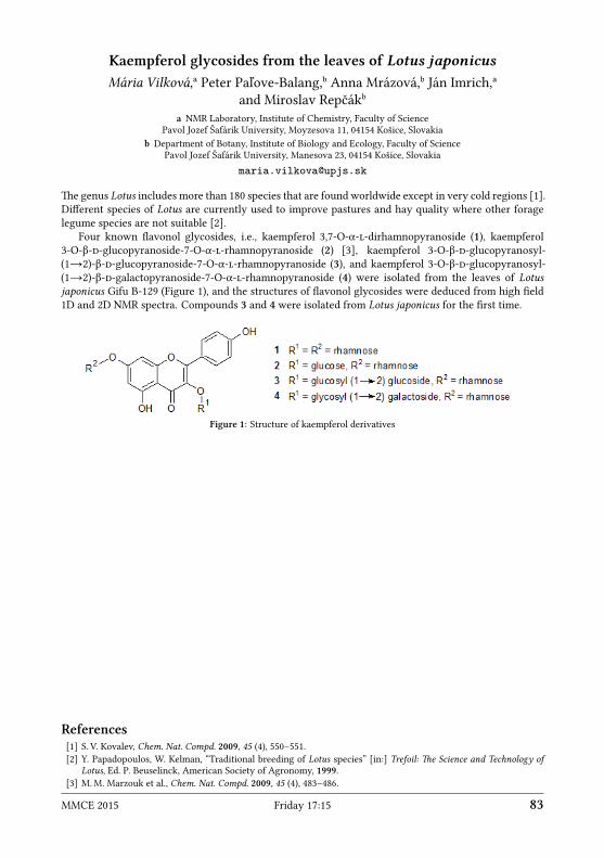
Kaempferol glycosides from the leaves of Lotus japonicusMária Vilková,ᵃ Peter Paľove-Balang,ᵇ Anna Mrázová,ᵇ Ján Imrich,ᵃ
and Miroslav Repčákᵇa NMR Laboratory, Institute of Chemistry, Faculty of Science
Pavol Jozef Šafárik University, Moyzesova 11, 04154 Košice, Slovakiab Department of Botany, Institute of Biology and Ecology, Faculty of Science
Pavol Jozef Šafárik University, Manesova 23, 04154 Košice, Slovakiamaria.vilkova upjs.sk
e genus Lotus includes more than 180 species that are found worldwide except in very cold regions [1].Different species of Lotus are currently used to improve pastures and hay quality where other foragelegume species are not suitable [2].
Four known flavonol glycosides, i.e., kaempferol 3,7-O-α--dirhamnopyranoside (1), kaempferol3-O-β--glucopyranoside-7-O-α--rhamnopyranoside (2) [3], kaempferol 3-O-β--glucopyranosyl-(1→2)-β--glucopyranoside-7-O-α--rhamnopyranoside (3), and kaempferol 3-O-β--glucopyranosyl-(1→2)-β--galactopyranoside-7-O-α--rhamnopyranoside (4) were isolated from the leaves of Lotusjaponicus Gifu B-129 (Figure 1), and the structures of flavonol glycosides were deduced from high field1D and 2D NMR spectra. Compounds 3 and 4 were isolated from Lotus japonicus for the first time.
Figure 1: Structure of kaempferol derivatives
References[1] S. V. Kovalev, Chem. Nat. Compd. 2009, 45 (4), 550–551.[2] Y. Papadopoulos, W. Kelman, “Traditional breeding of Lotus species” [in:] Trefoil: e Science and Technology of
Lotus, Ed. P. Beuselinck, American Society of Agronomy, 1999.[3] M.M. Marzouk et al., Chem. Nat. Compd. 2009, 45 (4), 483–486.
MMCE 2015 Friday 17:15 83

Solid state 13C NMR studies of modified poly(3-hydroxybutyrate)Peter Vrábel, Anton Baran, Viktor Hronský, Mária Kovaľaková, and Dušan Olčák
Department of Physics, Faculty of Elecrical Engineering and InformaticsTechnical University of Košice, Park Komenského 2, 04200 Košice, Slovakia
peter.vrabel.3 tuke.sk
e substitution of conventional polymeric materials by biodegradable materials is one of the mostaractive recent trends in material science. PHB (polyhydroxybutyrate) belongs to biologically syn-thesized polymers. It is non-toxic, biocompatible, biodegradable, and thermoplastic material. PHB issemicrystalline [1] polymer and its mechanical properties are similar to those of isotactic polypropy-lene, except for elongation at break leading to low toughness. Moreover, under ambient conditions itsproperties change significantly due to physical aging [2]. On the other hand, appropriate modificationsof PHB provide material with unique mechanical properties.
Properties of polymeric materials are determined by their structure and interactions at molecular aswell as supramolecular grade, and molecular dynamics which is closely related to the structure. iswork studies the influence of thermal treatment and plasticization on the structure and molecular dy-namics of PHB by means of nuclear magnetic resonance (NMR) spectroscopy, which is a unique toolfor the study of the structure and molecular dynamics of polymeric materials. For this purpose, the sin-gle pulse MAS 13C NMR spectra at ambient temperature and at 370 K, and spin-laice relaxation timesT1(13C) were measured for three PHB samples: virgin (PHB-v), quenched (PHB-Q) and plasticized withtriacetine (PHB-QT).
e solid-state 13C NMR spectra measured for all studied samples at both temperatures display car-bonyl (CO), methine (CH), methylene (CH2) andmethyl (CH3) resonances with narrow lines arising fromcrystalline domains superimposed on broad lines from amorphous domains. e spin-laice relaxationtime T1(13C) determined from measurements using the Torchia technique [3] showed bi-exponentialdecay for each resonance and besides that it was found out, that the used delay time of 120 s was longenough only for already relaxed CH3 group. Both used measurement techniques imply that all studiedPHB samples have semi-crystalline nature. e changes of the structure and molecular motion in PHBwere further analyzed by deconvolution of the methyl carbon resonance.
ermal treatment and plasticization slightly influence the crystallinity of PHB at ambient tempera-ture. Triacetine in PHB caused significant narrowing of the linewidth of the resonance line associatedwith amorphous domains at ambient temperature, which indicates the narrowing of the conformationdistribution. e same effect was observed for all samples at the temperature of 370 K, but this timeit could be related to the change of mobility in amorphous domains. e most significant change incrystallinity forced by temperature was found in PHB and PHB-Q.
Acknowledgement: is research has been supported by the Scientific Grant Agency (VEGA) of the Ministry of Educationof the Slovak Republic and the Slovak Academy of Sciences through the project No. 1/0492/13.
References[1] A. Hoffmann, S. Kreuzberger, G. Hinrichsen, Polym. Bull. 1994, 33, 355.[2] M. L. Di Lorenzo, Eur. Polym. J. 2013, 49, 510.[3] D. A. Torchia, J. Magn. Reson. 1978, 30, 613.
84 ursday 17:15 MMCE 2015

Application of alignment media in structural analysis of calix[4]arenederivatives
Lukas Vrzal,ᵃ Karolina Flidrová,ᵇ Jan Holub,ᵇ Pavel Lhotak,ᵇ and Hana Dvorakovaᵃa Laboratory of NMR Spectroscopy, University of Chemistry and Technology, Prague
Technicka 5, 166 28 Prague 6, Czech Republicb Deptartment of Organic Chemistry, University of Chemistry and Technology, Prague
Technicka 5, 166 28 Prague 6, Czech Republiclukas.vrzal vscht.cz
e aim of this work is the application of residual dipolar coupling constants (RDCs) [1, 2] measurementmethod in conformational behavior study of new inherently chiral calix[4]arene derivatives. RDC is ananisotropic direct through space dipole-dipole interaction, which is not observable in isotropic solutiondue to time averaging caused by fast molecular reorientations. is interaction could be reintroducedby partial alignment of the compound in external magnetic field and is manifested as the contributionto a scalar coupling constant. e magnitude of this interaction is dependent on the distance of couplednuclei and includes information about their spatial arrangement.
e study was started with the testing of the series of recently published liquid crystalline alignmentmedia based on polyglutamates and polyacetylenes on the model dipropoxycalix[4]arene with respect tothe ease of the preparation and the degree of the introduced alignment. Suitable alignment media wereused to determine or specify the spatial structure of three types of new inherently chiral calix[4]arenederivatives, whose structures could not be determined by standard NMR methods (NOE, J-interactions).
Using the method of RDC measurement we established that the phenoxanthiin derivative[3] oc-curs in 1,2-alternate conformation (Fig. 1-A) while dinitroso-derivative adopts pinched cone conforma-tion with nitroso groups on declined (flaened) aromatic rings (Fig. 1-B). In case of 2-pyridylsulfoxidecalix[4]arene with intramolecularly bridged meta positions[4] we succeeded to specify the X-RAY struc-ture (Fig. 1-C).
Figure 1: Structure of three types of new inherently chiral calix[4]arene derivatives obtained by the RDCmeasurementmethod. X-ray structure is shown of Fig. C in gray for comparison.
Acknowledgments: is research was supported by the Czech Science Foundation (P207/12/2027).
References[1] C.M. iele, Concepts Magn. Reson. Part A 2007, 30A, 65–80.[2] G. Kummerlowe, B. Luy, Annu. Rep. NMR 2009, 68, 193–230.[3] L. Vrzal et al., Chem. Commun. 2014, 50, 7590–7592.[4] J. Holub et al., Chem. Commun. 2013, 49, 2798–2800.
MMCE 2015 Friday 17:15 85

An efficient approa to 6D HNCO(NCA)CONHSzymon Żerko and Wiktor Koźmiński
Biological and Chemical Research CentreFaculty of Chemistry, University of WarsawŻwirki i Wigury 101, 02-089 Warsaw, Poland
szerko chem.uw.edu.pl
Intrinsically disordered proteins due to their low chemical shis dispersion are difficult target for NMRstudies. e resonance assignment is usually performed using 4D and 5D experiments which are neededto resolve overlapping signals. Approaches using even more dimensions are believed to be very insen-sitive, and thus leading to very longmeasurement times. What is more, performance of experimentsexploiting amide protons detection is additionally hampered by chemical exchange of well exposed tothe solvent amide protons. In this study we present that optimized 6D HNCO(NCA)CONH experimentcan be successfully acquired in about 13 hours using 800 MHz spectrometer with Room Temperatureprobe (15◦C sample temperature was set to suppress amide protons exchange). Such experiment allowto obtain nearly all possible correlation signals using standard 1 mM α-Synuclein sample leading to anearly full assignment of all non pre–proline residues.
86 ursday 17:15 MMCE 2015

Analysis of molecular mobility in pathogenic and protective mutants ofhuman prion protein from 15N relaxation data
Igor Zhukov,ᵃᵇ Ivana Biljan,ᶜ Gabriele Giachin,ᵈ Gregor Ilc,ᵉ Stefan Jurga,ᵇJanez Plavec,ᵉ and Guiseppe Legnameᵈ
a Institute of Biochemistry and Biophysics, Polish Academy of SciencesPawińskiego 5A, 02-106 Warsaw, Poland
b NanoBioMedical Center, Adam Mickiewicz UniversityUmultowska 85, 61-614 Poznań, Poland
c Department of Chemistry, Faculty of Science, University of ZagrebHorvatovac 102a, HR-10000 Zagreb, Croatia
d Laboratory of Prion Biology, Neurobiology SectorInternational School for Advanced Studies (SISSA)
via Bonomea 265, Trieste, Italye Slovenian NMR Center, National Institute of Chemistry
Hajdrihova ulica 19, SI-1001 Ljubljana, Sloveniaigor ibb.waw.pl
Prion diseases, also known as transmissible spongiform encephalopathies (TSE), are a group of neu-ropathies characterized by a fatal spongiform neurodegeneration of the brain, facilitated by conforma-tional changes in the prion protein (PrP) observed in humans and other animals. However, the initiationand propagation mechanisms by which PrP convert from normal cellular form (PrPC ) to the proteaseresistant PrPSc form is still not understood in detail. One of the important factors of this phenomenonare processes of molecular dynamics which facilitate and control the conversion of PrPC to PrPSc forms.
In the presented study, the backbone dynamics of the human pathogenic mutant (Q212P), protectivemutant (Q218K), and two wild-type constructs PrP(90-231) and PrP(23-231) were analyzed at pH 5.5. ehigh resolution 3D structures of all mutants were solved previously in our group [1, 2] with the excep-tion of the PrP(23-231) protein. ere were several studies focused on analysis of 15N relaxation dataperformed on mouse PrP protein [3, 4] demonstrated similarity of dynamic performed on the mousewild-type and several PrP mutants. Our studies focused on human pathological and protective PrPmutants. Experimental data (15N R1, R2 relaxation rates, and 1H-15N NOE) were acquired under twomagnetic fields (16.4 T and 18.8 T) and analyzed with both Spectral Density Mapping and ModelFreeformalisms. Moreover, results evaluated for the PrP(90-231) construct are analyzed together with wholelength PrP(23-231) protein, which provide possibilities to explore tiny changes in dynamic processescaused by a long, unstructured N-terminal domain. Our studies reveal the possibility to explore the ef-fects of molecular dynamics in the initial steps of the transformation of cellular PrPC into the pathogenicPrPSc form.
Acknowledgments: is work was partially supported by Polish National Centre for Research and Development underresearch grant 178479 (contract number PBS1/A9/13/2012).
References[1] G. Ilc et al., PLoS ONE 2010, 5 (7), e11715.[2] I. Biljan et al., Biochem. J. 2012, 446 (2), 243–251.[3] J. H. Viles, D. Donne et al., Biochemistry 2001, 40, 2743–2753.[4] S.-H. Bae, G. Legname et al., Biochemistry 2009, 48, 8120–8128.
MMCE 2015 Friday 17:15 87

List of Participants
Bruno AchardJEOL (Europe) SASFranceachard jeol.fr
Dimitris ArgyropoulosUnited Kingdomdargyro gmail.com
Anton BaranTechnical University of KošiceSlovakiaanton.baran tuke.sk
Michal BeckaP. J. Šafárik UniversitySlovakiamichal.becka student.upjs.sk
Nicholle BellUniversity of EdinburghUnited Kingdomn.g.a.bell sms.ed.ac.uk
Zoltán BéniGedeon Richter Plc.Hungaryz.beni richter.hu
Ivana BiljanUniversity of ZagrebCroatiaibiljan chem.pmf.hr
John BreslinUnited Kingdomjohn.breslin mac.com
Bernhard BrutscherInstitut de Biologie StructuraleFrancebernhard.brutscher ibs.fr
Tatiana BusovaP. J. Šafárik UniversitySlovakiatatiana.busova upjs.sk
Karolina CzajczyńskaInstitute of Bioorganic Chemistry, PASPolandkczajczynska ibch.poznan.pl
Rupashree DassUniversity of WarsawPolandr.dass cent.uw.edu.pl
Piotr DłużewskiLOT-antumDesign GmbHGermanypdluzewski lot-qd.com
Jana DoháňošováSlovak University of TechnologySlovakiajana.dohanosova stuba.sk
Hana DvořákováUniversity of Chemistry and Technology,PragueCzech Republicdvorakoh vscht.cz
Paweł DziekańskiCNBCh, University of WarsawPolandp.dziekanski student.uw.edu.pl
Isabella FelliCERM, University of FlorenceItalyfelli cerm.unifi.it
Andrea FlammUniversity ViennaAustriaandrea.flamm univie.ac.at
Petra GalerNational Institute of ChemistrySloveniapetra.galer ki.si
Zofia GdaniecInstitute of Bioorganic Chemistry, PASPolandzgdan ibch.poznan.pl
Leonhard GeistUniversity ViennaAustrialeonhard.geist univie.ac.at
88 MMCE 2015

Simon GlanzerUniversity of GrazAustriasimon.glanzer uni-graz.at
Michał GórkaUniversity of WarsawPolandmg219768 okwf.fuw.edu.pl
Christian GriesingerMax-Planck-Institute for Biophysical ChemistryGermanysekr nmr.mpibpc.mpg.de
Zuzana GrňováSlovak University of TechnologySlovakiazuzana.grnova stuba.sk
Katarzyna GrudziążCNBCh, University of WarsawPolandk.grudziaz student.uw.edu.pl
Lars G. HansonDanish Research Centre for MR and TechnicalUniversity of DenmarkDenmarklarsh drcmr.dk
Bert HeiseSpin-DocGermanybert.heise spin-doc.net
Gregor IlcNational Institute of ChemistrySloveniagregor.ilc ki.si
Jan ImrichP. J. Šafárik UniversitySlovakiajan.imrich upjs.sk
Agata JarzębowskaUniversity of WarsawPolandajarzebowska chem.uw.edu.pl
Tomislav JednačakUniversity of ZagrebCroatiatjednacak chem.pmf.hr
Krzysztof KazimierczukUniversity of WarsawPolandk.kazimierczuk cent.uw.edu.pl
Vojc KocmanNational Institute of ChemistrySloveniavojc.kocman ki.si
Robert KonratUniversity ViennaAustriarobert.konrat univie.ac.at
Georg KontaxisUniversity ViennaAustriageorg.kontaxis univie.ac.at
Katalin KövérUniversity of DebrecenHungarykover science.unideb.hu
Jozef KowalewskiStockholm UniversitySwedenjozef.kowalewski mmk.su.seWiktor KoźmińskiUniversity of WarsawPolandkozmin chem.uw.edu.pl
Petra KrafcikovaP. J. Šafárik UniversitySlovakiapkrafcikova gmail.com
Dennis KurzbachUniversity ViennaAustriadennis.kurzbach univie.ac.at
Karin LedolterUniversity ViennaAustriakarin.ledolter univie.ac.at
Tibor LiptajSlovak University of TechnologySlovakiatibor.liptaj stuba.sk
MMCE 2015 89

Peter Josef LukavskyCEITECCzech Republicpeter.lukavsky ceitec.muni.cz
Damjan MakucNational Institute of ChemistrySloveniadamjan.makuc ki.si
Gregor MaliNational Institute of ChemistrySloveniagregor.mali ki.si
Michał MalonJEOL Resonance Inc.Japanmmichal jeol.co.jp
Maja MarusicNational Institute of ChemistrySloveniamarusic.maja ki.si
Johannes MauhartUniversity of GrazAustriajohannes.mauhart edu.uni-graz.at
Vladimír MlynárikMedical University of ViennaAustriavladimir.mlynarik meduniwien.ac.at
Jerzy MorawiecJEOL (Europe) SASPolandjerzy jeol.fr
Jean-Pierre MunierJEOL (Europe) SASFrancejp.munier jeol.fr
Yusuke NishiyamaJEOL Resonance Inc.Japanyunishiy jeol.co.jp
Predrag NovakUniversity of ZagrebCroatiapnovak chem.pmf.hr
Michał NowakowskiCeNT, University of WarsawPolandlyam chem.uw.edu.pl
Keisuke OkayamaJEOL (Europe) SASCzech Republicokayama jeol.fr
Paweł OłówekJEOL (Europe) SASPolandpawel jeol.fr
Hartmut OschkinatFMP BerlinGermanyoschkinat fmp-berlin.de
Piotr PaluchCentre of Molecular and MacromolecularStudies, PASPolandppaluch cbmm.lodz.pl
Cuřínová PetraICPF Czech Academy of SciencesCzech Republiccurinova icpf.cas.cz
Janez PlavecNational Institute of ChemistrySloveniajanez.plavec ki.si
Radek PohlIOCB ASCRCzech Republicpohl uochb.cas.cz
Marek PotrzebowskiCentre of Molecular and MacromolecularStudies, PASPolandmarekpot cbmm.lodz.pl
Tomas SaraUniversity ViennaAustriatomas.sara univie.ac.at
Hiroaki SasakawaJEOL UKUnited Kingdomhiroaki.sasakawa jeoluk.com
90 MMCE 2015

Saurabh SaxenaCNBCh, University of WarsawPolandsaxena chem.uw.edu.pl
Harald SchwalbeGoethe UniversityGermanydathe nmr.uni-frankfurt.de
omas SchwarzUniversity ViennaAustriat.schwarz univie.ac.at
Alexandra ShchukinaUniversity of WarsawPolandaleksandra.shchukina gmail.com
Svetlana SimovaInstitute of Organic Chemistry with Centre ofPhytochemistryBulgariasds orgchm.bas.bg
Primoz SketNational Institute of ChemistrySloveniaprimoz.sket ki.si
Urška SlapšakNational Institute of ChemistrySloveniaurska.slapsak ki.si
Michal ŠoralSlovak University of TechnologySlovakiamichal.soral stuba.sk
Jan SykoraInstitute of Chemical Process FundamentalsCzech Republicsykora icpf.cas.cz
Csaba SzantayGedeon Richter Plc.Hungarycs.szantay richter.hu
Ján TarábekIOCB ASCRCzech Republictarabek uochb.cas.cz
Christina ieleTechnische Universität DarmstadtGermanycthiele thielelab.de
Marcela TkadlecováZentiva k.s.Czech Republicmarcela.tkadlecova zentiva.cz
Dusan UhrinUniversity of EdinburghUnited Kingdomdusan.uhrin ed.ac.uk
Mateusz UrbańczykUniversity of WarsawPolandmurbanczyk chem.uw.edu.pl
Agathe VanasUniversity ViennaAustriaagathe.vanas univie.ac.at
Maria VilkovaP. J. Šafárik UniversitySlovakiamaria.vilkova upjs.sk
Peter VrábelTechnical University of KošiceSlovakiapeter.vrabel.3 tuke.sk
Lukas VrzalUniversity of Chemistry and Technology,PragueCzech Republiclukas.vrzal vscht.cz
Władysław WęglarzInstitute of Nuclear Physics, PASPolandwladyslaw.weglarz ifj.edu.pl
Klaus ZanggerUniversity of GrazAustriaklaus.zangger uni-graz.at
Anna Zawadzka-KazimierczukCNBCh, University of WarsawPolandanzaw chem.uw.edu.pl
MMCE 2015 91

omas ZellhoferZellhofer ConsultingGermanythomas.zellhofer t-online.de
Igor ZhukovInstitute of Biochemistry and BiophysicsPolandigor ibb.waw.pl
Szymon ŻerkoCNBCh, University of WarsawPolandszerko chem.uw.edu.pl
Grzegorz ŻuchowskiCollegium Medicum, Jagiellonian UniversityPolandgrzegorz.zuchowski uj.edu.pl
Marek ŻylewskiJagiellonian Center of InnovationPolandmarek.zylewski jci.pl
92 MMCE 2015
Author Index
Kristina Djinović-Carugo, 77Frans A.A. Mulder, 73
Ralph W. Adams, 27Hope Y. Agbemenyah, 32Ümit Akbey, 42Jean-Paul Amoureux, 40
Viktor Bagutski, 45Anton Baran, 82Eva Baranovičová, 59Lubos Bauer, 36Stefan Becker, 32Nicholle G. A. Bell, 30, 60Dušan Berkeš, 78Wolfgang Bermel, 72Ivana Biljan, 50, 85Michal Bišanský, 59John W. T. Blackburn, 30, 60Bernhard Brutscher, 23Zoltán Béni, 48
Maria Cardona, 71Mirko Cevec, 51Jan Choutka, 52Nicolas Coudevylle, 65Petra Cuřínová, 61Karolina Czajczyńska, 62Ivana Císařová, 79
Rupashree Dass, 63Dušan Dobrota, 59Jana Doháňošová, 78Martin Dračínský, 79Sara Drmota, 77Hana Dvorakova, 83Paweł Dziekański, 49, 64, 68
Gregor Eichele, 32Stefan Eimer, 32
Kristina N. Farrugia, 71Isabella C. Felli, 22Claudio O. Fernandez, 32Gianni Ferrante, 47Juraj Filo, 67André Fischer, 32Andrea Flamm, 65Karolina Flidrová, 83Trent Franks, 42
Petra Galer, 66Nives Galić, 70Zofia Gdaniec, 35, 62Michel-Andreas Geiger, 42Leonhard Geist, 25Gabriele Giachin, 50, 85Armin Giese, 32Simon Glanzer, 26, 41, 72Margaret C. Graham, 30, 60Christian Griesinger, 32Katarzyna Grudziąż, 49, 64, 68Zuzana Grňová, 67Nina Gubensäk, 26Dorota Gudanis, 62
Lars G. Hanson, 28Bert Heise, 47Morkos Henen, 25Eva Hečková, 59Petra Hnilicová, 59Jan Holub, 83Zebin Hong, 73Viktor Hronský, 82Markus Huber, 25
Gregor Ilc, 50, 77, 85Tünde Z. Illyés, 27Ján Imrich, 69, 81
Pavel Janda, 80Patrik Jarvoll, 49, 64, 68Tomislav Jednačak, 70Jan K. Jensen, 73Stefan Jurga, 85Herbert Jäckle, 32
Lukas Kaltschnee, 27Paweł Kasprzak, 75Krzysztof Kazimierczuk, 53, 56, 63, 75Monika Klusáčková, 80Vojč Kocman, 37Andreas Kolmer, 27Robert Konrat, 24, 25, 65Tomaž Kos, 44Mária Kovaľaková, 82Jozef Kowalewski, 46Wiktor Koźmiński, 25, 49, 51, 56, 63, 64, 68, 73,
84Andraž Krajnc, 44Dennis Kurzbach, 24
MMCE 2015 93

Katalin E. Kövér, 27
Jurij Lah, 76Adam Lange, 32Giuseppe Legname, 50Guiseppe Legname, 85Brigita Lenarčič, 77Andrei Leonov, 32Johannes Levin, 32Martin Leyendecker, 45Pavel Lhotak, 83Tibor Liptaj, 67, 78Nataša Z. Logar, 44Peter J. Lukavsky, 38
David C. Magri, 71Damjan Makuc, 71Gregor Mali, 44Michal Malon, 29Jozef Markus, 78Ana Martinez Hernandez, 32Maja Marušič, 36Johannes Mauhart, 72Matjaž Mazaj, 44N. Helge Meyer, 26Nils-Christopher Meyer, 45Adam A. L. Michalchuk, 30, 60Toru Miura, 29Vladimír Mlynárik, 54Gareth A. Morris, 27Yulia Moskalenko, 45Anna Mrázová, 81Katharina Märker, 42Michael Müller, 32
Shinji Nakao, 29Armando Navarro-Vázquez, 32Marc Nazaré, 42Andy Nieuwkoop, 42Mathias Nilsson, 27Yusuke Nishiyama, 39Predrag Novak, 70Michał Nowakowski, 73
Dušan Olčák, 82Zsuzsanna Orban-Nemeth, 25Beáta Oroszová, 52Marcella Orwick-Rydmark, 42Hartmut Oschkinat, 42
Piotr Paluch, 40Kamil Parkan, 52Miha Pavšič, 77
Tomasz Pawlak, 40Peter Paľove-Balang, 81Jan S. Pedersen, 73Steen V Petersen, 73Gerald Platzer, 24Janez Plavec, 36, 37, 50, 51, 66, 70, 71, 76, 77, 85Jan Plšek, 80Radek Pohl, 52Tatyana Polenova, 40Marek J. Potrzebowski, 40, 43Iztok Prislan, 76Marianna Prokaiová, 69Friedrich Propst, 25
Uwe M. Reinscheid, 32Miroslav Repčák, 81Nasrollah Resaei-Ghaleh, 32Joren Retel, 42Mirta Rubčić, 70Lubomír Rulíšek, 80Sergey Ryazanov, 32
Peyman Sakhaii, 72Saurabh Saxena, 25, 51Manuel Schmidt, 32Volker Schmidts, 45Harald Schwalbe, 34omas Schwarz, 25omas C. Schwarz, 24Nina Schützenmeister, 32Alexandra Shchukina, 75Song Shi, 32Svetlana Simova, 31Urška Slapšak, 77Mahsheed Sorabi, 42Edgar Specker, 42Chris Spronk, 73Jan Stanek, 25, 51Jan Storch, 61, 79Takako Suematsu, 29Han Sun, 32Jan Sykora, 79Zoltán Szakács, 48Csaba Szantay, Jr., 21László Szilágyi, 27Zsuzsanna Sánta, 48Tomáš Sára, 74
Shinya Takaoka, 29Ján Tarábek, 80Hana Tarábková, 80Christina M. iele, 27, 45István Timári, 27
94 MMCE 2015

Katarina Tlučková, 36Julien Trébosc, 40Petra Tóthová, 36
Dušan Uhrín, 30, 60Mateusz Urbańczyk, 56
Barth-Jan van Rossum, 42Agathe Vanas, 24Viktor Viglasky, 36Mária Vilková, 69, 81Anja Voreck, 42Lukas Vrzal, 83Peter Vrábel, 82
Jens Wagner, 32Jochen Weishaupt, 32Władysław P. Węglarz, 55
Yuko Yamada, 29
Klaus Zangger, 26, 41, 72Anna Zawadzka-Kazimierczuk, 49, 64, 68Igor Zhukov, 85Markus Zwecksteer, 32
Slavko Čeru, 76
Boris Šket, 66Primož Šket, 36, 66, 70, 76Michal Šoral, 78Stanislava Šoralová, 78Szymon Żerko, 25, 84Jaroslav Žádný, 61
MMCE 2015 95

Notes
96 Notes MMCE 2015

MMCE 2015 Notes 97

98 Notes MMCE 2015