facet joint syndrome: from diagnosis to interventional
TRANSCRIPT

REVIEW
Facet joint syndrome: from diagnosis to interventional management
Romain Perolat1,2 & Adrian Kastler1 & Benjamin Nicot3 & Jean-Michel Pellat4 & Florence Tahon1& Arnaud Attye1
&
Olivier Heck1 & Kamel Boubagra1 & Sylvie Grand1& Alexandre Krainik1
Received: 9 January 2018 /Revised: 6 May 2018 /Accepted: 24 May 2018 /Published online: 8 August 2018# The Author(s) 2018
AbstractLow back pain (LBP) is the most common pain syndrome, and is an enormous burden and cost generator for society. Lumbarfacet joints (FJ) constitute a common source of pain, accounting for 15–45% of LBP. Facet joint degenerative osteoarthritis is themost frequent form of facet joint pain. History and physical examination may suggest but not confirm facet joint syndrome.Although imaging (radiographs, MRI, CT, SPECT) for back pain syndrome is very commonly performed, there are no effectivecorrelations between clinical symptoms and degenerative spinal changes. Diagnostic positive facet joint block can indicate facetjoints as the source of chronic spinal pain. These patients may benefit from specific interventions to eliminate facet joint pain suchas neurolysis, by radiofrequency or cryoablation. The purpose of this review is to describe the anatomy, epidemiology, clinicalpresentation, and radiologic findings of facet joint syndrome. Specific interventional facet joint management will also bedescribed in detail.
Teaching points• Lumbar facet joints constitute a common source of pain accounting of 15–45%.• Facet arthrosis is the most frequent form of facet pathology.• There are no effective correlations between clinical symptoms, physical examination and degenerative spinal changes.• Diagnostic positive facet joint block can indicate facet joints as the source of pain.• After selection processing, patients may benefit from facet joint neurolysis, notably by radiofrequency or cryoablation.
Keywords Low back pain . Facet joint . Block . Neurolysis . Radiofrequency . Cryoablation
AbbreviationsCN CryoneurolysisCT Computed tomographyFJ Facet jointLBP Low back painMAL Mamillo-accessory ligament
MBDR Medial branch of the dorsal ramiMRI Magnetic resonance imagingRF RadiofrequencyRFA Radiofrequency ablationSAP Superior articular processSPECT Single-photon emission computed tomography
Introduction
Chronic low back pain is one of the most common painsyndromes and represents an enormous burden and costgenerator for society [1]. Lumbar facet joints (FJs) constitutea common source of pain and remain a misunderstood,misdiagnosed and improperly treated pathology [2]. Facetosteoarthritis is the most frequent form of facet pathology [3].Although imaging for back pain syndrome is very common(radiographs, MRI, CT, SPECT), there is no effective correla-tion between clinical symptoms and degenerative spinalchanges [4], with some imaging findings that may, in specific
* Romain [email protected]
1 Clinique Universitaire de Neuroradiologie, Centre HospitalierUniversitaire A Michallon, Grenoble, France
2 Clinique Universitaire de Radiologie et Imagerie Médicale, CentreHospitalier Universitaire, A. Michallon, BP 217, 38043 GrenobleCedex 9, France
3 Service de Neurochirurgie, Centre Hospitalier Universitaire A.Michallon, Grenoble, France
4 Centre d’évaluation et du traitement de la douleur, Groupe hospitaliermutualiste, Grenoble, France
Insights into Imaging (2018) 9:773–789https://doi.org/10.1007/s13244-018-0638-x

cases, appear irrelevant to the clinical setting. Clinical facetjoint syndrome is defined as a unilateral or bilateral back painradiating to one or both buttocks, sides of the groin, and thighs,and stopping above the knee [5]. However, in some cases,patients’ symptoms in the setting of low back pain may lackspecificity, as facet joints may mimic the pain caused by her-niated discs or compressed roots. History and physical exam-ination may suggest, but not confirm FJs as the source of pain[6]. A diagnostic positive facet joint block can indicate facetjoints as the source of chronic spinal pain [7], but the rate offalse positives remains high. After conservative managementfailure, these patients may benefit from articular steroid injec-tions [8] and/or specific interventions to eliminate facet jointpain such as neurolysis [9]. Radiologists play an important rolein the management of back pain, as imaging of spinal disordershas become one of the keys to better patient management.Moreover, interventional radiology has become a keystone offacet joint management, as both a diagnostic and a therapeutictool. Therefore, this review aims to provide the radiologistwith specific information on facet joint epidemiology,anatomy and physiopathology, and its implication inchronic low back pain. Furthermore, the authors describethe essential knowledge of facet joint imaging modalitiesalong with a detailed description of existing interventionalmanagement.
Epidemiology
Chronic and recurrent pain has been defined as a specifichealth care problem and is considered a disease in its ownright [10]. A recent survey showed a high prevalenceof chronic pain of moderate to severe intensity in adultEuropeans, affecting the quality of their social and work-ing lives and is therefore a major health care problem inEurope [1]. Low back pain (LBP) is one of the mostcommon pain syndromes and is an enormous burdenand cost generator for society. The high health care costsmay be attributed to multiple factors, including lack ofan accurate diagnosis [2], imaging overuse, unwarrantedsurgery and working stoppages. LBP is responsible forfunctional limitations and causes difficulty in performingcommon daily life tasks, especially among the elderly[11]. Therefore, LBP is the most expensive disease inindustrialized countries, as has been reported inGermany at a total cost of 48.960 billion euros per year[12]. In the USA, the prevalence of LBP is reportedlybetween 15 and 45% according to cross-sectional studies[13]. Most spinal structures may be source of LBP, in-cluding intervertebral discs, FJs, sacroiliac joints andnerve roots, and may be accessible to diagnostic testsincluding imaging. Some disorders, particularly disc-related impairments, are reasonably easily diagnosed
and lead to definitive treatments. However, discogenicLBP without disc herniation, lumbar FJ, and sacroiliacjoint pain are difficult to diagnose with imaging only [2].The literature focuses on intervertebral discs as thesource of LBP; however, FJ pain also seems to play amajor role in generating LBP [8]. Among LBP patients,there are wide discrepancies in the reported prevalence ofFJ pain. Reviews implicate FJs as the primary pain gen-erator in 10–15% of young adult patients with chronicLBP and higher in older populations (15% among in-jured workers, 40% in older population without pre-existing trauma, 45% in a more heterogeneous popula-tion) [14]. Controlled diagnostic studies have shown aprevalence of lumbar FJ pain of 27–40% in patients withchronic LBP [15].
Anatomy of facet joints (FJs)
Each spinal segment consists of an intervertebral disc and pos-terior paired synovial joints (FJ) comprising a Bthree-jointcomplex^, where each component influences the other two,with degenerative changes in one joint affecting the biomechan-ics of the whole complex. FJs constitute the posterolateral artic-ulation connecting the posterior arch between vertebral levels.They are a paired and diarthrodial joint and are the only synovialjoints in the spine, with hyaline cartilage overlying subchondralbone, a synovial membrane and a joint capsule [16]. The jointspace presents capacity of 1–2mL [15]. Each joint comprises ananteriorly and laterally facing inferior articular process from thesuperior vertebral level and reciprocally a larger, posteriorly andmedially facing concave superior, articular process from theinferior vertebral level. Morphological variations may occurwithin the lumbar spine, as lumbosacral transitional vertebra(defined as either sacralization of the lowest lumbar segmentor lumbarization of the most superior sacral segment of thespine). They are common in the general population, with areported prevalence of 4–30%, with varying morphology, rang-ing from broadened transverse processes to complete fusion(Castellvi classification) [17]. Knowledge of such variations isessential to avoid an intervention at an incorrect level (see be-low). The axial morphology of the lumbar FJ from L3 to S1 hasbeen shown to assume a gradually more coronal orientationcompared to proximal lumbar levels, with a maximal transversearticular dimension to the distal end. The orientation of thelumbar FJ in the sagittal plane allows for a greater range offlexion motion and prevents gross rotatory instability [18].Facet joint tropism has been defined as an asymmetry betweenright and left FJ angles, with one joint having more of a sagittalorientation than the other. Some studies found a relationshipamong patients who had a symptomatic disc herniation or de-generative spondylolisthesis at L4–5 or L5–1 levels, and anincreased severity of facet joint tropism [19]. FJs play an
774 Insights Imaging (2018) 9:773–789

important role in load transmission, providing a posterior loadbearing helper, stabilizing the motion segment in flexion andextension. They are also involved in the mechanism of rotation-al kinematics by restricting the axial rotation [20]. This isachieved through a collagenous tissue of the fibrous capsulelayed in a transverse plane providing resistance to flexion mo-tions [16]. Because of their high level of mobility and the im-portant forces influencing in the lumbar area, they can developsignificant degenerative changes and be a potential source ofpain [21]. The capsule of the FJs, subchondral bone andsynovium are richly innervated with nociceptive and autonomicnerve fibres [22]. Substance P nerve fibres have been identifiedin subchondral bone in degenerative lumbar FJ [23].Inflammatory mediators such as prostaglandins and cytokines(IL6, TNFα) have been found in cases of degenerative disorders[24]. This partly explains the origin of LBP in case of FJ degen-eration. Bogduk et al. [25] were the first to describe three ram-ifications of the dorsal branch (medial, intermediate and lateralbranch) of the spinal nerve, which spread within the dorsalmuscles (Fig. 1). From L1 to L4 segments, each lumbar FJ isinnervated by the medial branch of the dorsal rami (MBDR). Itemerges from the inter-transversal ligament. This branch crossesthe superior margin of the medial termination of the transverseprocess, passing through the caudal root of the superior articu-late process (SAP) one level below (i.e. the MBDR of L4 levelpasses around the SAP of L5). At this level the nerve runsdownwards, and is fixed by the mamillo-accessory ligament(MAL). It then enters the multifidus muscle [26]. Intermediateand lateral branches emerge from the dorsal ramus, they runcaudally and laterally and enter respectively the longissimusand iliocostalis muscles. Each joint is innervated by a dual sup-ply from the medial branch at the same level and the level above[27] with ascending and descending branches. The L5 segmenthas a different distribution of the branches, which should beconsidered in FJ denervation [25]. First of all, the dorsal ramusis longer; it emerges dorsally and in the inferior region on top of
the sacrum wing, along the groove formed between theala of the sacrum at the root of the S1 SAP, and runsnear the inferior portion of the articular process. Thenerve then ramifies in an intermediate and a medialbranch. There is no lateral branch; the MBDR lies cau-dally to the process, running into a fibrous tissue equiv-alent to the MAL, with communicating branches withthe S1 dorsal ramus. Four factors were described foran anatomical structure to be deemed a cause of backpain: a nerve supply to the structure, the ability of thestructure to cause pain similar to that seen clinically innormal volunteers, the structure’s susceptibility to pain-ful diseases or injuries and demonstration that the struc-ture can be a source of pain in patients using diagnostictechniques of known reliability and validity [28]. Owingto this definition, lumbar FJ may be implicated in gen-erating low back pain due to their nerve supply, espe-cially in cases of capsular stretching [22]. The fact thatpain can originate in the FJ is widely accepted in theliterature and is supported by investigations employingarticular joint blocks [21]. Meanwhile, some patientsmay have variations or aberrant innervation of FJ,which may explain false-negative medial branch blocks[9]. Despite technical success, those considerationsshould be taken into account in patient selection andin FJ denervation procedures. (See below).
Etiologies of facet joints
Degenerative process (Fig. 2)
Facet joint degenerative osteoarthritis is the most frequentform of FJ pain, intimately tied to degeneration of theintervertebral discs. Like in all synovial lined joints, osteoar-thritis is a continuum between loss of joint space, narrowing,
Fig. 1 Innervation of facetjoints (L3–4, L4–5 levels). Vr:ventral ramus. Dr: Dorsal ramus.m: medial branch. i: intermediatebranch. l: lateral branch a:ascending branch. d: descendingbranch. Posterior (a) andposterolateral (b) view of thelumbar spine
Insights Imaging (2018) 9:773–789 775

loss of synovial fluid and loss of cartilage and bony over-growth. High-grade cartilage necrosis arises quite rapidly inFJ. Inflammation generated by degeneration of FJs and sur-rounding tissues is believed to be a cause of local pain.Prevalence of degenerative FJ is debated in the literature. Ina study on 647 cadaveric lumbar spine, Eubanks et al. foundthat degenerative changes are universal findings with a highestprevalence in L4-L5 spinal level [29]. Evidence of osteoarthri-tis may be found in early life, with more than one half of adultsyounger than 30 years and 100% after 60 years, highly sug-gestive of the major role played by FJs in back pain in theelderly population. In another study, Kalichman et al. showeda high prevalence of FJ osteoarthritis in a community-basedpopulation (59.6% of males and 6.7% of females) which in-creases with age and reaches 89.2% in individuals over60 years old [3]. Risk factors for lumbar FJ osteoarthritis in-clude: age, sex, spinal level (L4-L5), facet orientation (sagit-tally oriented) and associated background of intervertebraldisc degeneration. This last factor may is often related to theamount of heavy work done before the age of 20. However,the association between degenerative changes in the lumbarFJs and symptomatic low back pain remains unclear and sub-ject of an ongoing debate [3]. Synovial FJ cysts are also asso-ciated with radicular pain mimic rather than FJ pain. Indeed, inadvanced FJ osteoarthritis, a synovial cyst may appear by aherniation of the synovium through the facet capsule. In con-trast to primary facet osteoarthritis, which most often results inlow back pain, synovial cysts characteristically causeradiculopathy or symptomatic spinal stenosis by nerve rootimpingement, particularly in the lateral recesses [30]. LumbarFJ cysts is associated with higher rates of arthritis andcoronally orientated FJ [31]. Lumbar spinal canal or foraminalstenosis may result from degenerative changes in the posteriorlumbar spine structures, such as bony proliferation of the FJ
themselves (and/or associated with ligamentum flavum thick-ening) [32].
Spondylolisthesis (Fig. 3)
Degenerative spondylolisthesis is the displacement of one ver-tebra to another in the sagittal plane, which is related in themajority of cases to FJ osteoarthritis and failure of the motionsegment. It occurs as a result of subluxation of the FJs, relatedto an important and progressive loss of cartilage and articularremodelling, with segmental instability causing capsule ten-sion [22]. Spondylolisthesis most often occurs at the L4–5level, which is predominantly affected by osteoarthritis [33].In younger populations (30–40 years old), spondylolisthesiscan be due to congenital abnormalities, acute or stress-relatedfractures or isthmic spondylolisthesis. As opposed to itsdegenerative counterpart, L5–1 is the most affected level,and related instability seems to be more frequent [34].
Septic facet arthritis (Fig. 4)
Septic arthritis is a rare entity [35], which can show similarradiologic findings with more inflammation and a moreaggressive signs. It can be secondary to disc or vertebralinfection (spondylodiscitis). An isolated form shouldraise suspicion of tuberculosis or iatrogenic cause. Onecase of septic arthritis due to Kingella kingae has beendescribed [36].
Inflammatory conditions
Rheumatoid arthritis and ankylosing spondylitis, which areseronegative spondyloarthropathies, may also involve thelumbar FJs, as FJs are synovial joints [18].
Fig. 2 Degenerative facet jointosteoarthritis (FJOA): Sagittal(a) and axial (b, c) CT views.Hypertrophy of the posteriorarticular process (black arrow).Joint space narrowing (thin whitearrow). Joint capsule calcification(arrow head) and vacuumphenomenon (white arrow)
776 Insights Imaging (2018) 9:773–789

Clinical presentation and pain patterns
FJ as a source of LBP was first described by Goldthwaite in1911 [37], and Ghormhley who used the term Bfacetsyndrome^ to describe a symptom originating from the FJ[38]. It was initially described as lumbosacral pain with orwithout sciatica in 1933. Ten years later, Badgley et al. sug-gested FJ as the source of up to 80% of back pain [39]. Facetsyndrome included local pain and pseudo radicular radiationwith variability of the distribution of referral patterns of pain[3]. Most authors tried to classify the distribution of FJ painprovoked by an infiltration [40] or electrical stimulation [41].The majority of these studies have not found reliable referralpatterns of FJ pain. As suggested by Cohen, this may be ex-plained by the fact that stimulation does not reproduce physi-ological conditions [8]. FJ painmay be referred distally into thelower limb, thereby mimicking sciatica. BPseudo-radicular^lumbar pain typically radiates uni- or bilaterally to the buttockand the trochanteric region (from the L4 and L5 levels), the
groin and the thighs (from L2 to L5), ending above the knee,without neurological deficits (Fig. 5). However, radiating painmay reach the foot, mimicking sciatic pain, especially in casesof osteophytes or synovial cysts. Claudication is possible. Painis usually worse in the morning, during periods of inactivity,and following stress exercise, lumbar spine extension or rotarytrunk motions, is provoked by standing or sitting positions, andmay be elicited on FJ palpation [5]. Pain radiation can besubdivided into primary, secondary and least commonly painfulareas as previously described by Barlocher et al. [42].Abdominal and pelvic pain have also been described [43].Differential diagnoses include true sciatica, hip pathology (hiposteoarthritis or greater trochanteric bursitis) or sacroiliac im-pairment. However, lumbar FJ syndrome seems not to be areliable clinical diagnosis [44], and a specific etiology of backpain can be diagnosed in only about 15% of patients with cer-tainty based on clinical examination alone [45]. The results ofstudies investigating the FJ as the source of a patient’s symp-toms suggest that the currently available tests have limited or no
Fig. 3 Isthmic lysis. a: Axial CTview at L4–5 level; b: axial CTview at L5-S1 level c: X-ray sag-ittal view at L5-S1 level; d: sag-ittal CT view L4–5 level
Fig. 4 Septic facet jointarthritis. Axial (a) and coronal(b) T2 STIR views. Intra-articulareffusion (white arrow) and artic-ular process bone edema (whitestar). Unilateral signs should raisesuspicion of a septic cause
Insights Imaging (2018) 9:773–789 777

diagnostic validity. Moreover, history and physical examinationmay suggest but not confirm FJ as the source of pain [6].
Imaging findings (Table 1)
X-ray imaging: Radiographs and computedtomography (CT)
The initial radiographic assessment of patients presenting withlumbar facet-mediated pain includes AP, lateral, and obliqueviews [18]. Oblique radiographs are the best projections forassessing FJs of the lumbar spine because of their oblique posi-tion (BScottie dog^). Lateral films, however, may provide usefulinformation from the isthmus profile such as the parsinterarticularis defect. Because of its ability to provide cross-sectional images and to provide a higher contrast between bonystructures, CT improves anatomic evaluation of the FJs and isthe preferred method for imaging FJ osteoarthritis [46].However, standard radiographs can also show pathological
changes especially in severe disease. Degeneration is character-ized by joint space narrowing, sclerosis, subchondral sclerosisand erosions, cartilage thinning, calcification of the joint cap-sule, hypertrophy of articular processes and of the ligamentumflavum causing impingement of the foramina and osteophytes.Secondary signs include vacuum joint phenomenon (intra-artic-ular gas), joint effusion and associated degenerativespondylolisthesis. Synovial and subchondral cysts can extendposterior to the FJ but also anterior in the spinal cord orneuroforamen. Kalichman et al. showed 24% of X-rays FJOAbefore 40 years and 89% in the 60–69 years population, butonce again with no correlation between abnormal morphologyon radiologic findings and back pain [4].
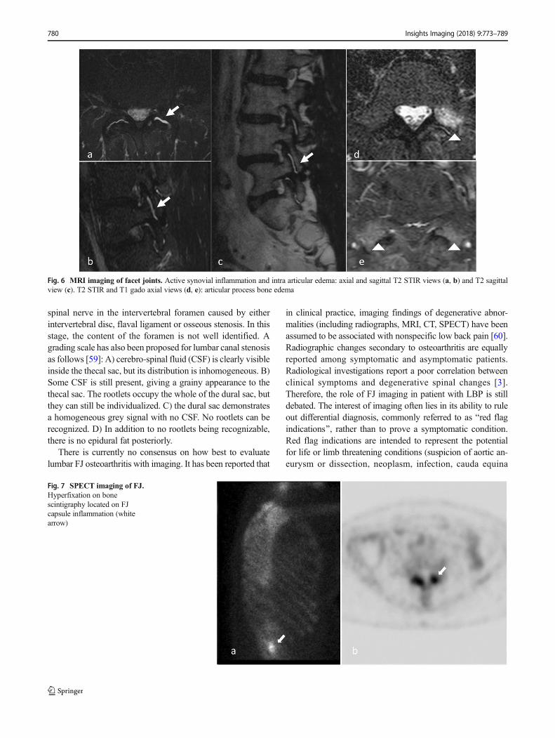
Magnetic resonance imaging (MRI) (Fig. 6)
MRI is a noninvasive and nonionizing modality that providesexcellent soft tissue resolution. The role of MRI in the evalu-ation of FJ degeneration is not proven. Osteoarthritis may bepresent in both symptomatic and asymptomatic patients (from8 to 14%) [47, 48]. Superior sensitivity of MRI compared toCT imaging is controversial [8]. CT and MRI are equallyuseful in demonstrating morphological changes in FJ. Oneof the two examinations is thus sufficient for assessing degen-erative changes [49]. MRI, however, clearly presents advan-tages of better assessing the immediate consequences of FJdegeneration, such as surrounding neural structure impinge-ment [50]. Chronic degenerative osteoarthritis processes inthese structures involve active synovial inflammation or adja-cent bone edema, which can be detected using MRI with a fatsaturation technique [51]. Exaggerated fluid in the facets andFJ synovial cysts seen on axial MRI seems to be significantlysuggestive of spondylolisthesis and its instability, but is notspecific of FJ origin of pain [52]. Recent studies using fat-suppressed MRI sequences have demonstrated thatsubchondral bone edema is present in the lumbar FJ articularprocesses in 14 to 41% of patients with back pain [53, 54].Enhancement of the FJ rim after gadolinium administrationwill establish a diagnosis of synovitis. Fujiwara et al. proposeda four-grade classification from 1 to 4 [55]: grade 1, normal;grade 2, joint space narrowing or mild osteophyte; grade 3,sclerosis or moderate osteophyte; and grade 4, marked osteo-phyte. They additionally described the wraparound bumperosteophyte formations which provides an additional stabiliz-ing effect in segmental degenerative disease. An importantobservation from the Fujiwara study is that MRI tends tounderestimate the severity of osteoarthritis of the FJs as com-pared to CT. The fluid-sensitive sequences on MRI are gener-ally preferred over CT for imaging FJ effusions and juxta-facet cysts; however, they are less sensitive in depicting thejoints’ bony cortices and are less accurate in quantifying theamount of sclerosis present. An additional limitation ofMRI isthat it cannot accurately measure cartilage thinning secondary
Fig. 5 Facet joint pain radiation. Posterior aspect of lower limb. Blue:from most frequent (dark blue), to less frequent (light blue) radiating painareas. Dark blue: pain limited to lower back. Intermediate blue: radiatingpain to the posterior aspect of the buttocks. Light blue: radiating pain to theposterior aspect of the lower limbs, may extend lower than the knee level.Green: anterior aspect of lower limb possible radiation areas. a anterioraspect of the lower limb (green). b posterior aspect of the lower limb etc
778 Insights Imaging (2018) 9:773–789

to the partial volume effect and chemical-shift artefactinherent in this type of imaging. CT is better able to demon-strate the degenerative changes of the FJs because of the highcontrast between bony structures and the surrounding softtissues [18]. However, some authors suggest that TSE T2 fatsaturation sequences and, when indicated, gadolinium admin-istration with T1 fat saturation sequences enhance the sensi-tivity and diagnostic specificity of MR scans. In particular,gadolinium will disclose the active inflammatory stage of adegenerative process thereby identifying new therapeutictargets for percutaneous treatment [51].
Single-photon emission computed tomography(SPECT)
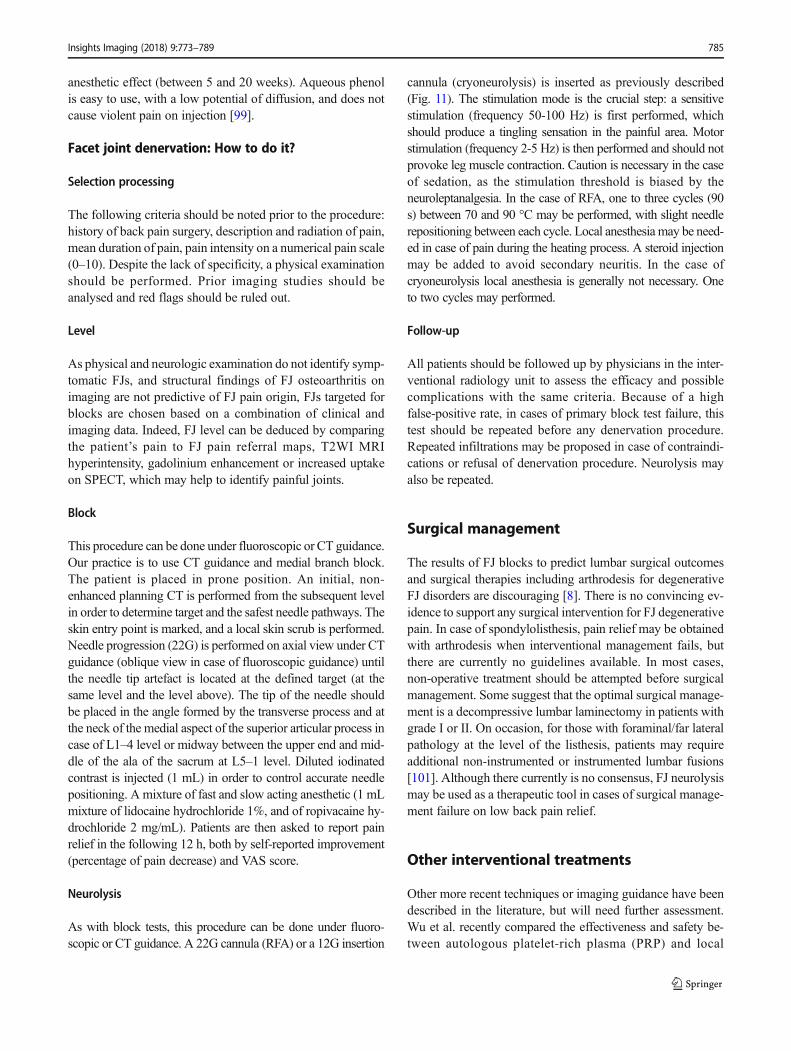
The detection of FJ inflammation may be more useful than mor-phological imaging of the joint itself. Radionuclide bone scintig-raphy, using 99mTc labelled bisphosphonates, show increasedosteoblastic activity along with synovial changes secondary toinflammation or hyperemia associated with bone remodelling(Fig. 7). It has been shown that patients present better improve-ment after FJ injection in case of positive SPECT findings [56].
Imaging classification of facet joint osteoarthritis
Two classifications of FJ degeneration are recommended forclinical use. Radiographically, Pathria’s classification clas-sifies FJ arthropathy as well: Facets with joint space
narrowing are classified as grade 1, facets with narrowingand sclerosis or hypertrophy as grade 2, and facets with severedegenerative disease encompassing narrowing, sclerosis, andosteophytes as grade 3 [57]. Standard radiographs (Meyerdingor Taillard classification) [33] also evaluate motion-relatedabnormalities in flexion or extension, and assess instabilityin cases of spondylolisthesis, thanks to dynamic studies. Inthe setting of degenerative spondylolisthesis, a weight-bearing lateral flexion-extension radiograph is most effectivefor grading spondylolisthesis and may be needed in additionto MRI and CT imaging. Anteroposterior translation of morethan a fewmillimetres is suggestive of lumbar spine instabilityin the sagittal plane, which in the appropriate clinical settingmay require surgical arthrodesis. In addition to Pathria’s clas-sification, Weishaupt’s grading scheme, based on the agree-ment between MRI and CT imaging, has been proposed.Facets were again graded from 0 to 3 depending on the degreeof joint space narrowing, hypertrophy, sclerosis, and osteo-phyte formation. The authors recommended against the rou-tine use of CT imaging in the presence of an adequate MRIscan [49]. Fujiwara et al. is credited with developing the stan-dard MRI-based classification system for lumbar FJ osteoar-thritis. An additional grading system for foramen stenosis,caused by disc and FJ degeneration can be used as well, basedon the depiction of the foraminal components: nerve, vesselsand fat [58]. First stage, the non stenotic stage: no modifica-tions depicted. Second stage corresponds to stenosis withoutevidence of root compression. Third stage, compression of the
Table 1 Main imaging findings in various imaging modalities
X-ray imaging MRI SPECT
Radiographs CT
AP, lateral (isthmus profile) andoblique views (BScottie dog^)
Highest contrast between bonystructures and adjacent softtissue
Active synovial inflammation,Adjacent bone edemaFat saturation technique ± Gadolinium
injection
99mTc labelled bisphosphonatesOsteoblastic activityHyperemia associated with
bone remodelling
Joint space narrowingSubchondral sclerosis and erosionsCartilage thinningCalcification of the joint capsuleHypertrophy of articular processesVacuum joint phenomenon joint effusion
Facet joint effusionSubchondral bone edemaEnhancement of the FJ rim (synovitis)Wraparound bumper osteophyte formation
Increased uptake (nonspecific)
Associated possible degenerative changes:
Degeneration of the intervertebral discs
Ligamentum flavum thickening
Degenerative spondylolisthesis (L4-L5 level)
Isthmic spondylolisthesis (L5-S1 level)
Facet joints cysts (coronally orientated FJ)
Lumbar spinal canal or foraminal stenosis
Neural structure impingement
AP antero posterior, CT computed tomography, MRI magnetic resonance imaging, SPECT single photon emission tomography
Insights Imaging (2018) 9:773–789 779
spinal nerve in the intervertebral foramen caused by eitherintervertebral disc, flaval ligament or osseous stenosis. In thisstage, the content of the foramen is not well identified. Agrading scale has also been proposed for lumbar canal stenosisas follows [59]: A) cerebro-spinal fluid (CSF) is clearly visibleinside the thecal sac, but its distribution is inhomogeneous. B)Some CSF is still present, giving a grainy appearance to thethecal sac. The rootlets occupy the whole of the dural sac, butthey can still be individualized. C) the dural sac demonstratesa homogeneous grey signal with no CSF. No rootlets can berecognized. D) In addition to no rootlets being recognizable,there is no epidural fat posteriorly.
There is currently no consensus on how best to evaluatelumbar FJ osteoarthritis with imaging. It has been reported that
in clinical practice, imaging findings of degenerative abnor-malities (including radiographs, MRI, CT, SPECT) have beenassumed to be associated with nonspecific low back pain [60].Radiographic changes secondary to osteoarthritis are equallyreported among symptomatic and asymptomatic patients.Radiological investigations report a poor correlation betweenclinical symptoms and degenerative spinal changes [3].Therefore, the role of FJ imaging in patient with LBP is stilldebated. The interest of imaging often lies in its ability to ruleout differential diagnosis, commonly referred to as Bred flagindications^, rather than to prove a symptomatic condition.Red flag indications are intended to represent the potentialfor life or limb threatening conditions (suspicion of aortic an-eurysm or dissection, neoplasm, infection, cauda equina
Fig. 7 SPECT imaging of FJ.Hyperfixation on bonescintigraphy located on FJcapsule inflammation (whitearrow)
780 Insights Imaging (2018) 9:773–789
Fig. 6 MRI imaging of facet joints. Active synovial inflammation and intra articular edema: axial and sagittal T2 STIR views (a, b) and T2 sagittalview (c). T2 STIR and T1 gado axial views (d, e): articular process bone edema

syndrome, fracture, motor weakness). Advanced diagnosticimaging of the symptomatic level is appropriate and/or work-up for a non-spinal source of spine pain [61].
Interventional management
First-line therapy consists in conservative multimodal manage-ment such as pain medication (acetaminophen, nonsteroidal anti-inflammatory drugs, muscle relaxants, antidepressants), physio-therapy, acupuncture, and, if necessary, psychotherapy [8].
As mentioned above, because radio-clinical correlation isnot reliable in patients with LBP, the diagnostic and therapeu-tic role of interventional procedures targeting the FJ have beenreported in chronic spinal pain in patients who have failedconservative management [62]. Whatever the technique used,it has been shown that the physician’s attitude seems to affectthe clinical outcome of a procedure by a hetero-suggestionphenomenon, with better results [63]. Imaging guidance hasshown to both to increase technical and clinical efficacy andreduce potential complications [64]. Common complicationsof FJ procedures include: hemorrhagic, infectious complica-tions, and vasovagal syncope [65].
Blocks
Because no clinical features or diagnostic imaging studies candetermine whether an FJ is painful or not, controlled blocksare the only reliable tool in the diagnosis of FJ pain as a causeof LBP [7]. Diagnostic blocks of nervous structures that aresuspected to generate pain can be performed to evaluate therole of the target structure in the painful syndrome [9].However, several debates exist on the technique and the def-inition of the performed block:
The degree of the relief that should occur
Bogduk defined specific criteria for an optimal selection as ananatomically accurate block under guidancewith ideally completerelief of pain following an MBDR block. Manchikanti et al. de-fined at least an 80% reduction of pain and the ability to performpreviously painfulmovements [66].More liberal criteria have alsobeen reported, such as greater than 50% relief of pain [9].
The target of the block (Fig. 8)
The first results comparing intra-articular and medial branchblock have reported similar outcomes [67, 68]. However, arecent review showed higher evidence in short- and long-term relief with medial branch blocks versus intra-articularblocks [2]. Moreover, intra-articular blocks appear lessanatomically accurate and have not been validated as predictiveof response to any form of treatment [9]. Medial branch block
also seem to present higher specificity to select patients formedial branch neurolysis [69]. Moreover, it seems technicallyeasier to perform using anatomic landmarks [70], than intra-articular injections, providing the use ofMDBRblock in patientselection before denervation procedures [8].
The number of blocks and the levels which should betargeted
A definitive diagnosis of FJ mediated pain may require blocksat two separate sessions. When performing a single-levelblock only, there is a high false-positive rate (30–45%).Some authors have therefore advocated the performance ofrepeated blocks [71]. Cohen et al. showed a success rate oflumbar FJ radiofrequency (RF) denervation patients of 39%after a single block and 64% after a double block [72].Because of the dual nerve supply of FJs, at the same leveland the level above, diagnostic blocks should be performedwith a minimum of two levels to block a single joint [66].
Injected drugs
Diagnostic blocks commonly include local anesthesia (lidocaineand/or bupivacaine) with or without steroids injections [8]. Somefind an advantage of adding steroid injection [66] (see below).
Steroid injections
In the majority of the reported studies, FJ injection include longacting corticosteroids (anti-inflammatory and antiedematous ef-fect, immunosuppressive action and inhibition of neural trans-mission within the C fibres) and local anesthetics [8]. FJ can beinfiltrated with intra-articular, periarticular and medial branchinjections. Due to the presence of inflammatory mediators intoand around degenerative FJ, short- to intermediate-term painrelief should occur after steroids injections. However, discrep-ancies persist in the literature about the efficacy of steroids forFJ pain [8]. Although intra-articular injections (with or withoutsteroids) have traditionally been used in the diagnosis of FJpain, a controlled trial by Lilius et al. reported no outcomedifferences between intra- and periarticular injections [73].European guidelines do not recommend the use of intra-articular steroids in management of chronic LBP [74].
Neurolysis
The ideal candidate for FJ denervation is a patient whounderwent medial branch infiltration with significant painrelief after failure of conservative management. Because ofthe dual nerve supply of a given FJ, electrodes or cryo-probes should be placed at two subsequent levels [41].Nerve fibres can either be destroyed by physical means: heat(radiofrequency) or cold (cryoneurolysis), or by chemical
Insights Imaging (2018) 9:773–789 781

means (alcohol/phenol). The main characteristics of thesetechniques are described in Table 2. Lumbar medial branchneurolysis achieves relief of pain, improvement in disability,and reduction of the need for analgesics [9]. Whatever thetechnique used, neurolysis does not allow definite pain relief.The destroyed nerve will eventually regenerate, and in conse-quence, recurrence of pain may occur. The procedure can berepeated [9]. Currently, the two most widely reported tech-niques are radiofrequency (RF) and cryoneurolysis (CN). Inboth techniques, before injection of local anesthetics and ther-mal lesions, electrical stimulation monitoring should be per-formed to ensure safety in performing thermal denervation
[75]. Currently, ISIS recommends a maximum of two FJdenervation per year [76]. Although RF techniques havebeen described in detail with a possibly longer effect thanCN, in our experience it may appear as a slightly morechallenging technique.
Physical neurolysis
Radiofrequency ablation (RFA) (Fig. 9)
Principle RF consists in the placement of electrodes under im-aging guidance, delivering a sinusoidal current (400–500 kHz).
Fig. 8 Medial branch blockunder CT guidance. a: L4–5level; b: L5-S1 level. c, d:Diffusion of contrast media priorto anesthetic injection confirmingoptimal needle tip placement(white arrows). Needle tip at thetarget point at injection should beplaced at the middle of the base ofthe transverse process at itsjunction with the superior processat the L4–5 level. An analogoustarget point should be used at theL5-S1 level midway between theupper end and middle of the ala ofthe sacrum (white stars). Vr:ventral ramus. Dr: Dorsal ramus.m: medial branch. i: intermediatebranch. l: lateral branch
Table 2 Main characteristics of the denervation procedure
Radiofrequency Cryoneurolysis Chemical neurolysis
Principle Sinusoidal currentIonic agitationTissular heating by frictionT > 45 C°
Joule–Thompson effectDecompression of CO2 or N20Ice ballT > −20 C°
Protein denaturation
Advantages Possibly longer effectTechnique described in more detailAbundant literatureWider range of needles available
NeuromaNeuritisLess tissue damageTechnically easier (bigger lesion)
CheapAvailable
Disadvantages Neuroma formation(rare)NeuritisMore tissue damageTechnically more challenging
Duration of effectiveness less assessedLarger probes and coaxial needles
Not widely used in this indicationNeuritisNeuromaTissue necrosisDeafferentation painUncontrolled diffusion
782 Insights Imaging (2018) 9:773–789

Regions crossed by the current undergo an ionic agitationwhich leads, through particle friction, to tissular heating. Thesought purpose is to expose nerve cells to a temperature > 45°C causing an irreversible cellular denaturation [77]. A widetemperature range (70–90 °C) has been reported in the literaturewith good results [78]. Another possibility is the use of pulsedRF (application of RF energy with pulsed time cycles at tem-peratures not exceeding 42 °C). The rationale for the use ofpulsed RF is to avoid any potential inadvertent damage to ad-jacent nerve roots as well as possible secondary spinal instabil-ity due to muscle denervation [14]. However, the use of thistechnique appears to be less effective in the long term [79].Therefore, pulsed RF does not appear as a substitute forconventional thermal lumbar medial branch neurotomy [9].
TechniqueBogduck et al. underlined the importance of patientselection and the use of a properly performed technique [80].Appropriate technique is described in the ISIS guidelines [81]where emphasis is made on the electrode placement: parallel tothe target nerve in order to achieve denervation along a sub-stantial length of the targeted nerve [26]. These considerationsseem more important to take into account with the RF tech-nique than with CN, where circumferential lesions are less ex-tensive than with cryoprobe [82]. RF probes produce transverselesions around the electrodes, but little lesioning at the needletip. Perpendicular placement may miss the targeted nerve [80].Moreover, operators should not rely on single placement of theelectrode, and multiple placements may be required in order tocover all possible variations of the nerve [9].
Results In a prospective study, Dreyfuss et al. showed thatunder these conditions some 60% of patients could expect atleast a 90% reduction in pain, and 87% could expect at least60% reduction lasting 12 months [77]. In Kessinger et al.’sstudy, conducted in patients with minor degenerativespondylolisthesis, 60% of patients sustained at least 80% painrelief lasting at least 12 months; 80% sustained at least 60%relief [83]. Several controlled studies confirmed this trend [69,84–87], with a mean decrease of 2–3 points on a visual ana-logue scale vs control groups. RF complications are uncom-mon (1% incidence), of limited duration and minor in nature[88]. Potential side effects include painful cutaneousdysesthesias or hyperesthesia increased pain due to neuritis,neuroma formation, and deafferentation pain. Unintentionaldamage to a spinal nerve causing a motor deficit, is also acomplication [89]. Sensory and motor stimulation during theprocedure may help to avoid this complication [75].
Cryoneurolysis (CN) (or Cryoneuroablation or Cryoanalgesia)(Fig. 10)
Principle Cryoneurolysis is an application of cold to the nerveto cause its denaturation. The physical principles relies on the
Joule–Thompson effect, which is based on a rapid decompres-sion of gas (either N2O or CO2) at the extremity of the probe,capable of delivering ice-cold temperatures of up to −70 °C[75]. At the tip of the needle, an ice ball is created in thesurrounding tissues. It induces a conduction block, similar tothe effect of local anesthetics (all nerves fibres stop conductingat −20 C°). Long-term pain relief from nerve freezing is ob-tained because of the vascular damage caused by ice crystalsto the vasa vasorum, which causes endo-neural edema and celldeath.
Technique As with RFA, the success of cryoneurolysis isdependent on patient selection and accurate probe place-ment, which should follow the same guidelines describedby the ISIS. The extent and duration of the effect is there-fore a function of the degree of cold obtained and thelength of cold application [73]. In contrast to RFA, a tan-gential approach of the probe is not essential [90].Minimal, if any, sedation should be used, as the patientmust be conscious to respond to sensory and motor stimu-lation [75]. Moreover, intra-procedural pain in CN appearsto be tolerable [91].
Results Lloyd proposed CN as superior to chemical neurolysis[90]. However, CN technique has been described as lessaccurate than RF. The lasting effect compared to RF alsoseems unclear. No studies comparing CN and RFA in FJpain management are available to date. Three recent pro-spective studies [42, 92, 93] showed a reduction of pain at6 weeks and 3 and 6 months, with a 50% pain decrease. Arecent retrospective study by Wölter et al. in 2011 con-firmed this trend [91]. Advantages of CN include less tis-sue damage, less risk of neuroma or neuritis, and a largerdenervation area at the needle tip [91, 94].
Chemical Neurolysis
This technique requires the administration of a chemicalagent able to destroy neural structures (protein denatur-ation) [95] involved in the perception of pain to promotelong lasting analgesia. The size of the lesions varies ac-cording to the concentration, and therefore the quantity.The two neurolytic agents most widely used in the treat-ment of chronic pain are phenol and alcohol, producing ablock that lasts 3–6 months [96]. Major drawbacks with theuse of these agents include: necrosis of surrounding tissue,neuritis, and uncontrolled diffusion (83). Furthermore,these powerful neurolytic agents may induce sequelae inthe axonal membrane, which might explain cases of pain-ful paresthesia observed several months following aneurolytic block: this is known to be deafferentation painsequelae [97]. These techniques are also associated with neu-roma formation [98].
Insights Imaging (2018) 9:773–789 783

Alcohol
The neurolytic effects of ethyl alcohol at a concentration great-er than 50% are well known, but higher concentrations (95–100%) are required for nerve destruction to be permanent [99].Alcohol is extremely irritating to both neural structures andsurrounding tissues, causing pain, burns, and local hypersen-sitivity. Alcohol neurolysis usually causes severe, intense
pain, which quickly disappears. Alcohol is associated with ahigher rate of neuritis than phenol [100].
Phenol
As with alcohol, neurolysis depends on the concentrationused: the efficacy of 3% phenol in saline is comparable to thatof 40% alcohol. Phenol is responsible for a transient local
Fig. 9 Facet jointradiofrequency ablation. a:Radiofrequency ablation at theright L5-S1 level. Appropriateelectrode placement (white arrow)parallel to the target nerve (whitestar) in order to achieve denerva-tion along a substantial segmentof the targeted nerve. b: 22GRadiofrequency needle showinguninsulated tip
Fig. 10 Facet jointcryoneurolysis. CT-guidedcryoablation at the right L5-S1level. a: Anatomically accuratecryoprobe (white arrow) place-ment with the target midway be-tween the upper end and middleof the ala of the sacrum (whitestar). b: 3D reconstruction show-ing the same cryoprobe place-ment. Cryoprobe needle tip (c):ice-ball formation at the cryo-probe tip (Joule–Thompson ef-fect) (d)
784 Insights Imaging (2018) 9:773–789
anesthetic effect (between 5 and 20 weeks). Aqueous phenolis easy to use, with a low potential of diffusion, and does notcause violent pain on injection [99].
Facet joint denervation: How to do it?
Selection processing
The following criteria should be noted prior to the procedure:history of back pain surgery, description and radiation of pain,mean duration of pain, pain intensity on a numerical pain scale(0–10). Despite the lack of specificity, a physical examinationshould be performed. Prior imaging studies should beanalysed and red flags should be ruled out.
Level
As physical and neurologic examination do not identify symp-tomatic FJs, and structural findings of FJ osteoarthritis onimaging are not predictive of FJ pain origin, FJs targeted forblocks are chosen based on a combination of clinical andimaging data. Indeed, FJ level can be deduced by comparingthe patient’s pain to FJ pain referral maps, T2WI MRIhyperintensity, gadolinium enhancement or increased uptakeon SPECT, which may help to identify painful joints.
Block
This procedure can be done under fluoroscopic or CT guidance.Our practice is to use CT guidance and medial branch block.The patient is placed in prone position. An initial, non-enhanced planning CT is performed from the subsequent levelin order to determine target and the safest needle pathways. Theskin entry point is marked, and a local skin scrub is performed.Needle progression (22G) is performed on axial view under CTguidance (oblique view in case of fluoroscopic guidance) untilthe needle tip artefact is located at the defined target (at thesame level and the level above). The tip of the needle shouldbe placed in the angle formed by the transverse process and atthe neck of the medial aspect of the superior articular process incase of L1–4 level or midway between the upper end and mid-dle of the ala of the sacrum at L5–1 level. Diluted iodinatedcontrast is injected (1 mL) in order to control accurate needlepositioning. A mixture of fast and slow acting anesthetic (1 mLmixture of lidocaine hydrochloride 1%, and of ropivacaine hy-drochloride 2 mg/mL). Patients are then asked to report painrelief in the following 12 h, both by self-reported improvement(percentage of pain decrease) and VAS score.
Neurolysis
As with block tests, this procedure can be done under fluoro-scopic or CT guidance. A 22G cannula (RFA) or a 12G insertion
cannula (cryoneurolysis) is inserted as previously described(Fig. 11). The stimulation mode is the crucial step: a sensitivestimulation (frequency 50-100 Hz) is first performed, whichshould produce a tingling sensation in the painful area. Motorstimulation (frequency 2-5 Hz) is then performed and should notprovoke leg muscle contraction. Caution is necessary in the caseof sedation, as the stimulation threshold is biased by theneuroleptanalgesia. In the case of RFA, one to three cycles (90s) between 70 and 90 °C may be performed, with slight needlerepositioning between each cycle. Local anesthesia may be need-ed in case of pain during the heating process. A steroid injectionmay be added to avoid secondary neuritis. In the case ofcryoneurolysis local anesthesia is generally not necessary. Oneto two cycles may performed.
Follow-up
All patients should be followed up by physicians in the inter-ventional radiology unit to assess the efficacy and possiblecomplications with the same criteria. Because of a highfalse-positive rate, in cases of primary block test failure, thistest should be repeated before any denervation procedure.Repeated infiltrations may be proposed in case of contraindi-cations or refusal of denervation procedure. Neurolysis mayalso be repeated.
Surgical management
The results of FJ blocks to predict lumbar surgical outcomesand surgical therapies including arthrodesis for degenerativeFJ disorders are discouraging [8]. There is no convincing ev-idence to support any surgical intervention for FJ degenerativepain. In case of spondylolisthesis, pain relief may be obtainedwith arthrodesis when interventional management fails, butthere are currently no guidelines available. In most cases,non-operative treatment should be attempted before surgicalmanagement. Some suggest that the optimal surgical manage-ment is a decompressive lumbar laminectomy in patients withgrade I or II. On occasion, for those with foraminal/far lateralpathology at the level of the listhesis, patients may requireadditional non-instrumented or instrumented lumbar fusions[101]. Although there currently is no consensus, FJ neurolysismay be used as a therapeutic tool in cases of surgical manage-ment failure on low back pain relief.
Other interventional treatments
Other more recent techniques or imaging guidance have beendescribed in the literature, but will need further assessment.Wu et al. recently compared the effectiveness and safety be-tween autologous platelet-rich plasma (PRP) and local
Insights Imaging (2018) 9:773–789 785

anesthesia/corticosteroid in intra-articular injection for thetreatment of FJ syndrome. They showed that prone autologousPRP presented a superior efficacy with longer duration [102].An observational retrospective study of 86 patients byKirchner et al. confirmed this trend [103].
Iwatsuki et al. performed laser radiation of the dorsal sur-face of the facet capsule in 21 patients and reported greaterthan 70% pain relief for at least 1 year in 81% (17 patients)[104]. Feasibility and safety of MRI-guided focused ultra-sound ablation of the lumbar medial branch nerve has beenshown in a swine model and thermal necrosis was confirmed[105].
Conclusion
Because chronic low back pain of facet joint pain origin rep-resents a major health care problem, diagnosis and manage-ment of such a high prevalent condition as facet joint syn-drome is a major socioeconomic burden. Because of the abil-ity of facet joint pathology to mimic spine root compression,the low specificity of FJ syndrome and inefficient use of lum-bar imaging, it appears as a misunderstood, misdiagnosed andimproperly treated pathology. Facet joint-related anatomical,clinical and radiologic knowledge is essential for successfulfacet joint syndrome management. Diagnostic blocks are akeystone of facet syndrome diagnosis. If diagnostic blocksof the nerves that supply specific facet joints relieve the pa-tient’s pain, denervation procedure lesioning of the samenerves can be offered to provide prolonged benefit. The roleof the radiologist is essential in the management of these pa-tients, and radiologists should embrace all aspects of facet
joint pain management, from diagnosis—enabled by high-performance modalities available—to interventional manage-ment. The radiologist can therefore play an active role in thedifficult task of alleviating patients’ chronic low back pain offacet joint origin.
Compliance with ethical standards
Conflict of interest The authors of this manuscript declare no relation-ships with any companies whose products or services may be related tothe subject matter of the article.
Open Access This article is distributed under the terms of the CreativeCommons At t r ibut ion 4 .0 In te rna t ional License (h t tp : / /creativecommons.org/licenses/by/4.0/), which permits unrestricted use,distribution, and reproduction in any medium, provided you give appro-priate credit to the original author(s) and the source, provide a link to theCreative Commons license, and indicate if changes were made.
References
1. Breivik H, Collett B, Ventafridda V, Cohen R, Gallacher D (2006)Survey of chronic pain in Europe: prevalence, impact on daily life,and treatment. Eur J Pain 10(4):287–333
2. Manchikanti L, Hirsch JA, Falco FJ, Boswell MV (2016)Management of lumbar zygapophysial (facet) joint pain. World JOrthop 7(5):315–337
3. Kalichman L, Li L, Kim DH et al (2008) Facet joint osteoarthritisand low back pain in the community-based population. Spine(Phila Pa 1976) 33(23):2560–2565
4. Kalichman L, Kim DH, Li L, Guermazi A, Hunter DJ (2010)Computed tomography-evaluated features of spinal degeneration:prevalence, intercorrelation, and association with self-reportedlow back pain. Spine J 10(3):200–208
Fig. 11 Photographs of thecoaxial needles: forcryodenervation (a, d) andradiofrequency (b, c),highlighting the difference indiameter 12G vs 22G (e)
786 Insights Imaging (2018) 9:773–789

5. Manchikanti L, Singh V, Pampati Vet al (2001) Evaluation of therelative contributions of various structures in chronic low backpain. Pain Physician 4(4):308–316
6. HancockMJ,Maher CG, Latimer J et al (2007) Systematic reviewof tests to identify the disc, SIJ or facet joint as the source of lowback pain. Eur Spine J 16(10):1539–1550
7. Falco FJ, Manchikanti L, Datta S et al (2012) An update of thesystematic assessment of the diagnostic accuracy of lumbar facetjoint nerve blocks. Pain Physician 15(6):E869–E907
8. Cohen SP, Raja SN (2007) Pathogenesis, diagnosis, and treatmentof lumbar zygapophysial (facet) joint pain. Anesthesiology106(3):591–614
9. Bogduk N, Dreyfuss P, Govind J (2009) A narrative review oflumbar medial branch neurotomy for the treatment of back pain.Pain Med 10(6):1035–1045
10. Declaration on Pain – EFIC. [En ligne]. Available via: http://www.europeanpainfederation.eu/about-efic/efic-declaration-on-pain/.Accessed: 8 February 2017
11. Edmond SL, Felson DT (2003) Function and back symptoms inolder adults. J Am Geriatr Soc 51(12):1702–1709
12. Wenig CM, Schmidt CO, Kohlmann T, Schweikert B (2009)Costs of back pain in Germany. Eur J Pain 13(3):280–286
13. Manchikanti L, Manchikanti KN, Cash KA, Singh V, Giordano J(2008) Age-related prevalence of facet-joint involvement inchronic neck and low back pain. Pain Physician 11(1):67–75
14. Saravanakumar K, Harvey A (2008) Lumbar zygapophyseal (fac-et) joint pain. Rev Pain 2(1):8–13
15. Datta S, Lee M, Falco FJ, Bryce DA, Hayek SM (2009)Systematic assessment of diagnostic accuracy and therapeuticutility of lumbar facet joint interventions. Pain Physician 12(2):437–460
16. Yahia LH, Garzon S (1993) Structure on the capsular ligaments ofthe facet joints. Ann Anat 175(2):185–188
17. Konin GP, Walz DM (2010) Lumbosacral transitional vertebrae:classification, imaging findings, and clinical relevance. AJNRAmJ Neuroradiol 31(10):1778–1786
18. Varlotta GP, Lefkowitz TR, Schweitzer M et al (2011) The lumbarfacet joint: a review of current knowledge: part 1: anatomy, bio-mechanics and grading. Skeletal Radiol 40(1):13–23
19. Gao T, Lai Q, Zhou S et al (2017) Correlation between facettropism and lumbar degenerative disease: a retrospective analysis.BMC Musculoskelet Disord 18:483
20. Adams MA, Hutton WC (1983) The mechanical function of thelumbar apophyseal joints. Spine (Phila Pa 1976) 8(3):327–330
21. Borenstein D (2004) Does osteoarthritis of the lumbar spine causechronic low back pain? Curr Pain Headache Rep 8(6):512–517
22. Cavanaugh JM, Ozaktay AC, Yamashita HT, King AI (1996)Lumbar facet pain: biomechanics, neuroanatomy and neurophys-iology. J Biomech 29(9):1117–1129
23. Beaman DN, Graziano GP, Glover RA, Wojtys EM, Chang V(1993) Substance P innervation of lumbar spine facet joints.Spine (Phila Pa 1976) 18(8):1044–1049
24. Igarashi A, Kikuchi S, Konno S, Olmarker K (2004) Inflammatorycytokines released from the facet joint tissue in degenerative lum-bar spinal disorders. Spine (Phila Pa 1976) 29(19):2091–2095
25. BogdukN,WilsonAS, TynanW (1982) The human lumbar dorsalrami. J Anat 134(Pt 2):383–397
26. Lau P, Mercer S, Govind J, Bogduk N (2004) The surgical anat-omy of lumbar medial branch neurotomy (facet denervation). PainMed 5(3):289–298
27. Clinical anatomy of the lumbar spine and sacrum- NLMCatalog -NCBI. [En ligne]. Available via: https://www.ncbi.nlm.nih.gov/nlmcatalog/9801582. Accessed: 31 January 2017
28. Bogduk N (1997) Clinical anatomy of the lumbar spine and sa-crum, 3rd edn. Churchill Livingstone, Edinburgh
29. Eubanks JD, Lee MJ, Cassinelli E, Ahn NU (2007) Prevalence oflumbar facet arthrosis and its relationship to age, sex, and race: ananatomic study of cadaveric specimens. Spine (Phila Pa 1976)32(19):2058–2062
30. Chazen JL, Leeman K, Singh JR, Schweitzer A (2017)Percutaneous CT-guided facet joint synovial cyst rupture: successwith refractory cases and technical considerations. Clin Imaging49:7–11
31. Ening G, Kowoll A, Stricker I, Schmieder K, Brenke C (2015)Lumbar juxta-facet joint cysts in association with facet joint ori-entation, −tropism and -arthritis: a case-control study. Clin NeurolNeurosurg 139:278–281
32. Yoshiiwa T, Miyazaki M, Notani N, Ishihara T, Kawano M,Tsumura H (2016) Analysis of the relationship betweenligamentum Flavum thickening and lumbar segmental instability,disc degeneration, and facet joint osteoarthritis in lumbar spinalstenosis. Asian Spine J 10(6):1132–1140
33. Kalichman L, Hunter DJ (2008) Diagnosis and conservative man-agement of degenerative lumbar spondylolisthesis. Eur Spine J17(3):327–335
34. Sun Y, Wang H, Yang D et al (2016) Characterization of radio-graphic features of consecutive lumbar spondylolisthesis.Medicine (Baltimore) 95(46):e5323
35. RajeevA, ChoudhryN, ShaikhM,NewbyM (2016) Lumbar facetjoint septic arthritis presenting atypically as acute abdomen - acase report and review of the literature. Int J Surg Case Rep 25:243–245
36. Le Hanneur M, Vidal C, Mallet C, Mazda K, Ilharreborde B(2016) Unusual case of paediatric septic arthritis of the lumbarfacet joints due to Kingella kingae. Orthop Traumatol Surg ResOTSR 102(7):959–961
37. Goldthwait JE (1911) The lumbosacral articulation: an explana-tion of many cases of lumbago, sciatica, and paraplegia. BostonMed Surg J 164:365–372
38. Ghormley RK (1933) Low back pain with special reference to thearticular facets, with presentation of an operative procedure.JAMA 101:773
39. Badgley CE (1941) The articular facets in relation to low backpain and sciatic radiation. J Bone Joint Surg Am 23:481–496
40. Fukui S, Ohseto K, Shiotani M, Ohno K, Karasawa H, NaganumaY (1997) Distribution of referred pain from the lumbarzygapophyseal joints and dorsal rami. Clin J Pain 13(4):303–307
41. Windsor RE, King FJ, Roman SJ et al (2002) Electrical stimula-tion induced lumbar medial branch referral patterns. PainPhysician 5(4):347–353
42. Bärlocher CB, Krauss JK, Seiler RW (2002) Kryorhizotomy: analternative technique for lumbar medial branch rhizotomy in lum-bar facet syndrome. J Neurosurg 98(1 Suppl):14–20
43. Piraccini E, Calli M, Corso RM, Byrne H, Maitan S (2017)Abdominal and pelvic pain: an uncommon sign in lumbar facetjoint syndrome. Minerva Anestesiol 83(1):104–105
44. Jackson RP (1992) The facet syndrome. Myth or reality? ClinOrthop Relat Res 279:110–121
45. Manchikanti L, Boswell MV, Singh V, Pampati V, Damron KS,Beyer CD (2004) Prevalence of facet joint pain in chronic spinalpain of cervical, thoracic, and lumbar regions. BMCMusculoskelet Disord 5(15)
46. Schwarzer AC,Wang SC, O’Driscoll D, Harrington T, Bogduk N,Laurent R (1995) The ability of computed tomography to identifya painful zygapophysial joint in patients with chronic low backpain. Spine (Phila Pa 1976) 20(8):907–912
47. Weishaupt D, Zanetti M, Hodler J, Boos N (1998) MR imaging ofthe lumbar spine: prevalence of intervertebral disk extrusion andsequestration, nerve root compression, end plate abnormalities,and osteoarthritis of the facet joints in asymptomatic volunteers.Radiology 209(3):661–666
Insights Imaging (2018) 9:773–789 787
48. Jensen MC, Brant-Zawadzki MN, Obuchowski N, Modic MT,Malkasian D, Ross JS (1994) Magnetic resonance imaging ofthe lumbar spine in people without back pain. N Engl J Med331(2):69–73
49. Weishaupt D, Zanetti M, Boos N, Hodler J (1999) MR imagingand CT in osteoarthritis of the lumbar facet joints. Skeletal Radiol28(4):215–219
50. Clarençon F, Law-Ye B, Bienvenot P, Cormier É, Chiras J (2016)The Degenerative Spine. Magn Reson Imaging Clin N Am 24(3):495–513
51. D’Aprile P, Tarantino A, Lorusso V, Brindicci D (2006) Fat satu-ration technique and gadolinium in MRI of lumbar spinal degen-erative disease. Neuroradiol J 19(5):654–671
52. Schinnerer KA, Katz LD, Grauer JN (2008) MR findings of ex-aggerated fluid in facet joints predicts instability. J Spinal DisordTech 21(7):468–472
53. Suri P, Miyakoshi A, Hunter DJ et al (2011) Does lumbar spinaldegeneration begin with the anterior structures? A study of theobserved epidemiology in a community-based population. BMCMusculoskelet Disord 12:202
54. Lakadamyali H, Tarhan NC, Ergun T, Cakir B, Agildere AM(2008) STIR sequence for depiction of degenerative changes inposterior stabilizing elements in patients with lower back pain.AJR Am J Roentgenol 191(4):973–979
55. Fujiwara A, Tamai K, Yamato M et al (1999) The relationshipbetween facet joint osteoarthritis and disc degeneration of thelumbar spine: an MRI study. Eur Spine J 8(5):396–401
56. Pneumaticos SG, Chatziioannou SN, Hipp JA, Moore WH, EssesSI (2006) Low back pain: prediction of short-termoutcome of facetjoint injection with bone scintigraphy. Radiology 238(2):693–698
57. Pathria M, Sartoris DJ, Resnick D (1987) Osteoarthritis of thefacet joints: accuracy of oblique radiographic measurement.Radiology 164:227–230
58. Splendiani A, Ferrari F, Barile A, Masciocchi C, Gallucci M(2014) Occult neural foraminal stenosis caused by associationbetween disc degeneration and facet joint osteoarthritis: demon-stration with dedicated upright MRI system. Radiol Med 119(3):164–174
59. Schizas C, Theumann N, Burn A et al (2010) Qualitative gradingof severity of lumbar spinal stenosis based on the morphology ofthe dural sac onmagnetic resonance images. Spine (Phila Pa 1976)35(21):1919–1924
60. Hofmann UK, Keller RL,Walter C,Mittag F (2017) Predictabilityof the effects of facet joint infiltration in the degenerate lumbarspine when assessing MRI scans. J Orthop Surg Res 12
61. Lateef H, Patel D (2009) What is the role of imaging in acute lowback pain? Curr Rev Musculoskelet Med 2(2):69–73
62. Filippiadis DK, Kelekis A (2015) A review of percutaneous tech-niques for low back pain and neuralgia: current trends in epiduralinfiltrations, intervertebral disk and facet joint therapies. Br JRadiol 20150357
63. Middendorp M, Kollias K, Ackermann H et al (2016) Does ther-apist’s attitude affect clinical outcome of lumbar facet joint injec-tions? World J Radiol 8(6):628–634
64. Manchikanti L, Boswell MV, Singh Vet al (2009) Comprehensiveevidence-based guidelines for interventional techniques in themanagement of chronic spinal pain. Pain Physician 12(4):699–802
65. Velickovic M, Ballhause TM (2016) Delayed onset of a spinalepidural hematoma after facet joint injection. SAGE Open MedCase Rep 4
66. Manchikanti L, Manchikanti KN, Manchukonda R et al (2007)Evaluation of lumbar facet joint nerve blocks in the managementof chronic low back pain: preliminary report of a randomized,double-blind controlled trial: clinical trial NCT00355914. PainPhysician 10(3):425–440
67. Dreyfuss PH, Dreyer SJ, Herring SA (1995) Lumbarzygapophysial (facet) joint injections. Spine (Phila Pa 1976)20(18):2040–2047
68. Dreyer SJ, Dreyfuss H (1996) Low back pain and thezygapophysial (facet) joints. Arch Phys Med Rehabil 77(3):290–300
69. van Wijk RMAW, Geurts JW, Wynne HJ et al (2005)Radiofrequency denervation of lumbar facet joints in the treat-ment of chronic low back pain: a randomized, double-blind, shamlesion-controlled trial. Clin J Pain 21(4):335–344
70. Taguchi T, Kawai S, Oda H, Kaneko K (2000) Anatomic basis forselective nervi-spinales infiltration in the treatment of articularback pain. J Neuroradiol 27(1):25–29
71. Manchukonda R, Manchikanti KN, Cash KA, Pampati V,Manchikanti L (2007) Facet joint pain in chronic spinal pain: anevaluation of prevalence and false-positive rate of diagnosticblocks. J Spinal Disord Tech 20(7):539–545
72. Cohen SP, Williams KA, Kurihara C et al (2010) Multicenter,randomized, comparative cost-effectiveness study comparing 0,1, and 2 diagnostic medial branch (facet joint nerve) block treat-ment paradigms before lumbar facet radiofrequency denervation.Anesthesiology 113(2):395–405
73. Lilius G, Laasonen EM, Myllynen P, Harilainen A, Grönlund G(1989) Lumbar facet joint syndrome. A randomised clinical trial. JBone Joint Surg Br 71(4):681–684
74. Airaksinen O, Brox JI, Cedraschi C et al (2006) Chapter 4.European guidelines for the management of chronic non-specificlow back pain. Eur Spine J 15:S192–S300
75. Trescot AM (2003) Cryoanalgesia in interventional pain manage-ment. Pain Physician 6(3):345–360
76. International Spine Intervention Society (2004) Lumbar medialneurotomy. In: Bogduk N (ed) Practice guidelines for spinal diag-nostic and treatment procedures. International Spinal InterventionSociety, San Francisco, pp 188–218
77. Dreyfuss P, Halbrook B, Pauza K, Joshi A, McLarty J, Bogduk N(2000) Efficacy and validity of radiofrequency neurotomy forchronic lumbar zygapophysial joint pain. Spine (Phila Pa 1976)25(10):1270–1277
78. Costandi S, Garcia-Jacques M, Dews T et al (2016) Optimal tem-perature for radiofrequency ablation of lumbar medial branches fortreatment of facet-mediated back pain. Pain Pract 16(8):961–968
79. Mikeladze G, Espinal R, Finnegan R, Routon J, Martin D (2003)Pulsed radiofrequency application in treatment of chroniczygapophyseal joint pain. Spine J 3(5):360–362
80. Bogduk N, Macintosh J, Marsland A (1987) Technical limitationsto the efficacy of radiofrequency neurotomy for spinal pain.Neurosurgery 20(4):529–535
81. International Spine Intervention Society (2004) Lumbar medialbranch blocks. In: Bogduk N (ed) Practice guidelines for spinaldiagnostic and treatment procedures. International SpinalIntervention Society, San Francisco, pp 47–65
82. Mattmüller R (2002) Radiofrequenzläsion und Kryoläsion. In:Hankemeier U, Hildebrandt J (eds) Neurodestruktive Verfahrenin der Schmerztherapie. Springer, Berlin, pp 19–32
83. Klessinger S (2012) Radiofrequency neurotomy for treatment oflow back pain in pat ients with minor degenerat ivespondylolisthesis. Pain Physician 15(1):E71–E78
84. Gallagher J, Petriccione di Valdo PL, Wedley JR et al (1994)Radiofrequency facet joint denervation in the treatment of lowback pain: a prospective controlled doubleblind study to assessits efficacy. Pain Clinic 7:193–198
85. Nath S, Nath CA, Pettersson K (2008) Percutaneous lumbarzygapophysial (facet) joint neurotomy using radiofrequency current,in the management of chronic low back pain: a randomized double-blind trial. Spine (Phila Pa 1976) 33(12):1291–1297 discussion 1298
788 Insights Imaging (2018) 9:773–789

86. van Kleef M, Barendse GA, Kessels A, Voets HM,Weber WE, deLange S (1999) Randomized trial of radiofrequency lumbar facetdenervation for chronic low back pain. Spine (Phila Pa 1976)24(18):1937–1942
87. Leclaire R, Fortin L, Lambert R, Bergeron YM, Rossignol M(2001) Radiofrequency facet joint denervation in the treatmentof low back pain: a placebo-controlled clinical trial to assess effi-cacy. Spine (Phila Pa 1976) 26(13):1411–1416 discussion 1417
88. Kornick C, Kramarich SS, Lamer TJ, Todd Sitzman B (2004)Complications of lumbar facet radiofrequency denervation.Spine (Phila Pa 1976) 29(12):1352–1354
89. Pacetti M, Fiaschi GS, Gennaro S (2016) Percutaneous radiofre-quency thermocoagulation of dorsal ramus branches as a treatmentof Blumbar facet syndrome^ how I do it. Acta Neurochir (Wien)158(5):995–998
90. Lloyd JW, Barnard JD, Glynn CJ (1976) Cryoanalgesia. A newapproach to pain relief. Lancet 2(7992):932–934
91. Wolter T, Deininger M, Hubbe U, Mohadjer M, Knoeller S (2011)Cryoneurolysis for zygapophyseal joint pain: a retrospective analysisof 117 interventions. Acta Neurochir (Wien) 153(5):1011–1019
92. Staender M, Maerz U, Tonn JC, Steude U (2005) Computerizedtomography-guided kryorhizotomy in 76 patients with lumbarfacet joint syndrome. J Neurosurg Spine 3(6):444–449
93. Birkenmaier C, Veihelmann A, Trouillier H et al (2007)Percutaneous cryodenervation of lumbar facet joints: a prospec-tive clinical trial. Int Orthop 31(4):525–530
94. Wolter T, Bozhkov Y, Knoeller SM (2017) An in vitro analysis ofthe size and shape of cryolesions for facet joint denervation. ClinNeurol Neurosurg 153:87–92
95. Kastler A, Cadel G, Comte A et al (2014) Alcohol percutaneousneurolysis of the sphenopalatine ganglion in the management ofrefractory craniofacial pain. Neuroradiology 56(7):589–596
96. Molloy R, Benzon H (2008) Neurolytic blocking agents: uses andcomplications. In: Raj’s practical management of pain. MosbyElsevier, Philadelphia, pp 839–850
97. Jain S, Gupta R (2007)Neural blockadewith neurolytic agents. In:Textbook of pain management. 1st ed, ed. Waldman. Elsevier,Philadelphia
98. Evans PJ, Lloyd JW, Jack TM (1981) Cryoanalgesia for intracta-ble perineal pain. J R Soc Med 74(11):804–809
99. Koyyalagunta D, Engle MP, Yu J, Feng L, Novy DM (2016) Theeffectiveness of alcohol versus phenol based splanchnic nerveneurolysis for the treatment of intra-abdominal cancer pain. PainPhysician 19(4):281–292
100. Raj PP (2004) Visceral pain. Agri 16:7–20101. Epstein NE, Hollingsworth RD (2017) Nursing review of diagno-
sis and treatment of lumbar degenerative spondylolisthesis. SurgNeurol Int 8
102. Wu J, Zhou J, Zhang J et al (2017) A Prospective StudyComparing Platelet-Rich Plasma and Local Anesthetic (LA)/Corticosteroid in Intra-Articular Injection for the Treatment ofLumbar Facet Joint Syndrome. Pain Pract 17(7):914–924
103. Kirchner F, Anitua E (2016) Intradiscal and intra-articular facetinfiltrations with plasma rich in growth factors reduce pain inpatients with chronic low back pain. J Craniovertebr JunctionSpine 7(4):250–256
104. Iwatsuki K, Yoshimine T, Awazu K (2007) Alternative denerva-tion using laser irradiation in lumbar facet syndrome. Lasers SurgMed 39(3):225–229
105. Kaye EA,Monette S, Srimathveeravalli G, MaybodyM, SolomonSB, Gulati A (2016) MRI-guided focused ultrasound ablation oflumbar medial branch nerve: feasibility and safety study in a swinemodel. Int J Hyperthermia 32(7):786–794
Publisher’s Note
Springer Nature remains neutral with regard to jurisdictional claims inpublished maps and institutional affiliations.
Insights Imaging (2018) 9:773–789 789