fabrication of hydroxyapatite with highly ordered macroporous frame by colloidal templating method
TRANSCRIPT
Fabrication of hydroxyapatite with highly ordered macroporous frame bycolloidal templating method
Liyun Zhou, Deping Wang, Wenhai Huang *, Aihua Yao, Chaohui Xia, Xiang Duan
The Institute of Bioengineering and Information Technology Materials, School of Material Science and Engineering, Tongji University, Shanghai 200092,
People’s Republic of China
Materials Research Bulletin 44 (2009) 259–262
A R T I C L E I N F O
Article history:
Received 12 December 2007
Received in revised form 22 April 2008
Accepted 13 June 2008
Available online 28 June 2008
Keywords:
A. Ceramics
A. Inorganic compound
B. Sol–gel chemistry
A B S T R A C T
Three-dimensional, highly ordered macroporous frame of hydroxyapatite has been fabricated via a
template-assisted colloidal processing technique. In the present method, colloidal template was first
prepared with SiO2 spheres by gravitational sedimentation, which was then infiltrated with
hydroxyapatite precursor prepared by the sol–gel process. The resulting hydroxyapatite replicated
the three-dimensionally ordered macroporous structure of SiO2 template. Modified by H2O2, the SiO2
spheres could be packed into better ordered templates. After removal of the template by immersing in
NaOH solution, the well-ordered macroporous frame made from HA was obtained. The arrangement of
the pore structure was hexagonal close-packed and pore sizes could be controlled by changing the sizes of
SiO2 spheres. The resulting highly ordered macroporous frame of hydroxyapatite could have potential
applications in the biomedical field.
� 2008 Elsevier Ltd. All rights reserved.
Contents lists available at ScienceDirect
Materials Research Bulletin
journa l homepage: www.e lsev ier .com/ locate /matresbu
1. Introduction
As high performance adsorption and separation materials, thematerials with ordered macroporous frame were found to exhibitmany unique properties [1,2]. With excellent connectivity amongpores [3], the materials exhibit improved adsorption anddesorption behavior. It has been demonstrated that an advancedchemical selectivity can also be achieved after a chemicalmodification to pore walls [4]. Therefore, the ordered macroporousmaterials and their preparation techniques are very attractive inrecent years.
So far, various kinds of fabrication methods have been appliedto prepare various ordered macroporous materials, of which thecolloidal templating approach is the most commonly used method[5–8]. The general concept of colloid templating is simple: (1) packthe spherical colloid into close-packed arrays, (2) fill the interstitialspaces of the colloid crystal with a fluid precursor capable ofsolidification, and (3) remove the template to obtain a porousinverse replica [9]. For the procedure, the materials precursors donot undergo a special pre-treatment process, and the pore sizes ofthe resulting material could be easily controlled. With these
* Corresponding author.
E-mail address: [email protected] (W. Huang).
0025-5408/$ – see front matter � 2008 Elsevier Ltd. All rights reserved.
doi:10.1016/j.materresbull.2008.06.016
merits, the method has been widely used to prepare various kindsof materials.
Hydroxyapatite (HA) is one of the major constituents of theinorganic component in human hard tissue (bones and teeth),and it is one of the most common biomaterials studied becauseof its great biomedical properties. With good biocompatibilityand bioactivity, HA would be preferred to be used as adsorptionand separation materials in biomedical fields [10]. However, HAwith three-dimensional ordered macroporous (3DOM) frame,prepared by colloidal templating method has not been reporteduntil now.
The goal of this work, therefore, was to prepare HA with 3DOMframe using monodisperse SiO2 spheres as a colloidal template.The resulting 3DOM HA would be promising for biomedicalapplications as drug carriers and bio-remarking materials.
2. Experimental
2.1. Materials
Tetraethyl orthosilicate (TEOS) of analytic purity was used asthe silica precursor, Trimethyl phosphate (TMP) and Calciumnitrate (Ca(NO3)2�4H2O) of analytic purity were used as thehydroxyapatite precursors, Hydrogen peroxide (H2O2) of analyticpurity was used to modify silica spheres.
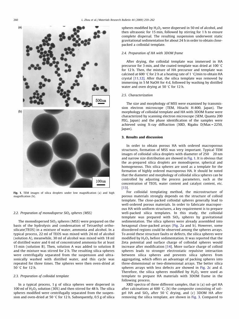
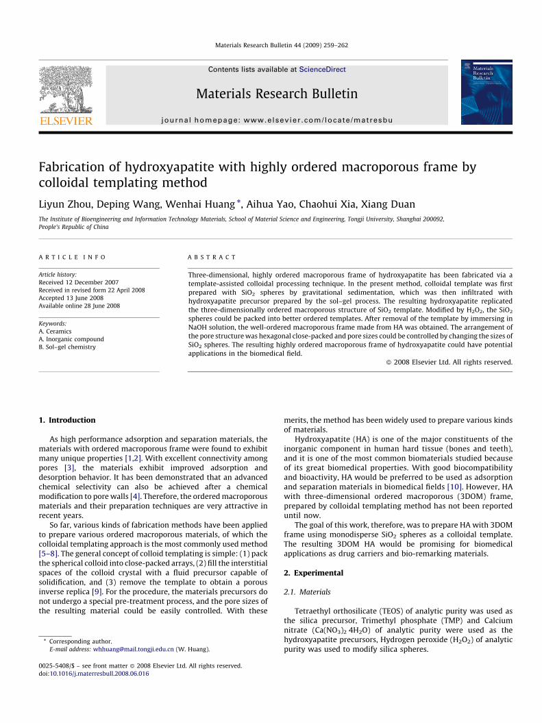
Fig. 1. TEM images of silica droplets under low magnification (a) and high
magnification (b).
L. Zhou et al. / Materials Research Bulletin 44 (2009) 259–262260
2.2. Preparation of monodisperse SiO2 spheres (MSS)
The monodispersed SiO2 spheres (MSS) were prepared on thebasis of the hydrolysis and condensation of Tetraethyl ortho-silicate(TEOS) in a mixture of water, ammonia and alcohol. In atypical process, 22 ml of TEOS was mixed with 24 ml of alcohol(solution A), meanwhile, 30 ml of alcohol was mixed with 18 mlof distilled water and 6 ml of concentrated ammonia for at least15 min (solution B). Then, solution A was added to solution Band the mixture was stirred for 2 h. The resulting silica sphereswere centrifugally separated from the suspension and ultra-sonically washed with distilled water, and this cycle wasrepeated for three times. The spheres were then oven-dried at50 8C for 12 h.
2.3. Preparation of colloidal template
In a typical process, 1 g of silica spheres were dispersed in100 ml of H2O2 solution (30%) and then stirred for 48 h. The silicaspheres modified were centrifugally separated from the suspen-sion and oven-dried at 50 8C for 12 h. Subsequently, 0.5 g of silica
spheres modified by H2O2 were dispersed in 50 ml of alcohol, andthen ultrasonic for 15 min, followed by stirring for 1 h to ensurecomplete dispersal. The resulting suspension underwent staticgravitational sedimentation for about 24 h in order to obtain close-packed a colloidal template.
2.4. Preparation of HA with 3DOM frame
After drying, the colloidal template was immersed in HAprecursor for 3 min, and the coated template was dried at 100 8Cfor 12 h. Then, the mixture of HA precursor and template wascalcined at 600 8C for 2 h at a heating rate of 1 8C/min to obtain HAcrystal [11,12]. After that, the silica template was removed byimmersing in 5 M NaOH for 4 d, followed by washing by distilledwater and oven drying at 50 8C for 12 h.
2.5. Characterization
The size and morphology of MSS were examined by transmis-sion electron microscope (TEM, Hitachi H-800, Japan). Themorphology of colloidal template and HA with 3DOM frame werecharacterized by scanning electron microscope (SEM, Quanta 200FEG, Japan) and the phase identification of the samples wereachieved using X-ray diffraction (XRD, Rigaku D/Max = 2250,Japan).
3. Results and discussion
In order to obtain porous HA with ordered macroporousstructures, formation of MSS was very important. Typical TEMimages of colloidal silica droplets with diameter of 250 � 20 nmand narrow size distribution are showed in Fig. 1. It is obvious thatthe as-prepared silica droplets are monodisperse, spherical andhomogeneous. This silica spheres are used as a template for theformation of highly ordered macroporous HA. It should be notedthat the diameter and morphology of colloidal silica spheres can becontrolled by adjusting the process parameters, such as theconcentration of TEOS, water content and catalyst content, etc.[13].
For colloidal templating method, the microstructure ofporous materials strongly depends on the structure of colloidtemplate. The close-packed colloidal spheres generally lead towell-ordered porous materials. In order to fabricate macropor-ous HA with uniform structures, a key requirement is to preparewell-packed silica templates. In this study, the colloidaltemplate was prepared with SiO2 spheres by gravitationalsedimentation. The silica spheres were already assembled intohexagonal close-packed arrays (Fig. 2a and b). However, somedisordered regions could be observed among the spheres arrays.To avoid these structure faults or defects, the silica spheres weremodified by H2O2 before sedimentation. It was reported that theZeta potential and surface charge of colloidal spheres wouldincrease after modification [14]. More surface charge of colloidspheres leads to stronger electrostatic repulsive interactionbetween silica spheres and prevents silica spheres fromaggregating, which offers an advantage of packing spheres intothree-dimensional or two-dimensional arrays. The better silicaspheres arrays with less defects are showed in Fig. 2c and d.Therefore, the silica spheres modified by H2O2 were used astemplate to prepare HA materials with 3DOM frame in thefollowing process.
XRD spectra of three different samples, that is (a) sol–gel HAafter calcinations at 600 8C, (b) the composite consisting of sol–gel HA and SiO2 after 50 8C drying, and (c) 3DOM HA afterremoving the silica template, are shown in Fig. 3. Compared to
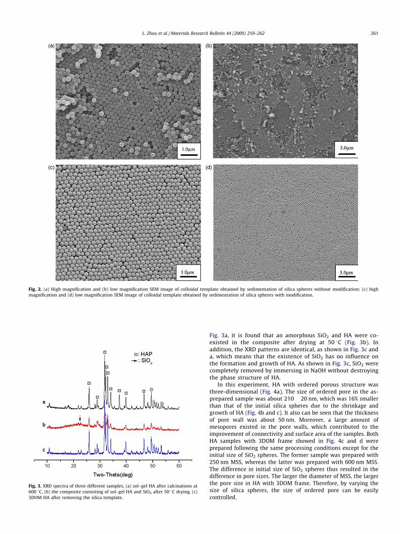
Fig. 2. (a) High magnification and (b) low magnification SEM image of colloidal template obtained by sedimentation of silica spheres without modification; (c) high
magnification and (d) low magnification SEM image of colloidal template obtained by sedimentation of silica spheres with modification.
Fig. 3. XRD spectra of three different samples, (a) sol–gel HA after calcinations at
600 8C, (b) the composite consisting of sol–gel HA and SiO2 after 50 8C drying, (c)
3DOM HA after removing the silica template.
L. Zhou et al. / Materials Research Bulletin 44 (2009) 259–262 261
Fig. 3a, it is found that an amorphous SiO2 and HA were co-existed in the composite after drying at 50 8C (Fig. 3b). Inaddition, the XRD patterns are identical, as shown in Fig. 3c anda, which means that the existence of SiO2 has no influence onthe formation and growth of HA. As shown in Fig. 3c, SiO2 werecompletely removed by immersing in NaOH without destroyingthe phase structure of HA.
In this experiment, HA with ordered porous structure wasthree-dimensional (Fig. 4a). The size of ordered pore in the as-prepared sample was about 210 � 20 nm, which was 16% smallerthan that of the initial silica spheres due to the shrinkage andgrowth of HA (Fig. 4b and c). It also can be seen that the thicknessof pore wall was about 50 nm. Moreover, a large amount ofmesopores existed in the pore walls, which contributed to theimprovement of connectivity and surface area of the samples. BothHA samples with 3DOM frame showed in Fig. 4c and d wereprepared following the same processing conditions except for theinitial size of SiO2 spheres. The former sample was prepared with250 nm MSS, whereas the latter was prepared with 600 nm MSS.The difference in initial size of SiO2 spheres thus resulted in thedifference in pore sizes. The larger the diameter of MSS, the largerthe pore size in HA with 3DOM frame. Therefore, by varying thesize of silica spheres, the size of ordered pore can be easilycontrolled.
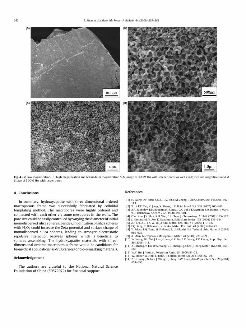
Fig. 4. (a) Low magnification, (b) high magnification and (c) medium magnification SEM image of 3DOM HA with smaller pores as well as (d) medium magnification SEM
image of 3DOM HA with larger pores.
L. Zhou et al. / Materials Research Bulletin 44 (2009) 259–262262
4. Conclusions
In summary, hydroxyapatite with three-dimensional orderedmacroporous frame was successfully fabricated by colloidaltemplating method. The macropores were highly ordered andconnected with each other via some mesopores in the walls. Thepore size could be easily controlled by varying the diameter of initialmonodispersed silica spheres. Besides, modification of silica sphereswith H2O2 could increase the Zeta potential and surface charge ofmonodispersed silica spheres, leading to stronger electrostaticrepulsive interaction between spheres, which is beneficial tospheres assembling. The hydroxyapatite materials with three-dimensional ordered macroporous frame would be candidates forbiomedical applications as drug carriers or bio-remarking materials.
Acknowledgement
The authors are grateful to the National Natural ScienceFoundation of China (50572072) for financial support.
References
[1] H. Wang, D.F. Zhao, X.D. Li, D.Z. Jin, C.M. Zheng, J. Chin. Ceram. Soc. 34 (2006) 107–113.
[2] X. Li, F.F. Tao, Y. Jiang, X. Zheng, J. Colloid. Interf. Sci. 308 (2007) 460–465.[3] A.A. Zakhidov, R.H. Baughman, Z. Iqbal, C.X. Cui, I. Khayrullin, S.O. Dantas, J. Marti,
V.G. Ralchenko, Science 282 (1998) 897–901.[4] C.W. Kuo, J.Y. Shiu, K.H. Wei, P.L. Chen, J. Chromatogr. A 1162 (2007) 175–179.[5] J. Hamagami, Y. Ato, K. Kanamura, Solid State Ionics 172 (2004) 331–334.[6] Z.F. Liu, Z.G. Jin, W. Li, J.J. Qiu, Mater. Res. Bull. 41 (2006) 119–127.[7] F.Q. Tang, T. Uchikoshi, Y. Sakka, Mater. Res. Bull. 41 (2006) 268–273.[8] Y. Sakka, F.Q. Tang, H. Fudouzi, T. Uchikoshi, Sci. Technol. Adv. Mater. 6 (2005)
915–920.[9] A. Stein, Microporous Mesoporous Mater. 44 (2001) 227–239.
[10] W. Wang, D.L. Shi, J. Lian, G. Yan, G.K. Liu, L.M. Wang, R.C. Ewing, Appl. Phys. Lett.89 (2006) 1–3.
[11] Z.L. Huang, Y. Lin, D.W. Wang, S.G. Zhang, L.J. Chen, J. Inorg. Mater. 16 (2001) 661–666.
[12] W.Y. Hu, J. Wuhan, Polytechn. Univ. 25 (2006) 21–23.[13] W. Stober, A. Fink, E. Bohn, J. Colloid. Interf. Sci. 26 (1968) 62–69.[14] Z.B. Huang, J.N. Gao, J. Wang, F.J. Tang, C.W. Yuan, Acta Phys.-Chim. Sin. 20 (2004)
651–655.