extramedullary plasmacytoma of the larynx - scielo · given the rarity of this lesion we report a...
TRANSCRIPT
410
Case Report Int. Arch. Otorhinolaryngol. 2012;16(3):410-413.
DOI: 10.7162/S1809-97772012000300019
Extramedullary plasmacytoma of the larynx
José Antônio Pinto1, Thiago Branco Sônego2, Marina Spadari Artico2, Carolina de Farias Aires Leal2, Silvana Bellotto3.
1) President of the Brazilian Society of Laryngology and Voice (2001-2003). Director of the Center for Otolaryngology and Head and Neck Surgery of São Paulo.
2) Resident Doctor of the Center for Otolaryngology and Head and Neck Surgery of São Paulo.
3) Otolaryngologist.
Institution: Center for Otolaryngology and Head and Neck Surgery of St. Paul.
Sao Paulo / SP - Brazil.
Address correspondence to: José Antonio Pinto - Alameda Nhambiquaras of 159 - Sao Paulo / SP - Brazil - CEP: 04090-010 - E-mail: [email protected]
Manuscript received June 8, 2010. Article accepted on August 1, 2010.
SUMMARY
Introduction: The extramedullary plasmocytoma is one of the localized forms of malignancy of the plasma cells, which has
multiple myeloma main diagnosis. Its main site to the head and neck, but with a rare presentation in the larynx.
Objective: To describe a case of extramedullary plasmocytoma of the larynx, with literature review.
Case Report: Patient female, 49, referring to intermittent dysphonia for 01 years with progressive worsening associated with
vocal fatigue and vocal effort, with reddish lesion, smooth edges fold left ventricular endoscopy. Being subjected to excisional
biopsy diagnosed with extramedullary histopathological plasmocytoma.
Conclusion: Extramedullary Plasmocytoma must be considered in the differential diagnosis of rare tumors of the larynx. It is
essential after the diagnosis of multiple myeloma research and a “follow up” appropriate.
Keywords: plasmocytoma, larynx, laryngeal diseases.
Int. Arch. Otorhinolaryngol., São Paulo - Brasil, v.16, n.3, p. 410-413, Jul/Aug/September - 2012.
INTRODUCTION
The extramedullary plasmocytoma is a malignancy
of the plasma cells, which together with the solitary
plasmocytoma bone sum less than 10% of this condition
located, which has multiple myeloma (MM) as the main
diagnosis.
MM is the systemic form of the disease, and cancer
is a B lymphocyte is characterized by proliferation of
malignant plasma cells and production of monoclonal
immunoglobulin. Its incidence among hematological
malignancies ranges from 10 to 15%, with increased
frequency in men by a ratio of 1.6:1, being more prevalent
in the sixth decade of life. The extramedullary
plasmocytoma (EMP), one of the localized forms, is a
neoplastic proliferation of monoclonal plasma cells. In
contradiction to other forms, the tumor may be confined to
their place of origin in 80% to 90% are located in the head
and neck, most commonly in the sub epithelial tissue of the
upper aerodigestive tract. It is estimated incidence 4-5% in
the nasal cavity, paranasal sinuses and nasopharynx (1.2).
The PEM of the larynx is a rare presentation of
unknown etiology that accounts for 0.04 to 0.19% of
malignant laryngeal neoplasias (3). The relationship
between men and women is 3:1, primarily affecting
patients over 50 years (4, 5, 6). and secondary symptoms
are often local invasion of tumor mass (7), with only 10-
20% with lymph node (2). Typically lesions are unique and
independent, but may be the first evidence of multiple
myeloma (6) and then progress 10-30% of the time for
diagnosis (4).
The solitary bone plasmocytoma (PSO), otherwise
located mainly affecting the pelvic and long bones of the
extremities with solitary bone lesions and without spinal
cord changes, the progression to the systemic form also
occurs, but more frequently, up to 60% cases up to 10
years.
Given the rarity of this lesion we report a case of
PEM of the larynx, taking into account its clinical, pathological
and therapeutic.
CASE REPORT
Female patient, aged 49, teacher, referring to our
service intermittent dysphonia a year ago with progressive
worsening associated with vocal effort and vocal fatigue.
During the physical examination Maximum phonation
time of 16 seconds [s] = 18 seconds, unable to perform the
[z]. It was showed slight harshness and breathiness of the
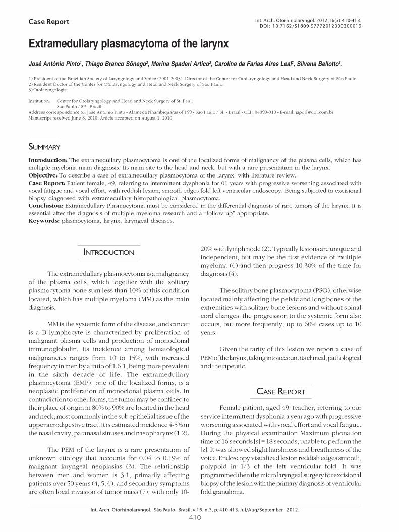
voice. Endoscopy visualized lesion reddish edges smooth,
polypoid in 1/3 of the left ventricular fold. It was
programmed then the micro laryngeal surgery for excisional
biopsy of the lesion with the primary diagnosis of ventricular
fold granuloma.
411
Int. Arch. Otorhinolaryngol., São Paulo - Brasil, v.16, n.3, p. 410-413, Jul/Aug/September - 2012.
Extramedullary plasmacytoma of the larynx. Pinto et al.
Patient undergoing the procedure where the
microscopic examination of the lesion damage was observed
in red, well vascularized, from third average throat left
ventricle, the consistency of soft tissue without significant
infiltration (Figure 1). 0.5 x0 resected lesion, 7 cm with the
help of CO2 laser material and sent for pathological
examination.
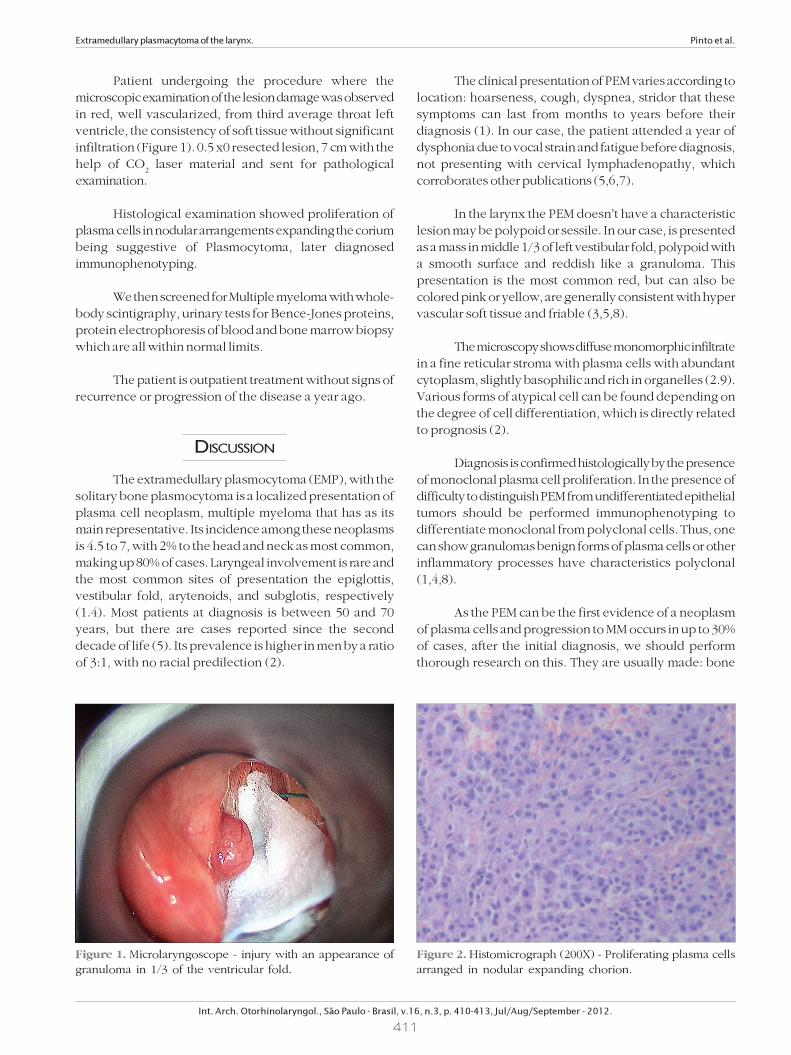
Histological examination showed proliferation of
plasma cells in nodular arrangements expanding the corium
being suggestive of Plasmocytoma, later diagnosed
immunophenotyping.
We then screened for Multiple myeloma with whole-
body scintigraphy, urinary tests for Bence-Jones proteins,
protein electrophoresis of blood and bone marrow biopsy
which are all within normal limits.
The patient is outpatient treatment without signs of
recurrence or progression of the disease a year ago.
DISCUSSION
The extramedullary plasmocytoma (EMP), with the
solitary bone plasmocytoma is a localized presentation of
plasma cell neoplasm, multiple myeloma that has as its
main representative. Its incidence among these neoplasms
is 4.5 to 7, with 2% to the head and neck as most common,
making up 80% of cases. Laryngeal involvement is rare and
the most common sites of presentation the epiglottis,
vestibular fold, arytenoids, and subglotis, respectively
(1.4). Most patients at diagnosis is between 50 and 70
years, but there are cases reported since the second
decade of life (5). Its prevalence is higher in men by a ratio
of 3:1, with no racial predilection (2).
The clinical presentation of PEM varies according to
location: hoarseness, cough, dyspnea, stridor that these
symptoms can last from months to years before their
diagnosis (1). In our case, the patient attended a year of
dysphonia due to vocal strain and fatigue before diagnosis,
not presenting with cervical lymphadenopathy, which
corroborates other publications (5,6,7).
In the larynx the PEM doesn’t have a characteristic
lesion may be polypoid or sessile. In our case, is presented
as a mass in middle 1/3 of left vestibular fold, polypoid with
a smooth surface and reddish like a granuloma. This
presentation is the most common red, but can also be
colored pink or yellow, are generally consistent with hyper
vascular soft tissue and friable (3,5,8).
The microscopy shows diffuse monomorphic infiltrate
in a fine reticular stroma with plasma cells with abundant
cytoplasm, slightly basophilic and rich in organelles (2.9).
Various forms of atypical cell can be found depending on
the degree of cell differentiation, which is directly related
to prognosis (2).
Diagnosis is confirmed histologically by the presence
of monoclonal plasma cell proliferation. In the presence of
difficulty to distinguish PEM from undifferentiated epithelial
tumors should be performed immunophenotyping to
differentiate monoclonal from polyclonal cells. Thus, one
can show granulomas benign forms of plasma cells or other
inflammatory processes have characteristics polyclonal
(1,4,8).
As the PEM can be the first evidence of a neoplasm
of plasma cells and progression to MM occurs in up to 30%
of cases, after the initial diagnosis, we should perform
thorough research on this. They are usually made: bone
Figure 1. Microlaryngoscope - injury with an appearance of
granuloma in 1/3 of the ventricular fold.
Figure 2. Histomicrograph (200X) - Proliferating plasma cells
arranged in nodular expanding chorion.

412
marrow biopsy, imaging tests to rule out lytic lesions,
electrophoresis and immunoelectrophoresis of serum
protein and urine test for Bence Jones proteins. The MRI
is the test of choice to highlight the lytic lesions (8).
There are studies that suggest an association between
laryngeal amyloidosis with extramedullary plasmocytoma.
VELEZ et al in 2008 reported a case that was confirmed by
histological examination of this correlation. In the literature
review conducted by the author shows that the first report
of correlation between these diseases happened in 2001
by Nagasaka et al. accordingly, in cases of laryngeal
amyloidosis is important to research pathological process
in lymphomatous laryngeal lesion (10). This association
was not observed in our case.
The differential diagnosis includes plasma cell
granuloma, pseudo lymphoma, undifferentiated carcino-
ma and metastases. The PEM has been differentiated from
the benign form of plasma cell granuloma by
immunophenotyping, in which this disease presents
polyclonal chains and other inflammatory cells (1).
Due to the low incidence of PEM, there is still no
consensus on optimal therapy. Radiation therapy is accepted
as the standard treatment, even if not standardized dose
and period, and no study showing the advantage over
surgery alone (2.11). The non-mutilating surgery, especially
in areas easily accessible to small and localized lesions,
promotes the same benefit with less morbidity (3,8). In our
case was chosen by the micro laryngeal surgery with CO2
laser and resection of the lesion, with subsequent
“screanning” for MM. The patient keeps up with no signs
of recurrence or disease progression.
Adjuvant chemotherapy may be used to prevent
progression to MM, however, its role remains uncertain.
Several studies suggest that chemotherapy increases the
clearance of proteins M and reduces progression to MM,
while others found no benefit (5).
The conversion of the PEM to MM ranges from 10 to
30%, significantly lower than the progression of the PSO.
Kapadia et al (1982) and HOLLAND et al (1992) observed
the progression to MM in their patients over a period of two
years, suggesting this period as high risk. Most authors
recommend a “follow-up” long since found cases of
conversion to MM 15 years after diagnosis of localized
disease (2).
The prognosis is related to the location of the tumor,
cartilage and bone destruction, and regional lymph node
involvement. Survival is higher in patients with localized
disease than in those with MM, with a five-year survival of
18% in patients with MM and approximately 66% among
those with PEM. (5). Patients who developed MM after the
diagnosis of PEM have a longer survival than those who
have MM as the initial diagnosis.
CONCLUSION
Extramedullary plasmocytoma should be considered
in the differential diagnosis of rare tumors of the larynx. It
is essential after the diagnosis of multiple myeloma research
and a “follow up” appropriate given that their progression
to the systemic form is well documented.
Radiotherapy is an effective therapy for PEM with
high rates of local control and the surgery is an option for
small and localized lesions, as described in our case.
REFERENCES
1. Zbären P, Läng H, Beer K, Becker M. Plasma cell granuloma
of the supraglottic larynx. J Laryngol Otol, 1995; 109(9):895-
8.
2. Susnerwala SS, Shanks JH, Banerjee SS, Scarffe JH,
Farrington WT, Slevin NJ. Extramedullary plasmocytoma of
the head and neck region: clinicopathological correlation in
25 cases. Br J Cancer, 1997; 75(6):921-7.
3. Bjelkenkratz K, Lundgren J, Olofsson J. Extramedullary
plasmocytoma of the larynx. J Otolaryngol, 1981; 10(1):28-
34.
4. Pratibha CB, Sreenivas V, Babu MK, Rout P, Nayar RC.
Plasmacytoma of Larynx - A Case Report. J. Voice, 2009;
23(6):735-8.
5. Gorenstein A, Bryan Neel H, Devine KD, Weiland LH.
Solitary extramedullary plasmocytoma of the larynx. Arch
Otolaryngol, 1977; 103(3):159-61.
6. Maniglia AJ, Xue JW. Plasmocytoma of the larynx.
Laryngoscope, 1983; 93(6):741-744.
7. Nakashima T, Matsuda K, Haruta A. Extramedullary
plasmocytoma of the larynx. Auris Auris Nasus Larynx, 2006;
33(2):219-22.
8. Hotz MA, Bosq J, Schwaab G, Munck J. Extramedullary
solitary plasmocytoma of the head and neck. Ann Otol Rhinol
Laryngol, 1999; 108(5):495-500.
9. Jyothirmayi R, Gangadharan VP, Nair MK, Rajan B.
Radiotherapy in the treatment of solitary plasmocytoma.
Br J Radiol, 1997; 70(833):511-6.
Extramedullary plasmacytoma of the larynx. Pinto et al.
Int. Arch. Otorhinolaryngol., São Paulo - Brasil, v.16, n.3, p. 410-413, Jul/Aug/September - 2012.

413
Extramedullary plasmacytoma of the larynx. Pinto et al.
Int. Arch. Otorhinolaryngol., São Paulo - Brasil, v.16, n.3, p. 410-413, Jul/Aug/September - 2012.
10. Velez D, Hinojar-Gutierrez Z, Nam-Cha S, Acevedo-
Barbera 2007 A. Laryngeal Plasmacytoma presenting as
amyloid tumour: a case report. Eur Arch Otorhinolaryngol,
2007; 264(8):959-61.
11. Creach KM, Foote RL, Neben-Wittich MA, Kyle RA.
Radiotherapy for extramedullary plasmocytoma of the head
and neck. Int J Radiat Oncol Biol Phys, 2009; 73(3):789-94.