case report spinal intradural, extramedullary ependymoma...
TRANSCRIPT
Case ReportSpinal Intradural, Extramedullary Ependymomawith Astrocytoma Component: A Case Report andReview of the Literature
Gene M. Weinstein,1 Knarik Arkun,2 James Kryzanski,3
Michael Lanfranchi,4 Gaurav K. Gupta,2 and Harprit Bedi1
1Department of Radiology, Tufts Medical Center, Boston, MA, USA2Department of Pathology and Laboratory Medicine, Tufts Medical Center, Boston, MA, USA3Department of Neurosurgery, Tufts Medical Center, Boston, MA, USA4Department of Radiology, Massachusetts General Hospital, Boston, MA, USA
Correspondence should be addressed to Gene M. Weinstein; [email protected]
Received 27 February 2016; Accepted 10 April 2016
Academic Editor: Yoji Nagashima
Copyright © 2016 Gene M. Weinstein et al. This is an open access article distributed under the Creative Commons AttributionLicense, which permits unrestricted use, distribution, and reproduction in any medium, provided the original work is properlycited.
Ependymomas are common spinal lesions, with the vast majority arising in an intramedullary location. Several cases have beendescribed in the literature of ependymomas in an intradural, extramedullary location. The authors present a case of a 56-year-oldfemale who presented with several weeks of lower back pain and weakness. MRI revealed an intradural, extramedullary enhancingmass at L1-L2.Themass was successfully resected surgically. Pathologic evaluation revealed a low grade glioma with components ofboth ependymoma and pilocytic astrocytomawithMUTYHG382Dmutation. Extramedullary ependymomas are very rare tumors.To the authors’ knowledge, this is the first case of ependymoma/astrocytoma collision tumors described in an extramedullarylocation.
1. Introduction
Most adult ependymomas are spinal lesions, with the vastmajority arising in an intradural, intramedullary location [1].Intradural, extramedullary ependymomas are exceptionallyrare tumors that have been reported in the literature [2, 3].Wereport a case of an intradural, extramedullary ependymomawith an astrocytoma component. To the best of our knowl-edge, this has not been described in the literature before. Boththe imaging and pathologic features are described here.
2. Case Report
A 56-year-old female presented to an outside hospital forseveral months of low back pain, bilateral hip pain, and legpain/weakness. She subsequently had an episode of severelow back pain that resulted in difficulty breathing and loss ofconsciousness. Review of systems revealed several episodes of
incontinence. Physical exam was remarkable for 5/5 strengthin all extremities, 2+ reflexes in the lower extremities, and anegative Babinski sign.
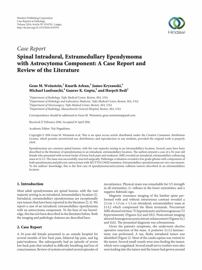
Magnetic resonance imaging of the lumbar spine per-formed with and without intravenous contrast revealed a2.1 cm × 1.5 cm × 1.3 cm intradural, extramedullary mass atL1-L2 which compressed the filum terminale. PrecontrastMRI showed intrinsic T1 hypointensity andheterogeneousT2hyperinstensity (Figures 1(a) and 1(b)). Postcontrast imagingshowed homogenous postcontrast enhancement (Figures 1(c)and 1(d)). The presumed diagnosis was schwannoma.
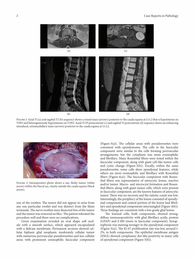
Given the patient’s symptoms, she underwent electiveoperative resection of the mass. A posterior L1-L2 laminec-tomy was performed. A tan, fleshy intradural tumor wasidentified (Figure 2). Most of the cauda equina was ventral tothe tumor. Several small vessels were seen feeding the tumor,which were coagulated. Several small nerve rootlets were alsoseen leading into the tumor and the tumor had grown around
Hindawi Publishing CorporationCase Reports in PathologyVolume 2016, Article ID 3534791, 5 pageshttp://dx.doi.org/10.1155/2016/3534791
2 Case Reports in Pathology
(a) (c)
(b) (d)
Figure 1: Axial T1 (a) and sagittal T2 (b) sequence shows a roundmass (arrows) posterior to the cauda equina at L1-L2 that is hypointense onT1WI and heterogeneously hyperintense on T2WI. Axial T1 FS postcontrast (c) and sagittal T1 postcontrast (d) sequence shows an enhancingintradural, extramedullary mass (arrows) posterior to the cauda equina at L1-L2.
Figure 2: Intraoperative photo shows a tan, fleshy tumor (whitearrow) within the thecal sac, clearly outside the cauda equina (blackarrow).
one of the rootlets. The tumor did not appear to arise fromany one particular rootlet and was distinct from the filumterminale.The nerve rootlets were dissected free of the tumorand the tumor was removed en bloc.The patient tolerated theprocedure well and there were no complications.
Gross examination revealed an oval shape soft nod-ule with a smooth surface, which appeared encapsulatedwith a delicate membrane. Permanent sections showed cel-lular biphasic glial neoplasm: moderately cellular tumorwith numerous perivascular pseudorosettes and less cellularareas with prominent eosinophilic fascicular component
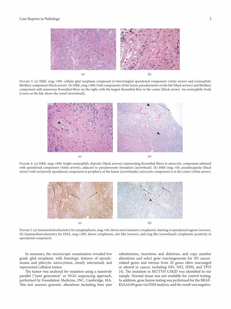
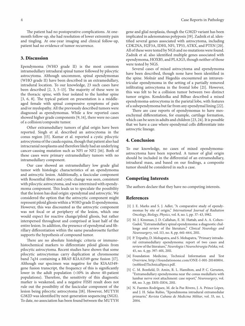
(Figure 3(a)). The cellular areas with pseudorosettes wereconsistent with ependymoma. The cells in the fascicularcomponent were similar to the cells forming perivasculararrangements, but the cytoplasm was more eosinophilicand fibrillary. Many Rosenthal fibers were noted within thefascicular component, along with giant cell-like tumor cellsand cystic change (Figure 3(b)). Focally, within the samepseudorosette, some cells show ependymal features, whileothers are more eosinophilic and fibrillary with Rosenthalfibers (Figure 4(a)). The fascicular component with Rosen-thal fibers was representative of astrocytic lesion, reactiveand/or tumor. Macro- and microcyst formation and Rosen-thal fibers, along with giant tumor cells, which were presentin fascicular component, are the known features of astrocytictumor. There was no necrosis and the mitotic count was low.Interestingly, the periphery of the lesion consisted of ependy-mal component and central portion of the lesion had fibril-lary and ependymal components intermingled (Figure 4(b)).These findings are consistent with a low grade glial lesion.
The lesional cells, both components, showed strong,diffuse immunopositivity with glial fibrillary acidic protein(GFAP) and S-100 (more in fascicular component). Synap-tophysin was staining stronger in the ependymal component(Figure 5(a)). The Ki-67 proliferation rate was low, around 1-2%, in both components. The epithelial membrane antigen(EMA) showed cytoplasmic dot-like positivity in many cellsof ependymal component (Figure 5(b)).
Case Reports in Pathology 3
(a) (b)
Figure 3: (a) H&E, mag ×100, cellular glial neoplasm composed of intermingled ependymal component (white arrow) and eosinophilicfibrillary component (black arrow). (b) H&E,mag ×400, both components of the lesion, pseudorosette on the left (black arrows) and fibrillarycomponent with numerous Rosenthal fibers on the right, with the largest Rosenthal fiber in the center (black arrow). An eosinophilic bodyis seen on the left, above the vessel (arrowhead).
(a) (b)
Figure 4: (a) H&E, mag ×400, bright eosinophilic deposits (black arrows) representing Rosenthal fibers in astrocytic component admixedwith ependymal component (white arrows), adjacent to pseudorosette formation (arrowhead). (b) H&E mag ×40, pseudocapsule (blackarrow) with exclusively ependymal component at periphery of the lesion (arrowheads); astrocytic component is in the center (white arrow).
(a) (b)
Figure 5: (a) Immunohistochemistry for synaptophysin,mag×40, showsmore intensive cytoplasmic staining in ependymal regions (arrows).(b) Immunohistochemistry for EMA, mag ×200, shows cytoplasmic, dot-like (arrows), and ring-like (arrowhead) cytoplasmic positivity inependymal component.
In summary, the microscopic examination revealed lowgrade glial neoplasm, with histologic features of ependy-moma and pilocytic astrocytoma, closely intermixed, andrepresented collision tumor.
The tumor was analyzed for mutation using a massivelyparallel (“next generation” or NGS) sequencing approach,performed by Foundation Medicine, INC, Cambridge, MA.This test assesses genomic alterations including base pair
substitutions, insertions and deletions, and copy numberalterations and select gene rearrangements for 315 cancer-related genes and introns from 28 genes often rearrangedor altered in cancer, including NF1, NF2, IDH1, and TP53[4]. The mutation in MUTYH G382D was identified in oursample. Normal tissue was not available for control testing.In addition, gene fusion testing was performed for the BRAF-KIAA1549 gene via FISH analysis, and the result was negative.
4 Case Reports in Pathology
The patient had no postoperative complications. At one-month follow-up, she had resolution of lower extremity painand tingling. At one-year imaging and clinical follow-up,patient had no evidence of tumor recurrence.
3. Discussion
Ependymoma (WHO grade II) is the most commonintramedullary intradural spinal tumor followed by pilocyticastrocytoma. Although uncommon, spinal ependymomas(WHO grade II) have been described in an extramedullary,intradural location. To our knowledge, 23 such cases havebeen described [2, 3, 5–13]. The majority of these were inthe thoracic spine, with four isolated to the lumbar spine[3, 6, 8]. The typical patient on presentation is a middle-aged female with spinal compressive symptoms of painand/or myelopathy. All the previously described tumors werediagnosed as ependymomas. While a few reported casesshowed higher grade components [9, 14], there were no casesof a collision/composite tumor.
Other extramedullary tumors of glial origin have beenreported. Singh et al. described an astrocytoma in theconus region [15]. Kumar et al. reported a cystic pilocyticastrocytoma of the cauda equina, though that patient also hadintracranial neoplasms and therefore likely had an underlyingcancer-causing mutation such as NF1 or NF2 [16]. Both ofthese cases were primary extramedullary tumors with nointramedullary component.
Our case showed an extramedullary low grade glialtumor with histologic characteristics of an ependymomaand astrocytic lesion. Additionally, a fascicular componentwith Rosenthal fibers and cystic change was seen, consistentwith pilocytic astrocytoma, andwas intermixed with ependy-moma component. This leads us to speculate the possibilitythat the lesion has dual origin: ependymal and astrocytic. Weconsidered the option that the astrocytic component mightrepresent piloid gliosis within aWHOgrade II ependymoma.However, this was discounted as the astrocytic componentwas not focal or at periphery of the lesion, which onewould expect for reactive change/piloid gliosis, but ratherinterspersed throughout and comprised at least half of theentire lesion. In addition, the presence of ependymal and fib-rillary differentiation within the same pseudorosette furthersupports the hypothesis of compound tumor.
There are no absolute histologic criteria or immuno-histochemical markers to differentiate piloid gliosis frompilocytic astrocytoma. Recent studies have shown that somepilocytic astrocytomas carry duplication at chromosomeband 7q34 containing a BRAF-KIAA1549-gene fusion [17].Although our specimen was negative for the KIAA1549-gene fusion transcript, the frequency of this is significantlylower in the adult population (<10% in above 40-patientpopulations). Therefore, the sensitivity of this diagnosticmarker is weakened, and a negative FISH result does notrule out the possibility of the fascicular component of thelesion being pilocytic astrocytoma [18]. However, MUTYHG382D was identified by next-generation sequencing (NGS).To date, no association has been found between theMUTYH
gene and glial neoplasia, though the G382D variant has beenimplicated in adenomatous polyposis [19]. Zadnik et al. iden-tified several genes associated with astrocytoma, includingCDK2NA, H2F3A, IDH1, NF1, TP53, ATRX, and PTEN [20].All of these were tested byNGS and nomutationswere found.Zadnik et al. also identified multiple genes associated withependymoma,HOXB5, and PLA2G5, though neither of thosewere tested by NGS.
Several cases of mixed astrocytoma and ependymomahave been described, though none have been identified inthe spine. Molnar and Hegedus encountered an intraven-tricular ependymoma in the setting of a partially removedinfiltrating astrocytoma in the frontal lobe [21]. However,this was felt to be a collision tumor between two distincttumor origins. Kondziolka and Bilbao described a mixedependymoma-astrocytoma in the parietal lobe, with featuresof a subependymoma but far from any ependymal lining [22].
There are case reports of ependymomas to have mes-enchymal differentiation, for example, cartilage formation,which can be seen in adults and children [23, 24]. It is possiblethat we have a case where ependymal cells differentiate intoastrocytic lineage.
4. Conclusion
To our knowledge, no cases of mixed ependymoma-astrocytoma have been reported. A tumor of glial originshould be included in the differential of an extramedullary,intradural mass, and based on our findings, a compositetumor should be considered in such a case.
Competing Interests
The authors declare that they have no competing interests.
References
[1] J. E. Marks and S. J. Adler, “A comparative study of ependy-momas by site of origin,” International Journal of RadiationOncology, Biology, Physics, vol. 8, no. 1, pp. 37–43, 1982.
[2] M. J. Kinsman, J. D. Callahan, E. M. Hattab, and A. A. Cohen-Gadol, “Extramedullary spinal ependymoma: a diagnostic chal-lenge and review of the literature,” Clinical Neurology andNeurosurgery, vol. 113, no. 8, pp. 661–664, 2011.
[3] P. Tripathy, D.Mohapatra, and S.Mohapatra, “Primary intradu-ral extramedullary ependymoma: report of two cases andreview of the literature,”Neurologia i Neurochirurgia Polska, vol.45, no. 4, pp. 397–401, 2011.
[4] Foundation Medicine, Technical Information and TestOverview, http://foundationone.com/ONE-I-001-20140804(nobleed)TechnicalSpecs.pdf.
[5] C. M. Bonfield, D. Amin, R. L. Hamilton, and P. C. Gerszten,“Extramedullary ependymoma near the conus medullaris withlumbar nerve root attachment: case report,” Neurosurgery, vol.68, no. 3, pp. E831–E834, 2011.
[6] N. Fuentes Rodrıguez, M. de la Paz Rivero, J. A. Prince Lopez,and J. H. Salas Rubio, “Ependimoma intradural extramedularprimario,” Revista Cubana de Medicina Militar, vol. 33, no. 1,2004.
Case Reports in Pathology 5
[7] J. Graca, N. Gultasli, N. D’Haene, J. Brotchi, I. Salmon, andD. Baleriaux, “Cystic extramedullary ependymoma,” AmericanJournal of Neuroradiology, vol. 27, no. 4, pp. 818–821, 2006.
[8] E. A. Iunes, J. N. Stavale, R. D. C. C. Pessoa et al., “Multifocalintradural extramedullary ependymoma: case report,” Journalof Neurosurgery: Spine, vol. 14, no. 1, pp. 65–70, 2011.
[9] T. Ikata, S. Katoh, A. Inou, and M. Takahashi, “Intraduralextramedullary ependymoma: a case report,” Spine, vol. 20, no.18, pp. 2036–2038, 1995.
[10] M. H. Li, S. Holtas, and E.-M. Larsson, “MR imaging ofintradural extramedullary tumors,”ActaRadiologica, vol. 33, no.3, pp. 207–212, 1992.
[11] T. Moriwaki, K. Iwatsuki, Y.-I. Ohnishi, M. Umegaki, M.Ishihara, and T. Yoshimine, “Intradural extramedullary spinalependymoma: a case report ofmalignant transformation occur-ring,” Asian Spine Journal, vol. 7, no. 2, pp. 139–142, 2013.
[12] B. Oliver, A. de Castro, M. A. Sarmiento, C. Arguello, and M.G. Blazquez, “Dorsal extramedullary ependymona,”Archivos deNeurobiologia, vol. 44, no. 4, pp. 215–224, 1981.
[13] M. Schuurmans, J. A. L. Vanneste, M. J. T. Verstegen, and W.R. van Furth, “Spinal extramedullary anaplastic ependymomawith spinal and intracranial metastases,” Journal of Neuro-Oncology, vol. 79, no. 1, pp. 57–59, 2006.
[14] A. Cerase, C. Venturi, G. Oliveri, D. De Falco, and C. Miracco,“Intradural extramedullary spinal anaplastic ependymoma.Case illustration,” Journal of Neurosurgery: Spine, vol. 5, no. 5,p. 476, 2006.
[15] P. Singh, A. P. Singh, T. Rajaram, and A. K. Sabhikhi,“Extramedullary astrocytoma of conus region: a short report,”Neurology India, vol. 49, no. 1, pp. 97–99, 2001.
[16] A. Kumar, A. Garg, M. C. Sharma, B. S. Sharma, and S. S. Kale,“Giant cystic intradural extramedullary pilocytic astrocytomaof Cauda equina,” Journal of Neurosciences in Rural Practice, vol.4, no. 4, pp. 453–456, 2013.
[17] M. Hasselblatt, B. Riesmeier, B. Lechtape et al., “BRAF-KIAA1549 fusion transcripts are less frequent in pilocyticastrocytomas diagnosed in adults,”Neuropathology and AppliedNeurobiology, vol. 37, no. 7, pp. 803–806, 2011.
[18] A. J. Sievert, S.-S. Lang, K. L. Boucher et al., “Paradoxicalactivation and RAF inhibitor resistance of BRAF protein kinasefusions characterizing pediatric astrocytomas,” Proceedings ofthe National Academy of Sciences of the United States of America,vol. 110, no. 15, pp. 5957–5962, 2013.
[19] V. Gismondi, M. Meta, L. Bonelli et al., “Prevalence of theY165C, G382D and 1395delGGA germline mutations of theMYH gene in Italian patients with adenomatous polyposis coliand colorectal adenomas,” International Journal of Cancer, vol.109, no. 5, pp. 680–684, 2004.
[20] P. L. Zadnik, Z. L. Gokaslan, P. C. Burger, and C. Bettegowda,“Spinal cord tumours: advances in genetics and their implica-tions for treatment,”Nature Reviews Neurology, vol. 9, no. 5, pp.257–266, 2013.
[21] P. Molnar and K. Hegedus, “Adjacent astrocytoma andependymoma: dependent mixed neoplasia or unusual collisiontumors?” Surgical Neurology, vol. 22, no. 5, pp. 455–460, 1984.
[22] D. Kondziolka and J. M. Bilbao, “Mixed ependymoma-astrocytoma (subependymoma?) of the cerebral cortex,” ActaNeuropathologica, vol. 76, no. 6, pp. 633–637, 1988.
[23] A. Coli, M. Novello, L. Massimi, M. Caldarelli, V. Ranucci,and L. Lauriola, “Cartilage differentiation in ependymoma:histogenetic considerations on a new case,” Child’s NervousSystem, vol. 30, no. 7, pp. 1301–1305, 2014.
[24] M.-M. Ruchoux, J. J. Kepes, P. Dhellemmes et al., “Lipomatousdifferentiation in ependymomas: a report on three cases andcomparison changes reported in other central nervous systemneoplasms of neuroectodermal origin,” American Journal ofSurgical Pathology, vol. 22, no. 3, pp. 338–346, 1998.

Submit your manuscripts athttp://www.hindawi.com
Stem CellsInternational
Hindawi Publishing Corporationhttp://www.hindawi.com Volume 2014
Hindawi Publishing Corporationhttp://www.hindawi.com Volume 2014
MEDIATORSINFLAMMATION
of
Hindawi Publishing Corporationhttp://www.hindawi.com Volume 2014
Behavioural Neurology
EndocrinologyInternational Journal of
Hindawi Publishing Corporationhttp://www.hindawi.com Volume 2014
Hindawi Publishing Corporationhttp://www.hindawi.com Volume 2014
Disease Markers
Hindawi Publishing Corporationhttp://www.hindawi.com Volume 2014
BioMed Research International
OncologyJournal of
Hindawi Publishing Corporationhttp://www.hindawi.com Volume 2014
Hindawi Publishing Corporationhttp://www.hindawi.com Volume 2014
Oxidative Medicine and Cellular Longevity
Hindawi Publishing Corporationhttp://www.hindawi.com Volume 2014
PPAR Research
The Scientific World JournalHindawi Publishing Corporation http://www.hindawi.com Volume 2014
Immunology ResearchHindawi Publishing Corporationhttp://www.hindawi.com Volume 2014
Journal of
ObesityJournal of
Hindawi Publishing Corporationhttp://www.hindawi.com Volume 2014
Hindawi Publishing Corporationhttp://www.hindawi.com Volume 2014
Computational and Mathematical Methods in Medicine
OphthalmologyJournal of
Hindawi Publishing Corporationhttp://www.hindawi.com Volume 2014
Diabetes ResearchJournal of
Hindawi Publishing Corporationhttp://www.hindawi.com Volume 2014
Hindawi Publishing Corporationhttp://www.hindawi.com Volume 2014
Research and TreatmentAIDS
Hindawi Publishing Corporationhttp://www.hindawi.com Volume 2014
Gastroenterology Research and Practice
Hindawi Publishing Corporationhttp://www.hindawi.com Volume 2014
Parkinson’s Disease
Evidence-Based Complementary and Alternative Medicine
Volume 2014Hindawi Publishing Corporationhttp://www.hindawi.com