extended report treatment outcome in early...
TRANSCRIPT

EXTENDED REPORT
Treatment outcome in early diffuse cutaneoussystemic sclerosis: the European SclerodermaObservational Study (ESOS)Ariane L Herrick,1,2 Xiaoyan Pan,3 Sébastien Peytrignet,3 Mark Lunt,3
Roger Hesselstrand,4 Luc Mouthon,5 Alan Silman,6 Edith Brown,7 László Czirják,8
Jörg H W Distler,9 Oliver Distler,10 Kim Fligelstone,11 William J Gregory,12
Rachel Ochiel,11 Madelon Vonk,13 Codrina Ancuţa,14 Voon H Ong,15
Dominique Farge,16 Marie Hudson,17 Marco Matucci-Cerinic,18 Alexandra Balbir-Gurman,19 Øyvind Midtvedt,20 Alison C Jordan,21 Paresh Jobanputra,21
Wendy Stevens,22 Pia Moinzadeh,23 Frances C Hall,24 Christian Agard,25 MarinaE Anderson,26 Elisabeth Diot,27 Rajan Madhok,28 Mohammed Akil,29 Maya H Buch,30
Lorinda Chung,31 Nemanja Damjanov,32 Harsha Gunawardena,33 Peter Lanyon,34
Yasmeen Ahmad,35 Kuntal Chakravarty,36 Søren Jacobsen,37
Alexander J MacGregor,38 Neil McHugh,39 Ulf Müller-Ladner,40
Gabriela Riemekasten,41 Michael Becker,42 Janet Roddy,43 Patricia E Carreira,44
Anne Laure Fauchais,45 Eric Hachulla,46 Jennifer Hamilton,47 Murat İnanç,48
John S McLaren,49 Jacob M van Laar,50 Sanjay Pathare,51 Susannah Proudman,52
Anna Rudin,53 Joanne Sahhar,54 Brigitte Coppere,55 Christine Serratrice,56
Tom Sheeran,57 Douglas J Veale,58 Claire Grange,59 Georges-Selim Trad,60
Christopher P Denton15
ABSTRACTObjectives The rarity of early diffuse cutaneoussystemic sclerosis (dcSSc) makes randomised controlledtrials very difficult. We aimed to use an observationalapproach to compare effectiveness of currently usedtreatment approaches.Methods This was a prospective, observational cohortstudy of early dcSSc (within three years of onset of skinthickening). Clinicians selected one of four protocols foreach patient: methotrexate, mycophenolate mofetil(MMF), cyclophosphamide or ‘no immunosuppressant’.Patients were assessed three-monthly for up to24 months. The primary outcome was the change inmodified Rodnan skin score (mRSS). Confounding byindication at baseline was accounted for using inverseprobability of treatment (IPT) weights. As a secondaryoutcome, an IPT-weighted Cox model was used to testfor differences in survival.Results Of 326 patients recruited from 50 centres,65 were prescribed methotrexate, 118 MMF, 87cyclophosphamide and 56 no immunosuppressant.276 (84.7%) patients completed 12 and 234 (71.7%)24 months follow-up (or reached last visit date). Therewere statistically significant reductions in mRSS at12 months in all groups: −4.0 (−5.2 to −2.7) units formethotrexate, −4.1 (−5.3 to −2.9) for MMF, −3.3(−4.9 to −1.7) for cyclophosphamide and −2.2 (−4.0to −0.3) for no immunosuppressant (p value forbetween-group differences=0.346). There were nostatistically significant differences in survival between
protocols before (p=0.389) or after weighting(p=0.440), but survival was poorest in the noimmunosuppressant group (84.0%) at 24 months.Conclusions These findings may support usingimmunosuppressants for early dcSSc but suggest thatoverall benefit is modest over 12 months and that bettertreatments are needed.Trial registration number NCT02339441.
INTRODUCTIONThe diffuse cutaneous subtype of systemic sclerosis(dcSSc) is rare (SSc incidence is around 10–20/million/year,1 of whom approximately 25% willhave diffuse disease) but carries high morbidity andmortality due to early internal organ involvementand rapidly progressive, painful skin thickening.Also, 5-year and 10-year survival rates, althoughimproving, are in the order of 68% and 50%,respectively.2 3
At present, there is no drug known to favourablyinfluence disease course. Randomised controlledtrials (RCTs) have historically been confounded bydisease rarity (only small numbers of patients arerecruited, often over long periods) and strict entrycriteria meaning that severe cases are oftenexcluded.4 These strict criteria further restrictsample sizes and limit generalisability. Therefore,although RCTs represent a gold standard for asses-sing drug efficacy, results may not be applicable to
1207Herrick AL, et al. Ann Rheum Dis 2017;76:1207–1218. doi:10.1136/annrheumdis-2016-210503
Clinical and epidemiological research
To cite: Herrick AL, Pan X, Peytrignet S, et al. Ann Rheum Dis 2017;76:1207–1218.
Handling editor Tore K Kvien
► Additional material is published online only. To view please visit the journal online (http://dx.doi.org/10.1136/annrheumdis-2016-210503).
For numbered affiliations see end of article.
Correspondence toProfessor Ariane Herrick, The University of Manchester, Salford Royal NHS Foundation Trust, Manchester Academic Health Science Centre, Manchester M13 9PT, UK; [email protected]
Received 12 September 2016Revised 24 November 2016Accepted 25 November 2016Published Online First 10 February 2017
on 24 June 2018 by guest. Protected by copyright.
http://ard.bmj.com
/A
nn Rheum
Dis: first published as 10.1136/annrheum
dis-2016-210503 on 10 February 2017. D
ownloaded from

real-life clinical settings.5 Small trials run the risk of beingunderpowered, thus potentially yielding false-negative results.6
The past three decades have seen a number of promising treat-ments for early dcSSc failing to meet efficacy end points inRCTs: examples include methotrexate (multinational, 71patients)7 and anti-transforming growth factor β1 antibodytherapy (multinational, 45 patients).8
A further difficulty in recruiting into RCTs of early dcSSc isthat many clinicians have reservations about placebo therapy ina potentially life-threatening disease and favour immunosup-pression, consistent with the European League AgainstRheumatism (EULAR) recommendations, which advocatemethotrexate for skin manifestations9 in early dcSSc, althoughthis agent has been shown to be of only limited efficacy.7
Immunosuppressants are potentially hazardous, especially inpatients prone to internal organ disease and infection.
Against this background, our aim was to compare, using anobservational approach, the effectiveness of standard treatmentapproaches (mainly immunosuppressant treatments but includ-ing a ‘no immunosuppressant’ option to reflect that somepatients or clinicians may choose this approach) in the earlymanagement of patients with dcSSc, capturing entry andoutcome data in a systematic way. Modern statistical approachesallow robust interrogations of prospective observational studies,as an adjunct to, or even substitute for, RCTs in rare diseases,10
although the potential of these novel approaches has not yetbeen realised.11
METHODSStudy designThe European Scleroderma Observational Study (ESOS) was aprospective, observational cohort study (ClinicalTrials.gov iden-tifier: NCT02339441), in which standardised data were col-lected at study entry and at follow-up visits, and enteredelectronically by investigators at each centre into an electroniccase record form. All data were checked by the project coordin-ator and any inconsistencies were discussed with the chief inves-tigator and (if appropriate) the local principal investigator. Themain inclusion criteria were early dcSSc (skin involvement prox-imal to elbow, knee, face, neck12 and within three years of theonset of skin thickening) and age >18 years. Exclusion criteriawere previous stem cell transplantation, previous immunosup-pressant treatment for >4 months or use of any immunosup-pressant drug other than methotrexate, mycophenolate mofetil(MMF) or cyclophosphamide within the month prior to studyentry.
Clinicians selected the protocol of their choice for eachpatient. The recommended treatment protocols, as decided bythe Steering Committee to reflect international best clinical prac-tice, were1. Methotrexate (oral or subcutaneous with a target dose of
20–25 mg weekly).2. MMF (500 mg twice daily for 2 weeks increasing to 1 g
twice daily).3. Cyclophosphamide.
Possible regimens included:i. Intravenous. Minimum monthly dose 500 mg/m2 with a
recommended duration of 6–12 months.ii. Oral. 1–2 mg/kg/day with a recommended duration of
12 months. Patients treated with cyclophosphamide werethen usually ‘transferred’ to a maintenance immunosup-pressive drug (methotrexate, MMF or azathioprine) asper the treating clinician’s choice.
4. No immunosuppressant treatment, to give the option ofincluding patients in whom immunosuppression was not feltindicated or appropriate (or declined by the patient).Patients were assessed at baseline, with subsequent visits
scheduled three-monthly for 24 months (or between 12 and24 months for those patients recruited after September 2013).
To have 80% power to detect a difference between two treat-ment arms of five modified Rodnan skin score (mRSS) units at12 months would require 63 patients per protocol. Allowing20% loss to follow-up, and varying numbers recruited to thedifferent protocols, recruitment target was 316 patients.
PatientsPatients were recruited between July 2010 and September 2014.Demographic characteristics including age, gender, smokinghabit, ethnicity, antibody status (anti-topoisomerase-1(anti-Scl70), anti-RNA III polymerase, anticentromere) and pres-ence of visceral organ involvement were recorded for allpatients. The algorithms to determine the presence of differenttypes of organ involvement are summarised in onlinesupplementary table S1.
Outcome measuresThe primary outcome measure, assessed at each visit, was thechange in mRSS over time. All mRSS assessments were per-formed by those experienced in skin scoring. The mRSS isassessed clinically at 17 body sites on a 0–3 scale (maximumscore 51) and measures the extent of skin thickening.13 It is themost commonly used primary outcome measure in RCTs ofdcSSc,4 7 8 reflecting disease severity and predicting mortality.14
All other outcomes/recorded variables were mainly part ofroutine clinical practice and are summarised in onlinesupplementary table S2. Secondary end points included pulmon-ary function (forced vital capacity (FVC: % predicted) andcarbon monoxide diffusing capacity (DLCO: % predicted)),quality of life15–18 (including the Health AssessmentQuestionnaire Disability Index (HAQ-DI)15 and Cochin HandFunction Scale18), occurrence of side effects and survival.
Statistical analysisIn an observational study, patient characteristics differ betweengroups and any differences in outcomes might be driven bythose characteristics rather than the treatments (confounding byindication). In each of the analyses (for the different outcomemeasures), all variables associated with the outcome were con-sidered as confounders.19 20
Differences between protocols at baselineKruskal-Wallis test was applied for continuous variables andFisher’s test for categorical variables.
Influence of baseline characteristics on mRSS at baselineand over timeThe association between baseline variables and mRSS wasassessed by simple linear regressions, entering each characteristicseparately as a predictor of mRSS. To examine how each vari-able affected the progression of mRSS, the regression equationwas modified by adding a term for time and its interaction withthe baseline predictor value.
Differences in the changes between groups for all outcomesInverse probability of treatment (IPT) weights equalise the dis-tributions of confounders between the treatment groups, thusremoving confounding by indication.21 Treatment probabilities
1208 Herrick AL, et al. Ann Rheum Dis 2017;76:1207–1218. doi:10.1136/annrheumdis-2016-210503
Clinical and epidemiological research on 24 June 2018 by guest. P
rotected by copyright.http://ard.bm
j.com/
Ann R
heum D
is: first published as 10.1136/annrheumdis-2016-210503 on 10 F
ebruary 2017. Dow
nloaded from

were computed using multinomial logistic regressions, with thebaseline values of the selected confounders as predictors.22
Censoring weights rebalance the data such that the distributionsof confounders remain unchanged throughout the study. Foreach observation, the probability of remaining uncensored giventhe baseline values of the confounders, the initial protocol and acubic spline for time was calculated using a pooled logisticregression model.23 Multiplying both weights yielded the IPTand inverse probability of censoring (IPTC) weights. Weights>20 were truncated at that value.24
Treatment effects were assessed using IPTC-weighted linearregression models, which include an intercept, a time term, indi-cator variables for treatment groups and interactions betweentime and treatments. The model followed an intention-to-treatapproach. Differences in the interaction terms reflected differ-ences in the evolution of outcome.
Cochin hand function data were log-transformed (afteradding one to each value) to correct for a highly left-skewed dis-tribution. CIs for the difference of logs were back-transformed,yielding a percentage difference between predicted baseline and12-month levels.
Because of missing data at baseline for confounders, multipleimputation by chained equations was applied with STATAV.13.1. Imputations were performed separately for each differ-ent outcome model. Moreover, each analysis was restricted tothe subset of patients with available outcome data at baseline.
Survival analysisKaplan-Meier curves, adjusted using IPT weights, provide esti-mates of the cumulative probability of surviving in each of theprotocols. An IPT-weighted Cox regression, including indicatorvariables for the protocols, was used to test for differences insurvival between protocols. Both overall and adverse event-freesurvival were examined.
RESULTSIn total, 326 patients from 50 centres (19 countries) wererecruited into the study (figure 1): 160 from mainland Europeand the Middle East, 134 from the UK, 15 from Australia and17 from North America (six centres from Australia and NorthAmerica joined after the initial recruitment wave). Not being arandomised study, the number of patients starting on eachprotocol differed: 65 (19.9%) methotrexate, 118 (36.2%)MMF, 87 (26.7%) cyclophosphamide and 56 (17.2%) noimmunosuppressant treatment. Median (IQR) doses are shownin online supplementary table S3.
Baseline characteristics of patientsThe median mRSS (21, IQR 16–27) and its distribution did notdiffer across all four treatment groups (p=0.306) (table 1).There were significant differences between treatment groups ingender (patients in the cyclophosphamide group less likely to befemale, p=0.003) and duration of skin thickening (the ‘noimmunosuppressant’ group had the longest, p=0.001). Also,patients in the cyclophosphamide group were more likely tohave had previous immunosuppression (p=0.007) or steroidtreatment (p=0.001). At baseline, 94 (28.8%) patients weretaking oral corticosteroids, with a median dose of 10 mg/day(range 2.5–60 mg/day).
Organ involvementThere were significant differences between groups for presenceof pulmonary fibrosis, cardiac, renal and muscle involvement.Patients on cyclophosphamide were more likely to have
pulmonary fibrosis (p=0.036 across groups) or cardiac involve-ment (p=0.009 across groups). Patients in the ‘no immunosup-pressant’ group were more likely to have renal involvement(p=0.039), and the methotrexate group had more frequentmuscle involvement (p=0.002).
Functional abilityScores for the HAQ-DI, Functional Assessment of ChronicIllness Therapy (FACIT) fatigue and Short-Form 36 (SF36)physical and mental indexes did not differ significantly betweengroups. However, there were significant differences acrossgroups in the cochin hand function scale (CHFS), which waspoorest in the cyclophosphamide group (p=0.025).
Concomitant medicationsAs anticipated in a study of patients with early dcSSc, there wassubstantial use of concomitant medications (see onlinesupplementary table S4).
Progression through the studyFigure 1 shows how patients progressed through the study.Overall, 276 patients (84.7%) remained in the study at12 months of follow-up and 234 (71.7%) completed 24 months(or reached the last study visit date of 30 September 2015).
Changes in protocolA total of 60 (18.4%), 12 (3.7%) and 1 (0.3%) patientschanged protocol one, two or three times during the study.Among patients still in the study, adherence to initial protocol at24 months for the different cohorts was 76.2% (methotrexate),79.7% (MMF), 79.2% (cyclophosphamide) and 73.3% (noimmunosuppressant) (see online supplementary figure S1). Inthe no immunosuppressant cohort, 10 out of 56 patients com-menced an immunosuppressant (figure 1).
Withdrawals and deathsIn total, 35 patients (10.7%) died and 42 (12.9%) withdrewfrom the study (including lost to follow-up). Of the 35 deceasedpatients, 31 cases were primarily attributed to SSc-related causes(26 most likely primarily cardiorespiratory, 2 renal crises, 2gastrointestinal (one aspiration) and 1 peritonitis (on peritonealdialysis following renal crisis)), 3 died of cancer (1 nasopharyn-geal, 1 rectal, 1 colorectal) and in 1 case the cause wasunknown.
Influence of baseline variables on the initial skin scoreand on skin score trajectoryTable 2 summarises the effect of different characteristics on theinitial mRSS and its subsequent trajectory, as analysed withlinear regression.
Using the associations described by table 2, the confoundersidentified for the skin score were age, duration of skin thicken-ing, current or previous steroid use, anti-topoisomerase,anti-RNA polymerase III, pulmonary fibrosis, pulmonary hyper-tension, cardiac, renal and muscle involvement, as well asHAQ-DI, Cochin hand function and FACIT fatigue scores (seeonline supplementary table S5 for lists of confounders andonline supplementary tables S6– S13 for each model’s confoun-der selection process).
Changes in skin score over time in the different treatmentgroupsThe mean change in mRSS after 12 and 24 months was −2.9 and−6.7 units. Based on a weighted regression model, there were
1209Herrick AL, et al. Ann Rheum Dis 2017;76:1207–1218. doi:10.1136/annrheumdis-2016-210503
Clinical and epidemiological research on 24 June 2018 by guest. P
rotected by copyright.http://ard.bm
j.com/
Ann R
heum D
is: first published as 10.1136/annrheumdis-2016-210503 on 10 F
ebruary 2017. Dow
nloaded from

Figure 1 Progression of patients through the study.
1210 Herrick AL, et al. Ann Rheum Dis 2017;76:1207–1218. doi:10.1136/annrheumdis-2016-210503
Clinical and epidemiological research on 24 June 2018 by guest. P
rotected by copyright.http://ard.bm
j.com/
Ann R
heum D
is: first published as 10.1136/annrheumdis-2016-210503 on 10 F
ebruary 2017. Dow
nloaded from
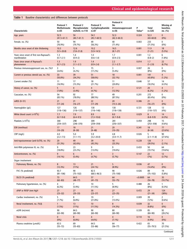
Table 1 Baseline characteristics and differences between protocols
Characteristic
Protocol 1Methotrexaten=65
Protocol 2Mycophenolatemofetil, n=118
Protocol 3Cyclophosphamiden=87
Protocol 4Noimmunosuppressantn=56
PValue*
Totaln=326
Missing atbaseline,no. (%)
Age, years 52.5(41.5–61.1)
50(40.7–61.1)
54.1(45.7–60.5)
52.3(42.3–60.3)
0.324 52.3(43–60.8)
0(0%)
Female, no. (%) 50(76.9%)
94(79.7%)
49(56.3%)
40(71.4%)
0.003 233(71.5%)
0(0%)
Months since onset of skin thickening 10.3(5.5–20.8)
12.6(7.8–21.8)
10.2(5.9–14.5)
16.5(8.7–27)
0.001 11.9(7–21)
18(5.5%)
Years since onset of first non-Raynaud’smanifestation
1.3(0.7–2.1)
1.4(0.9–2)
1.0(0.6–1.4)
1.7(0.9–2.5)
0.001 1.2(0.8–2)
6(1.8%)
Years since onset of Raynaud’sphenomenon
1.3(0.7–2.5)
1.9(1.1–3.1)
1.4(0.9–2.4)
2.2(1.1–3.7)
0.014 1.7(1–2.9)
22(6.7%)
Previous immunosuppressant use, no. (%)† 3(4.6%)
5(4.2%)
15(17.2%)
3(5.4%)
0.007 26(8%)
0(0%)
Current or previous steroid use, no. (%) 33(50.8%)
39(34.2%)
51(58.6%)
18(32.1%)
0.001 141(43.8%)
4(1.2%)
Current smoker (%) 11(16.9%)
17(15.3%)
18(21.7%)
11(20.8%)
0.646 57(18.3%)
14(4.3%)
History of cancer, no. (%) 5(7.8%)
4(3.4%)
4(4.7%)
7(12.5%)
0.121 20(6.2%)
4(1.2%)
Caucasian, no. (%) 54(83.1%)
92(78.0%)
77(88.5%)
49(87.5%)
0.201 272(83.4%)
0(0%)
mRSS (0–51) 21(17–24)
21(16–27)
22(17–29)
20(15.5–26)
0.306 21(16–27)
0(0%)
Haemoglobin (g/L) 127(118–136)
126(118–137)
130(116–140)
130(118–139)
0.721 128(118–137)
13(4%)
White blood count (×109/L) 7.7(6.7–9.4)
7.9(6.4–9.5)
8.9(7.3–10.6)
8.0(6.7–9.4)
0.029 8(6.8–9.9)
14(4.3%)
Platelets (×109/L) 300(254–337)
298(246–370)
309(259–359)
281(250–337)
0.459 298(253–358)
15(4.6%)
ESR (mm/hour) 17(10–29)
18(8–30)
21(9–48)
20(10–35)
0.341 18(8–34)
77(23.6%)
CRP (mg/L) 4.0(2.2–7.5)
5.0(1.1–11)
5.9(3.2–20.0)
4.8(3.0–11.7)
0.026 5(2.1–11.8)
90(27.6%)
Anti-topoisomerase (anti-Scl70), no. (%) 20(31.3%)
49(42.6%)
39(45.3%)
18(33.3%)
0.228 126(39.5%)
7(2.1%)
Anti-RNA polymerase III, no. (%) 9(18.8%)
23(23.2%)
9(13.0%)
9(19.6%)
0.433 50(19.1%)
64(19.6%)
Anticentromere, no. (%) 9(14.1%)
6(5.4%)
4(4.7%)
3(5.7%)
0.147 22(7%)
12(3.7%)
Organ involvement
Pulmonary fibrosis, no. (%) 8(12.3%)
13(11%)
21(24.1%)
5(8.9%)
0.036 47(14.4%)
0(0%)
FVC (% predicted) 93.7(81–106)
90(75–102)
82.5(68.5–96.5)
90(75–100)
0.026 89(75–102)
19(5.8%)
DLCO (% predicted) 73(62–83)
64.5(48–77)
57(41–73)
64(52–75)
<0.0005 64(50–78)
35(10.7%)
Pulmonary hypertension, no. (%) 4(6.3%)
7(5.9%)
10(11.5%)
5(8.9%)
0.488 26(8%)
1(0.3%)
sPAP or RVSP (mm Hg)‡ 27(23–32)
27(20–33)
30(23–37)
27(23–35)
0.472 29(21–34)
124(38%)
Cardiac involvement, no. (%) 5(7.7%)
8(6.8%)
19(21.8%)
7(13.0%)
0.009 39(12%)
2(0.6%)
Renal involvement, no. (%)§ 1(1.5%)
13(11%)
10(11.5%)
8(14.3%)
0.039 32(9.8%)
0(0%)
eGFR (mL/min) 90(63–90)
84.5(60–90)
90(60–90)
80(60–90)
0.339 85(60–90)
92(28.2%)
Renal crisis 0(0%)
8(6.8%)
4(4.6%)
4(7.1%)
0.110 16(4.9%)
0(0%)
Plasma creatinine (μmol/L) 63.5(55–72)
69(55–83)
64.5(53–86)
64(56–77)
0.422 65(55–79.5)
70(21.5%)
Continued
1211Herrick AL, et al. Ann Rheum Dis 2017;76:1207–1218. doi:10.1136/annrheumdis-2016-210503
Clinical and epidemiological research on 24 June 2018 by guest. P
rotected by copyright.http://ard.bm
j.com/
Ann R
heum D
is: first published as 10.1136/annrheumdis-2016-210503 on 10 F
ebruary 2017. Dow
nloaded from

statistically significant reductions in mRSS in all four treatmentgroups at 12 months (−4.0 (−5.2 to −2.7) units for methotrexate,−4.1 (−5.3 to −2.9) for MMF, −3.3 (−4.9 to −1.7) for cyclo-phosphamide and −2.2 (−4.0 to −0.3) for the no immunosup-pressant group), but the differences between treatments were notsignificant (p=0.346) (table 3 and figure 2).
Changes in secondary outcomes over time in the differenttreatment groupsLung functionAfter adjusting for potential confounders, the change rates ofFVC and DLCO were not significantly different in the fourtreatment groups (p=0.460 and 0.505) (table 3).
However, in a subset of patients with pulmonary fibrosis orsuspected pulmonary fibrosis (cases confirmed on high-resolution CT (HRCT) irrespective of FVC or DLCO, or withone of the following if HRCT not performed: FVC or DLCOunder 55% predicted or definite bibasal shadowing on X-ray),there was a significant difference in the change rate of FVC overtime (p=0.035). Patients initially prescribed cyclophosphamidedemonstrated 7.4% absolute increase in FVC (% predicted)compared with 2.0% decrease for methotrexate, 3.2% increasefor MMF and 4.0% increase for the ‘no immunosuppressant’group (table 3).
Functional ability and hand functionChanges over time for the HAQ-DI and CHFS did not differbetween protocols (p=0.130 and 0.073), regardless of adjusting(table 3).
Development of internal organ involvementThis is described in online supplementary figure S2.
Comparison of survival between treatment protocolsSurvival was lowest in the no immunosuppressant group at both12 and 24 months but differences between protocols were notstatistically significant either before (p=0.389) or after weight-ing (p=0.440). In the adjusted model, at 24 months, those inthe no immunosuppressant group had a predicted survival rateof 84.0% compared with 94.1% for methotrexate, 88.8% forMMF and 90.1% for cyclophosphamide (figure 3). Patientswith lung involvement (pulmonary fibrosis and/or hypertension)at baseline had significantly poorer survival than those without:at 24 months, their predicted survival rate was 74.6% versus91.7% (p<0.0005) and similarly for cardiac involvement,71.6% versus 90.7% (p<0.0005).
Adverse effectsOf the 75, 182 and 101 patients who were ever on methotrex-ate, MMF or cyclophosphamide, respectively, 29 (38.7%), 40(22.0%) and 23 (22.8%) were reported to have had side effects,necessitating drug discontinuation in 9 (12.0%), 14 (7.7%) and5 (4.5%) patients, respectively. A survival analysis on protocolexits due to adverse effects showed no differences in the toler-ability of the three treatments (p=0.212) (see onlinesupplementary figure S3).
DISCUSSIONOur main findings were, first, that there were no significant dif-ferences in outcome between the four treatment protocols(methotrexate, MMF, cyclophosphamide, no immunosuppres-sion), although there may be a signal in favour of immunosup-pression for early dcSSc. Although skin score improved in alltreatment groups, this was least in the no immunosuppressantcategory, who also had the highest mortality. Second, ESOS
Table 1 Continued
Characteristic
Protocol 1Methotrexaten=65
Protocol 2Mycophenolatemofetil, n=118
Protocol 3Cyclophosphamiden=87
Protocol 4Noimmunosuppressantn=56
PValue*
Totaln=326
Missing atbaseline,no. (%)
Any GI involvement, no. (%) 25(38.5%)
32(27.1%)
30(34.5%)
26(46.4%)
0.078 113(34.7%)
0(0%)
Muscle involvement, no. (%) 12(18.5%)
9(7.6%)
10(11.5%)
0(0%)
0.002 31(9.5%)
0(0%)
Current digital ulcers 10(15.4%)
17(14.4%)
17(19.5%)
11(19.6%)
0.705 55(16.9%)
0(0%)
Patient questionnaire data
HAQ-DI (0–3) 1.1(0.4–1.8)
1(0.5–1.6)
1(0.4–1.9)
0.7(0.1–1.5)
0.400 1(0.4–1.8)
19(5.8%)
FACIT fatigue score (0–52) 33(21.5–42.5)
30(20–37)
31(17–40)
37(21–44)
0.165 31(20–41)
16(4.9%)
SF36 physical score (0–100) 39.2(30.9–45.1)
36.4(30.8–43)
36(27.2–44.5)
39.2(32–48.4)
0.203 37.4(29.9–45)
15(4.6%)
SF36 mental score (0–100)¶ 37.2(33.3–44)
36.8(32.2–41.9)
39.5(35.7–44.7)
40.8(35.9–44.1)
0.029 38.3(34.3–44)
15(4.6%)
Cochin hand function score (0–90) ** 11.5(2–30.5)
10(4–24)
16(5–40)
6.5(0–23)
0.025 11(3–29)
96(29.4%)
Median (IQR) unless otherwise indicated.*p indicates significance of Kruskal-Wallis test (for continuous variables) or Fisher’s exact test (for categorical variables).†Of the 26 patients who had previously received immunosuppressant therapy, in 2 patients this was for cancer.‡86 patients had a sPAP/RVSP value assumed to be normal and thus not measured. If those cases are omitted, only 38 values of sPAP/RVSP are missing (11.7%). Median values are‘falsely’ high because calculation omits unmeasured (normal) values.§Renal involvement is defined as renal crisis and/or moderate-to-severe renal impairment.¶Despite the significant p-value for the Kruskal-Wallis test, post hoc tests reject any between-group differences in the SF36 mental scores.** Cochin hand function scores were not performed in all centres because of translational issues.CRP, C reactive protein; DLCO, carbon monoxide diffusing capacity; eGFR, estimated glomerular filtration rate; ESR, erythrocyte sedimentation rate; FACIT, Functional Assessment ofChronic Illness Therapy; FVC, forced vital capacity; GI, gastrointestinal; HAQ-DI, Health Assessment Questionnaire Disability Index; mRSS, modified Rodnan skin score (17 sites); RVSP, rightventricular systolic pressure; SF36, Short-Form 36; sPAP, systolic pulmonary artery pressure.
1212 Herrick AL, et al. Ann Rheum Dis 2017;76:1207–1218. doi:10.1136/annrheumdis-2016-210503
Clinical and epidemiological research on 24 June 2018 by guest. P
rotected by copyright.http://ard.bm
j.com/
Ann R
heum D
is: first published as 10.1136/annrheumdis-2016-210503 on 10 F
ebruary 2017. Dow
nloaded from

confirms the relative effectiveness of cyclophosphamide inpatients with pulmonary fibrosis.25 26
An important point when interpreting our findings (andtherefore a note of caution) is that the ‘no immunosuppressant’group was not a control group. Patients in this group had alonger disease duration than the other three groups and weremore likely to have renal involvement.
Our findings lend support to two recently published studies(the Autologous Stem Cell Transplantation InternationalScleroderma trial (ASTIS) trial of autologous stem cell trans-plantation27 and the Scleroderma Lung Study (SLS) II (compar-ing MMF and cyclophosphamide),26 which suggest benefit,including in mRSS, from immunosuppression (as did SLS 125).In ASTIS, those patients randomised to cyclophosphamide hadan 8.8 unit fall in mRSS (from 25.8) at 24 months (comparedwith 3.3 in ESOS over 12 months), but the cyclophosphamideprotocol was more intense, and the patients had more severedisease (patients with the highest mRSS at baseline tend toimprove most quickly4 as also demonstrated by our own find-ings (table 2)). MRSS fell by 19.9 units in those patients
randomised to stem cell transplantation27 (and therefore inten-sive immunosuppression). In SLS 1,25 patients with dcSSc ran-domised to cyclophosphamide experienced a 5.3 unit fall inmRSS at 12 months (compared with 3.3 in ESOS), whereasmRSS fell by 1.7 on placebo (compared with 2.2 units in theESOS ‘no immunosuppressant’ group). In SLS II,26 mRSS at24 months fell 4.9 units on MMF (compared with 4.1 units inESOS at 12 months) and by 5.4 after 12 months treatment withcyclophosphamide, although these values are not directly com-parable because they relate to patients with limited cutaneousand dcSSc combined.
The methodological strength of ESOS, which built uponexperience gained in a previous, smaller observational study,28
was its design: its standardised protocols emulated the condi-tions of a clinical trial, and although not randomised, patientswere enrolled into four homogenous treatment arms with well-defined interventions and a systematic record of protocolchanges and exits. Entry criteria were deliberately inclusive:RCTs often exclude patients with internal organ involvementand for whom immunosuppression is most likely to be
Table 2 Associations between baseline characteristics and skin score
Characteristic (A) Association with baseline mRSS (B) Effect on mRSS evolution
Baseline predictor coefficient(95% CI) p(1)
Time slope(12 months)
Time–predictor interaction coefficient(95% CI) p(2)
Age, per 10 years 0.9 (0.3 to 1.6) 0.004 −1.8 −0.3 (−0.6 to −0.1) 0.007
Female −1.8 (−3.7 to 0.2) 0.072 −3.5 0.1 (−0.5 to 0.8) 0.680
Months since onset of skin thickening −0.1 (−0.2 to 0) 0.156 −4.3 0.1 (0 to 0.1) 0.002
Previous immunosuppressant use −2.1 (−5.3 to 1.2) 0.205 −3.5 0.1 (−1 to 1.3) 0.808
Current or previous steroid use 1.2 (−0.6 to 3.0) 0.191 −2.9 −1.2 (−1.8 to −0.6) <0.0005
mRSS (0–51), per 5 units 0.9 −1 (−1.2 to −0.8) <0.0001
White blood count (×109/L) 0.5 (0.2 to 0.8) 0.002 −2.2 −0.1 (−0.3 to 0) 0.014
ESR (mm/hour) 0.1 (0 to 0.1) 0.003 −4.0 0 (0 to 0) 0.079
CRP (mg/L) 0.1 (0 to 0.2) 0.003 −4.4 0.1 (0 to 0.1) 0.001
Anti-topoisomerase (anti-Scl70) −2.6 (−4.4 to −0.8) 0.005 −4.3 2.2 (1.6 to 2.8) <0.0001
Anti-RNA polymerase III 4.5 (2.1 to 6.9) <0.0005 −3.0 −2.1 (−2.9 to −1.2) <0.0001
Anticentromere −0.4 (−3.9 to 3.1) 0.816 −3.4 0.5 (−0.8 to 1.9) 0.456
Organ involvement
Pulmonary fibrosis 2.9 (0.4 to 5.4) 0.021 −3.5 0.3 (−0.6 to 1.2) 0.534
FVC (% predicted) −0.1 (−0.1 to 0) 0.013 −5.5 0 (0 to 0) 0.005
DLCO (% predicted) 0 (−0.1 to 0) 0.105 −3.2 0 (0 to 0) 0.689
Pulmonary hypertension 2.5 (−0.7 to 5.8) 0.128 −3.3 −2.1 (−3.3 to −0.8) 0.001
Cardiac involvement 2.5 (−0.2 to 5.2) 0.075 −3.5 0 (−1.0 to 0.9) 0.929
Renal involvement* 2.2 (−0.7 to 5.2) 0.140 −3.3 −1.6 (−2.6 to −0.5) 0.004
Any GI involvement, no. (%) 2.2 (0.3 to 4.0) 0.021 −3.4 −0.1 (−0.8 to 0.5) 0.664
Muscle involvement 1.2 (−1.8 to 4.2) 0.425 −3.2 −2 (−2.9 to −1.0) <0.0005
Current digital ulcers 2.5 (0.1 to 4.8) 0.038 −3.3 −0.9 (−1.8 to 0) 0.047
Patient questionnaire data
HAQ-DI (0–3) 3.5 (2.5 to 4.6) <0.0001 −3.0 −0.4 (−0.8 to 0) 0.039
FACIT fatigue score (0–52), per 10 units −1.3 (−2.0 to −0.6) <0.0005 −3.1 −0.1 (−0.3 to 0.2) 0.484
SF36 physical score (0–100) −0.2 (−0.3 to −0.1) <0.0001 −3.3 0 (0 to 0) 0.873
SF36 mental score (0–100) 0.1 (−0.1 to 0.2) 0.297 −1.8 0 (−0.1 to 0) 0.081
Cochin hand function score (0–90), per10 units
1.4 (0.9 to 1.9) <0.0001 −3.7 0 (−0.2 to 0.2) 0.979
Example for interpretation of results: the presence of anti-RNA polymerase III is associated with (A) a higher mRSS by 4.5 units at baseline and (B) losing an extra 2.1 units per yearcompared with an average of −3.0 units per year for all patients.*Renal involvement is defined as renal crisis and/or moderate-to-severe renal impairment.p(1): Significance p value for characteristic coefficient in linear regression of baseline mRSS on baseline predictor.p(2): Significance p value for interaction coefficient between time and baseline characteristic in a longitudinal regression model.CRP, C reactive protein; DLCO, carbon monoxide diffusing capacity; ESR, erythrocyte sedimentation rate; FACIT, Functional Assessment of Chronic Illness Therapy; FVC, forced vitalcapacity; GI, gastrointestinal; HAQ-DI, Health Assessment Questionnaire Disability Index; mRSS, modified Rodnan skin score (17 sites); SF36, Short-Form 36.
1213Herrick AL, et al. Ann Rheum Dis 2017;76:1207–1218. doi:10.1136/annrheumdis-2016-210503
Clinical and epidemiological research on 24 June 2018 by guest. P
rotected by copyright.http://ard.bm
j.com/
Ann R
heum D
is: first published as 10.1136/annrheumdis-2016-210503 on 10 F
ebruary 2017. Dow
nloaded from

Table3
Predictedyearlychangesin
outcom
esandsurvivalratesaccordingto
initialprotocol,w
ithandwithoutadjusting(95%
CI)
Outcome
Mod
elspecificatio
npVa
lue
Metho
trexate
Mycop
heno
late
mofetil
Cyclop
hospha
mide
Noim
mun
osup
pressant
mRSS(0–51)
Noadjusting,
n=326
0.252
−4.4
(−5.7to
−3.2)
−3.8
(−4.9to
−2.8)
−3.5
(−5.0to
−2.0)
−2.4
(−3.9to
−1)
Adjustingforconfounding
(weightedmodel),n=
326
0.346
−4.0
(−5.2to
−2.7)
(n=65)
−4.1
(−5.3to
−2.9)
(n=118)
−3.3
(−4.9to
−1.7)
(n=87)
−2.2
(−4.0to
−0.3)
(n=56)
FVC(%
predicted)
Noadjusting,
n=307
0.045
−1.7
(−4.4to
1.0)
2.1
(−0.1to
4.3)
4.0
(0.5
to7.5)
2.7
(−0.8to
6.2)
Adjustingforconfounding
(weightedmodel),n=
307
0.460
−0.5
(−3.7to
2.6)
(n=59)
2.0
(−0.7to
4.6)
(n=111)
3.3
(−0.6to
7.2)
(n=84)
2.0
(−1.6to
5.6)
(n=53)
Subset
with
PFon
HRCT,w
eighted,
n=129†
0.035
−2.0
(−5.9to
2.0)
(n=19)
3.2
(−0.6to
7.0)
(n=31)
7.4
(2.2
to12.7)
(n=57)
4.0
(−1to
9.0)
(n=22)
DLCO
(%predicted)
Noadjusting,
n=291
0.703
−1.2
(−3.5to
1.0)
0.3
(−1.7to
2.3)
0.6
(−2.0to
3.3)
−0.3
(−4.2to
3.7)
Adjustingforconfounding
(weightedmodel),n=
291
0.505
−1.6
(−3.8to
0.6)
(n=51)
0.8
(−1.5to
3.1)
(n=110)
0.1
(−3.1to
3.2)
(n=79)
−0.1
(−3.7to
3.4)
(n=51)
Subset
with
PFon
HRCT,w
eighted,
n=116†
0.809
−0.8
(−5.4to
3.8)
(n=12)
1.9
(−1.8to
5.6)
(n=31)
1.8
(−1.9to
5.4)
(n=53)
1.6
(−2.6to
5.7)
(n=20)
HAQ-DI(0–3)
Noadjusting,
n=307
0.070
−0.1
(−0.2to
0.1)
0 (−0.1to
0.1)
−0.2
(−0.3to
−0.1)
0.1
(−0.1to
0.2)
Adjustingforconfounding
(weightedmodel),n=
307
0.130
−0.1
(−0.3to
0)(n=59)
0 (−0.1to
0.1)
(n=113)
−0.1
(−0.3to
0)(n=81)
0.1
(−0.1to
0.2)
(n=54)
Cochinhand
function(0–90)‡
Noadjusting,
n=230
0.072
−1.1
(−3.5to
2.2)
−0.3
(−1.6to
1.2)
−3.1
(−5.1to
−0.5)
1.1
(−0.3to
3.0)
Adjustingforconfounding
(weightedmodel),n=
230
0.073
−1.4
(−3.5to
1.4)
(n=36)
−0.6
(−2.0to
1.1)
(n=103)
−2.4
(−4.7to
0.6)
(n=49)
1.7
(-0.1
to4.0)
(n=42)
Survival
Noadjusting,
n=326
0.389
(12months)93.5%
(24months)93.5%
(12months)96.5%
(24months)89.3%
(12months)88.1%
(24months)85.4%
(12months)88.9%
(24months)85.1%
Adjustingforconfounding
(weightedmodel),n=
326
0.440
(12months)94.1%
(24months)94.1%
(12months)96.1%
(24months)88.8%
(12months)91.7%
(24months)90.1%
(12months)88.6%
(24months)84.0%
Significancep:
Fisher’stestfore
quality
ofchange
ratesbetweenprotocols,fore
achoutcom
evariable.
*Resultsarereporte
din
term
sof
changesafter12
months.Ho
wever,a
llstudydata
(from
baselineto
the24-m
onth
endpoint)wereused
inestim
ation.
Toobtain24-m
onth
changes,multiplyresults
aboveby
2.†Forthesubanalysis
involvingthesubset
ofpatientswith
pulmonaryfibrosis
atbaseline,patientswith
definite
bibasalp
ulmonaryfibrosis
confirm
edon
HRCT
wereincluded,irrespectiveof
FVCvalue.Ifno
HRCT
scan
was
perfo
rmed
atbaseline,an
FVC<
55%,D
LCO<55%
predictedor
definite
bibasalshadowingon
X-raywas
also
abasis
forinclusion.
‡Ch
angesexpressedin
units
fortheCochinregressio
narean
approximationderived
from
the95%
CIof
percentage
changesbetweenbaselineand12
months(onascaleshifted
byoneunit),a
ppliedto
thepredictedbaselinevalues
fore
achgroupinthe
originalscale.
DLCO
,carbonmonoxidediffu
singcapacity;FVC
,forcedvitalcapacity;H
AQ-DI,He
alth
AssessmentQu
estionnaire
DisabilityIndex;HR
CT,h
igh-resolutionCT;m
RSS,modified
Rodnan
skin
score(17sites);PF,p
ulmonaryfibrosis.
1214 Herrick AL, et al. Ann Rheum Dis 2017;76:1207–1218. doi:10.1136/annrheumdis-2016-210503
Clinical and epidemiological research on 24 June 2018 by guest. P
rotected by copyright.http://ard.bm
j.com/
Ann R
heum D
is: first published as 10.1136/annrheumdis-2016-210503 on 10 F
ebruary 2017. Dow
nloaded from

Figure 2 Modified Rodnan skin score (mRSS) during baseline and follow-up visits, by initial protocol. For each group of patients, according to theirinitial protocol, the distribution of the skin score is illustrated on the left-hand side by box and whisker plots (indicating the median and IQR) atbaseline, 12 and 24 months. On the right-hand side, the distribution of individual 1-year changes in the skin score is described by histograms and akernel density estimate. In addition, a vertical green line indicates the value of the average 1-year change in the skin score, irrespective of treatmentchoice. The bottom panel in the figure describes the estimated changes in mRSS (with 95% CI) according to initial protocol, based on the resultsfrom the adjusted model (described in table 3).
1215Herrick AL, et al. Ann Rheum Dis 2017;76:1207–1218. doi:10.1136/annrheumdis-2016-210503
Clinical and epidemiological research on 24 June 2018 by guest. P
rotected by copyright.http://ard.bm
j.com/
Ann R
heum D
is: first published as 10.1136/annrheumdis-2016-210503 on 10 F
ebruary 2017. Dow
nloaded from

beneficial. By recruiting 326 patients from 50 centres, ESOSrepresents a large cohort of patients with very early dcSSc(median duration of skin thickening 11.9 months): its data willserve as a benchmark when designing and interpreting futureclinical trials. This is especially relevant with a number of noveltreatment approaches currently being explored including bio-logical agents. For example, in a recent RCT of tocilizumab,29
mRSS fell over 24 weeks by 3.9 units from 26 in the 43tocilizumab-treated patients and by 1.2 units from 26 in the 44placebo-treated patients, this latter fall comparable to the ESOS‘no immunosuppressant’ response. In comparing between thesestudies, the higher baseline mRSS in the tocilizumab studyshould be borne in mind.
The main weakness of observational studies is that eachpatient’s outcome on her/his treatment arm cannot be com-pletely disentangled from her/his initial characteristics. Forinstance, ESOS has verified that patients with lung and cardiacinvolvement tend to be prescribed cyclophosphamide. However,adjusting using IPTweights minimises the problem of confound-ing by indication.
In conclusion, observational studies offer a rich population-wide perspective assessing treatment effects in a real-worldsetting. ESOS achieved its aim of following a large internationalcohort of patients with early dcSSc over 2 years, each of whomwas treated according to one of four protocols. The message forclinicians is that there is a weak signal to support using immuno-suppressants for early dcSSc (and in particular cyclophospha-mide for patients with pulmonary fibrosis). However, it is clearthat there remains a pressing need for the development of moreeffective and targeted treatments.
Author affiliations1Centre for Musculoskeletal Research, The University of Manchester, Salford RoyalNHS Foundation Trust, Manchester Academic Health Science Centre, Manchester, UK
2NIHR Manchester Musculoskeletal Biomedical Research Unit, Central ManchesterNHS Foundation Trust, Manchester Academic Health Science Centre, Manchester, UK3Centre for Musculoskeletal Research, The University of Manchester, ManchesterAcademic Health Science Centre, Manchester, UK4Department of Rheumatology, Lund University, Lund, Sweden5Service de Médecine Interne, Hôpital Cochin, Centre de Référence pour lesVascularites Nécrosantes et la Sclérodermie Systémique, Université Paris Descartes,Assistance Publique-Hôpitaux de Paris (AP-HP), Paris, France6Nuffield Department of Orthopaedics, Rheumatology and Musculoskeletal Sciences,University of Oxford, Oxford, UK7Member of Steering Committee, contact via Professor Herrick, The University ofManchester, Manchester, UK8Department of Rheumatology and Immunology, Medical Center, University of Pécs,Pecs, Hungary9Department of Internal Medicine 3, University of Erlangen-Nuremberg, Erlangen,Germany10Department of Rheumatology, University of Zurich, Zurich, Switzerland11Royal Free London NHS Foundation Trust, London, UK.12Rehabilitation Services, Salford Royal NHS Foundation Trust, Salford, UK13Department of the Rheumatic Diseases, Radboud University Nijmegen MedicalCentre, Nijmegen, The Netherlands14Rheumatology 2 Department, “Grigore T. Popa” University of Medicine andPharmacy, Clinical Rehabilitation Hospital, Iași, Romania15UCL Division of Medicine, Centre for Rheumatology and Connective TissueDiseases, London, UK16Unité Clinique de Médecine Interne, Maladies Auto-immunes et PathologieVasculaire, UF 04, Hôpital Saint-Louis, AP-HP Assistance Publique des Hôpitaux deParis, INSERM UMRS 1160, Paris Denis Diderot University, France17Jewish General Hospital, Lady Davis Institute and McGill University, Montreal,Canada18Department Experimental and Clinical Medicine, Division of Rheumatology AOUC,University of Florence, Florence, Italy19Shine Rheumatology Unit, Rambam Heath Care Campus; Rappaport Faculty ofMedicine, Haifa, Israel20Rheumatology Unit, Oslo University Hospital Rikshospitalet, Oslo, Norway21Queen Elizabeth Hospital Birmingham, UHB Foundation Trust, Birmingham, UK22St Vincent’s Hospital, Melbourne, Australia23Department for Dermatology, University of Cologne Kerpenerstr. 62, Köln,Germany24Cambridge University NHS Hospital Foundation Trust, Cambridge, UK25Department of Internal Medicine, Hôtel-Dieu Hospital, University of Nantes,Nantes, France
Figure 3 Kaplan-Meier estimated survival curves by treatment group.
1216 Herrick AL, et al. Ann Rheum Dis 2017;76:1207–1218. doi:10.1136/annrheumdis-2016-210503
Clinical and epidemiological research on 24 June 2018 by guest. P
rotected by copyright.http://ard.bm
j.com/
Ann R
heum D
is: first published as 10.1136/annrheumdis-2016-210503 on 10 F
ebruary 2017. Dow
nloaded from

26University of Liverpool, Aintree University Hospital, Liverpool, UK27Service de Médecine Interne, Hôpital Bretonneau Tours Cedex, France28Centre for Rheumatic Diseases, Glasgow Royal Infirmary, Glasgow, UK29Sheffield Teaching Hospitals, Sheffield, UK30Leeds Institute of Rheumatic and Musculoskeletal Medicine, University of Leedsand NIHR Leeds Musculoskeletal Biomedical Research Unit, Leeds Teaching HospitalsNHS Trust, UK31Stanford University, Stanford, California, USA.32University of Belgrade School of Medicine, Institute of Rheumatology, Belgrade,Serbia33Clinical and Academic Rheumatology, North Bristol NHS Trust, Bristol, UK34Nottingham University Hospitals NHS Trust, and Nottingham NHS TreatmentCentre, Nottingham, UK35Peter Maddison Rheumatology Centre, Llandudno, UK36Queens Hospital, Romford, UK37University of Copenhagen, Copenhagen Lupus and Vasculitis Clinic, Center forRheumatology and Spine Diseases, Rigshospitalet, Copenhagen, Denmark38Norwich Medical School, University of East Anglia, Norwich, UK39Royal National Hospital for Rheumatic Diseases, Bath, UK40Department of Rheumatology and Clinical Immunology, Justus-Liebig UniversityGiessen, Bad Nauheim, Germany41Department of Rheumatology, University of Lübeck, Lübeck, Germany42Department of Rheumatology and Clinical Immunology, University Hospital CharitéBerlin, Berlin, Germany43Department of Rheumatology, Royal Perth Hospital, Perth, Australia44Servicio de Reumatologia. Hospital Universitario 12 de Octubre, Madrid, Spain45Internal Medicine Unit, Limoges University Hospital, France46Département de Médecine Interne et Immunologie Clinique, Centre National deRéférence Maladies Systémiques etAuto-immunes Rares, Université de Lille, Inserm,U995, FHU Immune-Mediated Inflammatory Diseases and Targeted Therapies, Lille,France47Gateshead Hospitals Foundation Trust, Gateshead, UK48Department of Internal Medicine, Division of Rheumatology, Istanbul University,Istanbul, Turkey49Fife Rheumatic Diseases Unit, Whyteman’s Brae Hospital, Kirkcaldy, UK50Department of Rheumatology and Clinical Immunology, UMC Utrecht, Utrecht,The Netherlands51James Cook University Hospital, Middlesbrough, UK52Rheumatology Unit, Royal Adelaide Hospital, and Discipline of Medicine, Universityof Adelaide, Adelaide, South Australia53Department of Rheumatology and Inflammation Research, The SahlgrenskaAcademy at Gothenburg University, Gothenburg, Sweden54Monash Centre for Inflammatory Diseases, Monash University, Clayton,Melbourne, Australia55Department of Internal Medicine, Hôpital Edouard Herriot, Lyon, France56Department of Internal Medicine, Foundation Hospital Saint Joseph, Marseille,France57Cannock Chase Hospital, Cannock, UK58St Vincent’s University Hospital, Dublin, Ireland59 Department of Internal Medicine, Centre Hospitalier Lyon Sud, Pierre Benite,France60Internal Medecine, Ambroise Paré Hospital, Boulogne Billancourt, France
Acknowledgements The authors are grateful to Dr Holly Ennis for study set-upand to her and Dr Graham Dinsdale for project coordination during the earlierphases of the study. Thanks also to members of the independent oversight board:Stephen Cole, Dinesh Khanna and Frank Wollheim.
Contributors ALH, ML, RH, LM, AS, EB, LC, JHWD, OD, KF, WJG, RO, MV andCPD were members of the Steering Committee and designed the study. ALH, RH,LM, LC, JHWD, OD, MV, CA, VHO, DF, MH, MM-C, AB-G,OM, ACJ, PJ, WS, PM,FCH, CA, MEA, ED, RM, MA, MHB, LC, ND, HG, PL, YA, KC, SJ, AJM, NM, UM-L,GR, MB, JR, PEC, AF, EH, JH, MI, JSM, J van L, SP, SP, AR, JS, BC, CS, TS, DJV, CG,GT and CPD were principal investigators at the different sites and recruited patients.XP was study coordinator. SP and ML were responsible for the statistical analysis.ALH, XP, SP, ML, RH, LM, AS and CPD wrote the draft report, and all authorsreviewed the report, provided comments and approved the final report.
Funding ESOS was funded by a grant from the European League AgainstRheumatism (EULAR) Orphan Disease Programme. Additional funding fromScleroderma and Raynaud’s UK allowed a 1-year extension of the study.
Competing interests ALH has done consultancy work for Actelion, served on aData Safety Monitoring Board for Apricus, received research funding and speaker’sfees from Actelion, and speaker’s fees from GSK. JHWD has consultancyrelationships and/or has received research funding from Actelion, BMS, Celgene,Bayer Pharma, Boehringer Ingelheim, JB Therapeutics, Sanofi-Aventis, Novartis, UCB,GSK, Array Biopharma, Active Biotech, Galapagos, Inventiva, Medac, Pfizer, Anamarand RuiYi and is stock owner of 4D Science GmbH. OD has received consultancy
fees from 4D Science, Actelion, Active Biotech, Bayer, Biogenidec, BMS, BoehringerIngelheim, EpiPharm, Ergonex, espeRare Foundation, Genentech/Roche, GSK,Inventiva, Lilly, Medac, Medimmune, Pharmacyclics, Pfizer, Serodapharm, and Sinoxaand received research grants from Actelion, Bayer, Boehringer Ingelheim, Ergonex,Pfizer and Sanofi, and has a patent mir-29 for the treatment of systemic sclerosislicenced. WG has received teaching fees from Pfizer. FH has received researchfunding from Actelion. MEA has undertaken advisory board work and receivedhonoraria from Actelion, and received speaker’s fees from Bristol-Myers Squibb. LChas done advisory board work for Gilead and served Data Safety Monitoring Boardsfor Cytori and Reata. HG has done consultancy work and received honoraria fromActelion. UM-L is funded in part bu EUSTAR/EULAR. JMvL has received honorariafrom Eli Lilly, Pfizer, Roche, MSD and BMS. AR receives funding from AstraZeneca.CPD has done consultancy for GSK, Actelion, Bayer, Inventiva and Merck-Serono,received research grant funding from GSK, Actelion, CSL Behring and Inventiva,received speaker’s fees from Bayer and given trial advice to Merck-Serono.
Patient consent Obtained.
Ethics approval The Ethics Committee of each centre approved the study.
Provenance and peer review Not commissioned; externally peer reviewed.
Data sharing statement At present, unpublished data from the study are notavailable for sharing. This position may change in 6–12 months time.
Open Access This is an Open Access article distributed in accordance with theCreative Commons Attribution Non Commercial (CC BY-NC 4.0) license, whichpermits others to distribute, remix, adapt, build upon this work non-commercially,and license their derivative works on different terms, provided the original work isproperly cited and the use is non-commercial. See: http://creativecommons.org/licenses/by-nc/4.0/
REFERENCES1 Nikpour M, Stevens WM, Herrick AL, et al. Epidemiology of systemic sclerosis. Best
Practice Res Clin Rheumatol 2010;24:857–69.2 Rubio-Rivas M, Royo C, Simeon CP, et al. Mortality and survival in systemic
sclerosis : systematic review and meta-analysis. Sem Arthritis Rheum2014;44:208–19.
3 Nihtyanova SI, Schreiber BE, Ong VH, et al. Prediction of pulmonary complicationsand long-term survival in systemic sclerosis. Arthritis Rheum 2014;66:1625–35.
4 Merkel PA, Silliman NP, Clements PJ, et al. Patterns and predictors of change inoutcome measures in clinical trials in scleroderma: An individual patientmeta-analysis of 629 subjects with diffuse cutaneous systemic sclerosis. ArthritisRheum 2012;64:3420–9.
5 Silverman SL. From randomized controlled trials to observational studies. Am J Med2009;122:114–20.
6 Halpern SD, Karlawish JT, Berlin JA. The continuing unethical conduct ofunderpowered clinical trials. JAMA 2002;288:358–62.
7 Pope JE, Bellamy N, Seibold JR, et al. A randomized, controlled trial of methotrexateversus placebo in early diffuse scleroderma. Arthritis Rheum 2001;44:1351–8.
8 Denton CP, Merkel PA, Furst DE, et al. Recombinant human anti-transforminggrowth factor β1 antibody therapy in systemic sclerosis: a multicentre, randomized,placebo-controlled Phase I/II trial of CAT-192. Arthritis Rheum 2007;56:323–3.
9 Kowal-Bielecka O, Landewé R, Avouac J, et al. EULAR recommendations for thetreatment of systemic sclerosis: a report from the EULAR Scleroderma Trials andResearch Group (EUSTAR). Ann Rheum Dis 2009;68:620–8.
10 Rawlins MD. The Harveian Oration of 2008: On the evidence for decisions aboutthe use of therapeutic interventions. London: Royal College of Physicians, 2008.
11 Gagne J, Thompson L, O’Keefe K, et al. Innovative research methods for studyingtreatments for rare diseases: methodological review. BMJ 2014;349:g6802.
12 LeRoy EC, Black C, Fleischmajer R, et al. Scleroderma (systemic sclerosis):classification, subsets and pathogenesis. J Rheumatol 1988;15:202–5.
13 Clements P, Lachenbruch P, Siebold J, et al. Inter- and intraobserver variability oftotal skin thickness score (modified Rodnan TSS) in systemic sclerosis. J Rheumatol1995;22:1281–5.
14 Clements PJ, Hurwitz EL, Wong WK, et al. Skin thickness score as a predictor andcorrelate of outcome in systemic sclerosis. Arthritis Rheum 2000;43:2445–54.
15 Steen VD, Medsger TA. The value of the health assessment questionnaire andspecial patient-generated scales to demonstrate change in systemic sclerosis patientsover time. Arthritis Rheum 1997;40:1984–91.
16 Webster K, Cella D, Yost K. The Functional Assessment of Chronic Illness Therapy(FACIT) Measurement System: properties, applications, and interpretation. HealthQual Life Outcomes 2003;1:79.
17 Harel D, Thombs BD, Hudson M, et al. Measuring fatigue in SSc: a comparison ofthe Short Form-36 Vitality subscale and Functional Assessment of Chronic IllnessTherapy–Fatigue scale. Rheumatol 2012;51:2177–85.
18 Rannou F, Poiraudeau S, Berezne A, et al. Assessing disability and quality of life insystemic sclerosis: construct validities of the Cochin Hand Function Scale, Health
1217Herrick AL, et al. Ann Rheum Dis 2017;76:1207–1218. doi:10.1136/annrheumdis-2016-210503
Clinical and epidemiological research on 24 June 2018 by guest. P
rotected by copyright.http://ard.bm
j.com/
Ann R
heum D
is: first published as 10.1136/annrheumdis-2016-210503 on 10 F
ebruary 2017. Dow
nloaded from

Assessment Questionnaire (HAQ), Systemic Sclerosis HAQ, and Medical OutcomesStudy 36-Item Short Form Health Survey. Arthritis Rheum 2007;57:94–102.
19 Brookhart MA, Schneeweiss S, Rothman KJ, et al. Variable selection for propensityscore models. Amer J Epidemiol 2006;163:1149–56.
20 Austin PC, Grootendorst P, Normand S-LT, et al. Conditioning on the propensityscore can result in biased estimation of common measures of treatment effect: aMonte Carlo study. Stat Med 2007;26:754–68.
21 Sato T, Matsuyama Y. Marginal structural models as a tool for standardization.Epidemiology 2003;14:680–6.
22 Imbens G. The role of the propensity score in estimating dose-response functions.Biometrika 2000;87:706–10.
23 Fewell Z, Hernan MA, Wolfe F, et al. Controlling for time-dependant confoundingusing marginal structural models. Stata J 2004;4:402–20.
24 Cole SR, Hernán MA. Constructing inverse probability weights for marginalstructural models. Amer J Epidemiol 2008;168:656–64.
25 Tashkin DP, Elashoff R, Clements PJ, et al. Cyclophosphamide versus placebo inscleroderma lung disease. N Eng J Med 2006;354:2655–66.
26 Tashkin DP, Roth MD, Clements PJ, et al. Mycophenolate mofetil versus oralcyclophosphamide in scleroderma-related interstitial lung disease (SLS II):a randomsed controlled, double-blind, parallel group trial. Lancet Respir Med2016;4:708–19.
27 Van Laar JM, Farge D, Sont JK, et al. Autologous hematopoietic stem celltransplantation vs intravenous pulse cyclophosphamide in diffuse cutaneous systemicsclerosis. JAMA 2014;311:2490–8.
28 Herrick A, Lunt M, Whidby N, et al. Observational study of treatment outcomein early diffuse cutaneous systemic sclerosis. J Rheumatol 2010;37;116–24.
29 Khanna D, Denton CP, Jahreis A, et al. Safety and efficacy of subcutaneoustocilizumab in adults with systemic sclerosis (faSScinate): a phase 2, randomisedcontrolled trial. Lancet 2016;387:2630–40.
1218 Herrick AL, et al. Ann Rheum Dis 2017;76:1207–1218. doi:10.1136/annrheumdis-2016-210503
Clinical and epidemiological research on 24 June 2018 by guest. P
rotected by copyright.http://ard.bm
j.com/
Ann R
heum D
is: first published as 10.1136/annrheumdis-2016-210503 on 10 F
ebruary 2017. Dow
nloaded from